Published online Mar 26, 2022. doi: 10.12998/wjcc.v10.i9.2954
Peer-review started: November 9, 2021
First decision: December 27, 2021
Revised: January 5, 2022
Accepted: February 10, 2022
Article in press: February 10, 2022
Published online: March 26, 2022
In recent years, it has been recognized that transesophageal echocardiography (TEE) is of great value in resuscitation of cardiac arrest. However, its safety has rarely been reported.
We present a 59-year-old male patient scheduled to undergo cardiac surgery for rheumatic heart disease. Upper gastrointestinal bleeding from a Mallory-Weiss tear appeared following cardiopulmonary resuscitation, TEE, and percutaneous cardiopulmonary bypass resuscitation when he suffered from aesthesia-related cardiac arrest. Gastrointestinal injury was diagnosed promptly and treated effectively. However, the exact etiology of gastrointestinal injury was unclear; the interaction of closed-chest cardiac massage and the application of TEE may be involved as a most possible mechanism of injury.
Serious complications should be considered when TEE is used in patients with special pathophysiological conditions.
Core Tip: In recent years, transesophageal echocardiography (TEE) has been recognized as a valuable imaging tool to provide diagnostic and prognostic information for both cardiopulmonary resuscitation (CPR) cases and non-CPR cases. Although it is generally considered safe, some serious complications, including esophageal perforation and mucosal damage, have been observed in non-CPR cases. To our knowledge, there is no literature on the safety of TEE during CPR. We here report the first case of upper gastrointestinal bleeding from a Mallory-Weiss tear associated with TEE during successful CPR and percutaneous cardiopulmonary bypass resuscitation.
- Citation: Tang MM, Fang DF, Liu B. Upper gastrointestinal bleeding from a Mallory-Weiss tear associated with transesophageal echocardiography during successful cardiopulmonary resuscitation: A case report. World J Clin Cases 2022; 10(9): 2954-2960
- URL: https://www.wjgnet.com/2307-8960/full/v10/i9/2954.htm
- DOI: https://dx.doi.org/10.12998/wjcc.v10.i9.2954
Transesophageal echocardiography (TEE) can provide valuable diagnostic and prognostic information during cardiac arrest resuscitation[1]; but its safety in clinical practice has not been well reported[2,3]. Serious complications were observed in non-cardiopulmonary resuscitation (CPR) cases[4-7]. But in fact, the pathophysiological conditions that are unique to CPR: External chest compression, low cardiac output and percutaneous cardiopulmonary bypass resuscitation (pCPBR) may complicate the situation.
We here present a case of upper gastrointestinal bleeding from a Mallory-Weiss tear associated with TEE during successful CPR. The main purpose is to explore the pathogenesis of upper gastrointestinal bleeding in patients subjected to CPR, TEE and pCPBR. At the same time, we try to come up with some strategies that might help prevent such injury.
A 59-year-old man suffered from upper gastrointestinal bleeding after successful CPR using TEE.
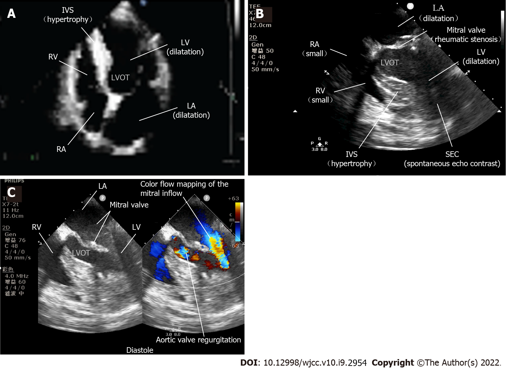
A 59-year-old man (168 cm, 76.5 kg) was scheduled to undergo aortic and mitral valve replacement as well as tricuspid valve repair for rheumatic heart disease causing progressive angina and recurrent syncope. His symptoms started two years ago with progressive angina and recurrent syncope, which had been worsened over the last six months. TTE revealed moderate aortic valve and mitral valve stenosis, moderate aortic valve regurgitation and mild tricuspid regurgitation, mild left ventricular dilatation, left ventricular hypertrophy, and left ventricular ejection fraction of 71% (Figure 1A). Electrocardiography showed normal sinus rhythm. Along with standard monitoring, an invasive intra-arterial blood pressure monitor was established under regional anesthesia. The patient was stable with a heart rate of 76 beats/min, blood pressure 138/60 mmHg, respiratory rate 18 breaths/min, and oxygen saturation 97% when breathing air. After peripheral venous access was secured, general anesthesia was induced with midazolam, propofol, sufentanil citrate and cisatracurium. Two minutes after successful endotracheal intubation, severe bradycardia (heart rate dropping from 70 beats/min to the 20 beats/min) followed by asystole and an undetectable blood pressure was noticed on the monitor. No skin or mucous membrane changes were observed. Cardiac arrest resuscitation was immediately initiated. Chest compressions were delivered. The patient was mechanically ventilated with 100% oxygen and his airway pressure was normal. Intravenous injection of 1mg epinephrine and 500 mg methylprednisolone were administered. A few minutes later, ventricular fibrillation was observed. Defibrillation with 200 J was applied and CPR was continued. Ventricular fibrillation recurred, amiodarone and sodium bicarbonate were given intravenously and defibrillations were applied for a total of 5 times. Arterial blood gas was drawn showing electrolyte disturbances and elevated glucose and hyperlactacidemia. The entire resuscitation lasted 15 min, while the patient still failed to return spontaneous circulation. A 2 MHz-7 MHz omniplane TEE probe (Philips X7-2T, Amsterdam, Netherlands) was blindly inserted with ease by an anesthesia fellow with more than 2-year experience in cardiac anesthesia to investigate the etiology without interfering with chest compressions. TEE was manipulated in the usual manner, including transgastric view. It detected that the cardiac motion ceased, but the ventricular fibrillation rhythm recurred. The right cardiac chambers were small, the rheumatic mitral stenosis and spontaneous echo contrast (SEC) indicated blood stasis in the left cardiac chambers (Figure 1B). The surgeon and attending anesthetist informed the family of the patient’s condition. With their consent, 750 IU/kg heparin was given intravenously and the pCPBR was successfully established by cannulating the arteria and vena femoralis 35 min after the cardiac arrest. The activated clotting time was 604 s during pCPBR. With all these efforts made, the spontaneous circulation with sinus rhythm was finally recovered 67 min after the cardiac arrest. The operation was thus cancelled. TEE imaging was performed to monitor the left ventricular function and to assist with the weaning of pCPBR. The accelerated mitral inflow through the stenosis and aortic valve regurgitation during diastole were shown in a four-chamber view with color Doppler (Figure 1C). After uneventful weaning from pCPBR was accomplished with epinephrine and phenylephrine infusions, protamine 400 mg was given to reverse the initial heparinization and the activated clotting time was 137 s. No blood transfusion was administered and his hematocrit was stable at about 40%. During preparation for transshipment, the TEE probe was removed with some bloodstains on the surface. About half an hour after arrival in the intensive-care unit (ICU), acute massive hematochezia appeared and a nasogastric tube was placed immediately, and a moderate amount of dark red bloody fluid was aspirated.
The patient denied a history of systemic drug allergy, gastrointestinal disease or coagulopathy.
The patient had a disease-free personal and family history.
When the patient was transferred to ICU, his temperature was 36.0°C, heart rate was 89 beats per minute, blood pressure was 122/68 mmHg, ventilator-controlled frequency was 20 bpm and oxygen saturation in 50% fractional concentration of inspired oxygen was 100%. The clinical neurological examination revealed a Glasgow Coma scale of E1VTM1, without any other pathological signs.
Preoperatively, the hemoglobin was 16.1 g/dL (normal range 13.0 g/dL-17.5 g/dL), the hematocrit was 42.9% (normal range 40.0%-50.0%), and the international normalized ratio was 0.9 (normal range 0.88-1.15). When the patient arrived at the ICU, the hemoglobin was 12.8 g/dL, the hematocrit was 39%, and the international normalized ratio was 1.76.
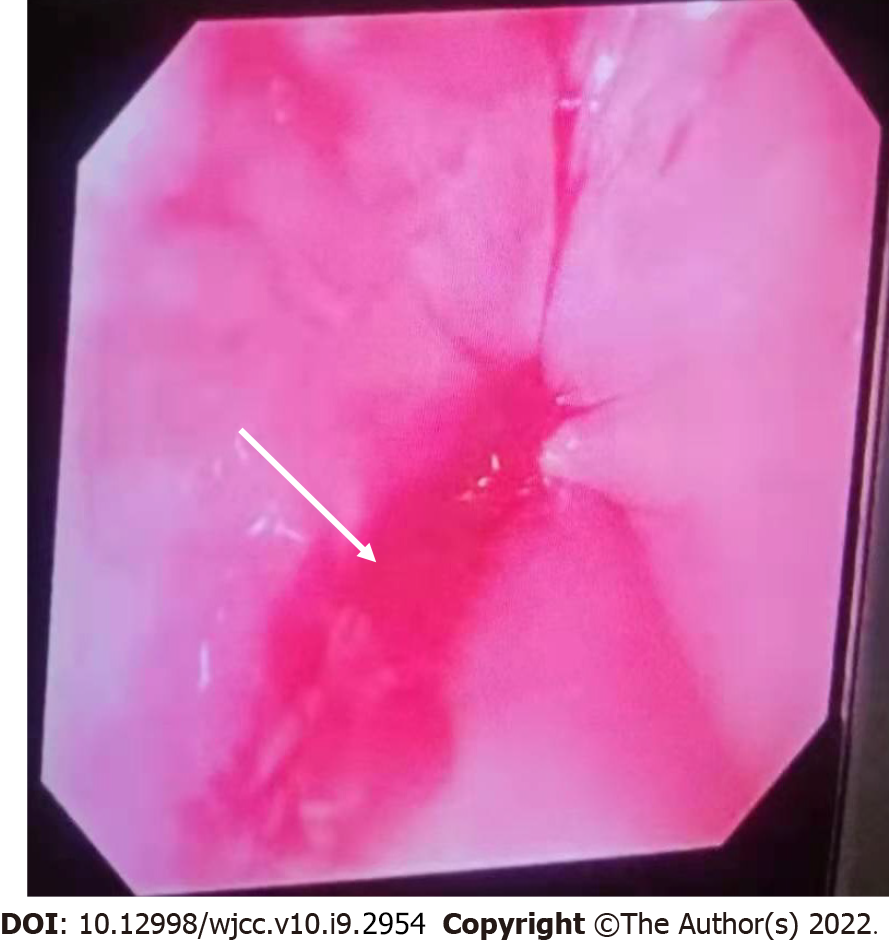
An emergent bedside esophagogastroscopy (GIF Type 2T200, Olympus, Tokyo, Japan) was performed and demonstrated a Mallory-Weiss linear mucosal tear at the gastroesophageal junction covered with a little fresh blood and no active bleeding was observed (Figure 2).
The final diagnosis of the presented case is an upper gastrointestinal bleeding from a Mallory-Weiss tear associated with TEE during successful CPR and pCPBR.
The therapy with intravenous administration of a histamine-2 receptor antagonist, proton pump inhibitor and thrombin product was stared. The lesion was successfully treated with mucosal clipping. As the patient was hemodynamically stable with a hematocrit of 39%, no packed red blood cells was given.
The patient was gradually awake several hours later and could move his limbs on command. He was weaned from mechanical ventilation on the fifth day after resuscitation. Endoscopy a week later revealed excellent mucosal healing. He achieved a satisfied recovery without any evidence of further upper gastrointestinal bleeding and discharged from the hospital 12 d after resuscitation with good neurological outcomes. The patient is currently followed up regularly to assess the best timing for cardiac surgery.
TEE was first described in 1976 and became a popular imaging modality in the mid-1980s[8]. Over the last four decades, TEE is used increasingly in the emergency department, perioperative procedure and critical care environment. Although generally considered to be safe, in a recent study of 22314 TEE examinations, there were 17 patients developed complications, including esophageal perforation and mucosal damage, and there were 7 deaths directly attributed to these complications, corresponding to an incidence of 0.08% and 0.03% respectively[7]. Because TEE can rapidly identify the reversible causes of arrest[9], guide the precise cannulation of extracorporeal cardiopulmonary resuscitation and optimize the treatment after the return of spontaneous circulation[10]. Therefore, TEE has been endorsed by reviews and guidelines as a well-suited imaging tool to improve cardiac arrest outcome[11-13]. While, little is known, about the rate of complications related to TEE during CPR. We found a few articles that described the safety of defibrillation during cardiac arrest while the TEE probe is in place[2]. Only in one case, in 2018, Marchandot et al[3] described a left atrial (LA) intramural hematoma detected by TEE during CPR. They considered that iterative contacts of the LA upon the TEE probe caused by chest compressions during CPR was the primary mechanism of injury. Therefore, the complications of TEE during CPR associated morbidity and mortality seems to be heavily underreported and should be watched out for.
Mallory-Weiss tear was first described in 1929 by Mallory et al[14] and Weiss et al[15]. It is defined as longitudinal, nonperforating mucosal lacerations in the gastroesophageal junction resulting in upper gastrointestinal bleeding. Both CPR and the use of TEE are considered to be risk factors for Mallory-Weiss tear[16]. In the present case, the patient had an acute onset of hematochezia, and a nasogastric tube was placed and aspirated gross blood and the endoscopic appearance was typical. All these factors jointly established the diagnosis.
The exact reason of the anesthesia-related cardiac arrest in this patient was unclear[17]. Anaphylactic shock after administering cis-atracurium during the induction of anesthesia was considered one of the most likely causes[18]. The exact etiology of gastrointestinal injury in this case was also unknown; however, the interaction of closed-chest cardiac massage and the application of TEE may be involved as a most possible mechanism of injury[14]. There may be several reasons. Firstly, it is much more difficult to insert or manipulate the TEE probe during chest compressions, causing iatrogenic trauma occasionally. Secondly, according to Urbanowicz et al[19] and Denault et al[20], the rigid echoprobe may produce a high contact pressure on the gastroesophageal junction both during manipulation and at rest. Thirdly, external chest compressions can further elevate intragastric pressure and contact pressure, which might result in laceration[21]. Low cardiac output syndrome, non-pulsatile flow from pCPBR, and administration of vasoconstrictors can cause gastrointestinal hypoperfusion and aggravate the injury to some extent[22].
Clinicians should take a risk vs benefit assessment on each individual patient before the application of TEE during CPR. The risk factors include but not limited to: Non-standard resuscitation; co-existing gastrointestinal disease or coagulopathy; periods of probe insertion and manipulation; periods of low cardiac output and non-pulsatile flow; increased frailty; and musculoskeletal disorders and the use of steroids[9,22]. TTE should be a priority. When really need to use TEE, a written or verbal informed consent is essential. TEE probe should be inserted gently with a visualization aid[23] and performed by experienced operator who have completed dedicated training[24]. For now, it is our view that a standardized and fast CPR-TEE assessment process should be established as soon as possible[25]. The transgastric view should be avoided because in this view the probe tip is closest to the site of compressions and there is a potential risk of gastric mucosal lacerations or perforation[26]. Indeed, ideally the TEE probe should be kept out of the patient's body during closed chest cardiac massage to minimize potential injury.
The use of TEE during CPR may cause gastrointestinal injury, especially when used in combination with external chest compressions and pCPBR. Therefore, particular caution should be exercised in patients with high risk factors. Avoiding TEE probe placement in the body during chest compressions may be the most critical precaution. In a word, the safety of TEE during CPR calls for more full attention and in-depth study.
We would like to express our gratitude to the cardiac perfusionist, Ming Luo, for her help in the analysis and understanding of the mechanism of CPR assisted by pCPBR.
Provenance and peer review: Unsolicited article; Externally peer reviewed.
Peer-review model: Single blind
Specialty type: Anesthesiology
Country/Territory of origin: China
Peer-review report’s scientific quality classification
Grade A (Excellent): A, A
Grade B (Very good): B
Grade C (Good): C
Grade D (Fair): 0
Grade E (Poor): 0
P-Reviewer: Chang A, Sharaf M, Tsou YK S-Editor: Chen YL L-Editor: A P-Editor: Chen YL
| 1. | Teran F, Prats MI, Nelson BP, Kessler R, Blaivas M, Peberdy MA, Shillcutt SK, Arntfield RT, Bahner D. Focused Transesophageal Echocardiography During Cardiac Arrest Resuscitation: JACC Review Topic of the Week. J Am Coll Cardiol. 2020;76:745-754. [PubMed] [DOI] [Cited in This Article: ] |
| 2. | Blaivas M. Transesophageal echocardiography during cardiopulmonary arrest in the emergency department. Resuscitation. 2008;78:135-140. [PubMed] [DOI] [Cited in This Article: ] |
| 3. | Marchandot B, Levy F, Santelmo N, Mertes PM, Morel O. Intramural atrial hematoma complicating transesophageal echocardiography during cardiac arrest. Heart Lung. 2018;47:248-249. [PubMed] [DOI] [Cited in This Article: ] |
| 4. | Daniel WG, Erbel R, Kasper W, Visser CA, Engberding R, Sutherland GR, Grube E, Hanrath P, Maisch B, Dennig K. Safety of transesophageal echocardiography. A multicenter survey of 10,419 examinations. Circulation. 1991;83:817-821. [PubMed] [DOI] [Cited in This Article: ] |
| 5. | Kallmeyer IJ, Collard CD, Fox JA, Body SC, Shernan SK. The safety of intraoperative transesophageal echocardiography: a case series of 7200 cardiac surgical patients. Anesth Analg. 2001;92:1126-1130. [PubMed] [DOI] [Cited in This Article: ] |
| 6. | Lennon MJ, Gibbs NM, Weightman WM, Leber J, Ee HC, Yusoff IF. Transesophageal echocardiography-related gastrointestinal complications in cardiac surgical patients. J Cardiothorac Vasc Anesth. 2005;19:141-145. [PubMed] [DOI] [Cited in This Article: ] |
| 7. | Ramalingam G, Choi SW, Agarwal S, Kunst G, Gill R, Fletcher SN, Klein AA; Association of Cardiothoracic Anaesthesia and Critical Care. Complications related to peri-operative transoesophageal echocardiography - a one-year prospective national audit by the Association of Cardiothoracic Anaesthesia and Critical Care. Anaesthesia. 2020;75:21-26. [PubMed] [DOI] [Cited in This Article: ] |
| 8. | Wann LS, Weyman AE, Feigenbaum H, Dillon JC, Johnston KW, Eggleton RC. Determination of mitral valve area by cross-sectional echocardiography. Ann Intern Med. 1978;88:337-341. [PubMed] [DOI] [Cited in This Article: ] |
| 9. | Hussein L, Rehman MA, Jelic T, Berdnikov A, Teran F, Richards S, Askin N, Jarman R; SHoC Investigators and the Resuscitative TEE Collaborative Registry Investigators. Transoesophageal echocardiography in cardiac arrest: A systematic review. Resuscitation. 2021;168:167-175. [PubMed] [DOI] [Cited in This Article: ] |
| 10. | Orihashi K. Transesophageal Echocardiography During Cardiopulmonary Resuscitation (CPR-TEE). Circ J. 2020;84:820-824. [PubMed] [DOI] [Cited in This Article: ] |
| 11. | Fair J, Mallin M, Mallemat H, Zimmerman J, Arntfield R, Kessler R, Bailitz J, Blaivas M. Transesophageal Echocardiography: Guidelines for Point-of-Care Applications in Cardiac Arrest Resuscitation. Ann Emerg Med. 2018;71:201-207. [PubMed] [DOI] [Cited in This Article: ] |
| 12. | Parker BK, Salerno A, Euerle BD. The Use of Transesophageal Echocardiography During Cardiac Arrest Resuscitation: A Literature Review. J Ultrasound Med. 2019;38:1141-1151. [PubMed] [DOI] [Cited in This Article: ] |
| 13. | Labovitz AJ, Noble VE, Bierig M, Goldstein SA, Jones R, Kort S, Porter TR, Spencer KT, Tayal VS, Wei K. Focused cardiac ultrasound in the emergent setting: a consensus statement of the American Society of Echocardiography and American College of Emergency Physicians. J Am Soc Echocardiogr. 2010;23:1225-1230. [PubMed] [DOI] [Cited in This Article: ] |
| 14. | Cappell MS, Dass K, Manickam P. Characterization of the syndrome of UGI bleeding from a Mallory-Weiss tear associated with transesophageal echocardiography. Dig Dis Sci. 2014;59:2381-2389. [PubMed] [DOI] [Cited in This Article: ] |
| 15. | Decker JP, Zamcheck N, Mallory GK. Mallory-Weiss syndrome: hemorrhage from gastroesophageal lacerations at the cardiac orifice of the stomach. N Engl J Med. 1953;249:957-963. [PubMed] [DOI] [Cited in This Article: ] |
| 16. | Rich K. Overview of Mallory-Weiss syndrome. J Vasc Nurs. 2018;36:91-93. [PubMed] [DOI] [Cited in This Article: ] |
| 17. | Ellis SJ, Newland MC, Simonson JA, Peters KR, Romberger DJ, Mercer DW, Tinker JH, Harter RL, Kindscher JD, Qiu F, Lisco SJ. Anesthesia-related cardiac arrest. Anesthesiology. 2014;120:829-838. [PubMed] [DOI] [Cited in This Article: ] |
| 18. | Harper NJN, Cook TM, Garcez T, Farmer L, Floss K, Marinho S, Torevell H, Warner A, Ferguson K, Hitchman J, Egner W, Kemp H, Thomas M, Lucas DN, Nasser S, Karanam S, Kong KL, Farooque S, Bellamy M, McGuire N. Anaesthesia, surgery, and life-threatening allergic reactions: epidemiology and clinical features of perioperative anaphylaxis in the 6th National Audit Project (NAP6). Br J Anaesth. 2018;121:159-171. [PubMed] [DOI] [Cited in This Article: ] |
| 19. | Urbanowicz JH, Kernoff RS, Oppenheim G, Parnagian E, Billingham ME, Popp RL. Transesophageal echocardiography and its potential for esophageal damage. Anesthesiology. 1990;72:40-43. [PubMed] [DOI] [Cited in This Article: ] |
| 20. | Côté G, Denault A. Transesophageal echocardiography-related complications. Can J Anaesth. 2008;55:622-647. [PubMed] [DOI] [Cited in This Article: ] |
| 21. | Spoormans I, Van Hoorenbeeck K, Balliu L, Jorens PG. Gastric perforation after cardiopulmonary resuscitation: review of the literature. Resuscitation. 2010;81:272-280. [PubMed] [DOI] [Cited in This Article: ] |
| 22. | Ashworth AD, Greenhalgh DL. Strategies for the prevention of peri-operative transoesophageal echocardiography-related complications. Anaesthesia. 2020;75:3-6. [PubMed] [DOI] [Cited in This Article: ] |
| 23. | Roscher C, Reidy C, Augoustides JG. Progress in perioperative echocardiography: focus on safety, clinical outcomes, 3-dimensional imaging, and education. J Cardiothorac Vasc Anesth. 2011;25:559-564. [PubMed] [DOI] [Cited in This Article: ] |
| 24. | Ogilvie E, Vlachou A, Edsell M, Fletcher SN, Valencia O, Meineri M, Sharma V. Simulation-based teaching versus point-of-care teaching for identification of basic transoesophageal echocardiography views: a prospective randomised study. Anaesthesia. 2015;70:330-335. [PubMed] [DOI] [Cited in This Article: ] |
| 25. | Lewiss RE, Hoffmann B, Beaulieu Y, Phelan MB. Point-of-care ultrasound education: the increasing role of simulation and multimedia resources. J Ultrasound Med. 2014;33:27-32. [PubMed] [DOI] [Cited in This Article: ] |
| 26. | Massey SR, Pitsis A, Mehta D, Callaway M. Oesophageal perforation following perioperative transoesophageal echocardiography. Br J Anaesth. 2000;84:643-646. [PubMed] [DOI] [Cited in This Article: ] |


