einstein (São Paulo). 28/Jan/2019;17(1):eAI4269.

Sellar and suprasellar arachnoid cyst
João Mangussi-Gomes
![]() , André Felix Gentil
, André Felix Gentil
![]() , Renée Zon Filippi
, Renée Zon Filippi
![]() , Rafael Almeida Momesso
, Rafael Almeida Momesso
![]() , Benjamin Wolf Handfas
, Benjamin Wolf Handfas
![]() , João Radvany
, João Radvany
![]() , Leonardo Balsalobre
, Leonardo Balsalobre
![]() , Aldo Cassol Stamm
, Aldo Cassol Stamm
![]()
DOI: 10.31744/einstein_journal/2019AI4269
A 36-year-old woman admitted with a 2-week history of headaches and blurred vision. Her medical history was positive for irregular menses and hypothyroidism. Visual field tests revealed defects in the upper quadrants bilaterally and blood tests indicated slightly elevated prolactin levels (24.4; range 4.8 to 23.3), reduced morning cortisol (3.8; range 4 to 22), and reduced growth hormone levels (<0.05; range 0.13 to 9.88).
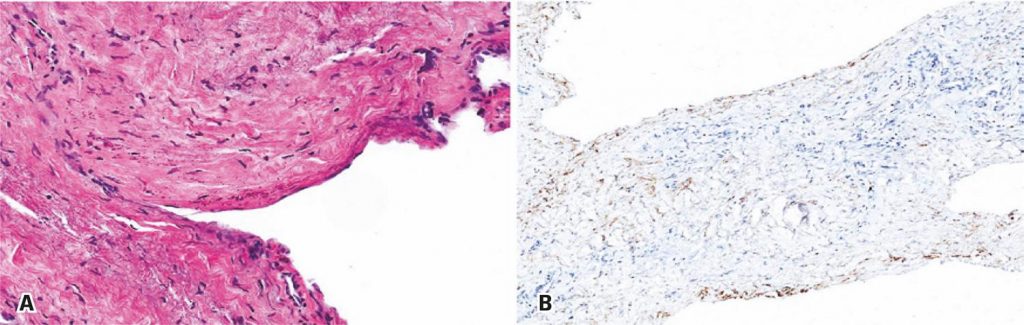
Magnetic resonance imaging identified a well-delineated, homogeneous, cystic sellar lesion with suprasellar extension and thin walls. The pituitary gland and stalk appeared to be stretched over the cyst boundaries and compressed against the dorsum sellae . No calcifications or solid areas were identified ( and ).
[…]
1,083