Phytochemical Analysis and Anticancer Activity of Seaweed Gracilaria Verrucosa Against Colorectal HCT-116 Cells
Kartika Dwi Kurniasari1, Ade Arsianti2,3 , Yully Astika Nugrahayning Aziza1, Baiq Kirana Dyahningrum Mandasari1, Riathul Masita1, Futihati Ruhama Zulfa1, Micheylla Kusumaning Dewi1,Cut Raisya Zahira Zagloel1, Norma Nur Azizah3 and Rista Putrianingsih2
1Medical Student, Faculty of Medicine University of Indonesia.
2Department of Medical Chemistry, Faculty of Medicine, University of Indonesia.
3Drug Development Research Cluster, Indonesia Medical Education and Research Institute (IMERI), Faculty of Medicine, University of Indonesia, Jalan Salemba Raya 6 Jakarta 10430, Indonesia.
Corresponding Author E-mail: arsi_ade2002@yahoo.com
DOI : http://dx.doi.org/10.13005/ojc/340308
Article Received on : April 03, 2018
Article Accepted on : May 21, 2018
Article Published : 04 Jun 2018
In this research, we develop Indonesia marine resource of seaweed Gracilaria verrucosa as an anti-colorectal cancer agent. Seaweed Gracilaria verrucosa which was collected from east Lombok beach, Nusa Tenggara Barat, Indonesia, were extracted into four different organic solvents, that is n-hexane, ethylacetate, chloroform and ethanol. The extracts were analyzed by Phytochemical test and Thin Layer Chromatography (TLC). Subsequently, anticancer activity of n-hexane, ethyl acetate, chloroform and ethanol extracts of Gracilaria verrucosa were evaluated against colorectal HCT-116 cells by MTT cell proliferation assay. Based on Phytochemical analysis, the extracts of Gracilaria verrucosa containing secondary metabolite of saponin, while TLC analysis indicated that the extracts were composed by five chemical compounds. Among four concentrated extracts of Gracilariaverrucosa, ethanolic extract showed the strongest anticancer activity against colorectal HCT-116 cells with IC50 of 43.9 μg/mL.Ethanolic extract of seaweed Gracilaria verrucosa is potential to be further developed as a promising anti-colorectal cancer agents.
KEYWORDS:Anticancer Activity; Colorectal HCT-116 Cells; Gracillaria Verrucosa; Phytochemistry
Download this article as:| Copy the following to cite this article: Kurniasari K. D, Arsianti A, Aziza Y. A. N, Mandasari B. K. D, Masita R, Zulfa F. R, Dewi M. K, Zagloel C. R. Z, Azizah N. N, Putrianingsih R. Phytochemical Analysis and Anticancer Activity of Seaweed Gracilaria Verrucosa Against Colorectal HCT-116 Cells. Orient J Chem 2018;34(3). |
| Copy the following to cite this URL: Kurniasari K. D, Arsianti A, Aziza Y. A. N, Mandasari B. K. D, Masita R, Zulfa F. R, Dewi M. K, Zagloel C. R. Z, Azizah N. N, Putrianingsih R. Phytochemical Analysis and Anticancer Activity of Seaweed Gracilaria Verrucosa Against Colorectal HCT-116 Cells. Orient J Chem 2018;34(3). Available from: http://www.orientjchem.org/?p=46489 |
Introduction
Colorectal cancer is one of malignant tumors which occurs in human’s colon. This cancer originates from adenoma polyp that develops into a carcinoma. Based on WHO record, colorectal cancer is the fourth biggest cancer in the world’s population. In Indonesia itself, colorectal cancer has the second position after the lung cancer. Based on data of Kementerian Kesehatan Indonesia, incidence of colorectal cancer is 1.8 per 100.000 population. For additional data, Dharmais Hospital in Jakarta-Indonesia, stated that colorectal cancer is sixth reason of man’s death and tenth reason of woman’s death. For the development of the cancer itself is still unknown.1
Asril Zahari on his book entitled “Early Detection, Diagnose and Treatment of Colorectal and Rectum Cancer”, stated that the patients of colorectal cancer are classified into three kind of groups which are familial, congenital and sporadic. This cancer can occur through two processes, LOH process and ROH process. LOH or known as Loss of Heterozygosity can develop from mutation of supressor genes such as APC genes. Meanwhile ROH (Retention of Heterozygosity) process can easily occur to the patient which has the mutation of germinativum cell HNPCC. There are some risk factors that will influence colorectal cancer, which are dietary pattern, tribe, lifestyle, history of familial cancer and history of polyp cases.2 National Cancer Institute has already declared that colorectal cancer can be treated in many ways, for example surgery, radiofrequency ablation, chemothrapy, radiation therapy and targeted therapy. Even if the treatment was done accordingly to the right procedure, there are so many complaints of the patient about relaps cases of their cancer. About 30-50% colorectal cancer patient are the relaps ones. To decrease the number of relaps case, ESMO Clinical Practice for diagnosis by Labianca stated that the patient of colorectal cancer that has been treated should follow the follow-up plans. The follow-up plans is usually done by using CEA or carcinoembryonic antigen and CT Scan. Not only the follow-up plans, the patient should also follow the survivorship care plans which consists of relap prevention method, rehabilitation process of the patient, side effect assesment, and health promotion to prevent relaps cases.3,4
In fact, the relaps cases occured caused by incompliance of the patient due to many side effects4, such as hemicolectomy and gastrointestinal dysfunction. Based on that reason, the researcher then thought about using some alternative medicine that will reduce the side effect. Macroalgae is one of natural resources that contains fiber, vitamin, mineral, fat, omega3 acid, protein, polisaccharides, essential amino acid and many others substances Not only that, macroalgae also know to have some activities which are useful for human’s helath such as anti-oxidant, anti-inflamation, anti-mirobial and anti-cancer.5
Gracilaria verrucosa is one of the species of Gracilaria sp. This macroalgae can be found in tropical or subtropical marine. Gracilaria verrucosa usually used for making jelly. But in fact, this species also produce bioactive substance and eicosanoid which is useful as anticancer agent.5Previous researcher reported that Gracilaria tenuispitata had an anti-cancer effect against oral cancer cells Ca9-22. Another previous experiment revealed that Gracilaria verrucosa can induce cell apoptosis by increasing γ-H2AX-based DNA double strand breakdown. That process would induce ROS to penetrate the cells via H2AX-Nox1/Rac1 pathway. Beside DNA breakdwon, Gracilaria tenuispitata can also reduce glutathione or GSH which caused ROS to easily produced in the cells. 6,7
Gracilaria verrucosa has a secondary metabolit of saponin, that is a substance which has aglycone hydrofobic structure with addtional part of sugar hyrofilic. This structure make saponin becomes sureface-active amphipathic compounds that has many benefits for its biological activity such as cell hemolytic. Francis et al revealed that aglycone hydrofobic of saponin make that substance becomes easier to be attached to the cholesterol of cell membrane. This attachment will induce pore formation and degradation of cell permeability. it will allows ROS to penetrates easily to the cells and it will induce apoptosis.6
As declared by NCBI, Gracilaria verrucosa did not have any toxic effect, analgesic effect and anti-convulsant effect for the patient. So it is assumed that Gracilaria verrucosa (Figure 1) is safe to be used as a new anti-cancer agent.
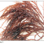 |
Figure 1 Click here to View figure |
Here is the taxonomy of Gracilaria verrucosa:8
Kingdom : Plantae
Subkingdom : Biliphyta
Divison : Rhodophyta
Subdivision : Eurhodophyta
Class : Florideophyceae
Subclass : Rhodymeniphycidae
Order : Gracilariales
Family : Gracilariceae
Genus : Gracilaria Grev.
Species : Gracilaria verrucosa
(Hudson) Papenfuss
The novelty of this study is to explore macroalgae Gracilaria verrucosa collected from Labuhan Haji beach, Lombok, Province of Nusa Tenggara Barat, Indonesia, as an anti-colorectal cancer against HCT 116 cells. This study will be conducted in several stages are as follows: the first stage is collection and identification of Gracilaria verrucosa. The second stage is extraction of Gracilaria verrucosa into four different polarity of organic solvents, that are n-hexane (non-polar) , chloroform (non-polar), ethylacetate (semipolar), and ethanol (polar). The third stage is phytochemical test and TLC (Thin Layer Chromatography) analysis of Gracilaria verrucosa extracts. The fourth stage is in vitro anticancer activity evaluation of n-hexane, chloroform, ethylacetate, and ethanol extracts of Gracilaria verrucosa against colorectal cancer cells HCT 116 by MTT assay.9,10
Experimental
Extraction and Fractionation Samples of Seaweeds
In this research, we use 1000 g dry powder of macroalgae Gracilaria verrucosa, which was macerated in ethyl acetate. Concentrated crude extract of ethylacetate of Gracillaria verrucosa was then fractioned by column chromatography on silica gel G60 (230-400 mesh ASTM) using non-polar solvent of chloroform and n-hexane, semi-polar solvent of ethylacetate, and polar solvent of ethanol, successively afforded n-hexane extract, chloroform extract, ethyl acetate extract and ethanol extract, respectively. Subsequently, these four kinds of Gracillaria verrucosa extract were analyzed using thin layer chromatography to identify the amount of chemical component containing in the extract, as well as to be subjected for phytochemical test to determine the secondary metabolites in four kinds extract of Gracillaria verrucosa. Furthermore, in vitro cytotoxic activities of those four extracts of Gracillaria verrucosa were evaluated against HCT 116 cells by MTT method.11
Phytochemistry Test
This method is done so that we can identify the secondary metabolite contained in concentrated fraction of macroalgae Gracilaria verrucosa. The procedure of phytochemical test are listed as following11:
Saponin Test
Saponin screening is done by using 10 mL solution placed in a tube; then is shaken for 10 seconds vertically before let it stand for 10 seconds. The presence of saponin is pointed out by a foam formation lasted more than 10 minutes which has 1-10 cm height.
Flavonoid Test
A total of 1 mL solution was evaporated until dry, then being moistened using acetone. A boric acid and oxalic acid powder then was added into the moistening dried-solution placed over a hot water; be careful not to be overheating the solution. The residual of the mixture then mingled together with 10 mL eter; afterward was identified under ultraviolet ray of 366nm. The presence of flavonoid is indicated by the presence of yellow fluorescence.
Triterpenoid and Steroid Test
The reaction used to indicate the presence of triterpenoid and steroid in Liebermann Burchard. This test used 2 mL solution that was evaporated in a procelain cup. The residual of evaporation process then was dissolved in 0.5 mL chloroform. Subsequently, we added acetic acid anhyride and 2 mL concentrated sulfuric acid through the wall of tube. The presence of triterpenoid was indicated by the formation of brownish or violet ring at the boundary of solution, meanwhile the presence of steroid was indicated by the formation of blue-greenish ring.
Alkaloid Test
A solution of 2 mL was placed in a porcelain cup to be evaporated until there was a residue; later mixed with 5 mL of 2 N-HCl. Later on, the mixture were divided into three tubes; the first one was a blanko which was mixed with HCl 2 N, the second tube was mixed with 3 drops of Dragendorff agent, and lastly we added 3 drops ofMayer reagents onto the third tube. The formation of an orange precipitate in the second tue and yellow precipitate in the third tube showed the presence of alkaloid.
Tannin Test
This procedure was conducted by reacting 1 mL solution with 10% iron (III) chloride solution. The formation of dark-blue or greenish black precipitation indicated the presence of tannin.
Glycosides Test
A total of 0.1 mL solution was evaporated in a water bath; afterward was dissolved with 5 mL acetic acid anhydride. Then, the solution was mixed with 10 drops of concentrated sulfuric acid. A bluish or greenish product indicated a glycoside presence.
Thin Layer Chromatography
Thin layer chromatography (TLC) is a procedure to identify the amount of substances contained in the extract sample, which is expressed by Rf values. This process was began by preparing TLC plate with size of 5cm x 20 cm x 0.2 mm. The extract sample was then applied on the plates by capillary pipe. The plate and mixture of solvent (CHCl3 :CH3OH = 3:1) as a mobile phase was added into the TLC chamber. The mobile phase will diffuse and move along the plate. After this process was complete, the plate were placed under ultraviolet lamp with wavelenght of 254 nm and 366 nm in order to visualize the spots of chemical component, and determine the Rf value of each spot.12,13
In Vitro Cytotoxicity Assay
Gracilaria verrucosa has a cytotoxic effect which inhibit the proliferation of HCT 116 cells. That effect can be evaluated by using MTT assay. We diluted the sample extract; later was added to target cells with triple methode in eight final concentration which were 200, 100, 50, 25, 12.5, 6.25, 3.125, and 1.5625 µg/ml, consecutively. The cells, later, were incubated for 48 hours, before was was mixed with 20 µg of 5 mg/mL solution of MTT in phosphate-buffered saline. The plates were incubated for 4 hours more; afterwards were centrifuged while the medium was removed. 200 µL of dimethyl sulphoxide was added in each well to dissolve the purple-blue sediment. Later, we identified the absorbance of the plates using 590nm microplate reader (Model 550, Bio Rad, USA). The inhibiton rate was calculated using this following formula.11
![]()
While the inhibitory concentration that diminished 50% of cancer cells, we called as IC50, are calculated with assay.
Result and Discussion
Phytochemical Composition
Phytochemistry test of Gracilaria verrucosa is displyed in Table 1, whic indicated that Gracilaria verrucosa extracts contained secondary metabolites of saponin and triterpenoid. Saponin was composed by hydrophobic aglycone with additional functional hydrofilic-sugar part. Because of this structure, saponin could do its biological function easily such as pore formation and degradation of cell permeability. The aglycone component in Gracilaria verrucosa consist of two structures which are steroid and triterpenoid. The steroid structure composed by six membered-ring of spirostane and five membered-ring of furostane, meanwhile triterpenoid consists of four and five membered-ring of olenan. Those structures made saponin can easily be engaged to membrane cholesterol; causing membrane distabilisation, furthermore the cell membrane get ruptured. Besides anti-cytotoxic effect, saponin in Gracilaria verrucosa has some other functions such as immune stimulator, anti-microbial agents, anti-protozoa, and anti-oxidant.14Besides saponin, Gracilaria verrucosa was also identified to have triterpenoid substances derived from isoprene unit through biosynthetic process, MVA pathway and catalase oxidative. Both triterpenoid and saponin seem to have the same effect to induce cell apoptosis process in HT 116 cells.
Thin layer Chromatography
TLC results of extracts of Gracilaria verrucosa is summarized in Table 2. As shown, Rf value of Gracilaria verrucosa extract in n-hexane, ethylacetate and chloroform has no signficant difference, which means that the secondary metabolit of these three solvents are similar. Meanwhile, the difference of Rf value was observed in ethanolic extract of Gracilaria verrucosa which indicated the difference secondary metabolit components.
Table 1: Phytochemicals analysis of Gracilaria verrucosa
| Secondary metabolites | Gracilaria verrucosa extract | ||||
| n-Hexane | Etyl Acetate | Chloroform | Ethanol | ||
| Saponin | + | + | + | + | |
| Flavonoid | – | – | – | – | |
| Triterpenoid | + | + | + | + | |
| Steroid | – | – | – | – | |
| Alkaloid | – | – | – | – | |
| Tannin | – | – | – | – | |
| Glycosides | – | – | – | – |
Tabel 2: TLC analysis and Retention factor (Rf) value of Gracilaria verrucosa extracts
| Extract |
Retention (Rf) value |
||||||
| Spot 1 | Spot 2 | Spot 3 | Spot 4 | Spot 5 | Spot 6 | Spot 7 | |
|
Ethanol |
0.4 |
0.7 |
|||||
|
n-hexane |
0.95 |
1.1 |
1.5 |
1.7 |
2 |
2.3 |
|
|
Chloroform |
1 |
1.1 |
1.3 |
1.7 |
2 |
2.3 |
2.7 |
|
Ethyl acetate |
0.9 |
1.1 |
1.8 |
2 |
2.2 |
2.7 |
2.7 |
*Rf =Retention factor
Anticancer Activity of Macrolagae Gracilaria Verrucosa
Cytotoxic effect of Gracilaria verrucosa extract as an anti-colorectal cancer agent is showed by its inhibitory acitivity and IC50 value on HCT 116 cells. The stronger inhibitory activity and the lower IC50 value indicated the higher anti-colorectal cancer activity of Gracilaria verrucosa against HT 116 cells. Table 3 summarizes anticancer activity of Gracilaria verrucosa extracts. As shown, inhibitory activity of Gracilaria verrucosa extracts will gradually increase after concentration of 6.25 μg/mL. Overall, the percentage of inhibition of Gracilaria verrucosa extracts varies from 21.2% to 68.4% .
Besides the percentage of inhibition, the anti-cancer activity of the extract is also represented by IC50 value. IC50 value below than 100 μg/mL indicates a good anticancer activity, meanwhile IC50 more than 300 μg/mL indicated no anticancer activity of the extract. The value of IC50 between 100 to 300 μg/mL indicates that the extract has a weak anticancer activity. In this research, n-hexane, ethylacetate, chloroform, and ethanol (alcohol) extracts have IC50 value of 49.9, 44.5, 44.9, 43.9 μg/mL, respectively. Therefore, all extracts of Gracilaria verrucosa have a good anti-colorectal cancer agents against HCT 116 cells. Figure 2 displays the percentage of inhibition (inhibition rate) which is proportional to the concentration of the extracts in μg/mL. It means, all extracts of Gracilaria verrucosa are concentration-dependent in terms of inhibiting the proliferation of HCT-116 cancer cells.
Table 3: Anticancer activity (IC50 value) of Gracilaria verrucosa extracts against colorectal HCT 116 cells
|
Tested extract |
IC50 (μg/mL) |
|
n-hexane |
49.9 |
|
Ethyl acetate |
44.5 |
|
Chloroform |
44.9 |
|
Ethanol |
43.9 |
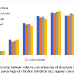 |
Figure 2: The relationship between relative concentrations of Gracilaria verrucosa extracts and their percentage of inhibition (inhibition rate) against colorectal HCT 116 cells. Click here to View figure |
Result findings in this research are similar and supported by previous research conducted by De Almeida and co-workers (2011) and Fitri (2015) that reported Gracilaria verrucosa showed anticancer activity and containing saponin which function to induce apoptosis of cancer cells.5,6
Conclusion
Seaweed Gracilaria verrucosa originated from Labuhan Aji beach, Lombok, Nusa Tenggara Barat, Indonesia, demonstrated anticancer activity against HCT 116 cancer cells (IC50 value: 43.9 to 49.9 μg/mL), which is potential to be developed as promising new anticolorectal cancer agent.
Acknowledgments
We wish to express our gratitute to to Directorate of research and public service University of Indonesia for PITTA (PublikasiInternasionalTerindeksUntukTugasAkhirMahasiswa) research grant for fiscal year 2017.
References
- PELNI Hospital. Kanker Colorectal, Peran Endoskopi Untuk Deteksi Dini [Internet]. Jakarta: RS Pelni. [cited 19 June 2016]. Available from: http://www.rspelni. co.id/?page_id=216.
- Zahari, A. Deteksi dini, Diagnosa dan Penatalaksanaan Kanker Kolon dan Rektum. Padang :Majalah Kedokteran Andalas. 2012.
- Colon Cancer Treatment [Internet]. Bethesda: National Cancer Institute. [updated June 30, 2016; cited 19 June 2016]. Available from: http://www.cancer.gov/types/colorectal/patient/colon-treatment-pdq.
- Labianca, R.; Nordlinger, B.; Beretta, G.; Mosconi, S.; Mandala, M.; Cervantes, A.; Arnold, D. Ann. Oncol. 2013, 24, 64-72.
CrossRef - De Almeida, C.L.F.; Falcão, H.S.; Lima, G.R.M.; Montenegro, C.A.; Lira, N.S.; De Athayde-Filho, P.F.; Souza, L.M.F.; Barbosa-Filho, J.M.; Batista, L.M. Int. J. Mol. Sci. 2011, 12, 4550-4573.
CrossRef - Fitria, W. F. Procedia Chemistry. 2015, 16, 407-412.
CrossRef - Yeh, C.; Yang, J.I.; Lee, J.C; Tseng, C.N.; Chan, Y.C.; Hseu, Y.C.; Tang, J.Y.; Chuang, L.Y.; Huang, H.W.; Chang, F.R.; Chang, H.W. BMC Complementary and Alternative Medicine. 2012, 12, 142-147.
CrossRef - ITIS Standard Report Page: Gracilaria verrucosa [Internet]. USA: Integrated Taxonomic Information System on-line database. 1996 [cited 20 June 2016]. Available from: http://www.itis.gov/servlet/SingleRpt/SingleRpt?search_topic=TSN&search_value=11985.
- Junedy, S.; Hermawan, A.; Ikawati, M. Uji sitotoksik metode MTT. Cancer Chemoprevention Research Center Faculty of Pharmacy, Gadjah Mada University. 2010.
- Depkes RI. Materia medika Indonesia. 5th ed. Jakarta: Departemen Kesehatan Republik Indonesia. 1989, 549-553.
- Arsianti, A.; Fadilah, F.; Yazid, F.; Suid, K.; Wibisono, L.K.; Azizah, N. A.; Putrianingsih, R.; Murniasih, T.; Rasyid, A.; Pangestuti, R. Asian J. Pharm. Clin. Res. 2016, 9, 116-119.
- Kromatografi Lapis (an) Tipis (KLT) [internet]. Yogyakarta : UGM [cited 1 Agustus 2017]. Available from : http://elisa.ugm.ac.id/user/archive/download/24048/a877915a150aeace10a
- Swieboda, R.; Jozwiak, A.; Joswiak, G.; Hajnos, M. Am. J. Anal. Chem. 2015, 5, 1109-1120.
CrossRef - Podolak, I.; Galanty, A.; Sabolewska, D. Phytochem. Rev. 2010, 9, 425-474.
CrossRef

This work is licensed under a Creative Commons Attribution 4.0 International License.
![]()
A New Edition of Web of Science
Journal Impact Factor
2022: 0.5
Five Year: 0.8
Journal is Indexed in

Cabells Whitelist
![]()


