Manuscript accepted on : 15-06-2021
Published online on: 20-07-2021
Plagiarism Check: Yes
Reviewed by: Dr. Yamini Tiwari
Second Review by: Dr. Salim Albukhaty
Final Approval by: Dr. Majid Sakhi Jabir
Turki M. Dawoud1 , Fatimah Alshehrei2
 , Khaizran Siddiqui3
 , Fuad Ameen1*, Jameela Akhtar3 and Afsheen Arif4
1Department of Botany and Microbiology, College of Science, King Saud University, Riyadh11451, Saudi Arabia.
2Department of Biology, Jumum college university, Umm Al-Qura University, P.O Box 7388, Makkah 21955, Saudi Arabia.
3Center of Molecular Genetics, University of Karachi, Pakistan.
4Karachi Institute of Biotechnology and Genetic Engineering (KIBGE), University of Karachi, Karachi, Pakistan.
Corresponding Author E-mail: fuadameen@ksu.edu.sa
DOI : http://dx.doi.org/10.13005/bbra/2915
ABSTRACT: Background: The wide use of dextran in many different applications, makes its industrial production a challenge and, hence, to obtain a control branched structure of this enzyme research is in progress. Objectives: In the present paper, the enzyme dextransucrase, produced by cultivation of the bacterium Leuconostoc mesenteroides CMG713, was purified and characterized. Methods: The produced dextransucrase was partially purified by PEG400 obtaining a purification factor of 29.4-fold and an overall yield of 18.3% from the initial crude enzymatic extract. Results: The partially purified dextransucrase had a specific activity of 24.0 U/mg and presented a molecular weight of about 200 kDa. In addition, the produced dextransucrase was stable at 30ºC and pH 5.5 for 3 days and led to a highly soluble dextran with wide potential industrial applications. The current study has successfully partial purification, characterization and conformation of dextransucrase produced by fermentation of the bacterium Leuconostoc mesenteroides CMG713.
KEYWORDS: Dextran; Dextransucrase; Leuconostoc m esenteroides
Download this article as:| Copy the following to cite this article: Dawoud T. M, Alshehrei F, Siddiqui K, Ameen F, Akhtar J, Arif A. Purification, Characterization and N-terminal Protein Sequencing of the Enzyme Dextransucrase Produced by Leuconostoc mesenteroides. Biosci Biotech Res Asia 2021;18(2) |
| Copy the following to cite this URL: Dawoud T. M, Alshehrei F, Siddiqui K, Ameen F, Akhtar J, Arif A. Purification, Characterization and N-terminal Protein Sequencing of the Enzyme Dextransucrase Produced by Leuconostoc mesenteroides. Biosci Biotech Res Asia 2021;18(2). Available from: https://bit.ly/3zsPgPP |
Introduction
Dextransucrase (sucrose: 1, 6-α-d-glucan 6-α-glucosyltransferase EC 2.4.1.5) is an extracellular enzyme that catalyzes the formation of dextran from sucrose by the polymerization of glucosyl moieties1,2. Generally, this enzyme is produced by lactic acid bacteria (LAB) such as Leuconostoc mesenteroides strains and Streptococcus sp., which are Gram positive cocci bacteria3,4. However, L. mesenteroides needs the presence of sucrose in its culture medium to produce dextransucrase enzymes whereas Streptoccoccus sp. as this genus is constitutive for dextransucrases5.
Dextran is a biodegradable glucose linear polymer consisting mainly of 1,6-α-d-glucosidic linkages as a backbone and some α-1,2, α-1,3 and α-1,4 as branching links6,7. The link frequency and type as well as its physical and chemical characteristics depend on the enzyme’s nature and microorganism’s type4,8. The different characteristics and properties of the purified and produced dextran will be used accordingly [9–11]. In this sense, Sarwat el al. 2 found that the strain Leuconostoc mesenteroides CMG713 produced a high molecular weight linear dextran with no branches. This dextran may present high solubility in water since low dextran solubility in water is related to a high amount of α-1,3 links[4]. Consequently, such a strain was selected to perform the present research. Given the industrial importance of dextran, monitoring the activity of dextransucrase enzymes to produce new dextran products with mastered characteristics is appalling 1,2, 6. Therefore, the aim of the present study was the partial purification, characterization and conformation of dextransucrase produced by fermentation of the bacterium Leuconostoc mesenteroides CMG713. To the author´s knowledge the dextransucrase produced by the above-mentioned strain has been hardly investigated.
Materials and Methods
Microorganism
Leuconostoc mesenteroides CMG713 was previously isolated and identified at the University of Karachi (Pakistan)[2]. The bacterium was maintained in modified MRS (yeast extract 0.4%; glucose 2.0%; sodium acetate trihydrate 0.5 %; Tween 80 0.1%) agar medium at 4ºC until used.
Dextransucrase production
Sterile mineral salt medium containing sucrose was prepared according to3 with minor modifications (0.18 g of MgSO4. 7H2O and 0.08 g NaCl were used instead of 0.2 g and 0.1g, respectively). Wire loops of slime producing colonies of L. mesenteroides were first used to inoculate (one loop per tube) test tubes containing 10 mL of MSM (mineral salt medium) broth and incubated at 30˚C for 20 h in the dark. To produce dextransucrase, the broth (final concentration 1%) was further transferred to 250-mL conical flasks containing 90 mL of fresh MSM broth and incubated as above. Then, the culture broth was collected and centrifuged at 10,000 rpm for 20 min at 4°C. The cell-free supernatant, containing the extra-cellular crude enzyme, was stored at -20˚C for further analysis.
Enzyme assay
Hydrolytic activity was determined by measuring the reducing sugars using the dinitrosalicylic acid (DNS) method[12]. Enzyme activity was expressed as dextransucrase units (DSU/mL/h) defined as the enzyme quantity that converts 1.0 mg of sucrose into fructose in 1 h under the standard assay conditions (30ºC, pH 5) against a blank.
The total protein content was measured spectroscopically at 560 nm by the method of Bradford 13 using bovine serum albumin (BSA) as a standard.
Enzyme purification and SDS-PAGE
The cell-free supernatant containing the extracellular enzyme produced by L. mesenteroides CMG713 was first purified by polyethylene glycol (PEG 400) fractionation as described by[14]. Briefly, different percentages of ice cold PEG 400 were added to 50 mL of cell-free supernatant in the range of 25-50%. They were incubated at 4 °C for 12 h. The mixture was centrifuged at 13 200×g for 30 min at 4 °C to separate the fractionated dextransucrase.
The molecular weight was determined using sodium dodecyl sulfate polyacrylamide gel electrophoresis (SDS-PAGE) according to[15–19].The resolving gel was prepared using 7% (w/v) acrylamide and the stacking gel using 4% (w/v) acrylamide. The loading buffer composition was Tris–HCl buffer (0.0625 M; pH 6.8), SDS (2.3% w/v), glycerol (10% w/v), 2- mercaptoethanol (5% w/v) and bromophenol blue (0.05% w/v). The partially-purified enzyme samples (fraction 50%) were boiled at 100ºC for 5 min and loaded onto the gel. The running buffer consisted of 0.56% glycine, 0.12% Tris-HCl buffer (1 M; pH 8.3) and 0.5% SDS. The electrophoresis was carried out with a current of 2 mA per lane. The protein bands were fixed with 40% ethanol and 10% acetic acid and then stained with 0.1% silver nitrate in 0.02% formaldehyde solution. The reaction was stopped by adding 1.46% disodium salt of ethylene diammine-tetra acetic acid and the protein bands were developed by adding 2.5% sodium carbonate in 0.01% formaldehyde solution. The molecular weight was determined with standard marker proteins (Promega Corporation, USA).
Identification of dextransucrase by activity staining
The identification of the dextransucrase enzyme by activity staining was conducted by electrophoresis (1.5 mm thick gels) following the method of Miller et al.20. After the electrophoresis, the gel was cut into half equal parts and both parts were subjected to activity staining. For the removal of SDS from the gel firstly, the gel was treated thrice with a solution containing 20 mM sodium acetate buffer (pH 5.4), 0.1% Triton-X-100 and 0.005% calcium chloride for 20 min each time. After the removal of SDS, one part of the gel was incubated with 5% raffinose solution in 20 mM sodium acetate buffer (pH=5.4) and the other part with 10% sucrose solution in 20 mM sodium acetate buffer (pH=5.4) for 10–12 h, After incubation, the gels were washed twice with 75% ethanol for 20 min each time and incubated in a solution consisting of 0.7% periodic acid in 5% acetic acid for 20 min at room temperature. The gels were then washed thrice with a solution of 0.2% sodium bisulfate in 5% acetic acid and finally stained with the Schiff’s reagent (0.5% w/v basic fuschin, 1% sodium bisulfite and 0.1 N HCl) until discrete magenta bands appeared.
For in situ detection of the dextransucrase activity, after electrophoresis the gel was washed thrice with sodium acetate buffer (20 mM, pH 5.4) containing 0.05 g of CaCl2 and 0.1% of Triton X-100 to remove the SDS. Then, the gel was incubated in the same buffer containing 100 g/L of sucrose at 30°C for 72 h. The active bands were detected by the formation of dextran as a white polymer inside the gel[21].
Electroblotting
The band showing dextransucrase activity was blotted onto a polyvinylidene diflouride membrane (PVDF) membrane using a semi-dry blotting device. After blotting, the transfer was performed by the method described by22. The membrane and the gel were removed, and the membrane was rinsed with deionized water. The PVDF membrane was soaked in methanol for few seconds before transferring it. The membrane was stained with Coomassie R-250 for no longer than one min. It was further distained in 50% methanol several times and finally rinsed with deionized water. The band of interest was cut and stored at 4°C for further analysis.
N-Terminal protein sequencing
The obtained protein band of 200 kDa was excised from the gel and sent for the N-terminal protein sequence determination (Proteome Factory AG, Berlin, Germany) using ABI procise491 protein sequencer. The obtained sequence was submitted to UniprotKB KB http:// www.uniprot.org and EMBL-EBI http://www.ebi.ac.uk. The sequence was analyzed using the Multiple sequence Alignment tool ClustalW2[23] that uses tree-based progressive alignments and can incorporate secondary structure information into the process.
General characteristics of dextran and partial characterization of dextransucrase
The dextran samples were analyzed for general characteristics like colour, texture and smell. For this, samples with different concentrations (i.e. 0.5, 1, 1.5 and 2 mg/mL) were used.
To determine the thermal stability of dextransucrase, 50 μL of the partially purified enzyme was incubated in a solution of 10% sucrose in 20 mM sodium acetate buffer (pH 5.4) at temperatures ranging from 20 to 60°C for 15 and 30 min. After each incubation time, the enzyme activity was determined using the DNS method as indicated previously.
For the pH stability, 50 μL of the partially purified enzyme was incubated in a solution of 10% sucrose in 20 mM sodium acetate buffer at pHs ranging from 3.5 to 6.5 for 15 min at 30ºC. After this time, the enzyme activity was determined as indicated previously.
Results
Dextransucrase production and partial purification
The dextransucrase activity of the crude enzyme before and after partial purification is shown in Table 1. Thus, before purification the dextransucrase activity was 167.5 U/mL (specific activity 20.42 U/mg) and after PEG400 purification, the dextransucrase activity was 220.5 U/mL (specific activity 23.96 U/mg). The purification fold was 29.4 with an overall yield of 18.9%.
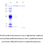
After SDS-PAGE of the partially purified dextransucrase, three protein bands were seen on the gel. Zymography indicated that the most intense coloured band of the partially purified dextransucrase was close to that of the standard strain L. mesenteroides NRRL-512F band. Therefore, the molecular weight of the obtained L. mesenteroides dextrosucrase was considered to be around 200 kDa (Figure 1).
Table 1: Purification of dextransucrase by PEG400
| PEG 400 (%) | Volume (mL) | Enzyme activity (U/mL) | Specific activity
(U/mg) |
Total units | Overall % yield | Protein (mg/mL) | Purification fold |
| Crude | 25 | 167.5 | 20.4 | 116 | 8.2 | ||
| Partially purified enzyme | |||||||
| 25% | 1.7 | 1.7 | 1.8 | 3.5 | 4.9 | 0.9 | 5.2 |
| 30% | 2.6 | 3.5 | 2.5 | 6.2 | 6.8 | 1.4 | 15.5 |
| 35% | 2.9 | 15.4 | 4.6 | 8.1 | 7.3 | 3.3 | 20.4 |
| 40% | 3.2 | 54.7 | 11.2 | 10.8 | 9.2 | 4.9 | 25.2 |
| 45% | 3.8 | 105.5 | 17.1 | 11.7 | 10.2 | 6.2 | 27.4 |
| 50% | 4.7 | 220.5 | 24.0 | 25.8 | 18.3 | 9.2 | 29.4 |
In situ detection of dextransucrase
The enzyme was confirmed to be dextransucrase by Schiff’s reagent staining. Thus, a bright magenta color band appeared in the gel incubated with sucrose whereas no band was detected in the gel incubated with raffinose.
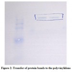
In situ electrophoresis was performed to distinguish the protein bands with dextransucrase activity and, thus, capable of synthesizing dextran from sucrose. After electrophoresis, an active white band of dextransucrase was detected after incubating the gel at 30°C for 18 h in the presence of sucrose confirming that the enzyme was active (Figure 2).
 |
Figure 2: Transfer of protein bands to the polyvinylidene difluoride (PVDF) membrane after SDS-PAGE. |
Electroblotting
An active band was observed after blotting the partially purified enzyme onto a polyvinylidene difluoride (PVDF) membrane using a semi-dry blotting device. The membrane was stained with Coomassie R-250 and distained in 50% methanol. The band of dextransucrase was excised from the gel and stored at 4°C for further analysis.
N-terminal sequencing analysis
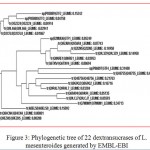
For this study, all the 22 dextransucrases from L. mesenteroides were selected and aligned using the alignment tool present on the UniprotKB (http://www.uniprot.org/align) (Table 2). In Figure 3 the multiple sequence alignment of the 22 dextransucrases from Leuconostoc mesenteroides is shown. The N-terminal amino acid sequencing analysis of the partially purified dextransucrase revealed that the first five amino acids were Asp-Ser-Thr-Asn-Tyr (D-S-T-N-Y) and the N-terminal molecular mass was 599 Da. UniProtKB P85089 was the accession number of the sequences and only nine dextransucrases (DsrF) were already reported from L. mesenteroides (Table 3a and 3b). The first four amino acids (i.e. D-S-T-N) were only found in four UniprotKB ID O52224, Q9L466, D2CFL0 and P85080 sequences. In addition, EBI database indicated that P85089 had an 80% homology with O52224, Q9L466, D2CFL0 and P85080 (Table 4).
 |
Figure 3: Phylogenetic tree of 22 dextransucrases of L. mesenteroides generated by EMBL-EBI. |
Table 2: Results for dextransucrase of Leuconostoc mesenteroides in UniProt KB databank sorted by descending score.
| Accession | Entry name | Protein names | Gene names | Organism | Length
(bp) |
| P85089 | GTF2_LEUME | Dextransucrase 2
Dextransucrase 2 (EC 2.4.1.5) (Dextransucrase DsrF) (Glucansucrase 2) (Sucrose 6-glucosyltransferase 2) |
dsrF | Leuconostoc mesenteroides | 5 |
| B2MUU6 | GTF1_LEUME | Dextransucrase 1
Dextransucrase 1 (EC 2.4.1.5) (Glucansucrase 1) (Sucrose 6-glucosyltransferase 1) |
dsrF | Leuconostoc mesenteroides | 284 |
| P86897 | GTF4_LEUME | Dextransucrase
Dextransucrase (EC 2.4.1.5) (Glucansucrase) (Sucrose 6-glucosyltransferase) |
— | Leuconostoc mesenteroides | 9 |
| P85080 | GTF3_LEUME | Dextransucrase
Dextransucrase (EC 2.4.1.5) (Glucansucrase) (Sucrose 6-glucosyltransferase) |
— | Leuconostoc mesenteroides | 6 |
| Q9L466 | Q9L466_LEUME | Dextransucrase
Dextransucrase (EC 2.4.1.5) (Mutant dextransucrase) |
dsrC dsrb742 | Leuconostoc mesenteroides | 1,477 |
| Q9ZAR4 | Q9ZAR4_LEUME | Dextransucrase
Dextransucrase |
DEX | Leuconostoc mesenteroides | 1,527 |
| Q48756 | Q48756_LEUME | Dextransucrase
Dextransucrase |
— | Leuconostoc mesenteroides | 1,290 |
| D2CFL0 | D2CFL0_LEUME | Dextransucrase
Dextransucrase |
dsrBCB4 | Leuconostoc mesenteroides | 1,505 |
| Q69A94 | Q69A94_LEUME | Dextransucrase
Dextransucrase (EC 2.4.1.5) |
dsrP | Leuconostoc mesenteroides | 1,454 |
| Q6TXV4 | Q6TXV4_LEUME | Dextransucrase
Dextransucrase (EC 2.4.1.5) |
dsrX | Leuconostoc mesenteroides | 1,522 |
| A0ELS0 | A0ELS0_LEUME | Dextransucrase
Dextransucrase |
— | Leuconostoc mesenteroides subsp. mesenteroides | 230 |
| Q8KRE1 | Q8KRE1_LEUME | Dextransucrase DsrD
Dextransucrase DsrD (EC 2.4.1.5) |
dsrD | Leuconostoc mesenteroides | 1,527 |
| Q8G9Q2 | Q8G9Q2_LEUME | Dextransucrase
Dextransucrase (EC 2.4.1.5) |
dsrE | Leuconostoc mesenteroides | 2,835 |
| Q84CN4 | Q84CN4_LEUME | Dextransucrase DsrR
Dextransucrase DsrR (EC 2.4.1.5) |
dsrR | Leuconostoc mesenteroides | 1,330 |
| Q9LCJ7 | Q9LCJ7_LEUME | Dextransucrase
Dextransucrase |
dsrT | Leuconostoc mesenteroides | 1,016 |
| Q9EZH5 | Q9EZH5_LEUME | Dextransucrase Dsrb742
Dextransucrase Dsrb742 |
dsrb742 | Leuconostoc mesenteroides | 1,508 |
| Q2I2N5 | Q2I2N5_LEUME | Dextransucrase
Dextransucrase (EC 2.4.1.5) |
dexYG | Leuconostoc mesenteroides | 1,527 |
| Q7M0M1 | Q7M0M1_LEUME | Dextransucrase
Dextransucrase (EC 2.4.1.5) |
— | Leuconostoc mesenteroides | 56 |
| Q9R4L7 | Q9R4L7_LEUME | Dextransucrase
Dextransucrase (EC 2.4.1.5) |
— | Leuconostoc mesenteroides | 20 |
| O52224 | O52224_LEUME | Glucosyltransferase
Glucosyltransferase (EC 2.4.1.5) |
dsrB | Leuconostoc mesenteroides | 1,508 |
| Q9L3Z9 | Q9L3Z9_LEUME | Dsr S protein
Dsr S protein |
dsr S | Leuconostoc mesenteroides | 66 |
| Q48755 | Q48755_LEUME | Putative uncharacterized protein
Putative uncharacterized protein |
— | Leuconostoc mesenteroides | 98 |
Table 3a: ClustalW2 multiple sequence alignment of 10 dextransucrase enzymes of Leuconostoc mesenteroides showing the homology to the N-terminal sequence of dextransucrase DsrF (P85089). The match peptides are highlighted in yellow.
| 1 ————— ———————————————————————————-DSTNY——— | 5 P85089
|
| 1 —— —————————————– ————————————————–DSTNTV——-
|
6 P85080
7 |
| 1 —– ——-MFMIKERNVRKKLYKSGKSWVIGGLILSTIMLSMTATSQNVNADSTNTVTDKS | 53 O52224
|
| 1———————————- ————————————-MLSMTATSQNVNADSTNTVTDKS | 23 Q9L466
|
| 1——————MIKERNVRKKLYKSGKSWVIGGLILSTIMLSMTATSQNVNADSTNTVTDKS
|
51 D2CFL0 |
| 1 – ————MFMIKERNVRKKLYKSGKSWVIGGLILSTIMLSMTATSQNVNAPSTNTVTDKS | 53 Q9EZH5
|
| 1———– —————– —————————————-MLIKERNVRITNSGDPNSGNAVTG | 24 Q84CN4
|
| 1 MRDMRVICDRKKLYKSGKVLVTAGIFALMMFGVTTASVSANTIAVDTNHSRTSAQINKS
|
59 Q8G9Q2 |
| 1 MRNRNVTSVFRKKMYKSGKMLVIAG–SVSIIGVTSFIQQAQADVSQKNGVVVTTAVNQS | 58 Q69A94 |
| 1————— ————-MYKSGKMLVIAG–SVSIIGVTSFIQQAQADVSQNNGVVVATAVDQS | 45 Q9LCJ7
|
Table 3b: Details of match N- terminal sequences of the 10 dextransucrase enzymes highlighted above.
| Accession | Organism and gene name | Reference | Sequence |
| P85089 | L. mesenteroides CMG713, dsrF | Current study | DSTNY |
| P85080 | L. mesenteroides AA1 | Aman et al., 2007 | DSTNTV |
| O52224 | L. mesenteroides NRRL B-1299, dsrB | Monchois et al., 1998 | DSTNTVTDKS |
| Q9L466 | L. mesenteroides NRRL B-1355 dsrC,dsrb742 | Arguello-Morales et al., 1999 | DSTNTVTDKS |
| D2CFL0 | L. mesenteroides NRRL B-1299CB4 dsrBCB4 | Kang et al.,2006 | DSTNTVTDKS |
| Q9EZH5 | L. mesenteroides B-742CB, dsrb742 | Kim et al.,2000 ) | PSTNTVTDKS |
| Q84CN4 | L. mesenteroides
NRRL-1501, dsrR |
Kim et al.,2002 | NSGNAVTG– |
| Q8G9Q2 | L. mesenteroides
NRRL B-1299 dsrE |
Bozonnet et al.,2002 | SRTSAQINKS |
| Q69A94 | L. mesenteroides IBT-PQ, dsrP | Olvera.et al.,2007 | VVVTTAVNQS |
| Q9LCJ7 | L. mesenteroides NRRL B-512F, dsrT | Funane et al.,2000 |
VVVATAVDQS |
Table 4: Length and molecular mass (Da) of dextransucrase.
| SeqB Name | Length (bp) | Mass (Da) |
| sp|P85089|GTF2_LEUME | 5 | 599 |
| sp|P85080|GTF3_LEUME | 6 | 636 |
| tr|Q9L466|Q9L466_LEUME | 1477 | 164,887 |
| tr|, |D2CFL0_LEUME | 1505 | 168,088 |
| tr|O52224|O52224_LEUME | 1508 | 168,511 |
General characteristics of dextran and partial characterization of dextransucrase
The dextran produced by L. masenteroides dextransucrase presented various characteristics of standard dextran such as colour, smell, texture and solubility. Thus, it was white in colour, had no specific smell, a powdery texture and its solubility in water and ethanol was 5% (Table 5).
On the other hand, the produced L. masenteroides dextransucrase was stable at 30oC and a pH of 5.5 for 3 days. In addition, the results illustrated that the enzyme produced was dextransucrase and not another enzyme.
Table 5: General characteristics of the produced dextran from Leuconostoc mesenteroides CMG713 dextransucrase.
| Characteristics | Dextransucrase |
| Color | White |
| Smell | Odorless |
| Texture | Powder |
| Solubility
(water & ethanol) |
5% |
Discussion
Exopolysaccharide dextrans from bacteria present better biodegradability and biocompatibility than those obtained from animals, plants and seaweeds. Dextrans differ in their glycosidic linkages, degree and/or type of branching, molecular mass and physical and chemical features depending on the producing bacterial strain14. Thus, as commented in the introduction section, each dextran is appropriate for different industries and applications depending on its characteristics. Consequently, it is essential to explore and identify dextransucrases produced by novel strains to obtain different types of dextran for multiple applications. The results obtained in the present study showed that L. mesenteroides CMG713 produced the enzyme dextransucrase. The partially purified enzyme showed a molecular weight of 200 kDa. This result is similar to that found by24 for a dextransucrase from L. dextranicum NRRL-B-1146, The molecular weight of most L. mesenteroides dextransucrases was reported to be around 180 kDa10,25,26. However, Florez-Guzman et al. [27] reported a molecular weight slightly lower (i.e. 170.1 kDa) for a dextransucrase produced by the strain L. mesenteroides IBUN 91.2.98. In addition, the purified enzyme remained stable at 30oC and a pH of 5.5 for 3 days.
On the other hand, the produced L. masenteroides dextransucrase was able to produce dextran which presented similar characteristics to the standard dextran but a higher solubility (5% in water and ethanol).
Conclusion
The isolation and characterization of the enzyme dextransucrase produced by L. mesenteroides CMG713 was partially purified using PEG400 and its molecular weight was found to be 200 kDa. The enzyme was subjected to in–situ renaturation and activity, which gave a white band in the presence of sucrose, due to in situ dextransucrase synthesis. A detailed comparison of the N-terminal sequencing of the obtained dextransucrase with other ones from databases showed that it was like at least nine dextransucrases from L. mesenteroides previously reported. In addition, the obtained dextransucrase from L. mesenteroides CMG713 produced a highly soluble dextran which makes it very interesting for various medical and industrial applications.
References
- Albukhaty, S., Naderi-Manesh, H., Tiraihi, T., & Sakhi Jabir, M. (2018). Poly-l-lysine-coated superparamagnetic nanoparticles: a novel method for the transfection of pro-BDNF into neural stem cells. Artificial cells, nanomedicine, and biotechnology, 46(sup3), S125-S132.
CrossRef - Albukhaty, S., Al-Musawi, S., Abdul Mahdi, S., Sulaiman, G. M., Alwahibi, M. S., Dewir, Y. H., … & Rizwana, H. (2020). Investigation of Dextran-Coated Superparamagnetic Nanoparticles for Targeted Vinblastine Controlled Release, Delivery, Apoptosis Induction, and Gene Expression in Pancreatic Cancer Cells. Molecules, 25(20), 4721.
CrossRef - Aman A, Siddiqui NN, Qader SAU. Characterization and potential applications of high molecular weight dextran produced by Leuconostoc mesenteroides AA1. Carbohydrate Polymers 2012;87(1):910–915.
CrossRef - Sarwat F, Qader SAU, Aman A, Ahmed N. Production & characterization of a unique dextran from an indigenous Leuconostoc mesenteroides CMG713. International Journal of Biological Sciences 2008;4(6):379.
CrossRef - Fu D, Robyt JF. A facile purification of Leuconostoc mesenteroides B-512FM dextransucrase. Preparative biochemistry 1990;20(2):93–106.
CrossRef - Jabir, M., Sahib, U. I., Taqi, Z., Taha, A., Sulaiman, G., Albukhaty, S., … & Rizwana, H. (2020). Linalool-Loaded Glutathione-Modified Gold Nanoparticles Conjugated with CALNN Peptide as Apoptosis Inducer and NF-κB Translocation Inhibitor in SKOV-3 Cell Line. International Journal of Nanomedicine, 15, 9025.
CrossRef - Jeanes A, Haynes WC, Wilham CA, Rankin JC, Melvin EH, Austin MJ, et al. Characterization and classification of dextrans from ninety-six strains of bacteria1b. Journal of the American Chemical Society 1954;76(20):5041–5052.
CrossRef - Robyt JF, Yoon S-H, Mukerjea R. Dextransucrase and the mechanism for dextran biosynthesis. Carbohydrate research 2008;343(18):3039–3048.
CrossRef - Iliev I, Vassileva T, Ignatova C, Ivanova I, Haertle T, Monsan P, et al. Gluco-oligosaccharides synthesized by glucosyltransferases from constitutive mutants of Leuconostoc mesenteroides strain Lm 28. Journal of applied microbiology 2008;104(1):243–250.
CrossRef - Khalikova E, Susi P, Korpela T. Microbial dextran-hydrolyzing enzymes: fundamentals and applications. Microbiol Mol Biol Rev 2005;69(2):306–25.
CrossRef - Vandamme E, De Baets S, Steinbuchel A. Polysaccharides I: polysaccharides and prokaryotes (biopolymers series). In: Biopolymers Vol. 5: Polysaccharides I, Vol. 5 10 vols/Vandamme, EJ, De Baets, S. and Steinbüchel, A.(eds), Wiley & Sons, 2002, p. 532.-. 2002.
- Miao M, Huang C, Jia X, Cui SW, Jiang B, Zhang T. Physicochemical characteristics of a high molecular weight bioengineered $α$-D-glucan from Leuconostoc citreum SK24. 002. Food Hydrocolloids 2015;50:37–43.
CrossRef - Purama RK, Goswami P, Khan AT, Goyal A. Structural analysis and properties of dextran produced by Leuconostoc mesenteroides NRRL B-640. Carbohydrate Polymers 2009;76(1):30–5.
CrossRef - Qader SAU, Aman A. Low molecular weight dextran: Immobilization of cells of Leuconostoc mesenteroides KIBGE HA1 on calcium alginate beads. Carbohydrate Polymers 2012;87(4):2589–92.
CrossRef - Graebin NG, de Andrades D, Barsé LQ, Rodrigues RC, Ayub MAZ. Preparation and characterization of cross-linked enzyme aggregates of dextransucrase from Leuconostoc mesenteroides B-512F. Process biochemistry 2018;71:101–8.
CrossRef - Bradford MM. A rapid and sensitive method for the quantitation of microgram quantities of protein utilizing the principle of protein-dye binding. Analytical biochemistry 1976;72(1–2):248–54.
CrossRef - Patel S, Kothari D, Goyal A. Purification and characterization of an extracellular dextransucrase from Pediococcus pentosaceus isolated from the soil of North East India. Food Technology and Biotechnology 2011;49(3):297
- Roy S, Kumar V. A practical approach on SDS PAGE for separation of protein. Int J Sci Res 2014;3(8):955–60.
- Prakash P, Singh HR, Jha SK. Production, purification and kinetic characterization of glutaminase free anti-leukemic L-asparaginase with low endotoxin level from novel soil isolate. Preparative Biochemistry & Biotechnology 2020;50(3):260–71.
CrossRef - Bala K, Husain I, Sharma A. Arginine deaminase from Pseudomonas aeruginosa PS2: purification, biochemical characterization and in-vitro evaluation of anticancer activity. 3 BIOTECH 2020;10(5).
CrossRef - Husain I, Sharma A, Kumar S, Malik F. Purification and characterization of glutaminase free asparaginase from Enterobacter cloacae: in-vitro evaluation of cytotoxic potential against human myeloid leukemia HL-60 cells. PLoS One 2016;11(2):e0148877.
CrossRef - Ito K, Matsushima K, Koyama Y. Gene cloning, purification, and characterization of a novel peptidoglutaminase-asparaginase from Aspergillus sojae. Applied and environmental microbiology 2012;78(15):5182–8.
CrossRef - Miller AW, Eklund SH, Robyt JF. Milligram to gram scale purification and characterization of dextransucrase from Leuconostoc mesenteroides NRRL B-512F. Carbohydrate research 1986;147(1):119–33.
CrossRef - Miller AW, Robyt JF. Detection of dextransucrase and levansucrase on polyacrylamide gels by the periodic acid-Schiff stain: staining artifacts and their prevention. Analytical biochemistry 1986;156(2):357–63.
CrossRef - Matsudaira P. Sequence from picomole quantities of proteins electroblotted onto polyvinylidene difluoride membranes. Journal of Biological Chemistry 1987;262(21):10035–8.
CrossRef - Larkin MA, Blackshields G, Brown NP, Chenna R, McGettigan PA, McWilliam H, et al. Clustal W and Clustal X version 2.0. bioinformatics 2007;23(21):2947–8.
CrossRef - Majumder A, Mangtani A, Goyal A. Purification, identification and functional characterization of glucansucrase from Leuconostoc dextranicum NRRL B-1146. Current Trends in Biotechnology and Pharmacy 2008;2(4):493–505.
- Miljković MG, Davidović SZ, Kralj S, Šiler-Marinković SS, Rajilić-Stojanović MD, Dimitrijević-Branković SI. Characterization of dextransucrase from Leuconostoc mesenteroides T3, water kefir grains isolate. _ HEMIJSKA INDUSTRIJA 2017;71(4):351–60.
CrossRef - Dols M, Remaud-Simeon M, Willemot RM, Vignon M, Monsan P. Characterization of the Different Dextransucrase Activities Excreted in Glucose, Fructose, or Sucrose Medium byLeuconostoc mesenteroides NRRL B-1299. Applied and Environmental Microbiology 1998;64(4):1298–302.
CrossRef - Guzman GYF, Hurtado GB, Ospina SA. New dextransucrase purification process of the enzyme produced by Leuconostoc mesenteroides IBUN 91.2. 98 based on binding product and dextranase hydrolysis. Journal of biotechnology 2018;265:8–14.
CrossRef

This work is licensed under a Creative Commons Attribution 4.0 International License.


