Published online May 16, 2024. doi: 10.12998/wjcc.v12.i14.2420
Revised: February 23, 2024
Accepted: April 1, 2024
Published online: May 16, 2024
A Bochdalek hernia (BH) is a congenital diaphragmatic hernia that often develops in the neonatal period. BH typically occurs on the left side of the diaphragm. A right-sided BH in an adult is rare.
A 45-year-old man was referred to our hospital because of an abnormal shadow seen on chest radiography during a medical check-up. A chest radiograph showed elevation of the right hemidiaphragm. Computed tomography showed prolapse of multiple intraabdominal organs into the right thoracic cavity, corresponding to a right-sided BH. The herniated contents included the stomach, transverse colon, and left lobe of the liver. The left lobe of the liver was enlarged, particularly the medial segment. Laparoscopic surgery was performed. However, the left lobe of the liver was completely trapped in the thoracic cavity. Therefore, thoracoscopic manipulation had to be performed to return the liver to the abdominal cavity. The hernia was repaired with interrupted nonabsorbable sutures and reinforced with mesh.
Combined laparoscopic and thoracoscopic surgery was successfully performed for right-sided BH with massive liver prolapse and abnormal liver morphology.
Core Tip: Bochdalek hernia in adults is rare, especially on the right side. We report an extremely rare case of an adult with a right-sided Bochdalek hernia in which the left lobe of the liver protruded into the right thoracic cavity. We successfully performed a combined laparoscopic and thoracoscopic surgery for an adult right-sided Bochdalek hernia with massive liver prolapse and abnormal liver morphology. The combination of laparoscopic and thoracoscopic surgery enabled safe surgery. To our knowledge, no such cases have ever been reported previously.
- Citation: Mikami S, Kimura S, Tsukamoto Y, Hiwatari M, Hisatsune Y, Fukuoka A, Matsushita T, Enomoto T, Otsubo T. Combined laparoscopic and thoracoscopic repair of adult right-sided Bochdalek hernia with massive liver prolapse: A case report. World J Clin Cases 2024; 12(14): 2420-2425
- URL: https://www.wjgnet.com/2307-8960/full/v12/i14/2420.htm
- DOI: https://dx.doi.org/10.12998/wjcc.v12.i14.2420
Bochdalek hernia (BH) is a congenital diaphragmatic hernia that typically develops in the neonatal period and presents with severe respiratory and circulatory disorders[1]. Most of these hernias are located on the left side. A BH in an adult may be asymptomatic and discovered incidentally[2,3], and a right-sided one is rare. Management of BH is performed exclusively through surgical treatment[4,5], specifically returning the herniated organs back to the abdominal cavity and repairing the diaphragmatic defect[6-8]. In this report, we describe a rare case of an adult who had right-sided BH with massive liver prolapse and was safely treated with combined laparoscopic and thoracoscopic surgeries.
A 45-year-old man was referred to our hospital because of an abnormal shadow discovered on chest radiography during a medical check-up.
The patient had no subjective symptoms at the time of the visit.
He had no history of illness.
There was no relevant family health history.
Routine physical examination revealed no unusual findings. The patient had no complaint of abdominal pain on examination.
Laboratory examinations revealed no abnormal findings.
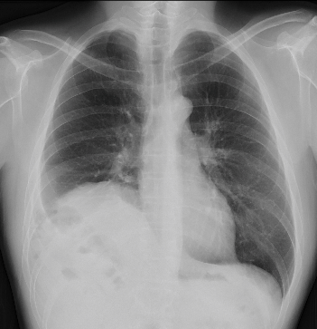
A chest radiograph showed elevation of the right hemidiaphragm (Figure 1). Computed tomography (CT) showed prolapse of multiple intraabdominal organs into the right thoracic cavity through a right-sided diaphragmatic defect (Figure 2A). The herniated contents included the stomach, transverse colon, gallbladder, and liver (Figure 2B). The left lobe of the liver was fitted into the right thoracic cavity in an anticlockwise rotation. The left lobe of the liver was enlarged, particularly the medial segment, and the surface was uneven and irregular (Figure 2C). The hernia measured approximately 10 cm in diameter. No significant organ damage was observed.
The patient was diagnosed with a right-sided BH, and surgery was planned.
We performed laparoscopic surgery with the patient in the supine position and the legs spread apart. We inserted a 12-mm camera port at the umbilicus, 5-mm ports into the right upper and left lateral abdomen, and a 12-mm port into the right lateral abdomen. The pneumoperitoneal pressure was 10 mmHg. Intraabdominal observation revealed an approximately 10 cm defect on the posterolateral side of the right diaphragm without a hernia sac. The stomach, transverse colon, greater omentum, liver, and gallbladder were herniated into the thoracic cavity through the diaphragmatic defect (Figure 3A and B). Laparoscopic forceps were used to pull the greater omentum, stomach, and transverse colon back into the abdominal cavity with relative ease. However, the left lobe of the liver was completely lodged in the thoracic cavity, and it was difficult to pull it back into the abdominal cavity, even when the patient was placed in the reverse Trendelenburg position (Figure 3C). Therefore, we decided to approach it thoracoscopically. We inserted an additional 12-mm port into the 7th intercostal space along the right midaxillary line. While pulling the liver toward the abdominal cavity, a tissue exclusion forceps (Endo Retract II®, Medtronic, Tokyo, Japan) was used to push the liver from the thoracic cavity, protecting the liver and allowing it to be returned safely into the abdominal cavity. The diaphragmatic defect measured 10 cm × 8 cm (Figure 3D). The hernia was repaired with interrupted 2-0 Prolene® (Medtronic, Tokyo, Japan) sutures (Figure 3E). In addition, Bard Composix E/X Mesh® (Bard, Tokyo, Japan) was fixed to the diaphragm using an Endo Universal 65 Hernia Stapler® (Medtronic, Tokyo, Japan) (Figure 3F). The operative time was 272 min, and blood loss was 145 mL.
The patient had an uncomplicated postoperative course and was discharged on the 7th postoperative day. Eight years have elapsed since the surgery, and there has been no recurrence.
A Bochdalek hernia is a congenital posterolateral hernia of the diaphragm that is usually observed during the neonatal period. BH occurs in approximately 1 in 2200-12500 live births but is rare in adults[1-3]. The incidence in adults is estimated to be around 0.17%-6%[9,10]. BH usually occurs on the left-side (80%-90%)[3]. A hernia sac has been reported to be present in 10%-38% of cases[3,11]. In our case, there was no hernia sac. Right-sided hernias are rare because of early closure of the right pleuroperitoneal canal and the location of the liver, which usually acts as a barrier against herniation on the right side[3]. In adults, BH is typically asymptomatic and is an incidental finding[9]. CT is the most useful tool for the diagnosis of BH, and multiple-detector CT is most effective in characterizing diaphragmatic defects[5,9].
Management of BH is performed exclusively through surgical treatment[4,5], specifically returning the herniated organs to the abdominal cavity and repairing the diaphragmatic defect[6-8]. Surgical treatment should be performed whether or not patients have symptoms because perforation or necrosis of the prolapsed organ can cause severe symptoms[12]. In recent years, the number of minimally invasive laparoscopic and thoracoscopic surgeries has increased[6,7,12].
The thoracic approach can be used to easily repair diaphragmatic defects and detach adhesions of the organs that have prolapsed into the thoracic cavity. However, it requires differential lung ventilation, making it difficult to confirm the status of the prolapsed organs. On the other hand, the abdominal approach can be used to easily return the organs if they are not adherent to the thoracic cavity. In this approach, the entire abdominal cavity can be observed, making it possible to confirm and repair damage to the organs. Furthermore, intestinal malrotation, which is often associated with BH, can be easily confirmed and can be repaired and fixed[5,9,10,13]. As there is no established consensus for choosing an approach, the surgical approach should be selected according to the clinical condition of the patient[10,12].
Generally, the diaphragmatic defect is sutured in either an interrupted or continuous manner using nonabsorbable sutures, depending on the size and location of the diaphragmatic defect[5]. The use of mesh reinforcement in the repair of defects depends on the size of the defect. Some reports recommend the use of mesh for defects larger than 20-30 cm, and others use mesh for defects larger than 8 cm or 25 cm2[5].
In this case, the diaphragmatic defect was 10 cm long and 80 cm2 in area; therefore, both direct suturing and mesh reinforcement were performed.
Ramspott et al[14] reviewed 44 patients with adult right-sided BH. The mean age was 58 years (22–98 years); 61% of the patients were women and 39% were men. The most common symptom was dyspnea (50%), followed by abdominal pain (43%) and nausea (23%); the condition was asymptomatic in 23% of patients. The most commonly herniated organs were the colon (52%), followed by the small intestine (43%), liver (27%), kidneys (18%), gallbladder (4%), and pancreas (2%). The abdominal approach (laparotomy, 34%; laparoscopy, 34%) was more commonly used than the thoracic approach (thoracotomy, 16%; thoracoscopy, 14%). Thoracoabdominal approaches were also performed in 16% of the cases. Recently, robot-assisted laparoscopic surgery (5%) and robot-assisted thoracoscopic surgery (5%) have also been reported. The diaphragmatic defect was repaired using direct suturing in 45% of the cases and mesh reinforcement in 43%. A combination of direct suturing and mesh was used in 16% of cases.
BH is frequently associated with other malformations, such as pulmonary hypoplasia, cardiac malformation, and intestinal malrotation. Furthermore, right-sided BHs are generally associated with hypoplasia or atrophy of the right lobe of the liver[13,15,16]. In these cases, atrophy and hypoplasia of the right lobe of the liver can cause herniation of abdominal organs through the defect into the thoracic cavity[13]. In this case, there was marked enlargement of the left lobe of the liver, but no hypoplasia or atrophy of the right lobe, which is considered to be extremely rare. In cases of BH in which the liver protrudes into the thoracic cavity, surgery can be performed safely by combining laparoscopic and thoracoscopic approaches. To our knowledge, no such cases have ever been reported previously.
Right-sided BH in adults with the liver protruding into the thoracic cavity is extremely rare. We successfully performed a combined laparoscopic and thoracoscopic surgery for an adult with right-sided BH with massive liver prolapse and abnormal liver morphology.
Provenance and peer review: Unsolicited article; Externally peer reviewed.
Peer-review model: Single blind
Specialty type: Surgery
Country/Territory of origin: Japan
Peer-review report’s scientific quality classification
Grade A (Excellent): 0
Grade B (Very good): 0
Grade C (Good): C
Grade D (Fair): 0
Grade E (Poor): 0
P-Reviewer: Liu T, China S-Editor: Liu JH L-Editor: A P-Editor: Chen YX
| 1. | Losanoff JE, Sauter ER. Congenital posterolateral diaphragmatic hernia in an adult. Hernia. 2004;8:83-85. [PubMed] [DOI] [Cited in This Article: ] |
| 2. | Rout S, Foo FJ, Hayden JD, Guthrie A, Smith AM. Right-sided Bochdalek hernia obstructing in an adult: case report and review of the literature. Hernia. 2007;11:359-362. [PubMed] [DOI] [Cited in This Article: ] |
| 3. | Laaksonen E, Silvasti S, Hakala T. Right-sided Bochdalek hernia in an adult: a case report. J Med Case Rep. 2009;3:9291. [PubMed] [DOI] [Cited in This Article: ] |
| 4. | Slesser AA, Ribbans H, Blunt D, Stanbridge R, Buchanan GN. A spontaneous adult right-sided Bochdalek hernia containing perforated colon. JRSM Short Rep. 2011;2:54. [PubMed] [DOI] [Cited in This Article: ] |
| 5. | Machado NO. Laparoscopic Repair of Bochdalek Diaphragmatic Hernia in Adults. N Am J Med Sci. 2016;8:65-74. [PubMed] [DOI] [Cited in This Article: ] |
| 6. | Settembre A, Cuccurullo D, Pisaniello D, Capasso P, Miranda L, Corcione F. Laparoscopic repair of congenital diaphragmatic hernia with prosthesis: a case report. Hernia. 2003;7:52-54. [PubMed] [DOI] [Cited in This Article: ] |
| 7. | Esmer D, Alvarez-Tostado J, Alfaro A, Carmona R, Salas M. Thoracoscopic and laparoscopic repair of complicated Bochdalek hernia in adult. Hernia. 2008;12:307-309. [PubMed] [DOI] [Cited in This Article: ] |
| 8. | Jambhekar A, Robinson S, Housman B, Nguyen J, Gu K, Nakhamiyayev V. Robotic repair of a right-sided Bochdalek hernia: a case report and literature review. J Robot Surg. 2018;12:351-355. [PubMed] [DOI] [Cited in This Article: ] |
| 9. | Costa Almeida CE, Reis LS, Almeida CM. Adult right-sided Bochdalek hernia with ileo-cecal appendix: Almeida-Reis hernia. Int J Surg Case Rep. 2013;4:778-781. [PubMed] [DOI] [Cited in This Article: ] |
| 10. | Moro K, Kawahara M, Muneoka Y, Sato Y, Kitami C, Makino S, Nishimura A, Kawachi Y, Gabriel E, Nikkuni K. Right-sided Bochdalek hernia in an elderly adult: a case report with a review of surgical management. Surg Case Rep. 2017;3:109. [PubMed] [DOI] [Cited in This Article: ] |
| 11. | Kocakusak A, Arikan S, Senturk O, Yucel AF. Bochdalek's hernia in an adult with colon necrosis. Hernia. 2005;9:284-287. [PubMed] [DOI] [Cited in This Article: ] |
| 12. | Debergh I, Fierens K. Laparoscopic repair of a Bochdalek hernia with incarcerated bowel during pregnancy: report of a case. Surg Today. 2014;44:753-756. [PubMed] [DOI] [Cited in This Article: ] |
| 13. | Enomoto N, Yamada K, Kato D, Yagi S, Wake H, Nohara K, Takemura N, Kiyomatsu T, Kokudo N. Right-sided Bochdalek hernia in an adult with hepatic malformation and intestinal malrotation. Surg Case Rep. 2021;7:169. [PubMed] [DOI] [Cited in This Article: ] |
| 14. | Ramspott JP, Jäger T, Lechner M, Schredl P, Gabersek A, Mayer F, Emmanuel K, Regenbogen S. A systematic review on diagnostics and surgical treatment of adult right-sided Bochdalek hernias and presentation of the current management pathway. Hernia. 2022;26:47-59. [PubMed] [DOI] [Cited in This Article: ] |
| 15. | Banchini F, Santoni R, Banchini A, Bodini FC, Capelli P. Right posterior diaphragmatic hernia (Bochdalek) with liver involvement and alteration of hepatic outflow in adult: a case report. Springerplus. 2016;5:1561. [PubMed] [DOI] [Cited in This Article: ] |
| 16. | Zenda T, Kaizaki C, Mori Y, Miyamoto S, Horichi Y, Nakashima A. Adult right-sided Bochdalek hernia facilitated by coexistent hepatic hypoplasia. Abdom Imaging. 2000;25:394-396. [PubMed] [DOI] [Cited in This Article: ] |


