Published online Oct 26, 2022. doi: 10.12998/wjcc.v10.i30.11185
Peer-review started: July 22, 2022
First decision: August 4, 2022
Revised: August 5, 2022
Accepted: September 16, 2022
Article in press: September 16, 2022
Published online: October 26, 2022
The objective of this work is displaying a successful treatment for an internal resorption case under operating microscope using bioceramic material.
Periapical radiograph showed radiolucent lesion representing large internal resorption of the root. The respective defect was obturated using endoscquence bioceramic material follow up at the month 18 after treatment revealed no abnormal finings clinically and radiographically.
New generations bioceramics have many advantages that internal root resorption cases can benefit from. The use of operating microscope helps to apply obturating materials with precision. However, long term study on a large sample is required in future studies.
Core Tip: New generations bioceramics have many advantages that internal root resorption cases can benefit from including easier handling ability and excellent seal. The objective of this work is displaying a successful treatment for an internal resorption case under operating microscope using bioceramic material. The use of operating microscope helps to apply obturating materials with precision. However, long term study on a large sample is required in future studies.
- Citation: Riyahi AM. Bioceramics utilization for the repair of internal resorption of the root: A case report. World J Clin Cases 2022; 10(30): 11185-11189
- URL: https://www.wjgnet.com/2307-8960/full/v10/i30/11185.htm
- DOI: https://dx.doi.org/10.12998/wjcc.v10.i30.11185
Internal resorption of the root is an inflammatory process that starts inside the pulp and results in dentin loss and potential cementum invasion[1]. Internal resorption can lead to perforation of the root if it progresses. Vital tissues apical to the resorptive area are required for the process of internal resorption to be active[2]. In its classical representation radiographically, the resorptive defect can be seen as round radiolucency with symmetrical enlargement of the canal space[3]. Internal resorption represents one of the treatment challenges in endodontics; therefore, many previous studies have been conducted on this subject, including case reports[4-7].
Endodontic treatment success relies on sufficient root canal system instrumentation, disinfection and obturation[8]. In general, bioceramics have demonstrated favorable physicochemical properties. Moreover, new generation bioceramics have improved on some of existing drawbacks of previous materials[9]. Promising results can be expected from the bioceramics due the antibacterial and anti-biofilm characteristics in addition to the biocompatible nature of the material[10]. This study aims to demonstrate a comprehensive management solution for internal root resorption with the use of a bioceramic material based as an obturation material for the resorptive defect.
A 51-year-old female who was referred for evaluation of tooth 21 presented with a chief complaint of discomfort associated with an upper front tooth.
Some discomfort associated with upper front teeth.
The patient has history of multiple dental caries which was treated with restorations and crowns.
The patient has no relevant medical history.
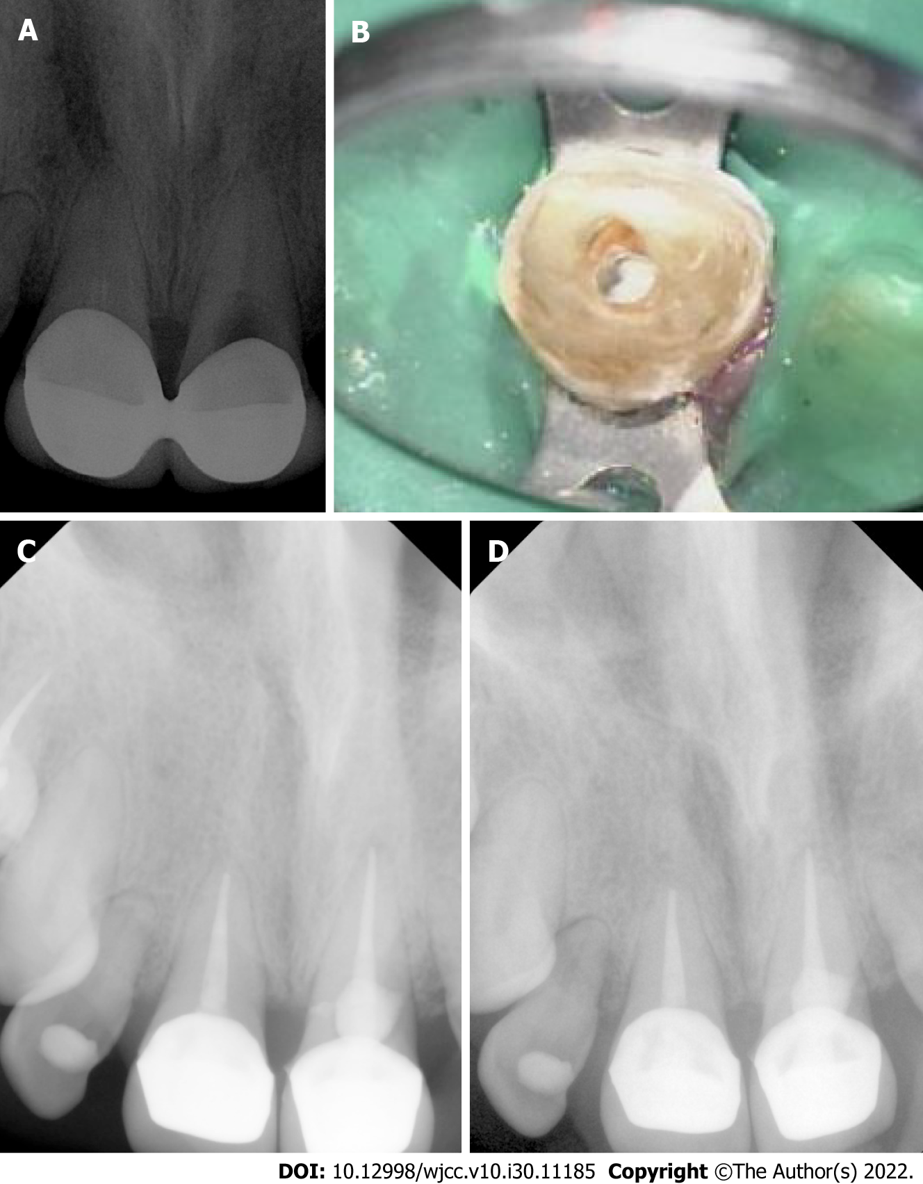
Upon clinical assessment, tooth 21 responded negatively to both cold and electrical pulp tests. In addition, no mobility or deep probing were detected. However, the tooth was tender to percussion and bite upon testing. A periapical radiograph of the tooth in question is shown in Figure 1A. Radiographic examination showed what appeared to be a large internal resorptive defect related to tooth 21. The endodontic diagnosis was necrotic pulp with symptomatic apical periodontitis. The patient wanted to try to save the tooth if possible. Planned treatment was nonsurgical endodontics and final restoration if the tooth was found to be restorable.
In addition, the clinical examination of tooth 11 showed no response to vitality testing. Although slight tenderness to percussion was reported, the tooth responded normally to palpation. The tooth also had no deep probing or mobility. Radiographic evaluation showed that tooth 11 underwent previous endodontic treatment with a fill short of the apex.
There is no laboratory examinations.
Periapical radiographs were obtained for teeth in question.
The diagnosis was previously treated tooth, with symptomatic apical periodontitis. Planned treatment included endodontic retreatment and final restoration.
To provide local anesthesia, 1.8 mL cartridge of xylocaine 2% with epinephrine 1:80000 was administered as buccal infiltration to tooth 21. After rubber dam isolation, the access cavity was prepared under an operating microscope. The working length was found to be 19 mm. Hand files were used to create the glide path. K3 (Sybron Endo, Orange, CA) Rotary Files were used to prepare the root canal at a speed of 300 rpm. Irrigation was carefully performed using 5.25% sodium hypochlorite. The canal was dried using paper points and obturated with gutta-percha and AH plus sealer (Dentsply International Inc., York, PA, United States) using the vertical compaction obturation technique. The resorptive defect was obturated with EndoSequence® BC RRM-Fast Set Putty (Brasseler United States, Savannah, GA) using pluggers of different sizes under an operating microscope (Figure 1B). The provisional crown was cemented, and occlusion was checked.
After one week, the patient had no complaints related to tooth 21. The tooth responded negatively to both percussion and palpation. Rubber dam isolation was obtained, and the access cavity was prepared for tooth 11. The previously placed obturation material was removed from the canal. A ProTaper Universal (Tulsa Dental, Tulsa, OK) rotary retreatment system was used for this purpose. Subsequently, instrumentation was completed using K3 rotary files and irrigation using sodium hypocrite. The canal was obturated using gutta-percha and AH Plus sealer. The access cavity was temporized using Cavit (3M ESPE, St. Paul, MN, United States), followed by glass ionomer restoration. The provisional crown was cemented, and occlusion was checked. The patient was referred for the final restoration of both teeth 11 and 21. A periapical radiograph was obtained after the crowns cemented by the restorative dentist, as shown in Figure 1C.
The patient was seen at the month 18 after treatment for evaluation. The patient had no complaints. Clinical examination showed no abnormal findings. Teeth 11 and 21 responded normally to percussion and palpation with no mobility detected. Periapical Radiographs showed no abnormalities as seen in Figure 1D.
Internal resorption of the root poses an endodontics treatment challenge for various reasons. The irregular nature of the resorptive lesion and the possible perforation externally of the root surface are among the factors of difficulty in these situations[11]. However, salvaging a functional tooth remains one of the main objectives of endodontic treatment. The utilization of new materials with favorable properties can be beneficial in these cases.
Various studies have examined the characteristics of bicoeramics and the potential advantages of these materials[12,13]. Bioceramics are bio-inert, biocompatible, and non-toxic[14]. In addition, a previous study found similar sealing capability when evaluating Endosequence Bioceramic Root Repair Material Putty and Mineral Trioxide Aggregate[15]. A new generation of bioceramics have been reported to be clinical used for treatment of internal resorption that cause perforations. In a previous study[16], bioceramic sealer was utilized for management of perforating internal resorption in chamber/coronal canal region.
Microscope-improved visualization is one of the advantages of using magnification[17]. Using the operating microscope throughout the course of treatment in this case was helpful in visualizing and precisely applying the material to the resorptive defect. Obtaining cone beam computed tomography (CBCT) is valuable in identifying the perforation in the internal root resorption[18]. In this case, CBCT image preoperatively could be beneficial for determining the size and the extension of the resorptive lesion prior treatment. Furthermore, the use of bioceramic sealer instead of the resin based sealer could be advantageous in such case.
A combination of correct diagnosis and proper management is important for the success of root canal treatment. The use of an operating microscope, bioceramics, and coronal seal after endodontic treatment can be important for treating internal root resorption. However, future studies with representative samples are required to evaluate long-term outcomes.
Internal resorption represents one of many endodontic treatment challenges. The use of a new generation of bioceramics in such cases has potential benefits that can add to overall treatment success. Further studies are required to evaluate the outcomes.
Provenance and peer review: Unsolicited article; Externally peer reviewed.
Peer-review model: Single blind
Specialty type: Medicine, research and experimental
Country/Territory of origin: Saudi Arabia
Peer-review report’s scientific quality classification
Grade A (Excellent): 0
Grade B (Very good): B
Grade C (Good): C
Grade D (Fair): 0
Grade E (Poor): 0
P-Reviewer: Brkanović S, Croatia; Reda R, Italy S-Editor: Wang JJ L-Editor: A P-Editor: Wang JJ
| 1. | American Association of Endodontists. Glossary of Endodontic Terms. [cited 17 July 2022]. Available from: https://www.aae.org/. [Cited in This Article: ] |
| 2. | Tronstad L. Root resorption--etiology, terminology and clinical manifestations. Endod Dent Traumatol. 1988;4:241-252. [PubMed] [DOI] [Cited in This Article: ] [Cited by in Crossref: 394] [Cited by in F6Publishing: 358] [Article Influence: 9.9] [Reference Citation Analysis (0)] |
| 3. | Haapasalo M, Endal U. Internal inflammatory root resorption: the unknown resorption of the tooth. Endod Topics. 2008;14:60. [DOI] [Cited in This Article: ] [Cited by in Crossref: 67] [Cited by in F6Publishing: 69] [Article Influence: 3.8] [Reference Citation Analysis (0)] |
| 4. | Yıldırım S, Elbay M. Multidisciplinary Treatment Approach for Perforated Internal Root Resorption: Three-Year Follow-Up. Case Rep Dent. 2019;2019:5848272. [PubMed] [DOI] [Cited in This Article: ] [Cited by in F6Publishing: 2] [Reference Citation Analysis (0)] |
| 5. | Sanaei-Rad P, Bolbolian M, Nouri F, Momeni E. Management of internal root resorption in the maxillary central incisor with fractured root using Biodentine. Clin Case Rep. 2021;9:e04502. [PubMed] [DOI] [Cited in This Article: ] [Cited by in Crossref: 3] [Cited by in F6Publishing: 1] [Article Influence: 0.3] [Reference Citation Analysis (0)] |
| 6. | Mehra N, Yadav M, Kaushik M, Roshni R. Clinical Management of Root Resorption: A Report of Three Cases. Cureus. 2018;10:e3215. [PubMed] [DOI] [Cited in This Article: ] [Cited by in Crossref: 4] [Cited by in F6Publishing: 5] [Article Influence: 0.8] [Reference Citation Analysis (0)] |
| 7. | Yang Y, Zhang B, Huang C, Ye R. Intentional Replantation of a Second Premolar with Internal Resorption and Root Fracture: A Case Report. J Contemp Dent Pract. 2021;22:562-567. [PubMed] [DOI] [Cited in This Article: ] |
| 8. | Torabinejad M, Corr R, Buhrley M, Wright K, Shabahang S. An animal model to study regenerative endodontics. J Endod. 2011;37:197-202. [PubMed] [DOI] [Cited in This Article: ] [Cited by in Crossref: 29] [Cited by in F6Publishing: 27] [Article Influence: 2.1] [Reference Citation Analysis (0)] |
| 9. | Wang ZJ. Bioceramic materials in endodontics. Endod Topics. 2015;32:3. [DOI] [Cited in This Article: ] [Cited by in Crossref: 49] [Cited by in F6Publishing: 52] [Article Influence: 5.8] [Reference Citation Analysis (0)] |
| 10. | Wang Z, Shen Y, Haapasalo M. Antimicrobial and Antibiofilm Properties of Bioceramic Materials in Endodontics. Materials (Basel). 2021;14. [PubMed] [DOI] [Cited in This Article: ] [Cited by in Crossref: 12] [Cited by in F6Publishing: 6] [Article Influence: 2.0] [Reference Citation Analysis (0)] |
| 11. | Berman LH, Hargreaves KM. Cohen’s Pathways of the Pulp-E-book. Netherlands: Elsevier Health Sciences, 2020. [Cited in This Article: ] |
| 12. | Motwani N, Ikhar A, Nikhade P, Chandak M, Rathi S, Dugar M, Rajnekar R. Premixed bioceramics: A novel pulp capping agent. J Conserv Dent. 2021;24:124-129. [PubMed] [DOI] [Cited in This Article: ] [Cited by in Crossref: 12] [Cited by in F6Publishing: 9] [Article Influence: 3.0] [Reference Citation Analysis (0)] |
| 13. | Chybowski EA, Glickman GN, Patel Y, Fleury A, Solomon E, He J. Clinical Outcome of Non-Surgical Root Canal Treatment Using a Single-cone Technique with Endosequence Bioceramic Sealer: A Retrospective Analysis. J Endod. 2018;44:941-945. [PubMed] [DOI] [Cited in This Article: ] [Cited by in Crossref: 67] [Cited by in F6Publishing: 49] [Article Influence: 8.2] [Reference Citation Analysis (0)] |
| 14. | Raghavendra SS, Jadhav GR, Gathani KM, Kotadia P. Bioceramics in endodontics - a review. J Istanb Univ Fac Dent. 2017;51:S128-S137. [PubMed] [DOI] [Cited in This Article: ] [Cited by in Crossref: 37] [Cited by in F6Publishing: 48] [Article Influence: 6.9] [Reference Citation Analysis (0)] |
| 15. | Antunes HS, Gominho LF, Andrade-Junior CV, Dessaune-Neto N, Alves FR, Rôças IN, Siqueira JF Jr. Sealing ability of two root-end filling materials in a bacterial nutrient leakage model. Int Endod J. 2016;49:960-965. [PubMed] [DOI] [Cited in This Article: ] [Cited by in Crossref: 22] [Cited by in F6Publishing: 26] [Article Influence: 2.9] [Reference Citation Analysis (0)] |
| 16. | Haapasalo M, Parhar M, Huang X, Wei X, James L, Shen Y. Clinical use of bioceramic materials. Endod Topics. 2015;32:97. [DOI] [Cited in This Article: ] [Cited by in Crossref: 21] [Cited by in F6Publishing: 22] [Article Influence: 2.4] [Reference Citation Analysis (0)] |
| 17. | Low JF, Dom TNM, Baharin SA. Magnification in endodontics: A review of its application and acceptance among dental practitioners. Eur J Dent. 2018;12:610-616. [PubMed] [DOI] [Cited in This Article: ] [Cited by in Crossref: 16] [Cited by in F6Publishing: 21] [Article Influence: 4.2] [Reference Citation Analysis (0)] |
| 18. | Khojastepour L, Moazami F, Babaei M, Forghani M. Assessment of Root Perforation within Simulated Internal Resorption Cavities Using Cone-beam Computed Tomography. J Endod. 2015;41:1520-1523. [PubMed] [DOI] [Cited in This Article: ] [Cited by in Crossref: 4] [Cited by in F6Publishing: 4] [Article Influence: 0.4] [Reference Citation Analysis (0)] |


