Article

Genetic and Physiological Characterization of Soybean-Nodule-Derived Isolates from Bangladeshi Soils Revealed Diverse Array of Bacteria with Potential Bradyrhizobia for Biofertilizers

1
United Graduate School of Agricultural Science, Tokyo University of Agriculture and Technology (TUAT), Saiwai-cho 3-5-8, Fuchu-shi, Tokyo 183-8509, Japan
2
Institute of Food and Radiation Biology, Atomic Energy Research Establishment, Bangladesh Atomic Energy Commission, Ganakbari, Savar, Dhaka 1207, Bangladesh
3
Faculty of Agriculture, Tokyo University of Agriculture and Technology (TUAT), Saiwai-cho 3-5-8, Fuchu-shi, Tokyo 183-8509, Japan
4
Institute of Global Innovation Research, Tokyo University of Agriculture and Technology (TUAT), Harumi-cho 3-8-1, Fuchu-shi, Tokyo 183-8509, Japan
5
Faculty of Agriculture, Utsunomiya University, Utsunomiya-shi, Tochigi 321-8505, Japan
6
Institute of Agriculture, Tokyo University of Agriculture and Technology (TUAT), Saiwai-cho 3-5-8, Fuchu-shi, Tokyo 183-8509, Japan
7
Faculty of Food and Agricultural Sciences, Fukushima University, Kanayagawa 1, Fukushima-shi, Fukushima 960-1248, Japan
*
Author to whom correspondence should be addressed.
Microorganisms 2022, 10(11), 2282; https://doi.org/10.3390/microorganisms10112282
Submission received: 27 September 2022 / Revised: 5 November 2022 / Accepted: 15 November 2022 / Published: 17 November 2022
(This article belongs to the Section Plant Microbe Interactions)

Abstract

:
Genetic and physiological characterization of bacteria derived from nodules of leguminous plants in the exploration of biofertilizer is of paramount importance from agricultural and environmental perspectives. Phylogenetic analysis of the 16S rRNA gene of 84 isolates derived from Bangladeshi soils revealed an unpredictably diverse array of nodule-forming and endosymbiotic bacteria—mostly belonging to the genus Bradyrhizobium. A sequence analysis of the symbiotic genes (nifH and nodD1) revealed similarities with the 16S rRNA gene tree, with few discrepancies. A phylogenetic analysis of the partial rrn operon (16S-ITS-23S) and multi-locus sequence analysis of atpD, glnII, and gyrB identified that the Bradyrhizobium isolates belonged to Bradyrhizobium diazoefficiens, Bradyrhizobium elkanii, Bradyrhizobium liaoningense and Bradyrhizobium yuanmingense species. In the pot experiment, several isolates showed better activity than B. diazoefficiens USDA110, and the Bho-P2-B2-S1-51 isolate of B. liaoningense showed significantly higher acetylene reduction activity in both Glycine max cv. Enrei and Binasoybean-3 varieties and biomass production increased by 9% in the Binasoybean-3 variety. Tha-P2-B1-S1-68 isolate of B. diazoefficiens significantly enhanced shoot length and induced 10% biomass production in Binasoybean-3. These isolates grew at 1–4% NaCl concentration and pH 4.5–10 and survived at 45 °C, making the isolates potential candidates for eco-friendly soybean biofertilizers in salty and tropical regions.

1. Introduction

Bacterial diversity analysis is important to understand, maintain, and conserve global genetic resources. Bacteria are not only a vital part of soil and environment, but also the main agents of different nutrient cycling [1]. As new environments and regions are explored, the abundance and fruitfulness of microbial diversity will become increasingly evident [2]. Root nodule bacteria, comprising different types of Alpha-proteobacteria, Beta-proteobacteria, and possibly Gamma-proteobacteria, are of significant importance in biodiversity studies [3]. These soil bacteria are capable of forming root nodules and establishing symbiosis with the roots or stems of leguminous plants [4]. During the symbiotic association process, rhizobia reduce atmospheric nitrogen into ammonium, a usable nitrogen resource for plants, while, concurrently, some compounds are exchanged between the bacteroid and its plant host [5]. The ability of rhizobia to fix nitrogen has a significant effect on reducing the utilization of chemical nitrogen fertilizers in agriculture. The biodiversity of rhizobia represents a valuable bio-resource for the exploration of bacterial strains with suitable traits that can increase legume production [6].
At present, all symbiotic nitrogen-fixing bacteria are categorized in the vast phylum Proteobacteria, within the classes Alphaproteobacteria (α-rhizobia), Beta-proteobacteria (β-rhizobia), and possibly Gamma-proteobacteria (γ-rhizobia), with approximately 180 nodulating species in 21 genera [3,7,8]. The symbiotic bacteria in the class Alpha-proteobacteria are the most common nitrogen-fixing bacteria, which are distributed in 16 genera of seven families: Agrobacterium, Allorhizobium, Ensifer (previously Sinorhizobium), Neorhizobium, Pararhizobium, Rhizobium, and Shinella of the family Rhizobiaceae; Aminobacter, Phyllobacterium, and Mesorhizobium of Phyllobacteriaceae; Bradyrhizobium of Bradyrhizobiaceae; Microvirga and Methylobacterium of Methylobacteriaceae; Ochrobactrum of Brucellaceae; Devosia of Hyphomicrobiaceae; and Azorhizobium of Xanthobacteraceae. All are members of the order Rhizobiales, in which some of the other families are Bartonellaceae, Beijerinckiaceae, Cohaesibacteraceae, Methylocystaceae, Rhodobiaceae, and Roseiarcaceae [3]. Compared with the rhizobia in the class Alpha-proteobacteria, the symbiotic bacteria in Beta-proteobacteria and Gamma-proteobacteria were recognized much later [9,10] and are less diverse, including approximately 20 species in four genera: Cupriavidus, Paraburkholderia, and Trinickia, belonging to the family Burkholderiaceae [11], and Herbaspirillum, in the family Oxalobacteraceae [9].
All these bacteria form symbioses with different types of leguminous plants. One of those is soybean (Glycine max L. Merr.), which belongs to the tribe of Phaseoleae, sub-tribe of Glycininae, and genus Glycine, which originated and was domesticated in China around 5000 years ago [12,13]. The highest diversity of soybean-nodulating rhizobia is found in China and countries in the Americas [12,13,14]. Among rhizobia, Bradyrhizobium is the most established soybean microsymbiont and is used as a biofertilizer. The genus Bradyrhizobium was established in early 1982, and, to date, 73 species have been recognized worldwide [15,16]. Of the 73 species, B. elkanii, B. japonicum, B. diazoefficiens, B. daqingense, B. liaoningense, B. huanghuaihaiense, and B. ottawaense are seven slow-growing rhizobial species that have been described to nodulate soybean [3]. Some species of the genus Sinorhizobium (syn. Ensifer) nodulate soybean [3], whereas Mesorhizobium tianshanense, formerly known as Rhizobium tianshanense, from the genus Mesorhizobium, has also been reported to be a soybean microsymbiont [17].
Root nodule bacterial diversity associated with soybean (Glycine max) is immensely important because soybean is one of the most important legume crops in the world, representing 50% of global area planted with crop legumes and 68% of global legume crop production [7,12,18]. The characterization of soybean-associated bacteria from unexplored regions and environments can aid in the discovery of novel bacteria with potential biofertilizer activity. The application of local or indigenous bacteria associated with soybeans can increase crop productivity more than foreign inoculants because of their adaptation to the local environment and compatibility with local soybean varieties. Thus, the isolation of suitable and potential local bacteria, by analyzing the diversity of soybean root nodule bacteria from Bangladeshi soil samples, can facilitate the development of efficient biofertilizers for soybean.
Moreover, the study of bacterial diversity in Bangladeshi soils associated with soybean is scarce. Therefore, the objectives of this study are as follows:
(i) To achieve a fundamental understanding of the diversity and taxonomic identity of rhizobia associated with soybean root nodules from Bangladeshi soil.
(ii) To estimate the stress tolerance of the isolates and identify suitable stress-tolerant bacteria.

2. Materials and Methods

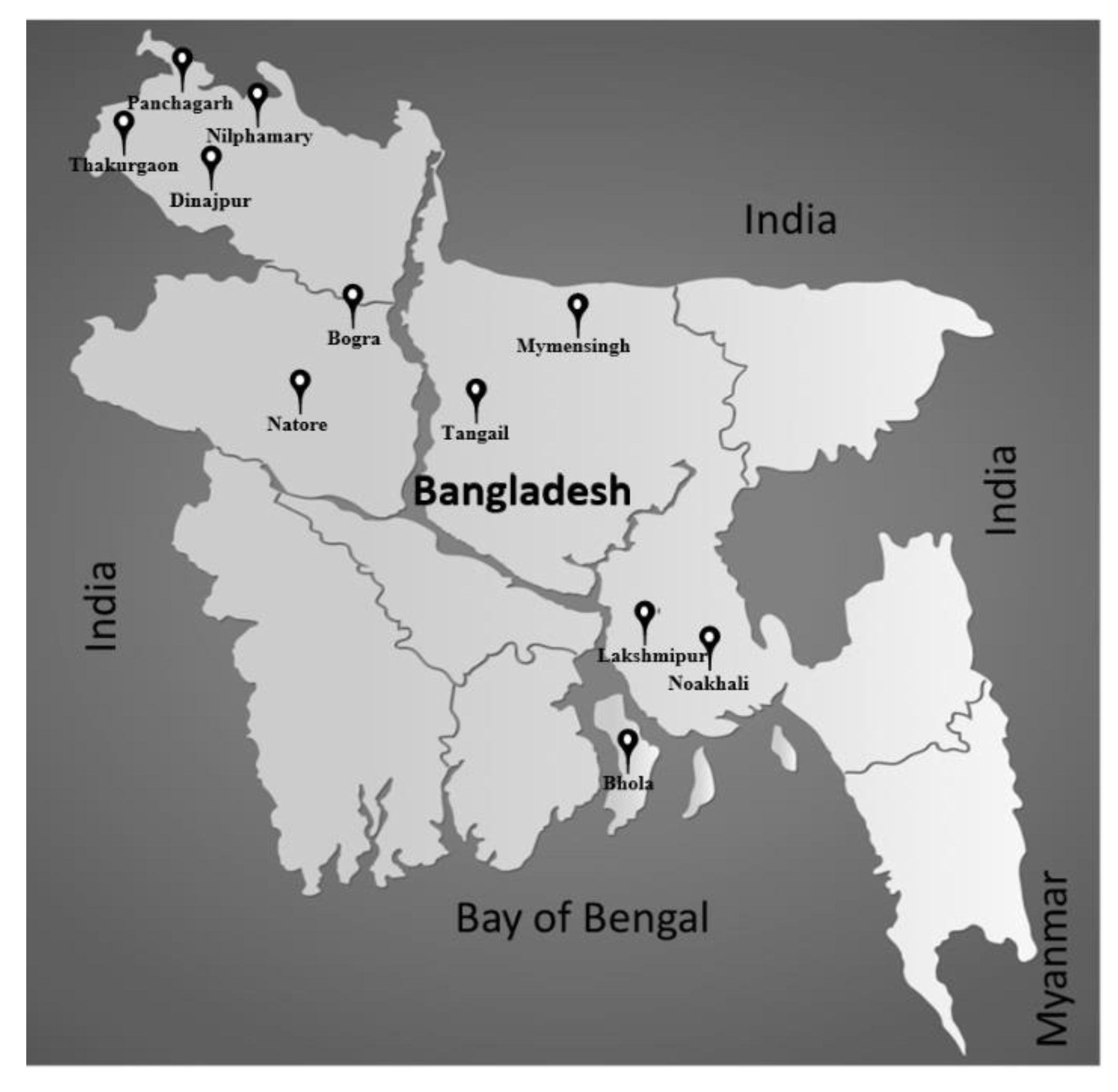
2.1. Collection of Soil Samples

Soil samples were collected from 11 districts of different agro-ecological zones in Bangladesh, focusing mainly on areas where soybeans have been cultivated (Table 1 and Figure 1). For each location, soil sample was collected from a depth of ~10–15 cm from different positions of a field and mixed. The soil samples were stored at 4 °C until use.

2.2. Collection of Nodules Using Soil Samples in Pot Experiment

The seeds of soybean cultivar Glycine max c.v. Enrei (Japanese variety) were surface-sterilized by immersion in 70% ethanol for 30 s, and then in 3% sodium hypochlorite solution for 3 min, and the seeds were washed exhaustively with sterile water [19]. For seed germination, the sterilized seeds were incubated for two days at 28 °C. From each soil sample, 1 g of soil was suspended in 5 mL of sterilized water and soil suspensions were used as inoculants. Each inoculant was applied to 300 mL glass jars containing germinated seeds and sterilized vermiculite, and the jars were placed in growth chamber and kept under controlled conditions with 12 h light/dark photoperiod at temperatures of 25 °C and 18 °C in the light and dark hours, respectively [19]. Plant growth was supported by adding a sterilized N-free nutrient solution [20] to the jars. After four weeks, whole plants were uprooted from the jars, washed in running tap water to remove vermiculite, and the nodules were harvested. Root nodules were surface-sterilized by immersion in 70% ethanol for 30 s and in 3% sodium hypochlorite for 3 min, then washed five times with sterile water [19]. Each nodule was crushed in 100 μL of glycerol solution (15%, v/v) to obtain a turbid suspension. An aliquot (10 μL) of suspension was plated onto 1.5% yeast extract mannitol agar (YEM) [21] plates and incubated for 3–7 days at 28 °C. The remaining suspension was maintained at −30 °C. Single colonies were selected and checked for purity by repeated streaking onto fresh YEM medium. The isolates were maintained for the long-term into 50% glycerol stocks at −80 °C and the short-term into slant stocks at 4 °C.

2.3. Temperature Tolerance Test of Isolates

The isolates used in this study were examined for growth under high-temperature conditions. For the temperature tolerance test, isolates were inoculated on YMA plates and incubated at 10 °C, 20 °C, 28 °C, 37 °C, 40 °C, and 45 °C for 3–7 days [22]. After incubation, bacterial growth was examined.

2.4. pH Tolerance Test of Isolates

The isolates used in this study were examined for growth at different pHs. For pH tolerance tests, isolates were inoculated on YMA plates at pH 4.5, 6, 7, 8.5, and 10. The pH was adjusted to pH 4.5 and 6 using 0.5 M HCl, while the pH was adjusted to pH 7, 8.5, and 10 using 0.5 M NaOH. After bacterial inoculation, the plates were incubated at 28 °C for 3–7 days [22]. After incubation, bacterial growth was examined.

2.5. Salinity Tolerance Test of Isolates

The isolates used in this study were examined for growth under high-salinity conditions. For the salt tolerance test, isolates were inoculated on YMA plates containing 1%, 2%, 3%, and 4% (w/v) NaCl concentration and incubated at 28 °C for 3–7 days [22]. After incubation, bacterial growth was examined.

2.6. DNA Extraction, Amplification and Sequencing

Genomic DNA for each isolate was extracted from YEM broth culture following the standard protocol using the Wizard® Genomic DNA Purification Kit (WI 53711-5399; Promega Corporation, Madison, WI, USA). DNA sequences corresponding to the 16S rRNA, rrn operon (16S-ITS-23S), nodD1, nifH, atpD, glnII, and gyrB genes were amplified by PCR using the KOD-Plus-Neo enzyme system (Toyobo Co., Ltd., Osaka, Japan) and sequenced using an Applied Biosystems 3500 Genetic Analyzer using the BigDye® Terminator v3.1 Cycle Sequencing Kit Protocol (Thermo Fisher Scientific, Life Technologies Corporation, California, CA, USA) with the primer pairs shown in Table S1 [23,24,25,26,27,28].

2.7. Analysis of DNA Sequences and Phylogeny Genomic

An analysis and quality check of DNA sequences were performed primarily using Sequence Scanner 2.0 software (Thermo Fisher Scientific) and 4Peaks (Nucleobytes). Sequences derived from forward and reverse primers were individually joined by identifying the overlapping sequence between them, using GENETYX version 11 software (Genetyx Corp., Tokyo, Japan) and the online tool Merger (http://www.bioinformatics.nl/cgi-bin/emboss/merger; accessed on 17 June 2019). Multiple sequence alignments of nucleotide sequences and bootstrapping to create maximum likelihood and neighbor-joining phylogenetic trees were performed using MEGA X [29]. The evolutionary history of all phylogenetic trees in this study was inferred by the maximum likelihood method using MEGA X software [29]. The percentage of trees in which the associated taxa clustered together is shown next to the branches. The initial tree(s) for the heuristic search were obtained by applying the neighbor-joining method to a matrix of pairwise distances, estimated using the maximum composite likelihood approach. The tree was drawn to scale, with branch lengths measured in terms of the number of substitutions per site [29].

2.8. Analysis of Inoculation Effects of Selected Isolates on Plants

Isolates were grown in yeast-extract mannitol (YM) broth for three days at 28 °C, after which the cells were collected by centrifugation. The cells were then washed and diluted with sterile, ultrapure Milli-Q water. The absorbance of the diluted cells was measured using a spectrophotometer (Ultrospec 3300 Pro; Amersham Biosciences, Cambridge, UK) at 600 nm. For each isolate, the amount of bacterial inoculation was kept almost equal by maintaining (absorbance at 600 nm × volume of inoculation = 1) the same scale and measuring the colony-forming unit (CFU) by plate count. The concentration of cells in the solution used for inoculation was maintained at approximately 108 cells mL−1 [24,25]. Surface-sterilized seeds of Glycine max cv. Enrei and Binasoybean-3 (Bangladeshi soybean variety, Table S2) were sown in 300 mL of sterile vermiculite (Yoshino Gypsum Co., Ltd., Tokyo, Japan) in glass jars. After sowing, 1 mL of the cell suspension was applied to the seeds. Three replicates were performed for each isolate. Subsequently, the jars were transferred to a growth chamber. Sterilized N-free nutrient solution [20] was added to the glass jars. The moisture content was maintained at ~70% throughout the growth period. Plants were grown in a growth room under a 12 h light (25 °C)/dark (18 °C) photoperiod. After five weeks of incubation, whole plants were removed and washed from the vermiculite and different parameters, e.g., shoot length, root length, nodule color, nodule size and number, shoot weight, root weight, nodule weight, and ARA, were measured [23,30]. Experiments were performed using a randomized complete block design with 2 soybean varieties, 14 bacteria and 3 replicates for each treatment [31].

2.9. Acetylene Reduction Assay

For the acetylene reduction assay (ARA), whole plant roots with root nodules from each tested plant were introduced into a 300 mL airtight jar. Subsequently, 30 mL of air from the jar was replaced with 30 mL of acetylene, and the whole roots with root nodules were further incubated for 1 h at 30 °C to evaluate nitrogenase activity in the root nodules [19]. The amount of ethylene produced owing to the activity of nitrogenase on acetylene enclosed in the jar was determined using a Shimadzu GC-8A gas chromatograph (Shimadzu, Tokyo, Japan) equipped with a Porapak N column (Chrompack, Middelburg, the Netherlands). The roots of plants without bacterial inoculation were used as controls [19].

2.10. Nucleotide Sequence Accession Numbers

DNA sequences were deposited in the DNA Data Bank of Japan (DDBJ) under the accession numbers LC652665 to LC652748 for the 16S rRNA gene, LC658965 to LC658985 for 16S rRNA, 16S-23S rRNA internal transcribed spacer, tRNA-Ile, tRNA-Ala, 23S rRNA gene, LC670617 to LC670629 for atpD, LC670630 to LC670642 for glnII, LC670643 to LC670655 for gyrB, LC670656 to LC670720 for nifH, and LC671364-LC671428 for nodD1.

2.11. Statistical Analysis

The experimental data obtained from plant tests were subjected to statistical analyses, such as multiple comparison using general linear model with Dunnett’s Post Hoc test (with 95% confidence level), a correlation analysis using the bivariate correlations method with Pearson Correlation Coefficient and two-tailed test of significance, and regression analysis using the linear regression method with 95% confidence intervals level using IBM SPSS Statistics, Version 23.0.

3. Results

3.1. Isolation of Bacteria from Root Nodule

Soil samples from 11 districts of Bangladesh were used for root-nodule bacterial isolation. Soybean (Glycine max cv. Enrei) plants were grown in vermiculite using the soil samples. A total of 84 isolates were obtained from the selected root nodules of soybean plants.

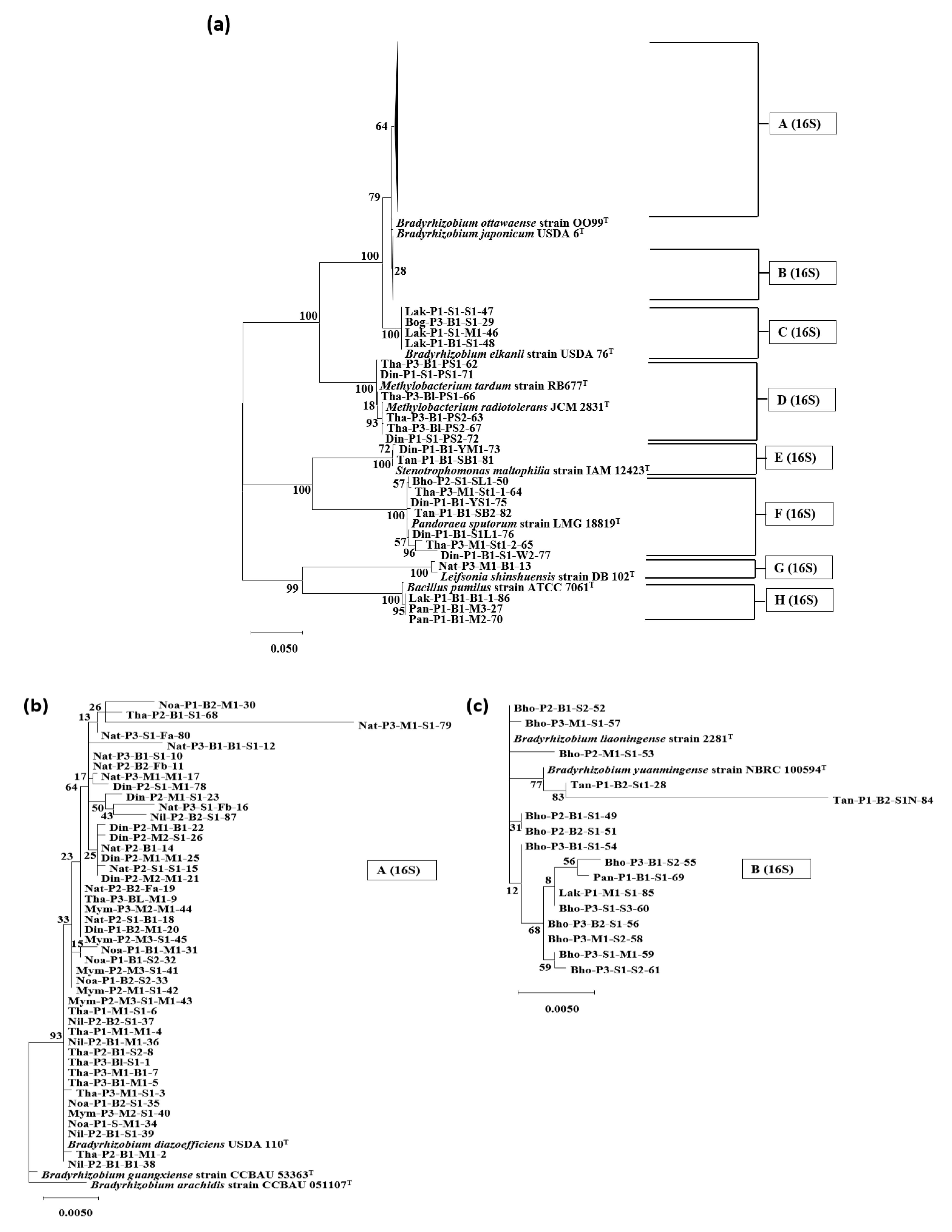
3.2. Phylogenetic Analysis Based on the 16S rRNA Genes

To determine the phylogenetic position of the isolates, 16S rRNA gene sequencing and phylogenetic analysis with highly similar reference strains were performed using a 1348 nt sequence for each bacterium.
Subsequently, the 84 isolates were classified into eight groups (Figure 2a) belonging to the Alpha-proteobacteria, Beta-proteobacteria, Gamma-proteobacteria, Firmicutes, and Actinobacteria phylum/class. The majority of isolates (65/84; 77.4%), showed a close relationship with the genus Bradyrhizobium, belonging to the class Alphaproteobacteria and family Rhizobiales. The Bradyrhizobium isolates were classified into three groups: A (16S), B (16S), and C (16S). Forty-five isolates showing a close relationship with Bradyrhizobium diazoefficiens were grouped in A (16S) (Figure 2b). Group B (16S), consisting of 16 isolates, was found to belong to the same clade as Bradyrhizobium liaoningense and Bradyrhizobium yuanmingense (Figure 2c). Four isolates belonging to group C (16S) were observed to have the highest similarity to Bradyrhizobium elkanii. Group D (16S), with six isolates, showed a close relationship with the genus Methylobacterium, which also belongs to the class Alpha-proteobacteria and family Rhizobiales.
Two isolates of group E (16S) and seven isolates of group F (16S) were observed in the same branch as the genus Stenotrophomonas of the Gamma-proteobacteria class and genus Pandoraea of the Beta-proteobacteria class, respectively. Group G (16S), comprising only one isolate, was observed to reside in the same branch as Leifsonia lichenia of the Actinobacteria class. Three isolates of group H (16S) belonged to the genus Bacillus and Firmicutes.

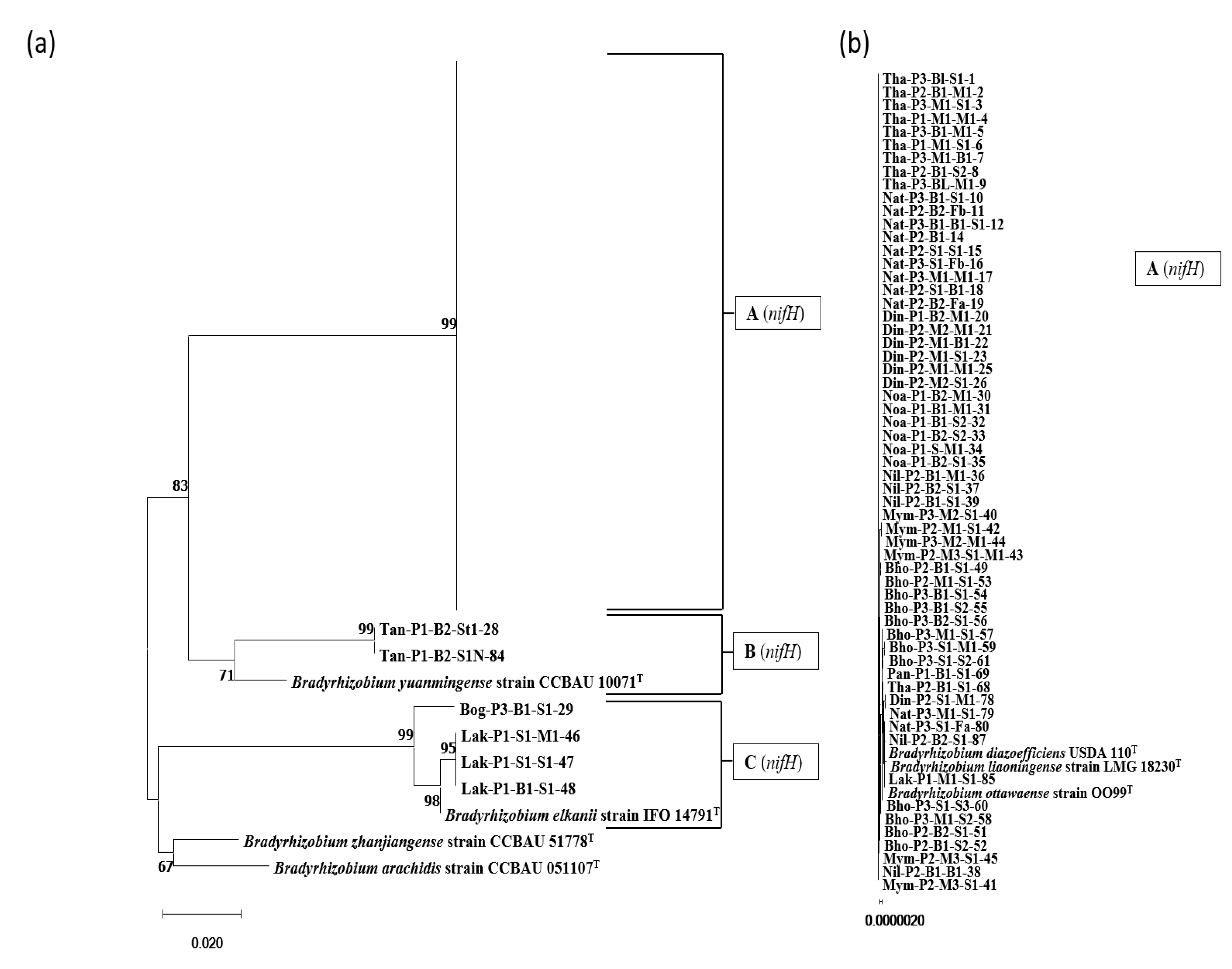
3.3. Phylogenetic Analysis Based on the nifH Gene

A total of 65 isolates and seven closely similar type strains were phylogenetically characterized based on 718 nt long DNA fragments from the nifH region. As shown in Figure 3a, the isolates were classified into three groups. Group A (nifH) comprised the majority of the isolates (59/65) positioned in the same branch as the reference strains of B. diazoefficiens (Figure 3b). Isolates of group B (nifH) were spotted in the same clade as the strains of B. yuanmingense. Four isolates from group C (nifH) resembled the B. elkanii strains.

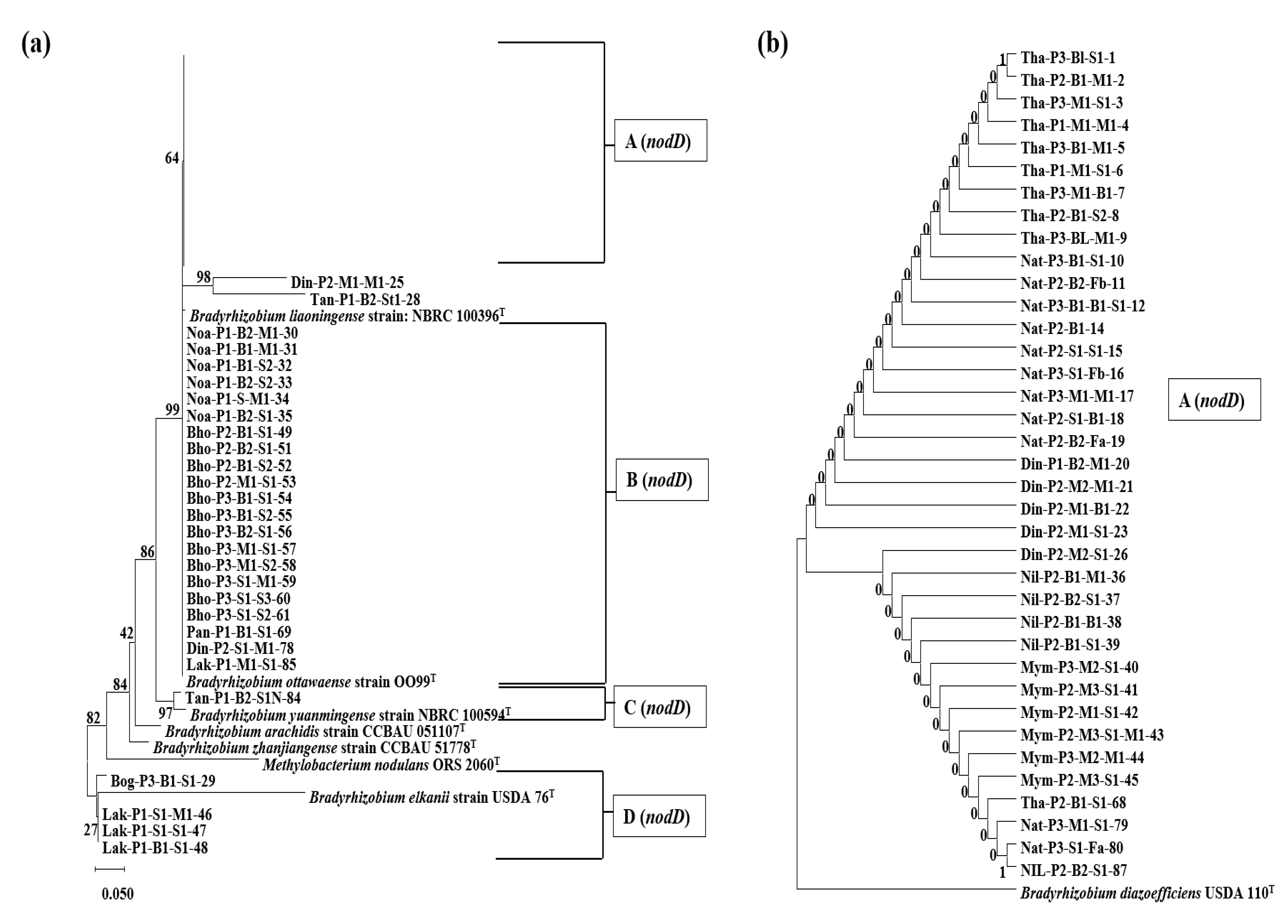
3.4. Phylogenetic Analysis Based on the nodD1 Gene

A total of 65 isolates and eight reference type strains were phylogenetically classified based on 353 nt long DNA fragments from the nodD1 region. As shown in Figure 4, four groups were identified. Group A (nodD) consisted of 37 isolates clustered in the same branch as the B. diazoefficiens strains, with B. liaoningense in the neighboring branch, and 21 isolates of group B (nodD) were spotted in the same branch as B. ottawaense. Group C (nodD) and group D (nodD), containing one and four isolates, were formed with type strains of B. yuanmingense and B. elkanii, respectively, which was almost similar to the results of previous phylogenetic trees (Figure 2 and Figure 3). One of the isolates, Tan-P1-B2-St1-28, which showed similarity with B. yuanmingense in the 16S rRNA- and nifH-based trees, and Din-P2-M1-M1-25 isolate, a sequence similarity of ~83% (100% query coverage) with B. diazoefficiens, were found in a separate branch, with B. diazoefficiens, B. ottawaense, and B. liaoningense strains in the neighboring branches.

3.5. Selection of Isolates for Further Genetic and Physiological Characterization

From the 16S rRNA sequence analysis, it was found that, among the 84 isolates, 71 were from Alpha-proteobacteria, seven from Beta-proteobacteria, two from Gamma-proteobacteria, three from Firmicutes, and one from Actinobacteria (Figure S1). Of the 84 isolates from 11 districts/locations in Bangladesh, 13 representative Bradyrhizobium isolates were selected based on location and the phylogenetic analysis of the 16S rRNA, nifH, and nodD1 genes for pot experiments, as well as further genetic and physiological characterization.

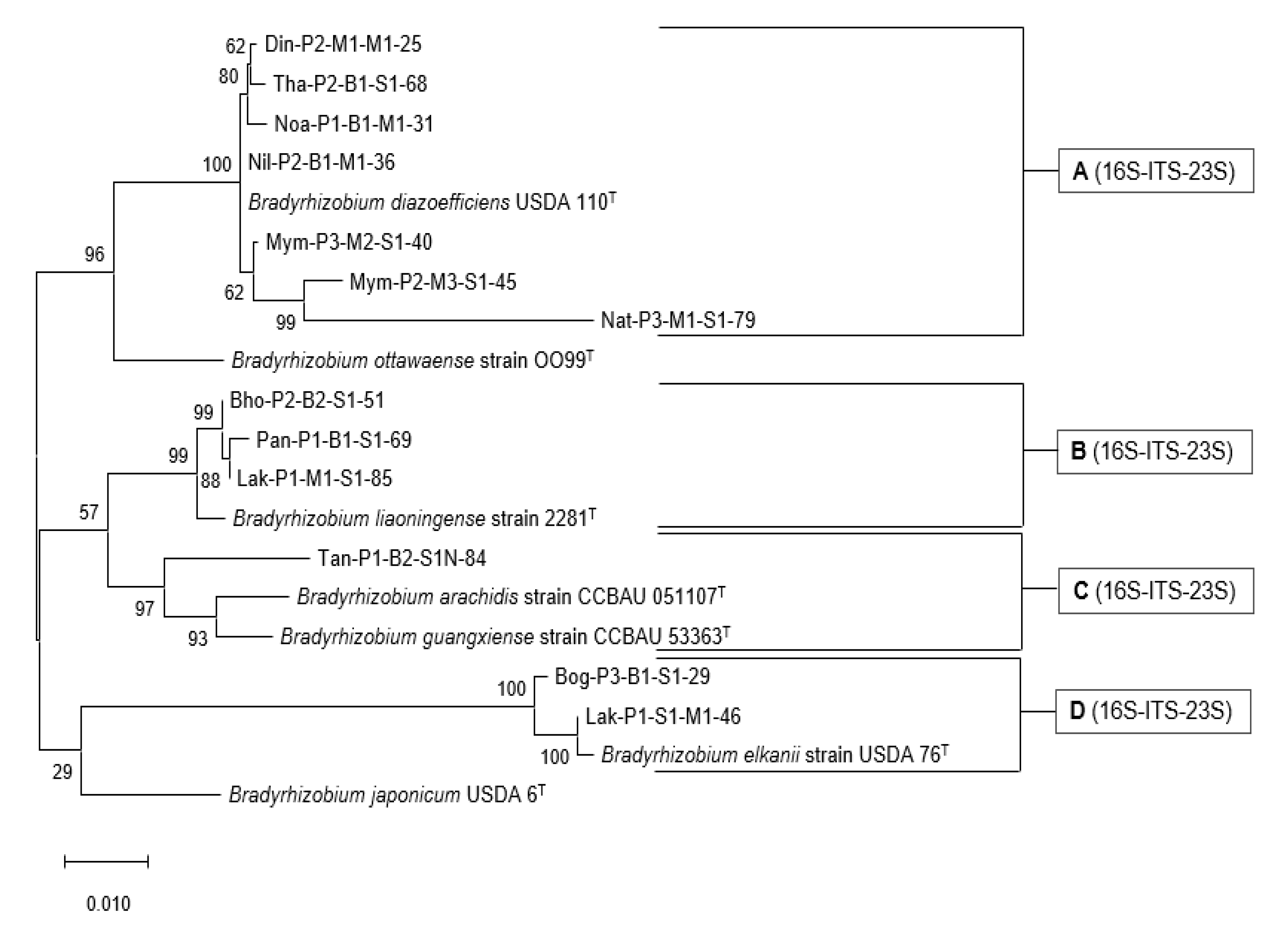
3.6. Phylogenetic Analysis of the rrn Operon (16S-ITS-23S)

Phylogenetic analysis of the partial rrn operon (16S-ITS-23S) was performed by concatenating the almost complete 16S rRNA gene, full-length 16S-23S internal transcribed spacer (ITS) region, and partial 23S rRNA gene sequences of the selected isolates (Figure 5). The length of the sequences of the isolates was 2610-2685 nt. The selected isolates and seven reference type strains were phylogenetically characterized using the maximum likelihood method, and four groups were obtained. Group A (16S-ITS-23S) comprised seven isolates that branched with B. diazoefficiens. Three isolates from group B (16S-ITS-23S) were closely related to B. liaoningense, and one isolate from group C (16S-ITS-23S) was closely related to Bradyrhizobium arachidis and Bradyrhizobium guangxiense. Two isolates clustered with B. elkanii in Group D (16S-ITS-23S).

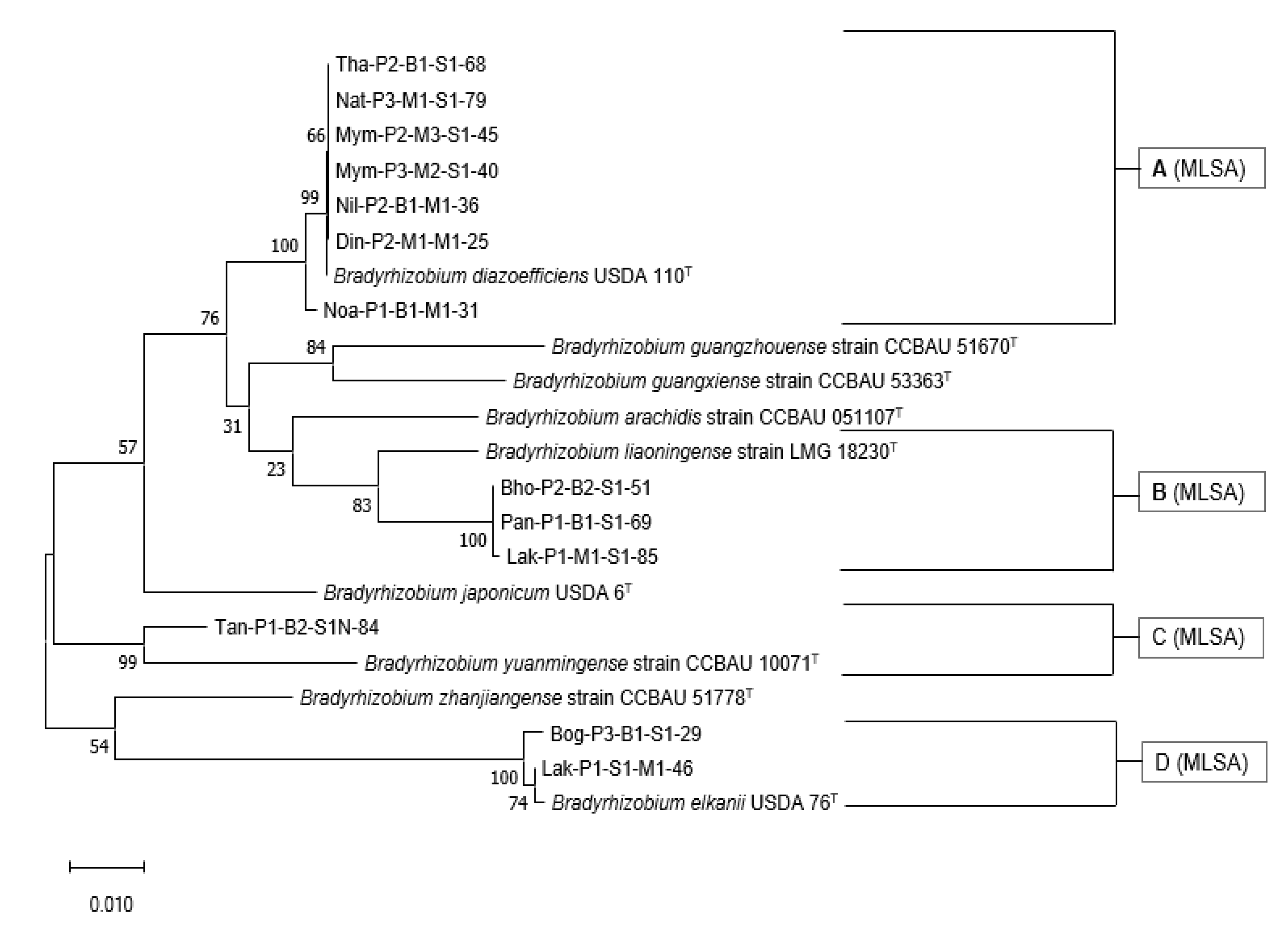
3.7. Multi-Locus Sequence Analysis of the Housekeeping Genes

Multi-locus sequence analysis (MLSA) was performed by concatenating the almost complete atpD, glnII, and gyrB genes (total of 2559 nt length of sequence of each isolate) of selected Bradyrhizobium isolates with reference strains using the maximum likelihood method (Figure 6). The selected isolates and nine reference strains were phylogenetically characterized using the maximum likelihood method, and four groups were obtained.
Group A (MLSA) comprised of seven isolates that branched with B. diazoefficiens. Three isolates from Group B (MLSA) and one isolate from Group C (MLSA) were closely related to B. liaoningense and B. yuanmingense, respectively. Two isolates clustered with B. elkanii in Group D (MLSA).

3.8. Symbiotic Performances

To assess the symbiotic ability of the 13 selected isolates, pot experiments were performed under controlled laboratory conditions using two soybean varieties, Glycine max cv. Enrei and Binasoybean-3. Different parameters, such as shoot length, main root length, nodule color (Figure S2), nodule size and number, shoot weight, root weight, nodule weight, and ARA, were measured.
The data of nodule number of different sizes, (e.g., medium size (2–5 mm) and small size (<2 mm)) of both varieties inoculated with bacteria are presented in Table 2. Isolates Bho-P2-B2-S1-51 and Mym-P2-M3-S1-45 produced the highest number of medium- and small-sized nodules in Enrei plants, respectively. Similar to the Enrei variety, in the case of Binasoybean-3, Bho-P2-B2-S1-51 produced the highest number of medium-sized nodules, whereas Din-P2-M1-M1-25 produced the highest number of small-sized nodules. The total number of nodules produced was higher in the Binasoybean-3 variety than that in the Enrei variety.
The shoot and root lengths of soybean plants (Enrei variety) inoculated with selected 13 isolates and B. diazoefficiens USDA110 are shown in Figure S3a. Most of the isolated bacteria (11/13) enhanced shoot growth in inoculated plants in comparison to B. diazoefficiens USDA110, and Lax-P1-M1-S1-46 isolates increased shoot growth and Bog-P3-B1-S1-29 demonstrated the highest root length development, but none of the isolates produced statistically significantly higher shoot or root growth than B. diazoefficiens USDA110.
In the Binasoybean-3 variety, most of the isolates (10/13) enhanced shoot growth in inoculated plants compared to B. diazoefficiens USDA110. However, among them, only the Tha-P2-B1-S1-68 isolate showed significantly higher shoot length than B. diazoefficiens USDA110 (Figure S3b). In the case of root length, no significant increase was observed in comparison with B. diazoefficiens USDA110, and Bog-P3-B1-S1-29 produced the highest root length among all bacteria.
The data on shoot dry weight (DW), root DW, and nodule DW of soybean plants inoculated with isolates and B. diazoefficiens USDA110 are presented in the bar chart in Figure S4. With regard to the soybean Enrei variety, most of the plants inoculated with isolated bacteria accumulated similar or higher amounts of shoot dry weight in comparison to B. diazoefficiens USDA110. However, in the case of root DW and nodule DW, only a few isolates produced higher amounts (Figure S4a). None of the isolates showed significantly higher shoot, root, or nodule DW gain than B. diazoefficiens USDA110. Din-P2-M1-M1-25, Bog-P3-B1-S1-29, and Nat-P3-M1-S1-79 isolates stimulated the highest shoot DW, root DW, and nodule DW, respectively.
The shoot DW, root DW, and nodule DW of the Binasoybean-3 variety measured from pot experiments are displayed in Figure S4b. Compared to B. diazoefficiens USDA110, a slight increase in shoot DW yield was observed in plants inoculated with some isolates (6/13). Pan-P1-B1-S1-69 isolate stimulated the highest shoot DW. In contrast to the Enrei variety, more isolates induced a higher root and nodule DW in the Binasoybean-3 variety and the Nil-P2-B1-M1-36 isolate produced a significantly higher amount of nodule DW in comparison to B. diazoefficiens USDA110 (Figure S4b). This isolate also produced the highest root DW.
The results of biomass production (shoot + root + nodule DW) and ARA activity in the Enrei variety and Binasoybean-3 inoculated with isolated bacteria and B. diazoefficiens USDA110 are shown in Table 3. In the case of the Enrei variety, no substantial difference in biomass production was observed between plants inoculated with isolates and B. diazoefficiens USDA110. Some isolates produced higher amounts of plant biomass in comparison with B. diazoefficiens USDA110, and Din-P2-M1-M1-25 isolate produced the highest plant biomass among isolates, which was 7.4% higher than B. diazoefficiens USDA110. Regarding ARA activity, 8 out of 13 isolates demonstrated higher ARA activity than B. diazoefficiens USDA110, and Bho-P2-B2-S1-51 showed significantly higher ARA activity than B. diazoefficiens USDA110.
For the Binasoybean-3 variety, considerable differences were not observed between plants inoculated with isolates and B. diazoefficiens USDA110. Six isolates produced equal or higher amounts of plant biomass in comparison with B. diazoefficiens USDA110, and Nil-P2-B1-M1-36 isolate produced the highest plant biomass among all the isolates, which was 14% higher than B. diazoefficiens USDA110. Regarding ARA activity, six isolates out of 13 demonstrated higher ARA activity than B. diazoefficiens USDA110, and Bho-P2-B2-S1-51 showed significantly higher ARA activity than B. diazoefficiens USDA110. Bho-P2-B2-S1-51 was the only isolate that produced significant ARA in both the Enrei and Binasoybean-3 varieties.

3.9. Stress Tolerance Tests

Data from the stress tolerance tests (salinity, temperature, and pH) are shown in Table 4. In the salinity tolerance test, all the investigated isolates grew at 1–4% NaCl concentration. In the temperature tolerance test, all the isolates grew at 20 °C, 28 °C, and 37 °C. Interestingly, all the isolates grew at 40 °C, furthermore, the Bho-P2-B2-S1-51, Bog-P3-B1-S1-29, Mym-P2-M3-S1-45, Nat-P3-M1-S1-79, and Tha-P2-B1-S1-68 isolates survived at 45 °C. Some of the isolates grew at 10 °C. In the pH tolerance test, all isolates developed colonies at pH 6.0–10.0. Except for Mym-P3-M2-S1-40 and Noa-P1-B1-M1-31, all isolates also grew at pH 4.5.

4. Discussion

The principal objective of this study was to achieve a fundamental understanding of the diversity, physiology, and symbiotic characteristics of nitrogen-fixing bacteria associated with soybean root nodules in Bangladeshi soil samples. To accomplish these goals, experiments related to bacterial isolation from root nodules, the phylogenetic characterization of bacterial strains with the 16S rRNA, rrn operon (16S-ITS-23S), nifH, nodD1, atpD, glnII, and gyrB genes, stress-tolerance tests, and plant tests in pots were performed.
Soil samples were collected from 11 different districts in Bangladesh, focusing on areas where soybean had been cultivated. Soybean plants were grown in pots using suspensions of the soil samples, and 84 isolates were isolated from the nodules of the soybean plants. Phylogenetic analysis of 84 soybean root-nodule isolates using the 16S rRNA gene revealed that the majority of the isolates (65/84) showed a phylogenetic resemblance with the genus Bradyrhizobium (Figure 2). Among these isolates, 45 showed a close relationship with B. diazoefficiens and B. japonicum, 16 with B. liaoningense and B. yuanmingense, and four with B. elkanii. The prevalence of Bradyrhizobium strains, especially B. diazoefficiens and B. japonicum, in root nodules of soybean is a common scenario and is scientifically expected. B. diazoefficiens (and/or B. japonicum) and B. elkanii species have been found in diverse climatic regions across the world, and strains of B. liaoningense are abundantly found in alkaline soils of temperate to subtropical climates in South and Southeast Asia, while the warm tropical climates of India and Nepal support B. yuanmingense [32]. Furthermore, in a research expedition for the genetic analysis of 76 bacteria isolated from root nodules of soybean inoculated with soil samples from Myanmar, India, Nepal, and Vietnam, Vinuesa et al. (2008) showed that 75 of the isolates were phylogenetically similar to B. japonicum (USDA110) or currently, B. diazoefficiens (in Nepal), B. liaoningense (in Myanmar, Vietnam), B. yuanmingense (in India, Myanmar, and Vietnam), and B. elkanii (in Myanmar) [28]. Another study on root-nodule isolates of soybean from five regions in India, performed by Appunu et al. (2008), showed that 36% of isolates were identified as B. yuanmingense, 26% as B. liaoningense, and 38% were different from all described Bradyrhizobium species but showed the same symbiotic genotype as B. liaoningense and B. japonicum [33].
Besides these slow-growing Bradyrhizobium isolates of the Alpha-proteobacteria class, some other bacterial strains, such as Methylobacterium of Alpha-proteobacteria, Pandoraea of Beta-proteobacteria, Stenotrophomonas of Gamma-proteobacteria, Bacillus of Firmicutes, and Leifsonia of Actinobacteria phylum/class, were also found in this study. Methylobacterium of the Methylobacteriaceae family and Rhizobiales order, belonging to the class Alpha-proteobacteria, is a well-established group of bacteria associated with the root nodules of leguminous plants [34,35,36,37]. Bacterial strains of the genus Burkholderia of the Burkholderiaceae family of Beta-proteobacteria have also been isolated by many researchers from root nodules [6,8,38,39]. Therefore, the isolation of bacteria belonging to the Pandoraea genus of the Burkholderiaceae family of the Beta-proteobacteria class is new but not unnatural. Stenotrophomonas bacteria of the Gamma-proteobacteria class have been isolated from the rhizosphere soil of leguminous plants [40]. Therefore, the existence of bacteria of this genus in root-nodules is also reasonable. Different species of Bacillus are commonly found in the root nodules of different leguminous plants and are thus treated as a well-known phenomenon [24,25,41]. In addition, many bacterial strains belonging to the phylum Actinobacteria, such as Frankia, Rhodococcus, Arthrobacter, Brevibacterium, Micromonospora, and Streptomyces, have also been isolated from root nodules and characterized for their nitrogen-fixing or plant-growth-promoting capabilities [3,42,43,44,45,46].
According to researchers, the analysis of symbiotic genes, such as nifH, nifD, nifK, nodA, nodB, nodC, and nodD, of root nodule bacteria can provide valuable insights into their host range and symbiotic relationships, which may vary from those expected by rRNA-based phylogenies [6,20,38,47]. In the nifH phylogenetic analysis, 59 out of 65 Bradyrhizobium isolates possessed the nifH gene, similar to that of B. diazoefficiens (Figure 3). Except for the four isolates that showed 16S rRNA gene resemblance with B. elkanii (group C (16S) of Figure 2), and two isolates out of 16 of Bradyrhizobium liaoningense and Bradyrhizobium yuanmingense (group B (16S) of Figure 2c), all other isolates showed nifH gene similarity to B. diazoefficiens. In an analysis of a database consisting of 32,954 aligned sequences of the nifH gene, Gaby et al. (2014) revealed that genomes that had >97% similarity in the 16S rRNA gene could possess up to 23% dissimilarity in the nifH sequences [48]. Therefore, B. yuanmingense and B. liaoningense, which have a 16S rRNA gene similarity of approximately 99% with B. diazoefficiens, can logically possess the nifH gene resembling B. diazoefficiens, which has approximately 10% sequence dissimilarity. This transfer of the nifH gene may be the outcome of vertical gene transfer in the symbiotic region of Bradyrhizobium species. In the case of nodD1 gene phylogeny, almost similar results were obtained for the nifH gene tree, with some discrepancies. Most isolates (n = 37) clustered in the same clade as B. diazoefficiens (in group A (nodD) of Figure 4b). The Tan-P1-B2-St1-28 and Din-P2-M1-M1-25 isolates showed a sequence similarity of ~83% (100% query coverage) with B. diazoefficiens and were placed in a different branch adjacent to Bradyrhizobium branches. No change was observed in the four isolates that showed similarity with B. elkanii in the nodD1 gene tree or the 16S rRNA and nifH tree, suggesting a strong phylogenetic relation with B. elkanii. Therefore, further analysis should be performed to verify the symbiotic gene transfer process.
Some researchers have shown that the analysis of the rrn operon (16S–ITS–23S) signifies more than the analysis of the 16S rRNA gene for the resolution of taxa at the species level [49,50]. Phylogenetic analysis of the partial rrn operon consisting of the almost complete 16S rRNA gene, full-length 16S-23S internal transcribed spacer (ITS) region, and partial 23S rRNA gene sequences for the selected isolates was congruent with the 16S rRNA gene-based tree (Figure 5). MLSA was also performed by concatenating the atpD, glnII, and gyrB genes of selected Bradyrhizobium isolates with type strains using the maximum likelihood method (Figure 6). Seven of the 13 isolates branched with B. diazoefficiens, three branched with B. liaoningense, and one branched with B. yuanmingense. Two isolates clustered with B. elkanii. For all these isolates, the MLSA position could be considered the true taxonomic identity. The use of MLSA of the housekeeping genes in bacterial species definition and identification has been strongly endorsed by the scientific community [51]. Unless whole-genome sequencing is performed, MLSA is considered a distinctive identification process for exploring the genetic diversity within a proposed species [51]. Therefore, based on 16S rRNA gene, MLSA and rrn operon phylogenetic analysis, except for the Tan-P1-B2-S1N-84, the other 12 isolates can be classified as B. diazoefficiens sp. Din-P2-M1-M1-25, B. diazoefficiens sp. Mym-P2-M3-S1-45, B. diazoefficiens sp. Mym-P3-M2-S1-40, B. diazoefficiens sp. Nat-P3-M1-S1-79, B. diazoefficiens sp. Nil-P2-B1-M1-36, B. diazoefficiens sp. Noa-P1-B1-M1-31, B. diazoefficiens sp. Tha-P2-B1-S1-68, B. elkanii sp. Bog-P3-B1-S1-29, B. elkanii sp. Lak-P1-S1-M1-46, B. liaoningense sp. Bho-P2-B2-S1-51, B. liaoningense sp. Lak-P1-M1-S1-85, B. liaoningense sp. Pan-P1-B1-S1-69 for taxonomic identity. For Tan-P1-B2-S1N-84 isolate further analysis is needed to confirm the taxonomic identity.
In this study, among the different phylogenetic methods, including Bayesian inference, maximum-likelihood, maximum-parsimony, and neighbour-joining, the maximum-likelihood method was applied for phylogenetic reconstruction. Although each of these techniques has its strengths and weaknesses, maximum-likelihood (ML) and Bayesian methods typically provide more realistic and robust phylogenies because they employ explicit models of molecular evolution [51]. The analysis of the highest DNA sequence similarity of the selected isolates with Bradyrhizobium type strains showed no variation in the case of Bradyrhizobium diazoefficiens isolates for all analyzed genes. However, some discrepancies were observed for isolates of other genera (Table 5). Among the housekeeping genes, gyrB was found to be more decisive in identifying the isolates than atpD and glnII, and showed similarities with rrn operon sequence data.
To evaluate the symbiotic ability of the 13 selected isolates, pot experiments were performed using two soybean varieties, Glycine max cv. Enrei and Binasoybean-3, and different parameters were measured. All isolates formed red or pink (cross-sections) nodules, indicating the presence of leghemoglobin in the soybean Enrei variety and Binasoybean-3 variety, except Tan-P1-B2-S1N-84 belonging to B. yuanmingense, which did not form any nodules in the Enrei variety (Figure S2, Table 2). The variation in nodule production could be due to the variation in the agronomic traits of the two soybean cultivars and differences in the compatibility of the isolates with the cultivars. Wang et al. (2019) described that soybean cultivars can influence nodule formation, and some bacteria can form nodules in some varieties, while others cannot [3]. The number of isolates that formed nodules and the number of nodules formed were higher in the Binasoybean-3 variety than those in the Enrei variety (Table 2). This could be due to the fact that Binasoybean-3 is a local Bangladeshi variety, meaning that isolates of Bangladeshi soil samples have a higher affinity towards it.
The performance of the isolates varied in terms of effective symbiosis and biomass production. The nodulation and nitrogen fixation depends on many aspects, especially the symbiotic genes (such as such as nifH, nifD, nifK, nodA, nodB, nodC, and nod, etc.), which can vary from rhizobia to rhizobia; thus, different rhizobial species show different host-specificity and nitrogen fixation performances [3,7]. When inoculated, some of the isolates enhanced biomass production in both the Enrei and Binasoybean-3 varieties in comparison to B. diazoefficiens USDA110, whereas some decreased. In the case of Enrei, the Din-P2-M1-M1-25 isolate produced the highest plant biomass among isolates, which was 7.4% higher than B. diazoefficiens USDA110. For Binasoybean-3, Nil-P2-B1-M1-36 isolate produced the highest plant biomass among all the isolates, which was 14% higher than B. diazoefficiens USDA110. In comparison to B. diazoefficiens USDA110, three isolates produced 10% or higher biomass gain in Binasoybean-3 variety, compared to no isolate in the case of the Enrei variety. These results suggest that the isolates were more compatible with Bangladeshi local varieties. Having a phylogenetic similarity with B. liaoningense, Bho-P2-B2-S1-51 was the only isolate that produced a significant increase in ARA compared to B. diazoefficiens USDA110 in both the Enrei and Binasoybean-3 varieties, increasing plant biomass production by 9% in case of Binasoybean-3 variety. Therefore, this isolate could be a potential biofertilizer candidate. Regarding Binasoybean-3, the Tha-P2-B1-S1-68 isolate belonging to B. diazoefficiens significantly increased shoot length compared to B. diazoefficiens USDA110 (Figure S3) and enhanced biomass production by 10% (Table 3), making the isolate a suitable candidate as biofertilizer for local varieties. According to Chen et al. (2021), the highly effective symbiosis could only be obtained when the host specificity and habitat specificity cooperated in a rhizobial symbiont; therefore, the rhizobia and legume variety of same locality can demonstrate better symbiotic effects [7].The relationships between different parameters of pot experiments, e.g., nodule number, small and medium size nodule, nodule dry weight, ARA activity, and biomass, were observed to be non-linear and varied from isolate to isolate (Table 6). Among the different parameters, it was observed that, for both soybean varieties, nodule dry weight had a significant positive correlation with ARA (in µmol/h/plant) and biomass DW, suggesting that with an increase in nodule dry weight, ARA (in µmol/h/plant) and biomass production may increase. It is known that nodule dry weight is a good indicator of symbiotic efficacy, and an important parameter in strain evaluation [52]. Allito et al. also observed similar correlation and mentioned nodule dry weight as one of the key indicators of effective legume-rhizobia symbiosis [53]. Although the medium-size nodule number showed a significantly high positive correlation with biomass DW and ARA (in µmol/h/plant) in Binasoybean-3, small nodule numbers displayed insignificant and/or negative correlations, suggesting that a larger nodule size can significantly contribute to biomass production and ARA (in µmol/h/plant) than immature/smaller ones. No significant positive correlations were observed with other parameters. Similar to these results, some researchers did not find any clear relationship between ARA activity, nodule numbers and plant biomass in their studies [25,54].
In the stress tolerance tests, all the selected isolates grew at 1–4% NaCl concentration. Similarly, most isolates grew at pH 4.5, except four isolates, and almost all isolates grew at pH 4.5–10, which demonstrate that the isolates are tolerant to high salt and pH conditions. Under saline conditions, salt-tolerant rhizobia are beneficial for host plant growth. For instance, the growth of alfalfa is enhanced when inoculated with the salt-tolerant Rhizobium strain Rm1521 rather than the salt-sensitive strain A2 under NaCl treatment [55]. Among the isolates, five isolates, namely, Bho-P2-B2-S1-51, Bog-P3-B1-S1-29, Mym-P2-M3-S1-45, Nat-P3-M1-S1-79, and Tha-P2-B1-S1-68, survived at 45 °C, demonstrating their suitability as biofertilizer candidates for tropical environments. Chen et al. (2002) isolated Rhizobia from soybeans that could grow at 40 °C [56]. Correspondingly, Phaseolus vulgaris nodule bacteria were found to survive at 47 °C [57], and Rhizobium isolated from Sesbania aculeata plants were observed to grow at 50 °C [58,59]. As temperature, salinity, origin of cultivar, etc., are prominent effectors of rhizobial symbiosis [19], the isolates with temperature and salinity tolerance could be potential biofertilizers, especially for local legume varieties.

5. Conclusions

This study represents the first genetic and physiological characterization of soybean-nodulating isolates from different regions of Bangladesh, one of the top soybean oil consumer countries of the world. Surprisingly, diverse arrays of both nodule-forming and endosymbiotic bacteria belonging to Bradyrhizobium and Methylobacterium of Alphaproteobacteria, Pandoraea of Beta-proteobacteria, Stenotrophomonas of Gamma-proteobacteria, Bacillus of Firmicutes, and Leifsonia of Actinobacteria phylum/class were found to be present in the root nodules of soybean plants grown in pots using a suspension of soil samples from Bangladesh, with some potential isolates showing a higher symbiotic ability than B. diazoefficiens USDA110 in pot experiments. One of the selected isolates, Bho-P2-B2-S1-51, which showed a phylogenetic similarity to B. liaoningense, produced a significant increase in ARA compared to B. diazoefficiens USDA110 in both the Enrei and Binasoybean-3 varieties, induced plant biomass production by 9% in case of Binasoybean-3 variety, and survived at 45 °C. Therefore, this isolate could be a potential biofertilizer candidate. With regard to Binasoybean-3, the Tha-P2-B1-S1-68 isolate, which showed a phylogenetic similarity to B. diazoefficiens, also significantly increased shoot length compared to B. diazoefficiens USDA110, enhanced biomass production by 10%, and could grow at 45 °C, making this isolate a suitable candidate for use as biofertilizer for local Bangladeshi soybean varieties. Further studies, such as whole-genome sequence analysis and field trials, will need to be performed for prospective candidate isolates as biofertilizers to acquire an in-depth understanding of the genetic make-up and functional characteristics of soybean-rhizobium symbiosis.

Supplementary Materials

The following supporting information can be downloaded at: https://www.mdpi.com/article/10.3390/microorganisms10112282/s1, Table S1: Information of primers used in this study.; Table S2: Basic information of soybean varieties used in this study.; Table S3: List of selected Bradyrhizobium isolates for pot experiments and further characterization.; Figure S1: Doughnut graph (a) and map of Bangladesh (b) showing the distribution of different types of bacteria.; Figure S2. Pictures of dissected nodules of soybean Enrei variety and Binasoybean-3 variety inoculated with selected isolates and B. diazoefficiens USDA110.; Figure S3: Shoot and root length of soybean Enrei variety (a) and Binasoybean-3 variety (b) inoculated with selected isolates and B. diazoefficiens USDA110. Data are presented in average value with error bar denoting the STDEV of three plants each. ‘*’ Denotes significance with “B. diazoefficiens USDA110” at 95% confidence level using Dunnett’s test.; Figure S4: Shoot dry weight (DW), root DW and nodule DW of soybean Enrei variety (a) and Binasoybean-3 variety (b) inoculated with isolates and B. diazoefficiens USDA110. Data are presented in average value with error bar denotes STDEV of three plants each. ‘*’ Denotes significance with “B. diazoefficiens USDA110” at 95% confidence level using Dunnett’s test.

Author Contributions

Conceptualization, M.F.M., S.D., H.S., T.Y., S.O. and N.O.-O.; methodology, M.F.M., S.D., T.I., S.-i.A. and S.O.; formal analysis, M.F.M.; investigation, M.F.M., S.D. and T.I.; resources, T.Y.; data curation, M.F.M.; writing—original draft preparation, M.F.M.; writing—review and editing, S.D., S.-i.A. and N.O.-O.; visualization, M.F.M.; supervision, H.S., T.Y., S.O. and N.O.-O.; project administration, N.O.-O.; funding acquisition, T.Y. and N.O.-O. All authors have read and agreed to the published version of the manuscript.

Funding

This work was supported by a Grant-in-Aid for Scientific Research (20KK0136) from the Japan Society for the Promotion of Science (JSPS) and the Ministry of Education, Culture, Sports, Science, and Technology (MEXT).

Data Availability Statement

All relevant data can be accessed in the manuscript and Supporting Information files.

Acknowledgments

The authors thank the Ministry of Science and Technology, Bangladesh and the Bangladesh Atomic Energy Commission for their momentous help and support.

Conflicts of Interest

The authors declared that there are no conflict of interest.

References

  1. Liu, M.; Shi, X.; Peng, K.; Xiao, F.; Sun, M.; Liu, X.; Shi, K.; Peng, F.; Xiao, Y. Effects of Root Zone Aeration on Soil Microbes Species in a Peach Tree Rhizosphere and Root Growth. Microorganisms 2022, 10, 1879. [Google Scholar] [CrossRef]
  2. Colwell, R.R. Microbial Diversity: The Importance of Exploration and Conservation. J. Ind. Microbiol. Biotechnol. 1997, 18, 302–307. [Google Scholar] [CrossRef] [PubMed]
  3. Wang, E.T.; Tian, C.F.; Chen, W.F.; Young, J.P.W.; Chen, W.X. Ecology and Evolution of Rhizobia; Springer: Singapore, 2019. [Google Scholar] [CrossRef]
  4. Martínez-Hidalgo, P.; Hirsch, A.M. The Nodule Microbiome: N2 fixing Rhizobia Do Not Live Alone. Phytobiomes J. 2017, 1, 70–82. [Google Scholar] [CrossRef] [Green Version]
  5. Wagner, S.C. Biological Nitrogen Fixation. Nat. Educ. Knowl. 2011, 3, 15. [Google Scholar]
  6. Binde, D.R.; Menna, P.; Bangel, E.V.; Barcellos, F.G.; Hungria, M. Rep-PCR Fingerprinting and Taxonomy Based on the Sequencing of the 16S RRNA Gene of 54 Elite Commercial Rhizobial Strains. Appl. Microbiol. Biotechnol. 2009, 83, 897–908. [Google Scholar] [CrossRef] [PubMed]
  7. Chen, W.F.; Wang, E.T.; Ji, Z.J.; Zhang, J.J. Recent Development and New Insight of Diversification and Symbiosis Specificity of Legume Rhizobia: Mechanism and Application. J. Appl. Microbiol. 2021, 131, 553–563. [Google Scholar] [CrossRef]
  8. Cevallos, M.A.; Degli Esposti, M. New Alphaproteobacteria Thrive in the Depths of the Ocean with Oxygen Gradient. Microorganisms 2022, 10, 455. [Google Scholar] [CrossRef]
  9. Moulin, L.; Munive, A.; Dreyfus, B.; Boivin-Masson, C. Nodulation of Legumes by Members of the Beta-Subclass of Proteobacteria. Nature 2001, 411, 948–950. [Google Scholar] [CrossRef]
  10. Shiraishi, A.; Matsushita, N.; Hougetsu, T. Nodulation in Black Locust by the Gammaproteobacteria Pseudomonas Sp. and the Betaproteobacteria Burkholderia sp. Syst. Appl. Microbiol. 2010, 33, 269–274. [Google Scholar] [CrossRef]
  11. Estrada-de los Santos, P.; Palmer, M.; Chávez-Ramírez, B.; Beukes, C.; Steenkamp, E.; Briscoe, L.; Khan, N.; Maluk, M.; Lafos, M.; Humm, E.; et al. Whole Genome Analyses Suggests That Burkholderia Sensu Lato Contains Two Additional Novel Genera (Mycetohabitans gen. nov., and Trinickia gen. nov.): Implications for the Evolution of Diazotrophy and Nodulation in the Burkholderiaceae. Genes 2018, 9, 389. [Google Scholar] [CrossRef]
  12. Iturralde, E.T.; Covelli, J.M.; Alvarez, F.; Pérez-Giménez, J.; Arrese-Igor, C.; Lodeiro, A.R. Soybean-Nodulating Strains with Low Intrinsic Competitiveness for Nodulation, Good Symbiotic Performance, and Stress-Tolerance Isolated from Soybean-Cropped Soils in Argentina. Front. Microbiol. 2019, 10, 1061. [Google Scholar] [CrossRef] [PubMed]
  13. Jaiswal, S.K.; Dakora, F.D. Widespread Distribution of Highly Adapted Bradyrhizobium Species Nodulating Diverse Legumes in Africa. Front. Microbiol. 2019, 10, 310. [Google Scholar] [CrossRef] [PubMed] [Green Version]
  14. Zhang, Y.M.; Li, Y.; Chen, W.F.; Wang, E.T.; Tian, C.F.; Li, Q.Q.; Zhang, Y.Z.; Sui, X.H.; Chen, W.X. Biodiversity and Biogeography of Rhizobia Associated with Soybean Plants Grown in the North China Plain. Appl. Environ. Microbiol. 2011, 77, 6331–6342. [Google Scholar] [CrossRef] [Green Version]
  15. Mousavi, S.A.; Young, J.P.W. International Committee on Systematics of Prokaryotes, Subcommittee on the Taxonomy of Rhizobia and Agrobacteria, Minutes of the Annual Meeting by Videoconference, 5 July 2021, Followed by Online Discussion until 31 December 2021. Int. J. Syst. Evol. Microbiol. 2022, 72, 005453. [Google Scholar] [CrossRef]
  16. Parte, A.C.; Carbasse, J.S.; Meier-Kolthoff, J.P.; Reimer, L.C.; Göker, M. List of Prokaryotic Names with Standing in Nomenclature (LPSN) Moves to the DSMZ. Int. J. Syst. Evol. Microbiol. 2020, 70, 5607–5612. [Google Scholar] [CrossRef] [PubMed]
  17. Chen, W.; Wang, E.; Wang, S.; Li, Y.; Chen, X.; Li, Y. Characteristics of Rhizobium tianshanense sp. nov., a Moderately and Slowly Growing Root Nodule Bacterium Isolated from an Arid Saline Environment in Xinjiang, People’s Republic of China. Int. J. Syst. Bacteriol. 1995, 45, 153–159. [Google Scholar] [CrossRef] [Green Version]
  18. Tian, C.F.; Zhou, Y.J.; Zhang, Y.M.; Li, Q.Q.; Zhang, Y.Z.; Li, D.F.; Wang, S.; Wang, J.; Gilbert, L.B.; Li, Y.R.; et al. Comparative Genomics of Rhizobia Nodulating Soybean Suggests Extensive Recruitment of Lineage-Specific Genes in Adaptations. Proc. Natl. Acad. Sci. USA 2012, 109, 8629–8634. [Google Scholar] [CrossRef] [PubMed] [Green Version]
  19. Yuan, K.; Reckling, M.; Ramirez, M.D.A.; Djedidi, S.; Fukuhara, I.; Ohyama, T.; Yokoyama, T.; Bellingrath-Kimura, S.D.; Halwani, M.; Egamberdieva, D.; et al. Characterization of Rhizobia for the Improvement of Soybean Cultivation at Cold Conditions in Central Europe. Microbes Environ. 2020, 35, ME19124. [Google Scholar] [CrossRef] [Green Version]
  20. Somasegaran, P.; Hoben, H.J. Handbook for Rhizobia: Methods in Legume-Rhizobium Technology; Springer-Verlag: New York, NY, USA, 1994. [Google Scholar]
  21. Somasegaran, P.; Hoben, H.J.; Somasegaran, P.; Hoben, H.J. Quantifying the Growth of Rhizobia. In Handbook for Rhizobia; Springer: New York, NY, USA, 1994; pp. 47–57. [Google Scholar] [CrossRef]
  22. Djedidi, S.; Yokoyama, T.; Ohkama-Ohtsu, N.; Risal, C.P.; Abdelly, C.; Sekimoto, H. Stress Tolerance and Symbiotic and Phylogenic Features of Root Nodule Bacteria Associated with Medicago Species in Different Bioclimatic Regions of Tunisia. Microbes Environ. 2011, 26, 36–45. [Google Scholar] [CrossRef]
  23. Risal, C.P.; Yokoyama, T.; Ohkama-Ohtsu, N.; Djedidi, S.; Sekimoto, H. Genetic Diversity of Native Soybean Bradyrhizobia from Different Topographical Regions along the Southern Slopes of the Himalayan Mountains in Nepal. Syst. Appl. Microbiol. 2010, 33, 416–425. [Google Scholar] [CrossRef]
  24. van Berkum, P.; Fuhrmann, J.J. Evolutionary Relationships among the Soybean Bradyrhizobia Reconstructed from 16S RRNA Gene and Internally Transcribed Spacer Region Sequence Divergence. Int. J. Syst. Evol. Microbiol. 2000, 50 Pt 6, 2165–2172. [Google Scholar] [CrossRef] [PubMed] [Green Version]
  25. Mortuza, M.F.; Tomooka, N.; Habibi, S.; Akatsu, T.; Djedidi, S.; Naito, K.; Sekimoto, H.; Okazaki, S.; Ohkama-Ohtsu, N.; Yokoyama, T. Multiphase Characterization of Wild Vigna Associated Root Nodule Bacteria from Japanese Subtropical Islands Unveiled Novel High Temperature Resistant Bradyrhizobium Strains Having High Symbiotic Compatibility with Soybean and Mungbean. Soil Sci. Plant Nutr. 2020, 66, 285–298. [Google Scholar] [CrossRef]
  26. Laguerre, G.; Nour, S.M.; Macheret, V.; Sanjuan, J.; Drouin, P.; Amarger, N. Classification of Rhizobia Based on NodC and NifH Gene Analysis Reveals a Close Phylogenetic Relationship among Phaseolus vulgaris Symbionts. Microbiology 2001, 147, 981–993. [Google Scholar] [CrossRef] [PubMed] [Green Version]
  27. Martens, M.; Dawyndt, P.; Coopman, R.; Gillis, M.; De Vos, P.; Willems, A. Advantages of Multilocus Sequence Analysis for Taxonomic Studies: A Case Study Using 10 Housekeeping Genes in the Genus Ensifer (Including Former Sinorhizobium). Int. J. Syst. Evol. Microbiol. 2008, 58, 200–214. [Google Scholar] [CrossRef] [Green Version]
  28. Vinuesa, P.; Rojas-Jiménez, K.; Contreras-Moreira, B.; Mahna, S.K.; Prasad, B.N.; Moe, H.; Selvaraju, S.B.; Thierfelder, H.; Werner, D. Multilocus Sequence Analysis for Assessment of the Biogeography and Evolutionary Genetics of Four Bradyrhizobium Species That Nodulate Soybeans on the Asiatic Continent. Appl. Environ. Microbiol. 2008, 74, 6987–6996. [Google Scholar] [CrossRef] [Green Version]
  29. Kumar, S.; Stecher, G.; Li, M.; Knyaz, C.; Tamura, K. MEGA X: Molecular Evolutionary Genetics Analysis across Computing Platforms. Mol. Biol. Evol. 2018, 35, 1547–1549. [Google Scholar] [CrossRef]
  30. Habibi, S.; Djedidi, S.; Prongjunthuek, K.; Mortuza, M.F.; Ohkama-Ohtsu, N.; Sekimoto, H.; Yokoyoma, T. Physiological and Genetic Characterization of Rice Nitrogen Fixer PGPR Isolated from Rhizosphere Soils of Different Crops. Plant Soil 2014, 379, 51–66. [Google Scholar] [CrossRef]
  31. Woliy, K.; Degefu, T.; Frostegård, Å. Host Range and Symbiotic Effectiveness of N2O Reducing Bradyrhizobium Strains. Front. Microbiol. 2019, 10, 2746. [Google Scholar] [CrossRef]
  32. Naamala, J.; Jaiswal, S.K.; Dakora, F.D. Microsymbiont Diversity and Phylogeny of Native Bradyrhizobia Associated with Soybean (Glycine max L. Merr.) Nodulation in South African Soils. Syst. Appl. Microbiol. 2016, 39, 336–344. [Google Scholar] [CrossRef]
  33. Appunu, C.; N’Zoue, A.; Laguerre, G. Genetic Diversity of Native Bradyrhizobia Isolated from Soybeans (Glycine max L.) in Different Agricultural-Ecological-Climatic Regions of India. Appl. Environ. Microbiol. 2008, 74, 5991–5996. [Google Scholar] [CrossRef] [Green Version]
  34. Ardley, J.K.; Reeve, W.G.; O’Hara, G.W.; Yates, R.J.; Dilworth, M.J.; Howieson, J.G. Nodule Morphology, Symbiotic Specificity and Association with Unusual Rhizobia Are Distinguishing Features of the Genus Listia within the Southern African Crotalarioid Clade Lotononis s.l. Ann. Bot. 2013, 112, 1–15. [Google Scholar] [CrossRef] [PubMed] [Green Version]
  35. Madhaiyan, M.; Poonguzhali, S.; Senthilkumar, M.; Sundaram, S.; Sa, T. Nodulation and Plant-Growth Promotion by Methylotrophic Bacteria Isolated from Tropical Legumes. Microbiol. Res. 2009, 164, 114–120. [Google Scholar] [CrossRef] [PubMed]
  36. Renier, A.; Jourand, P.; Rapior, S.; Poinsot, V.; Sy, A.; Dreyfus, B.; Moulin, L. Symbiotic Properties of Methylobacterium Nodulans ORS 2060T: A Classic Process for an Atypical Symbiont. Soil Biol. Biochem. 2008, 40, 1404–1412. [Google Scholar] [CrossRef]
  37. Sy, A.; Giraud, E.; Jourand, P.; Garcia, N.; Willems, A.; De Lajudie, P.; Prin, Y.; Neyra, M.; Gillis, M.; Boivin-Masson, C.; et al. Methylotrophic Methylobacterium Bacteria Nodulate and Fix Nitrogen in Symbiosis with Legumes. J. Bacteriol. 2001, 183, 214–220. [Google Scholar] [CrossRef] [PubMed] [Green Version]
  38. Dobritsa, A.P.; Samadpour, M. Transfer of Eleven Species of the Genus Burkholderia to the Genus Paraburkholderia and Proposal of Caballeronia gen. nov. to Accommodate Twelve Species of the Genera Burkholderia and Paraburkholderia. Int. J. Syst. Evol. Microbiol. 2016, 66, 2836–2846. [Google Scholar] [CrossRef]
  39. Gyaneshwar, P.; Hirsch, A.M.; Moulin, L.; Chen, W.M.; Elliott, G.N.; Bontemps, C.; De Los Santos, P.E.; Gross, E.; Dos Reis, F.B.; Janet, I.S.; et al. Legume-Nodulating Betaproteobacteria: Diversity, Host Range, and Future Prospects. Mol. Plant-Microbe Interact. 2011, 24, 1276–1288. [Google Scholar] [CrossRef] [Green Version]
  40. Hoque, M.S.; Broadhurst, L.M.; Thrall, P.H. Genetic Characterization of Root-Nodule Bacteria Associated with Acacia salicina and A. stenophylla (Mimosaceae) across South-Eastern Australia. Int. J. Syst. Evol. Microbiol. 2011, 61, 299–309. [Google Scholar] [CrossRef] [Green Version]
  41. Bai, Y.; D’Aoust, F.; Smith, D.L.; Driscoll, B.T. Isolation of Plant-Growth-Promoting Bacillus Strains from Soybean Root Nodules. Can. J. Microbiol. 2002, 48, 230–238. [Google Scholar] [CrossRef]
  42. Ampomah, O.Y.; Huss-Danell, K. Genetic Diversity of Root Nodule Bacteria Nodulating Lotus corniculatus and Anthyllis vulneraria in Sweden. Syst. Appl. Microbiol. 2011, 34, 267–275. [Google Scholar] [CrossRef]
  43. Palaniappan, P.; Chauhan, P.S.; Saravanan, V.S.; Anandham, R.; Sa, T. Isolation and Characterization of Plant Growth Promoting Endophytic Bacterial Isolates from Root Nodule of Lespedeza sp. Biol. Fertil. Soils 2010, 46, 807–816. [Google Scholar] [CrossRef]
  44. Barnawal, D.; Bharti, N.; Maji, D.; Chanotiya, C.S.; Kalra, A. ACC Deaminase-Containing Arthrobacter protophormiae Induces NaCl Stress Tolerance through Reduced ACC Oxidase Activity and Ethylene Production Resulting in Improved Nodulation and Mycorrhization in Pisum sativum. J. Plant Physiol. 2014, 171, 884–894. [Google Scholar] [CrossRef]
  45. Sreevidya, M.; Gopalakrishnan, S.; Kudapa, H.; Varshney, R.K. Exploring Plant Growth-Promotion Actinomycetes from Vermicompost and Rhizosphere Soil for Yield Enhancement in Chickpea. Brazilian J. Microbiol. 2016, 47, 85–95. [Google Scholar] [CrossRef] [PubMed] [Green Version]
  46. Tokala, R.K.; Strap, J.L.; Jung, C.M.; Crawford, D.L.; Salove, M.H.; Deobald, L.A.; Bailey, J.F.; Morra, M.J. Novel Plant-Microbe Rhizosphere Interaction Involving Streptomyces lydicus WYEC108 and the Pea Plant (Pisum sativum). Appl. Environ. Microbiol. 2002, 68, 2161–2171. [Google Scholar] [CrossRef] [Green Version]
  47. Moulin, L.; Béna, G.; Boivin-Masson, C.; Stepkowski, T. Phylogenetic Analyses of Symbiotic Nodulation Genes Support Vertical and Lateral Gene Co-Transfer within the Bradyrhizobium Genus. Mol. Phylogenet. Evol. 2004, 30, 720–732. [Google Scholar] [CrossRef]
  48. Gaby, J.C.; Buckley, D.H. A Comprehensive Aligned NifH Gene Database: A Multipurpose Tool for Studies of Nitrogen-Fixing Bacteria. Database 2014, 2014, bau001. [Google Scholar] [CrossRef] [PubMed] [Green Version]
  49. Cuscó, A.; Catozzi, C.; Viñes, J.; Sanchez, A.; Francino, O. Microbiota Profiling with Long Amplicons Using Nanopore Sequencing: Full-Length 16S RRNA Gene and the 16S-ITS-23S of the Rrn Operon. F1000Research 2019, 7, 1755. [Google Scholar] [CrossRef]
  50. Nygaard, A.B.; Tunsjø, H.S.; Meisal, R.; Charnock, C. A Preliminary Study on the Potential of Nanopore MinION and Illumina MiSeq 16S RRNA Gene Sequencing to Characterize Building-Dust Microbiomes. Sci. Rep. 2020, 10, 3209. [Google Scholar] [CrossRef] [Green Version]
  51. De Lajudie, P.M.; Andrews, M.; Ardley, J.; Eardly, B.; Jumas-Bilak, E.; Kuzmanović, N.; Lassalle, F.; Lindström, K.; Mhamdi, R.; Martínez-Romero, E.; et al. Minimal Standards for the Description of New Genera and Species of Rhizobia and Agrobacteria. Int. J. Syst. Evol. Microbiol. 2019, 69, 1852–1863. [Google Scholar] [CrossRef]
  52. Graham, P.H.; Hungria, M.; Tlusty, B. Breeding for Better Nitrogen Fixation in Grain Legumes: Where Do the Rhizobia Fit In? Crop Manag. 2004, 3, 1–6. [Google Scholar] [CrossRef]
  53. Allito, B.B.; Ewusi-Mensah, N.; Logah, V.; Hunegnaw, D.K. Legume-Rhizobium Specificity Effect on Nodulation, Biomass Production and Partitioning of Faba Bean (Vicia faba L.). Sci. Rep. 2021, 11, 3678. [Google Scholar] [CrossRef]
  54. Habibi, S.; Ayubi, A.G.; Ohkama-Ohtsu, N.; Sekimoto, H.; Yokoyama, T. Genetic Characterization of Soybean Rhizobia Isolated from Different Ecological Zones in North-Eastern Afghanistan. Microbes Environ. 2017, 32, 71–79. [Google Scholar] [CrossRef] [PubMed] [Green Version]
  55. Dong, R.; Zhang, J.; Huan, H.; Bai, C.; Chen, Z.; Liu, G. High Salt Tolerance of a Bradyrhizobium Strain and Its Promotion of the Growth of Stylosanthes guianensis. Int. J. Mol. Sci. 2017, 18, 1625. [Google Scholar] [CrossRef] [PubMed] [Green Version]
  56. Chen, L.S.; Figueredo, A.; Pedrosa, F.O.; Hungria, M. Genetic Characterization of Soybean Rhizobia in Paraguay. Appl. Environ. Microbiol. 2000, 66, 5099–5103. [Google Scholar] [CrossRef]
  57. Karanja, N.K.; Wood, M. Selecting Rhizobium phaseoli Strains for Use with Beans (Phaseolus vulgaris L.) in Kenya: Tolerance of High Temperature and Antibiotic Resistance. Plant Soil 1988, 112, 15–22. [Google Scholar] [CrossRef]
  58. Zhang, J.; Singh, D.; Guo, C.; Shang, Y.; Peng, S. Rhizobia at Extremes of Acidity, Alkalinity, Salinity, and Temperature. In Microbial Versatility in Varied Environments; Springer: Singapore, 2020; pp. 51–65. [Google Scholar] [CrossRef]
  59. Kulkarni, S.; Surange, S.; Shekhar Nautiyal, C. Crossing the Limits of Rhizobium Existence in Extreme Conditions. Curr. Microbiol. 2000, 41, 402–409. [Google Scholar] [CrossRef] [PubMed]
Figure 1. Map of soil sample collection sites of Bangladesh.
Microorganisms 10 02282 g001
Figure 2. Phylogenetic analysis of the 16S rRNA gene of 84 isolated bacteria with reference strains. The percentage of trees in which the associated taxa clustered together after 1000 Bootstrap replications is shown next to the branch points. (a) The main tree with two compressed sub-trees is shown with (b,c) the respective sub-trees.
Microorganisms 10 02282 g002
Figure 3. Phylogenetic analysis of the nifH gene of isolated bacteria with reference strain. The percentage of trees in which the associated taxa clustered together after 1000 Bootstrap replications is shown next to the branch points. (a) Main tree with one compressed sub-tree has been shown with (b) the respective sub-tree.
Microorganisms 10 02282 g003
Figure 4. Phylogenetic analysis of the nodD1 gene of isolated bacteria with reference strain. The percentage of trees in which the associated taxa clustered together after 1000 Bootstrap replications is shown next to the branch points. (a) Main tree with one compressed sub-tree has been shown with (b) the respective sub-tree.
Microorganisms 10 02282 g004
Figure 5. Sequence analysis of the rrn operon (16S-ITS-23S) of selected bacteria with reference strains. The percentage of trees in which the associated taxa clustered together after 1000 Bootstrap replications is shown next to the branch points.
Microorganisms 10 02282 g005
Figure 6. Multi-locus sequence analysis of the atpD, glnII, and the gyrB gene of selected Bradyrhizobium isolates with reference strains. The percentage of trees in which the associated taxa clustered together after 1000 Bootstrap replications is shown next to the branch points.
Microorganisms 10 02282 g006
Table 1. Information of soil sample collection sites.
No.Name of SiteCollection Date
(dd/mm/yy)
Soil TypeCrop HistorySoil pHLatitudeLongitudeSoybean Cultivation
1Bhola4 February 2018Clay loamSoybean–rice–rice–soybean–rice–rice6.8522.688390.5975Yes
2Bogra2 February 2018LoamyMustard–rice–rice6.9224.888189.3869No
3Dinajpur5 February 2018Sandy loamSoybean–rice–potato–vegetable6.3025.837388.4794Yes
4Lakshmipur6 February 2018Sandy loamSoybean–rice–soybean–rice–so–bean6.1922.709790.9952Yes
5Mymensingh4 February 2018LoamySoybean–soybean–soybean6.4924.724490.4300Yes
6Natore2 February 2018LoamySoybean–rice–soybean–rice6.4124.372088.8991Yes
7Nilphamary28 January 2018Sandy loamCorn–rice–jute–soybean6.8726.110288.8506Yes
8Noakhali6 February 2018Sandy loamSoybean–rice–soybean–rice–soybean6.6822.738891.0677Yes
9Panchagarh5 February 2018Sandy loamSoybean–rice–groundnut–rice–groundnut6.0426.244088.5598Yes
10Tangail8 February 2018loamyMustard–jute–rice–mustard–jute–rice6.0424.234189.8598No
11Thakurgaon3 February 2018LoamyMustard–rice–soybean–rice6.3925.920888.4702Yes
Table 2. Data of medium size (2–5 mm) and small size (<2 mm) nodule number of soybean Enrei variety and Binasoybean-3 variety Inoculated with selected isolates and B. diazoefficiens USDA110. All experiments were performed in triplicate and the data are presented as the mean ± STDEV.
Enrei VarietyBinasoybean-3 Variety
Bacteria NameMedium
Size Nodule
Small
Size Nodule
Medium
Size Nodule
Small
Size Nodule
USDA11010.00 ± 1.009.00 ± 6.9316.33 ± 3.227.67 ± 4.04
Bho-P2-B2-S1-5113.00 ± 5.575.00 ± 4.3619.33 ± 4.731.33 ± 0.58
Nat-P3-M1-S1-7912.67 ± 2.086.67 ± 2.8914.00 ± 4.3611.33 ± 4.73
Lak-P1-M1-S1-8510.33 ± 1.535.00 ± 3.6112.33 ± 1.531.67 ± 0.58
Lak-P1-S1-M1-469.67 ± 3.5112.00 ± 5.2011.67 ± 4.733.00 ± 3.00
Tha-P2-B1-S1-688.67 ± 1.165.33 ± 4.5116.33 ± 7.0211.67 ± 13.28
Nil-P2-B1-M1-368.33 ± 1.537.33 ± 0.5813.67 ± 3.795.00 ± 2.65
Pan-P1-B1-S1-697.67 ± 2.086.33 ± 4.7312.33 ± 3.221.67 ± 2.89
Din-P2-M1-M1-257.33 ± 2.0813.00 ± 2.6511.00 ± 3.0014.33 ± 8.08
Mym-P2-M3-S1-457.00 ± 4.0014.33 ± 16.6514.33 ± 3.798.00 ± 4.36
Mym-P3-M2-S1-406.67 ± 1.164.00 ± 1.7313.00 ± 6.9310.67 ± 11.72
Noa-P1-B1-M1-314.00 ± 1.001.00 ± 1.7313.00 ± 1.007.00 ± 4.00
Bog-P3-B1-S1-291.00 ± 0.006.67 ± 7.6410.33 ± 3.0613.00 ± 11.27
Tan-P1-B2-S1N-840.00 ± 0.000.00 ± 0.000.33 ± 0.5811.00 ± 12.29
Table 3. Biomass dry weight (DW) and ARA activity in soybean Enrei variety and Binasoybean-3 variety inoculated with isolated bacteria and B. diazoefficiens USDA110. All experiments were performed in triplicate and the data are expressed as the mean ± STDEV.
Enrei VarietyBinasoybean-3 Variety
Bacteria NameBiomass DW (g)ARA (µmol/h/g Nodule DW)Biomass DW (g)ARA (µmol/h/g Nodule DW)
USDA1100.95 ± 0.0422.37 ± 20.180.52 ± 0.0435.83 ± 12.78
Bho-P2-B2-S1-510.87 ± 0.20109.76 * ± 7.040.57 ± 0.0486.77 * ± 27.27
Bog-P3-B1-S1-290.96 ± 0.0550.83 ± 88.410.50 ± 0.0244.98 ± 11.93
Din-P2-M1-M1-251.02 ± 0.0221.74 ± 29.170.46 ± 0.1242.59 ± 31.99
Lak-P1-M1-S1-850.96 ± 0.0618.71 ± 28.870.54 ± 0.0764.66 ± 16.53
Lak-P1-S1-M1-460.95 ± 0.0644.53 ± 41.290.49 ± 0.0724.95 ± 12.07
Mym-P2-M3-S1-450.93 ± 0.0330.92 ± 27.520.52 ± 0.0521.80 ± 9.19
Mym-P3-M2-S1-400.89 ± 0.0460.12 ± 20.580.50 ± 0.0428.98 ± 7.37
Nat-P3-M1-S1-790.95 ± 0.1112.53 ± 21.650.51 ± 0.1069.23 ± 21.22
Nil-P2-B1-M1-360.98 ± 0.0236.72 ± 39.180.60 ± 0.0019.55 ± 11.68
Noa-P1-B1-M1-310.92 ± 0.075.01 ± 4.480.57 ± 0.0624.58 ± 2.93
Pan-P1-B1-S1-690.79 ± 0.0160.53 ± 60.330.59 ± 0.0448.34 ± 21.62
Tan-P1-B2-S1N-840.85 ± 0.080.00 ± 0.000.50 ± 0.046.43 ± 5.84
Tha-P2-B1-S1-680.96 ± 0.0830.77 ± 51.640.57 ± 0.0513.20 ± 6.80
* Denotes significance with “B. diazoefficiens USDA110” at 95% confidence level using Dunnett’s test.
Table 4. Data of stress-tolerance tests of selected isolates.
NaClTemperaturepH
0%1%2%3%4%10 °C20 °C28 °C37 °C40 °C45 °C4.5678.510
USDA110(+)(+)(+)(+)(−)(−)(+)(+)(+)(−)(−)(+)(+)(+)(+)(+)
Bho-P2-B2-S1-51(+)(+)(+)(+)(+)(−)(+)(+)(+)(+)(+)(+)(+)(+)(+)(+)
Bog-P3-B1-S1-29(+)(+)(+)(+)(+)(−)(+)(+)(+)(+)(+)(+)(+)(+)(+)(+)
Din-P2-M1-M1-25(+)(+)(+)(+)(+)(−)(+)(+)(+)(+)(−)(+)(+)(+)(+)(+)
Lak-P1-M1-S1-85(+)(+)(+)(+)(+)(−)(+)(+)(+)(+)(−)(+)(+)(+)(+)(+)
Lak-P1-S1-M1-46(+)(+)(+)(+)(+)(+)(+)(+)(+)(+)(−)(+)(+)(+)(+)(+)
Mym-P2-M3-S1-45(+)(+)(+)(+)(+)(−)(+)(+)(+)(+)(+)(+)(+)(+)(+)(+)
Mym-P3-M2-S1-40(+)(+)(+)(+)(+)(−)(+)(+)(+)(+)(−)(−)(+)(+)(+)(+)
Nat-P3-M1-S1-79(+)(+)(+)(+)(+)(−)(+)(+)(+)(+)(+)(+)(+)(+)(+)(+)
Nil-P2-B1-M1-36(+)(+)(+)(+)(+)(−)(+)(+)(+)(+)(−)(+)(+)(+)(+)(+)
Noa-P1-B1-M1-31(+)(+)(+)(+)(+)(−)(+)(+)(+)(+)(−)(−)(+)(+)(+)(+)
Pan-P1-B1-S1-69(+)(+)(+)(+)(+)(+)(+)(+)(+)(+)(−)(+)(+)(+)(+)(+)
Tan-P1-B2-S1N-84(+)(+)(+)(+)(+)(+)(+)(+)(+)(+)(−)(+)(+)(+)(+)(+)
Tha-P2-B1-S1-68(+)(+)(+)(+)(+)(−)(+)(+)(+)(+)(+)(+)(+)(+)(+)(+)
(+) denotes growth, (−) denotes no growth.
Table 5. Highest similarity of genes sequences of selected isolates with type strains.
Bacteria Name16S rRNA Gene Similarity ITS Region Similarity23S rRNA Gene SimilaritySymbiotic Genes SimilarityHouse-Keeping Gene Similarity
nifH Gene SimilaritynodD1 Gene SimilarityatpDglnIIgyrBMLSA
Bho-P2-B2-S1-51B. liaoningenseB. liaoningenseB. liaoningenseB. diazoefficiensB. diazoefficiensB. diazoefficiensB. guangxienseB. liaoningenseB. guangzhouense
Bog-P3-B1-S1-29B. elkaniiB. elkaniiB. elkaniiB. elkaniiB. zhanjiangenseB. japonicumB. paxllaeriB. elkaniiB. elkanii
Din-P2-M1-M1-25B. diazoefficiensB. diazoefficiensB. diazoefficiensB. diazoefficiensB. diazoefficiensB. diazoefficiensB. diazoefficiensB. diazoefficiensB. diazoefficiens
Lak-P1-M1-S1-85B. liaoningenseB. liaoningenseB. liaoningenseB. diazoefficiensB. ottawaenseB. diazoefficiensB. guangxienseB. liaoningenseB. diazoefficiens
Lak-P1-S1-M1-46B. elkaniiB. elkaniiB. elkaniiB. elkaniiB. zhanjiangenseB. japonicumB. paxllaeriB. elkaniiB. elkanii
Mym-P2-M3-S1-45B. diazoefficiensB. diazoefficiensB. diazoefficiensB. diazoefficiensB. diazoefficiensB. diazoefficiensB. diazoefficiensB. diazoefficiensB. diazoefficiens
Mym-P3-M2-S1-40B. diazoefficiensB. diazoefficiensB. diazoefficiensB. diazoefficiensB. diazoefficiensB. diazoefficiensB. diazoefficiensB. diazoefficiensB. diazoefficiens
Nat-P3-M1-S1-79B. diazoefficiensB. diazoefficiensB. diazoefficiensB. diazoefficiensB. diazoefficiensB. diazoefficiensB. diazoefficiensB. diazoefficiensB. diazoefficiens
Nil-P2-B1-M1-36B. diazoefficiensB. diazoefficiensB. diazoefficiensB. diazoefficiensB. diazoefficiensB. diazoefficiensB. diazoefficiensB. diazoefficiensB. diazoefficiens
Noa-P1-B1-M1-31B. diazoefficiensB. diazoefficiensB. diazoefficiensB. diazoefficiensB. diazoefficiensB. diazoefficiensB. diazoefficiensB. diazoefficiensB. diazoefficiens
Pan-P1-B1-S1-69B. liaoningenseB. liaoningenseB. liaoningenseB. diazoefficiensB. ottawaenseB. guangzhouenseB. guangxienseB. liaoningenseB. guangzhouense
Tan-P1-B2-S1N-84B. yuanmingenseB. arachidisB. japonicumB. yuanmingenseB. yuanmingenseB. diazoefficiensB. arachidisB. diversitatisB. diazoefficiens
Tha-P2-B1-S1-68B. diazoefficiensB. diazoefficiensB. diazoefficiensB. diazoefficiensB. diazoefficiensB. diazoefficiensB. diazoefficiensB. diazoefficiensB. diazoefficiens
Table 6. Results of regression analysis of different parameters of pot experiment.
NameDependent VariableIndependent VariablePearson Correlation/
Standardized Coefficient Beta
Significance (p-Value)
EnreiBiomass DWNodule dry weight0.3440.026 *
Nodule number0.2190.163
Medium size nodule0.0320.841
Small size nodule0.2510.108
ARA (in µmol/h/g nodule DW)−0.1640.301
ARA (in µmol/h/plant)−0.0970.539
ARA (in µmol/h/plant)Nodule dry weight0.3250.035 *
Nodule number0.2400.125
Medium size nodule0.2930.060
Small size nodule0.1060.506
Binasoybean-3Biomass DWNodule dry weight0.3370.029 *
Nodule number−0.0880.578
Medium size nodule0.3430.026 *
Small size nodule−0.3230.037 *
ARA (in µmol/h/g nodule DW)−0.1010.524
ARA (in µmol/h/plant)0.0370.817
ARA (in µmol/h/plant)Nodule dry weight0.3580.020 *
Nodule number0.2130.176
Medium size nodule0.4310.004 *
Small size nodule−0.0930.558
* Denotes significance at 95% confidence level. The correlation analysis and pot experiment results indicated that the isolated bacteria have showed better activity in the Bangladeshi local variety, Binasoybean-3 than the Japanese variety, Enrei. The reason for this could be the higher symbiotic affinity, compatibility and effectiveness between isolates and plant variety of same origin—Bangladesh.
Publisher’s Note: MDPI stays neutral with regard to jurisdictional claims in published maps and institutional affiliations.

