Article

Removal of Pb (II) from Aqueous Solution by a Pectin-Producing Alga, Penium margaritaceum, Immobilized on Filter Paper

1
Department of Applied Life Science, United Graduate School of Agricultural Science, Tokyo University of Agriculture and Technology, Fuchu 183-8509, Japan
2
Advanced Instrumental Analysis Department, Collaboration Center for Research and Development, Utsunomiya University, Utsunomiya 321-8585, Japan
3
Department of Applied Biological Chemistry, School of Agriculture, Utsunomiya University, Utsunomiya 321-8505, Japan
*
Author to whom correspondence should be addressed.
Microbiol. Res. 2022, 13(4), 1007-1017; https://doi.org/10.3390/microbiolres13040073
Submission received: 15 November 2022 / Revised: 30 November 2022 / Accepted: 7 December 2022 / Published: 10 December 2022

Abstract

:
Lead (Pb) pollution from local mines and industrial use increases risks for human, animal, and plant health. Pectin is an effective chelator of Pb, and it has been shown that a unicellular green alga, Penium margaritaceum, synthesizes pectin in the cell wall. In this study, the ability of P. margaritaceum to remove Pb from an aqueous solution was investigated. Energy dispersive X-ray spectroscopy revealed that two strains of P. margaritaceum accumulated Pb on the cell surface. Hence, P. margaritaceum cells were immobilized on cellulose filter paper. The immobilized algal cells were soaked in 1.0 mg/L Pb solution with gentle shaking for 8 h, and Pb in the solution and on the filter paper was measured by flame atomic absorption spectrometry. The immobilized algal cells continuously decreased the Pb concentration to less than 0.5 mg/L and recovered 31.8–32.7% of added Pb. The specific decrease in Pb and increase in Ca were observed in the presence of 1.0 mg/L each of Ca, Mg, Na, and K. Fourier transform infrared spectra suggested that the carboxylic acid group would be responsible for the adsorption of Pb. This study is the first to demonstrate the effectiveness of the immobilized P. margaritaceum cell in removing Pb from aqueous solutions with simple solid–liquid separation.

1. Introduction

Among heavy metal pollution, much attention has been paid to lead (Pb) pollution because of its global anthropogenic emission from mining, processing, production, and use, and its risks to health due to its poisonous properties [1]. Pb pollution in water has been widely and globally reported in countries such as China [2], Mongolia [3], Russia [4], and Bangladesh [5]. The accumulation of Pb induces the overproduction of reactive oxygen species in many organisms and this overproduction results in oxidative stress [6]. In addition, hepatotoxicity [7], nephrotoxicity [8], and neurotoxicity [9] resulting from Pb exposure have been reported.
The removal and recycling of heavy metal ions from wastewaters have been achieved by numerous methods, including liquid–liquid extraction and chemical precipitation, coagulation, ion-exchange, adsorption, and coordination complexation by solid–liquid separation [10]. However, some of these methods are not appropriate for the removal of heavy metals at low levels [11]. Therefore, cost-effective methods to remove heavy metals at low levels need to be developed.
In solid–liquid separation using adsorbent materials, the chemical structures of ligands and the morphologies of carriers have been shown to determine their capacity and affinity for heavy metal ions [12]. The algal cell walls contain polysaccharides and proteins with functional groups, such as carboxy, amino, sulfhydryl, and hydroxy groups, that function to bind heavy metals [13]. For this reason, many studies have shown the high abilities of algal cell walls to bind heavy metals. Pectins are complex polysaccharides found in the cell walls of higher plants [14], and have the ability to bind divalent and trivalent metal cations [15]. The “egg box” structure of low-methylesterified homogalacturonan explains the fundamental mechanism for how pectin molecules connect with metal ions. The ability of pectin to bind metals is affected by a degree of methoxylation on homogalacturonan. Almost all aquatic algae are regarded as lower plants and do not generally synthesize pectins in the cell wall because they do not have pectin biosynthetic enzymes, which have been shown to be located in the Golgi apparatus [16]. Although P. margaritaceum is a freshwater unicellular alga belonging to the phylum Streptophyta [17], it is known to synthesize pectins as the main component of the algal cell wall [18], which indicates that this was a function of the alga prior to the movement of plants onto land. Pectin has a straight chain structure of poly-galacturonic acid, in which galacturonic acid and galacturonic acid methyl ester (pectinic acid) are linked by an α-1,4-glycoside bond. Its properties vary depending on the degree of methoxylation [19]. High-methoxy pectin is composed of polymers with a degree of methoxylation of at least 50%, and has a characteristic gelling ability.
Microalgae are suitable for large-scale wastewater treatment processes due to their ability to grow effectively using solar energy and that treatment processes can be constructed at low cost. Numerous researchers have used Ca–alginate gels to immobilize algal cells in removing heavy metals from aqueous solutions [20,21,22]. However, alginate needs to be extracted from brown algae [23,24], and the Ca–alginate gel itself has the ability to bind divalent cations [25,26]. Therefore, an immobilization carrier that is prepared using a simple method that does not have the capacity to bind divalent cations is needed in order to evaluate the ability of P. margaritaceum as a biosorbent for the removal of Pb from aqueous solutions.
In this study, the adsorption of Pb and immobilization on cellulose filter paper in P. margaritaceum cells were examined. Furthermore, the removal efficiency and specificity at a low concentration of Pb were evaluated using the immobilized P. margaritaceum cell on the filter paper. The ability of the immobilized P. margaritaceum cell as a Pb biosorbent for the removal of Pb from polluted river water and groundwater was discussed.

2. Materials and Methods

2.1. Algal Strains and Reagents

P. margaritaceum NIES-217 and NIES-303 were purchased from the National Institute for Environment Studies (Tsukuba, Ibaraki, Japan). All solutions were prepared using deionized distilled water. Lead (II) acetate trihydrate (Special grade, Fuji Film Wako Pure Chemical, Osaka, Japan) was used to prepare the Pb solution. A mixture of monovalent and divalent cations was prepared using CaCl2·2H2O, MgSO4·7H2O, K2SO4, and NaCl each at a concentration of 1.0 mg/L.

2.2. Culture Conditions and Exposure of Algal Cells to Pb Solution

The algal cell was cultured in Woods Hole medium [27] at 18 °C with gentle shaking. Light was illuminated using fluorescent lamps at a photon flux density of 22 µmol/m2/s in cycles of 14 h light and 10 h dark. Algal cells at the stationary phase were exposed to Pb solution directly after removing the culture medium by decantation or with an immobilized form that was prepared by vacuum filtration with cellulose filter paper cut into a 47 mm circular form (quantitative No. 2, Advantec, Tokyo, Japan). Algal cells that were not immobilized were incubated in 50 mg/L of Pb at room temperature. In order to immobilize the algal cells, the filer paper was dried at 50 °C, cooled in a desiccator containing silica gel at room temperature, weighed, and set in a vacuum filtration device (filter holders with receiver 300–4000, Thermo Scientific Nalgene, Tokyo, Japan). Filtration was performed using the P. margaritaceum culture until the filter was clogged up. After removing the residual culture from the filtration device, the filter paper with the immobilized algal cells was pinched off from the filtration device. The filter paper was soaked in 100 mL of 1.0 mg/L Pb solution with gentle shaking at room temperature for 8 h. The OD600 was measured for the Pb solution at 0 h and after the immobilized cells were soaked using a microplate reader (SH-1000, Hitachi High-Technologies, Tokyo, Japan) by pouring 200 μL of solution into a 96-well transparent microplate well. After pinching off the alga-immobilized filter paper, the cellular debris and insoluble materials at the bottom of the flask were collected by vacuum filtration of the Pb solution.

2.3. Dry Ashing of Filter Paper and Algal Cell

Filter papers pinched off from aqueous solutions or filtration devices were placed in petri dishes, dried at 50 °C, cooled in a desiccator containing silica gel at room temperature, and weighed. The algal cell dry weight was calculated by subtracting the weight of the dried filter paper from that of the dried filter paper containing algal cells. The dried filter paper was cut into small pieces and put into a crucible. Dry ashing was performed at 500 °C for 1 h by placing the crucibles containing filter paper in an electric muffle furnace (FUW220PA, Advantec). The ash in the crucible was dissolved in 10 mL of 1.0 N nitric acid in order to measure the amount of Pb on the filter paper.

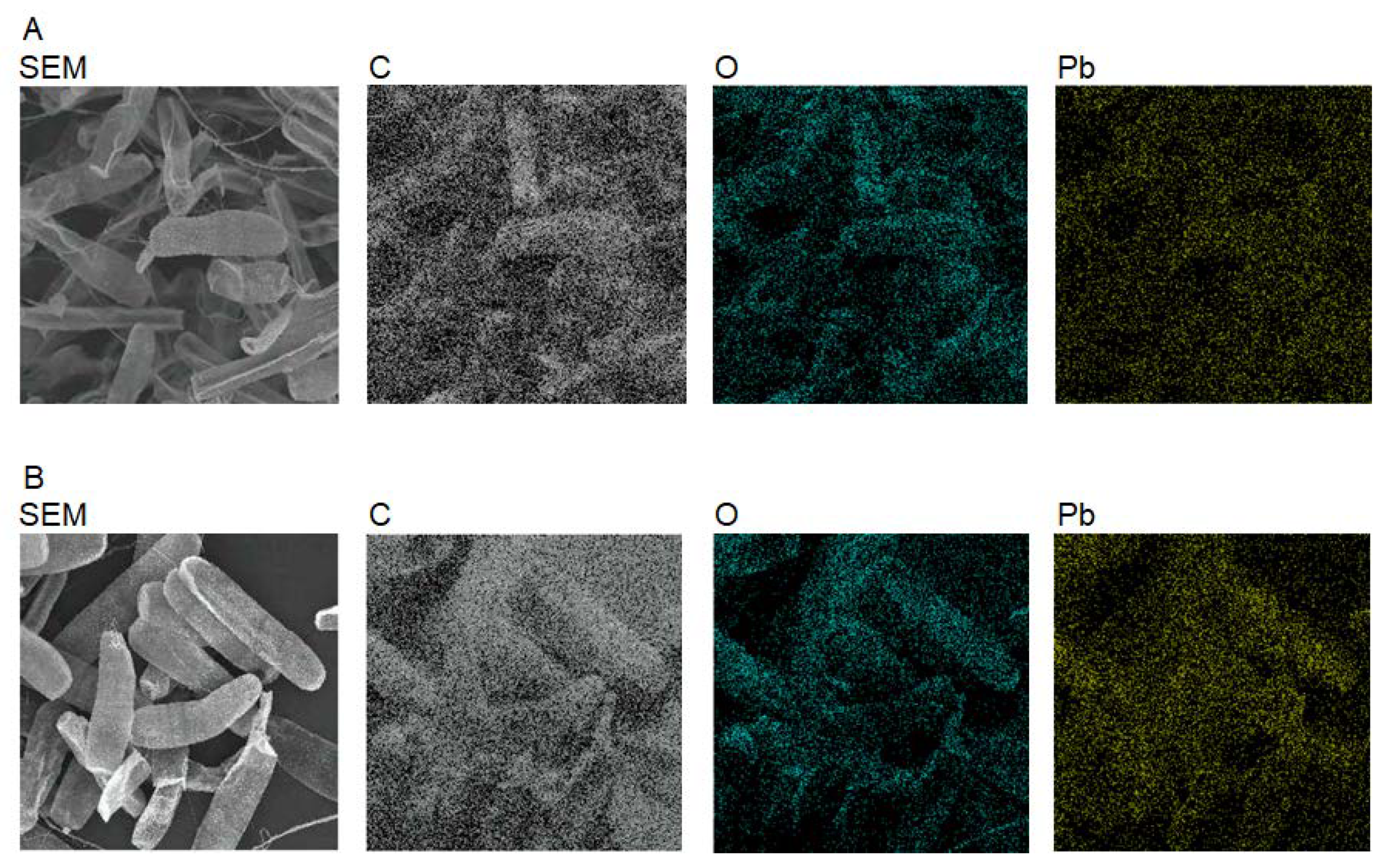
2.4. Elemental Mapping of Algal Cells and Measurements of Pb, Ca, Mg, K, and Na

P. margaritaceum cells were incubated in 50 mg/L of Pb solution for 2 h at room temperature. After collecting the cells by decantation, the cells were resuspended in water. The cell suspension was frozen at −25 °C and lyophilized with a freeze-dryer (VD-500F, Taitec, Koshigaya, Saitama, Japan). The dried cells were coated with Pt–Pd using an ion sputter (E-1030, Hitachi, Tokyo, Japan). The pretreated cells were observed with a field emission scanning electron microscope (SEM) (S-4500, Hitachi, Tokyo, Japan) equipped with a line scanning energy dispersive X-ray (EDX) spectrometer (EMAX-5770; Horiba, Kyoto, Japan). This was used to perform the elemental mapping of C, O, and Pb at the cell surface.
Pb, Ca, Mg, K, and Na concentrations were determined by flame atomic absorption spectrophotometer (AA-6200, Shimadzu, Kyoto, Japan) equipped with a hollow cathode lamp of Pb (L233-82NQ), Ca (L233-20NU), Mg (L233-12NU), K (L233-19NB) (Hamamatsu Photonics, Hamamatsu, Shizuoka, Japan), or Na (Atomax N202-5333, PerkinElmer, Winter Street Waltham, MA, USA).

2.5. Acquisition of Fourier Transform Infrared (FT-IR) Spectra

The P. margaritaceum cells were collected before and after suspension in 50 mg/L of Pb solution for 2 h at room temperature by centrifugation at 130× g for 10 min at 20 °C. The collected algal cells were dried at 50 °C overnight. The algal powder was placed between two KBr plates (ϕ8 mm) and was compressed by a hand press (JASCO Engineering, Tokyo, Japan). FT-IR spectra were obtained using a spectrometer (FT/IR-4100; JASCO, Tokyo, Japan).

2.6. Measurements of Pectin Content

Pectin content was measured as previously described [28]. The algal cells were collected from 300 mL of culture by centrifugation at 130× g for 10 min at 20 °C, lyophilized as described above, and kept in a desiccator containing silica gel for 1 h. The dried cells were weighed, transferred to a glass test tube, and suspended in 1.0 N nitric acid at a ratio of 1:40 (w/v). The test tube was heated in boiling water for 10 min and cooled on ice. Ethanol was added in order to adjust the ethanol concentration to 66%. The cellular debris was removed by centrifugation at 130× g for 10 min at 20 °C, and the pectin gel was separated from the ethanol solution by filtration. After drying at 50 °C for 24 h and cooling down in a desiccator containing silica gel at room temperature, the pectin gel was weighed. The pectin content (%) was calculated by dividing the pectin gel dry weight with the cellular dry weight, multiplied by 100.

3. Results

3.1. Adsorption of Pb on Algal Cells

P. margaritaceum NIES-217 and NIES-303 accumulated pectin at 19.6 and 25.1% cellular dry weight, respectively. In order to confirm the ability of P. margaritaceum to adsorb Pb, the algal cells were suspended in the Pb solution. After the collection and lyophilization of the cells, the cell shape and distribution were firstly observed using SEM, and the spatial distribution of C, O, and Pb was observed by EDX. The SEM images revealed the existence of extracellular polymeric substances (EPSs) in both NIES-217 and NIES-303 (Figure 1 and Figure S1). The distribution of C, O, and Pb in EDX corresponded to the cell shape and distribution in SEM (Figure 1). As the cellular components are mainly composed of major elements, such as C and O, it is conceivable that the P. margaritaceum cells are able to adsorb Pb.

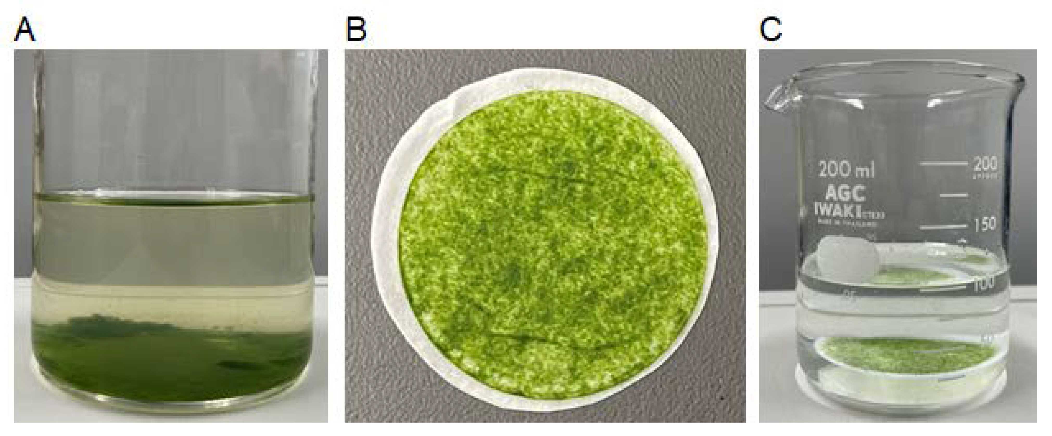
3.2. Immobilization of Algal Cells on Filter Paper

The coagulation of P. margaritaceum cells was observed as they grew in liquid culture (Figure 2A). However, the cell coagulates were easily dispersed by shaking and decantation. Though it was confirmed that Pb bound to the P. margaritaceum cell surface, the cells required collection using centrifugation in order to resuspend them in Pb solution. Therefore, the P. margaritaceum cells were collected on filter paper from liquid culture using vacuum filtration (Figure 2B). After soaking in Pb solution with gentle shaking for 8 h, the immobilized cells were still retained on the filter paper (Figure 2C) and could be separated from the solution simply by pinching off the filter paper. However, small amounts of insoluble materials, including cells and their debris, were observed at the bottom of the flask after the filter paper was removed. After soaking for 8 h, the dry weights of the immobilized cells were 9.5 mg (NIES-217) and 17.5 mg (NIES-303), whereas the dry weights of the insoluble materials at bottom of the flask were 1.6 mg (NIES-217) and 0.4 mg (NIES-303), which were less than 15% of the total dry weights (Table 1). The OD600 of the Pb solution at 0 h and after the immobilized cells were soaked did not change. Therefore, it was conceivable from these results that the algal cells were not suspended in the solution after soaking and were immobilized on the filter paper.

3.3. Removal and Recovery of Pb by the Immobilized Algal Cells

The immobilized cells were soaked in 100 mL of aqueous solution containing 100 µg of Pb. The filter paper itself did not reduce the amount of Pb in the solution nor did it adhere to Pb after soaking for 8 h (Table 2). The amount of Pb in the aqueous solution started to decrease immediately after the immobilized algal cells were soaked at 0 h. After soaking for 8 h, the amount of Pb in the aqueous solution was reduced to less than half of the initial amount, and more than 30% of the added Pb was recovered by the immobilized algal cells. The insoluble materials at the bottom of the flask after soaking also bound 22.7 µg (NIES-217) and 20.4 µg (NIES-303) of Pb. The total recovery of Pb from the solution, immobilized cells, and insoluble materials was 96.3% and 98.9% in NIES-217 and NIES-303, respectively.

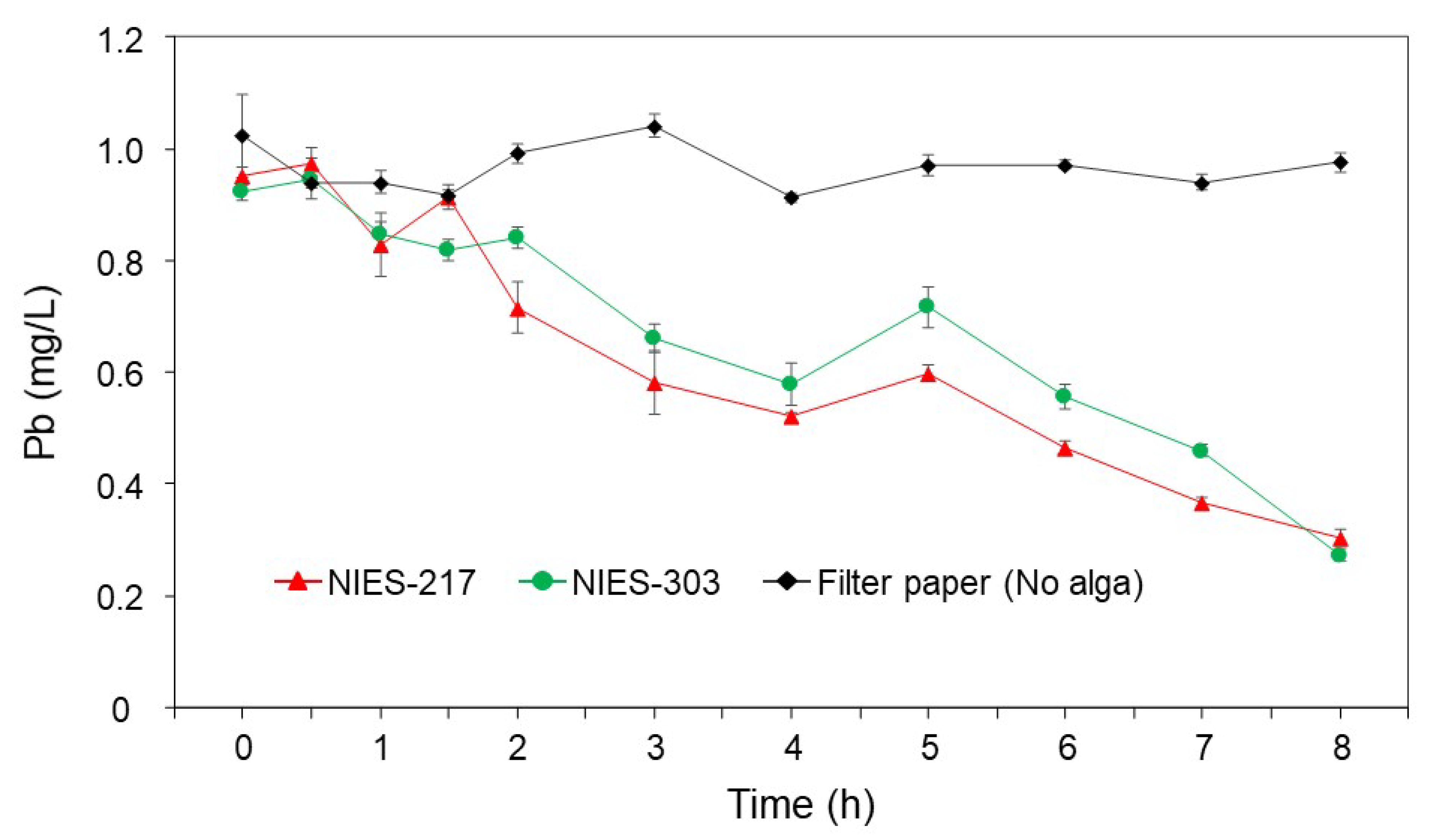
3.4. Time Course and the Specificity of Pb Removal

The concentration of Pb in the aqueous solution decreased over the course of soaking for 8 h (Figure 3). No obvious difference was observed between the decreasing rates of NIES-217 and NIES-303. A marked decrease took place during 4 h after the start of soaking, and then the concentrations gradually decreased until 8 h.
The specificity of Pb removal by the immobilized cells was evaluated in the presence of divalent and monovalent cations. When the immobilized cells were soaked in the solution for 0 h, the Pb concentrations were lower than the added concentration (1.0 mg/L) because of the immediate removal of Pb by the immobilized cells (Figure 4). On the other hand, the Na concentrations were higher than the added concentration due to the presence of the immobilized cells or substances carried in the aqueous solution. After soaking for 8 h, a marked increase in the concentration of Ca and decrease in the concentration of Pb were observed in both NIES-217 and NIES-303.

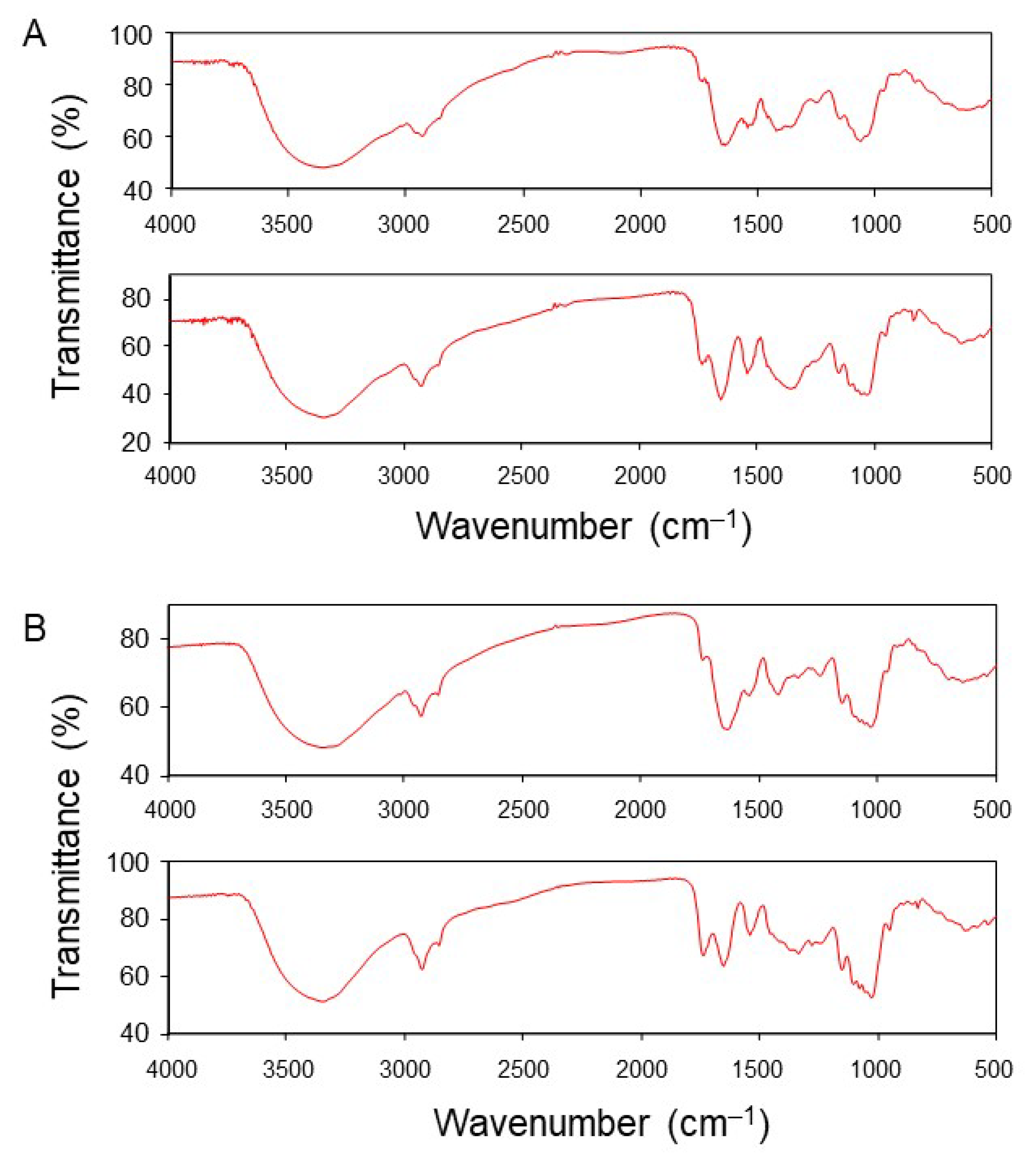
3.5. Detection of a Functional Group Involved in the Adsorption of Pb

In order to detect the functional groups involved in the adsorption of Pb, the FT-IR spectra of NIES-217 and NIES-303 cells were analyzed before and after the suspension of cells in Pb solution. The absorbance peaks around 1640 cm−1 shifted from 1635 cm−1 to 1652 cm−1, and increases in the absorbance of Pb at 1732 cm−1 and 1737 cm−1 were observed for NIES-217 and NIES-303, respectively (Figure 5). The absorbance peaks around 1740 cm−1 were considered to have derived from the C=O bond in non-ionic carboxylic acids [29] and their increases might be due to the partial increase of –COOH under acidic conditions caused by the addition of Pb stock solution. The peak shift from 1637 to 1644 was observed for the nitric-acid-treated pectin-rich orange peel through the binding of Pb (II) [29]. It has been shown that the absorption maxima of metal–ethylenediaminetetraacetic acid complexes are found to range from 1590 to 1650 cm−1 [30]. The results indicate that the carboxylic acid group of the algal cell wall is responsible for the adsorption of Pb.

4. Discussion

In this study, we demonstrated that immobilized P. margaritaceum cells could be applied as an efficient biosorbent to remove Pb from aqueous solutions. The filter paper retained the algal cells during soaking for 8 h, with the loss of less than 15% of the algal cells, and enabled the recovery of Pb simply by pinching off the alga-immobilized filter paper. It has been demonstrated that an aerial microalga, Trentepohlia aurea, that is immobilized on the surface of filter paper grows on paper that is dampened with medium for 40 d [31]. The soaking period of immobilized algal cells might be extended to longer than 8 h. The gelling feature of P. margaritaceum, which is due to the cell wall layers and EPSs, may be suitable for increasing immobilization stability on the filter paper. The immobilization of algal cells on the filter paper enables more cost-effective and simple procedures that remove and recover Pb from aqueous solutions.
In the preceded studies, microalgae have been examined as a conventional Pb adsorption material. A green microalga, Chlamydomonas reinhardtii, exhibited a higher affinity for Pb(II) than for Hg(II) and Cd(II), ranging from 20 to 400 mg/L each [32]. The biosorption by microalga biomass increased as the initial concentration of metal ions increased in the biosorption medium. The removal potential of lead by the EPSs of unicellular green algae, Parachlorella kessleri and Chlorella vulgaris, ranges from 15 to 50% at concentrations of 10 to 150 mg/L [33]. A green microalga, Pseudochlorococcum typicum, showed high efficiencies of Hg (II), Cd (II), and Pb (II) biosorption. Its maximum removal of metal ions occurred during the first 30 min of contact with a removal efficiency of 70% for Pb (II) at 10 mg/L, which was calculated based on the concentrations in solution before and after contact [34]. On the other hand, Pb concentrations in river water and groundwater at polluted areas have been reported to be 0.040 mg/L [3] and 0.02–0.40 mg/L, respectively [5]. Therefore, the removal of Pb at lower concentrations needs to be demonstrated in the context of bioremediation for polluted river water and groundwater using microalgal cells. In this view, a green microalga, Scenesesmus sp., has been examined at a lower Pb concentration of 0.05 mg/L [35]. The Pb removal capacity of 70% was achieved at 0.05 mg/L with a 7 d growth process. This removal capacity was determined using the amount of Pb in all the insoluble materials collected from the culture by glass filter paper. In this study, using the immobilized P. margaritaceum cells, the recovery efficiencies of 32.7% (NIES-217) and 31.8% (NIES-303) were achieved in 1.0 mg/L Pb solution with 8 h of soaking simply by pinching off the alga-immobilized filter paper. These efficiencies increased to 55.4% (NIES-217) and 52.2% (NIES-303) when the amounts of Pb collected from the bottom of the flask by filtration were included. The amounts of Pb adsorbed on the immobilized algal biomass were 3.4 mg/g (NIES-217) and 1.8 mg/g (NIES-303). These adsorption capacities on the algal biomass may be reduced by immobilization on filter paper, delaying the progress of adsorption until equilibrium compared to the capacity and progress of adsorption in the suspended cells.
Pectins in plant cells can bind and sequester Pb within the cell walls [36,37]. The thick cell walls act as a barrier that limits Pb from entering the protoplasm. Pb exposure induces the modification of pectin distribution, especially the distribution of homogalacturonan with high levels of methyl-esterification in the roots of sunflowers [38]. As seen in land plant cell walls, the cell wall and EPS of P. margaritaceum contains pectin polymers [39,40]. The Ca-cross-linked homogalacturonan-rich lattice structure is formed on its cell surface [41]. The lattice structure on the cell surface was also observed with SEM in the NIES strains (Figure S1). The concentration of Ca in the solution containing Pb and mineral ions increased after the immobilized P. margaritaceum cells were soaked for 8 h. It has been demonstrated that the adsorption of Pb by Ca–alginate beads takes place through ion-exchange between Ca and Pb on the alginate beads [42,43]. Therefore, the increase in the concentration of Ca after soaking for 8 h could be explained by the ion-exchange between Ca and Pb on homogalacturonan in the cell surface lattice and EPSs. The FT-IR spectra indicating the participation of carboxylic acid groups in the adsorption of Pb also support the scenario that pectin polymers in the algal cell wall are responsible for the removal of Pb from aqueous solutions.
Increasing the surface-area-to-volume ratio through the introduction of the mesoporous silica monolith was effective in the detection and adsorption of Cd (II) by organic ligand-based materials [44]. In order to increase the surface-area-to-volume ratio, the exploration of P. margaritaceum strains producing dense lattice structures and abundant EPSs may improve the capacity and affinity of the immobilized algal cells to Pb. The ability of P. margaritaceum to specifically adsorb Pb at low concentrations in the presence of minerals might be suitable for the removal of Pb from polluted river water and groundwater.

5. Conclusions

The immobilized P. margaritaceum cells were retained on the filter paper after soaking for 8 h and 31.8–32.7% of the added Pb was recovered. The specific decrease in Pb in the presence of minerals was associated with an increase in Ca, suggesting that the pectin polymers produced by P. margaritaceum might be responsible for the adsorption of Pb.

Supplementary Materials

The following supporting information can be downloaded at: https://www.mdpi.com/article/10.3390/microbiolres13040073/s1, Figure S1: SEM images of lattice structures on NIES-217 and NIES-303 cell surfaces.

Author Contributions

Conceptualization, T.B. and I.M.; methodology, I.M.; formal analysis, T.B. and K.H.; resources, I.M.; data curation, T.B. and K.H.; writing—original draft preparation, T.B. and I.M.; writing—review and editing, I.M.; funding acquisition, I.M. All authors have read and agreed to the published version of the manuscript.

Funding

This research received no external funding.

Institutional Review Board Statement

Not applicable.

Informed Consent Statement

Not applicable.

Data Availability Statement

Not applicable.

Acknowledgments

The authors thank David S. Domozych for providing P. margaritaceum strain 8 (Skidmore Algal Culture Facility), which was used to confirm Pb adsorption, and also thank the Advanced Instrumental Analysis Department, Utsunomiya University for their technical support.

Conflicts of Interest

The authors declare no conflict of interest.

References

  1. Sevak, P.I.; Pushkar, B.K.; Kapadne, P.N. Lead pollution and bacterial bioremediation: A review. Environ. Chem. Lett. 2021, 19, 4463–4488. [Google Scholar] [CrossRef]
  2. Yang, J.; Li, X.; Xiong, Z.; Wang, M.; Liu, Q. Environmental pollution effect analysis of lead compounds in China based on life cycle. Int. J. Environ. Res. Public Health 2020, 17, 2184. [Google Scholar] [CrossRef] [PubMed] [Green Version]
  3. Sikder, M.T.; Kihara, Y.; Yasuda, M.; Mihara, Y.; Tanaka, S.; Odgerel, D.; Mijiddorj, B.; Syawal, S.M.; Hosokawa, T.; Saito, T. River water pollution in developed and developing countries: Judge and assessment of physicochemical characteristics and selected dissolved metal concentration. CLEAN–Soil Air Water 2013, 41, 60–68. [Google Scholar] [CrossRef]
  4. Kasimov, N.; Kosheleva, N.; Gunin, P.; Korlyakov, I.; Sorokina, O.; Timofeev, I. State of the environment of urban and mining areas in the Selenga Transboundary River Basin (Mongolia Russia). Environ. Earth Sci. 2016, 75, 1283. [Google Scholar] [CrossRef]
  5. Rahman, M.A.; Paul, M.; Bhoumik, N.; Hassan, M.; Alam, M.; Aktar, Z. Heavy metal pollution assessment in the groundwater of the Meghna Ghat industrial area, Bangladesh, by using water pollution indices approach. Appl. Water Sci. 2020, 10, 186. [Google Scholar] [CrossRef]
  6. Yin, Y.; Zhang, P.; Yue, X.; Du, X.; Li, W.; Yin, Y.; Yi, C.; Li, Y. Effect of sub-chronic exposure to lead (Pb) and Bacillus subtilis on Carassius auratus gibelio: Bioaccumulation, antioxidant responses and immune responses. Ecotoxicol. Environ. Saf. 2018, 161, 755–762. [Google Scholar] [CrossRef]
  7. Luo, T.; Shen, M.; Zhou, J.; Wang, X.; Xia, J.; Fu, Z.; Jin, Y. Chronic exposure to low doses of Pb induces hepatotoxicity at the physiological, biochemical, and transcriptomic levels of mice. Environ. Toxicol. 2019, 34, 521–529. [Google Scholar] [CrossRef]
  8. Satarug, S.; Gobe, G.C.; Vesey, D.A.; Phelps, K.R. Cadmium and lead exposure, nephrotoxicity, and mortality. Toxics 2020, 8, 86. [Google Scholar] [CrossRef]
  9. Lee, J.-W.; Choi, H.; Hwang, U.-K.; Kang, J.-C.; Kang, Y.J.; Kim, K.I.; Kim, J.-H. Toxic effects of lead exposure on bioaccumulation, oxidative stress, neurotoxicity, and immune responses in fish: A review. Environ. Toxicol. Pharmacol. 2019, 68, 101–108. [Google Scholar] [CrossRef]
  10. Salman, M.S.; Znad, H.; Hasan, M.N.; Hasan, M.M. Optimization of innovative composite sensor for Pb (II) detection and capturing from water samples. Microchem. J. 2021, 160, 105765. [Google Scholar] [CrossRef]
  11. Awual, M.R.; Hasan, M.M.; Islam, A.; Rahman, M.M.; Asiri, A.M.; Khaleque, M.A.; Sheikh, M.C. Offering an innovative composited material for effective lead (II) monitoring and removal from polluted water. J. Clean. Prod. 2019, 231, 214–223. [Google Scholar] [CrossRef]
  12. Awual, M.R. Mesoporous composite material for efficient lead (II) detection and removal from aqueous media. J. Environ. Chem. Eng. 2019, 7, 103124. [Google Scholar] [CrossRef]
  13. Crist, D.; Crist, R.; Martin, J.; Watson, J. Ion exchange systems in proton—Metal reactions with algal cell walls. FEMS Microbiol. Rev. 1994, 14, 309–313. [Google Scholar] [CrossRef]
  14. Thakur, B.R.; Singh, R.K.; Handa, A.K.; Rao, M. Chemistry and uses of pectin—A review. Crit. Rev. Food Sci. Nutr. 1997, 37, 47–73. [Google Scholar] [CrossRef]
  15. Krzesłowska, M. The cell wall in plant cell response to trace metals: Polysaccharide remodeling and its role in defense strategy. Acta Physiol. Plant. 2011, 33, 35–51. [Google Scholar] [CrossRef] [Green Version]
  16. Atmodjo, M.A.; Hao, Z.; Mohnen, D. Evolving views of pectin biosynthesis. Annu. Rev. Plant Biol. 2013, 64, 747–779. [Google Scholar] [CrossRef] [Green Version]
  17. Fürst-Jansen, J.M.; de Vries, S.; de Vries, J. Evo-physio: On stress responses and the earliest land plants. J. Exp. Bot. 2020, 71, 3254–3269. [Google Scholar] [CrossRef] [Green Version]
  18. Zanchetta, E.; Damergi, E.; Patel, B.; Borgmeyer, T.; Pick, H.; Pulgarin, A.; Ludwig, C. Algal cellulose, production and potential use in plastics: Challenges and opportunities. Algal Res. 2021, 56, 102288. [Google Scholar] [CrossRef]
  19. Chan, S.Y.; Choo, W.S.; Young, D.J.; Loh, X.J. Pectin as a rheology modifier: Origin, structure, commercial production and rheology. Carbohydr. Polym. 2017, 161, 118–139. [Google Scholar] [CrossRef]
  20. Ahmad, A.; Bhat, A.; Buang, A. Enhanced biosorption of transition metals by living Chlorella vulgaris immobilized in Ca-alginate beads. Environ. Technol. 2019, 40, 1793–1809. [Google Scholar] [CrossRef]
  21. Bayramoğlu, G.; Tuzun, I.; Celik, G.; Yilmaz, M.; Arica, M.Y. Biosorption of mercury (II), cadmium (II) and lead (II) ions from aqueous system by microalgae Chlamydomonas reinhardtii immobilized in alginate beads. Int. J. Miner. Process. 2006, 81, 35–43. [Google Scholar] [CrossRef]
  22. Moreno-Garrido, I.; Campana, O.; Lubián, L.; Blasco, J. Calcium alginate immobilized marine microalgae: Experiments on growth and short-term heavy metal accumulation. Mar. Pollut. Bull. 2005, 51, 823–829. [Google Scholar] [CrossRef] [PubMed]
  23. Latifi, A.M.; Sadegh Nejad, E.; Babavalian, H. Comparison of extraction different methods of sodium alginate from brown alga Sargassum sp. localized in the Southern of Iran. J. Appl. Biotechnol. Rep. 2015, 2, 251–255. [Google Scholar]
  24. Papageorgiou, S.; Kouvelos, E.; Katsaros, F. Calcium alginate beads from Laminaria digitata for the removal of Cu+2 and Cd+2 from dilute aqueous metal solutions. Desalination 2008, 224, 293–306. [Google Scholar] [CrossRef]
  25. Al-Rub, F.A.; El-Naas, M.; Benyahia, F.; Ashour, I. Biosorption of nickel on blank alginate beads, free and immobilized algal cells. Process Biochem. 2004, 39, 1767–1773. [Google Scholar] [CrossRef]
  26. Hameed, M.A. Continuous removal and recovery of lead by alginate beads, free and alginate-immobilized Chlorella vulgaris. Afr. J. Biotechnol. 2006, 5, 1819–1823. [Google Scholar]
  27. Nichols, H.W. Growth media-freshwater. In Handbook of Phycological Methods: Culture Methods and Growth Measurements, 1st ed.; Stein, J.R., Ed.; Cambridge University Press: New York, NY, USA, 1973; Volume 1, pp. 39–78. [Google Scholar]
  28. Sato, M.d.F.; Rigoni, D.C.; Canteri, M.H.G.; Petkowicz, C.L.d.O.; Nogueira, A.; Wosiacki, G. Chemical and instrumental characterization of pectin from dried pomace of eleven apple cultivars. Acta Scientiarum. Agron. 2011, 33, 383–389. [Google Scholar]
  29. Lasheen, M.R.; Ammar, N.S.; Ibrahim, H.S. Adsorption/desorption of Cd (II), Cu (II) and Pb (II) using chemically modified orange peel: Equilibrium and kinetic studies. Solid State Sci. 2012, 14, 202–210. [Google Scholar] [CrossRef]
  30. Kołodyńska, D.; Hubicki, Z.; Pasieczna-Patkowska, S. FT-IR/PAS Studies of Cu (II)-EDTA Complexes Sorption οn the Chelating Ion Exchangers. Acta Phys. Pol. A 2009, 116, 340–343. [Google Scholar] [CrossRef]
  31. Abe, K.; Matsumura, I.; Imamaki, A.; Hirano, M. Removal of inorganic nitrogen sources from water by the algal biofilm of the aerial microalga Trentepohlia aurea. World J. Microbiol. Biotechnol. 2003, 19, 325–328. [Google Scholar] [CrossRef]
  32. Tüzün, I.; Bayramoğlu, G.; Yalçın, E.; Başaran, G.; Celik, G.; Arıca, M.Y. Equilibrium and kinetic studies on biosorption of Hg (II), Cd (II) and Pb (II) ions onto microalgae Chlamydomonas reinhardtii. J. Environ. Manag. 2005, 77, 85–92. [Google Scholar] [CrossRef] [PubMed]
  33. Ciempiel, W.; Czemierska, M.; Szymańska-Chargot, M.; Zdunek, A.; Wiącek, D.; Jarosz-Wilkołazka, A.; Krzemińska, I. Soluble Extracellular Polymeric Substances Produced by Parachlorella kessleri and Chlorella vulgaris: Biochemical Characterization and Assessment of Their Cadmium and Lead Sorption Abilities. Molecules 2022, 27, 7153. [Google Scholar] [CrossRef]
  34. Shanab, S.; Essa, A.; Shalaby, E. Bioremoval capacity of three heavy metals by some microalgae species (Egyptian Isolates). Plant Signal. Behav. 2012, 7, 392–399. [Google Scholar] [CrossRef] [PubMed]
  35. Pham, T.-L.; Dao, T.-S.; Bui, H.N.; Pham, T.K.N.; Ngo, T.T.H.; Bui, H.M. Lipid production combined with removal and bioaccumulation of Pb by Scenedesmus sp. Green Alga. Pol. J. Environ. Stud. 2020, 29, 1785–1791. [Google Scholar] [CrossRef]
  36. Sumranwanich, T.; Boonthaworn, K.; Singh, A. The roles of plant cell wall as the first-line protection against lead (Pb) toxicity. Appl. Sci. Eng. Prog. 2018, 11, 239–245. [Google Scholar] [CrossRef]
  37. Zhang, X.; Hu, W.; Xie, X.; Wu, Y.; Liang, F.; Tang, M. Arbuscular mycorrhizal fungi promote lead immobilization by increasing the polysaccharide content within pectin and inducing cell wall peroxidase activity. Chemosphere 2021, 267, 128924. [Google Scholar] [CrossRef]
  38. Chudzik, B.; Szczuka, E.; Leszczuk, A.; Strubińska, J. Modification of pectin distribution in sunflower (Helianthus annuus L.) roots in response to lead exposure. Environ. Exp. Bot. 2018, 155, 251–259. [Google Scholar] [CrossRef]
  39. Domozych, D.; Kort, S.; Benton, S.; Yu, T. The extracellular polymeric substance of the green alga Penium margaritaceum and its role in biofilm formation. Biofilms 2005, 2, 129–144. [Google Scholar] [CrossRef]
  40. Domozych, D.; Serfis, A.; Kiemle, S.; Gretz, M. The structure and biochemistry of charophycean cell walls: I. Pectins of Penium margaritaceum. Protoplasma 2007, 230, 99–115. [Google Scholar] [CrossRef]
  41. Domozych, D.S.; Sørensen, I.; Popper, Z.A.; Ochs, J.; Andreas, A.; Fangel, J.U.; Pielach, A.; Sacks, C.; Brechka, H.; Ruisi-Besares, P. Pectin metabolism and assembly in the cell wall of the charophyte green alga Penium margaritaceum. Plant Physiol. 2014, 165, 105–118. [Google Scholar] [CrossRef] [Green Version]
  42. Mohammed, C.; Mahabir, S.; Mohammed, K.; John, N.; Lee, K.-Y.; Ward, K. Calcium alginate thin films derived from Sargassum natans for the selective adsorption of Cd2+, Cu2+, and Pb2+ ions. Ind. Eng. Chem. Res. 2018, 58, 1417–1425. [Google Scholar] [CrossRef]
  43. Mousa, N.E.; Simonescu, C.M.; Pătescu, R.-E.; Onose, C.; Tardei, C.; Culiţă, D.C.; Oprea, O.; Patroi, D.; Lavric, V. Pb2+ removal from aqueous synthetic solutions by calcium alginate and chitosan coated calcium alginate. React. Funct. Polym. 2016, 109, 137–150. [Google Scholar] [CrossRef]
  44. Hasan, M.N.; Salman, M.S.; Islam, A.; Znad, H.; Hasan, M.M. Sustainable composite sensor material for optical cadmium (II) monitoring and capturing from wastewater. Microchem. J. 2021, 161, 105800. [Google Scholar] [CrossRef]
Figure 1. Images of SEM and the elemental mapping of C, O, and Pb in P. margaritaceum cells. Pb-treated NIES-217 (A) and NIES-303 (B) cells were observed.
Microbiolres 13 00073 g001
Figure 2. Observations of P. margaritaceum NIES-303 culture, the immobilized cells, and soaking of the immobilized cells in the Pb solution. The algal culture (A) was filtrated using filter paper to prepare the immobilized cells (B), which was soaked in the Pb solution with gentle shaking for 8 h (C).
Microbiolres 13 00073 g002
Figure 3. Time course of Pb concentration as the immobilized cells were soaked in the aqueous solution. Plots with an error bar show means (n = 3) ± SD.
Microbiolres 13 00073 g003
Figure 4. Specificity of Pb removal in the presence of divalent and monovalent cations during the soaking immobilized cells in the aqueous solution. Blue (Na), green (K), gray (Ca), yellow (Mg), and red (Pb) bars with an error bar show the means (n = 3) ± SD at 0 h and after soaking for 8 h. ND indicates that Pb was not detected.
Microbiolres 13 00073 g004
Figure 5. FT-IR spectra of the algal cells treated with and without the Pb solution. Cells of NIES-217 (A) and NIES-303 (B) were compressed in KBr plates before (upper spectra) and after (lower spectra) suspension of cells in the Pb solution.
Microbiolres 13 00073 g005
Table 1. Dry weights of the immobilized algal cells and insoluble materials at the bottom of the flask after soaking, and the optical densities at 0 h and after soaking.
ImmobilizationFilter Paper (mg) *Flask Bottom (mg) *OD600 at 0 hOD600 at 8 h
NIES-2179.5 ± 1.11.6 ± 0.70.018 ± 0.0020.016 ± 0.001
NIES-30317.5 ± 3.00.4 ± 0.40.016 ± 0.0010.016 ± 0.002
* Means (n = 3) ± SD are shown.
Table 2. Removal and recovery of Pb by the immobilized cells at 0 h and after soaking for 8 h.
ImmobilizationTimeSolution (µg) *Filter Paper (µg) *Flask Bottom (µg) *Recovery (µg) *Recovery %
NIES-2170 h90.9 ± 1.4
8 h41.0 ± 11.632.7 ± 1.822.7 ± 6.496.3 ± 16.096.3
NIES-3030 h92.0 ± 2.1
8 h46.7 ± 10.031.8 ± 6.420.4 ± 15.698.9 ± 6.198.9
No alga0 h96.9 ± 5.8
8 h96.2 ± 1.20.2 ± 0.4ND96.4 ± 2.796.4
* Means (n = 3) ± SD are shown. ND indicates that Pb was not detected.
Publisher’s Note: MDPI stays neutral with regard to jurisdictional claims in published maps and institutional affiliations.

