Quantitative assessment of Fgfr1 expression in neurons and glia
- Published
- Accepted
- Subject Areas
- Cell Biology, Developmental Biology, Neuroscience, Psychiatry and Psychology
- Keywords
- astrocyte, SOX2, SVZ, hippocampus, cortex, Fibroblast Growth Factor, doublecortin, oligodendrocyte, FGF, hypothalamus
- Copyright
- © 2016 Choubey et al.
- Licence
- This is an open access article distributed under the terms of the Creative Commons Attribution License, which permits unrestricted use, distribution, reproduction and adaptation in any medium and for any purpose provided that it is properly attributed. For attribution, the original author(s), title, publication source (PeerJ Preprints) and either DOI or URL of the article must be cited.
- Cite this article
- 2016. Quantitative assessment of Fgfr1 expression in neurons and glia. PeerJ Preprints 4:e2343v1 https://doi.org/10.7287/peerj.preprints.2343v1
Abstract
Fibroblast growth factors (FGFs) and their receptors (FGFRs) have numerous functions in the developing and adult CNS. For example, the FGFR1 receptor is important for proliferation of radial glial cells in the cortex and hippocampus, oligodendrocyte proliferation and regeneration, midline glia morphology and soma translocation, Bergmann glia morphology, and cerebellar morphogenesis. In addition, FGFR1 signaling in astrocytes is required for postnatal maturation of interneurons expressing parvalbumin (PV). FGFR1 is implicated in synapse formation in the hippocampus, and alterations in the expression of Fgfr1 and its ligand, Fgf2 accompany major depression. Understanding which cell types express Fgfr1 during development may elucidate its roles in normal development of the brain as well as illuminate possible causes of certain neuropsychiatric disorders. Here, we used a BAC transgenic reporter line to trace Fgfr1 expression in the developing murine CNS. The specific transgenic line employed was created by the GENSAT project, tgFGFR1-EGFPGP338Gsat, and includes a gene encoding enhanced green fluorescent protein (EGFP) under the regulation of the Fgfr1 promoter, to trace Fgfr1 expression in the developing CNS. This model reveals that Fgfr1 is primarily expressed in glial cells, in both astrocytes and oligodendrocytes, along with some neurons. Dual labeling experiments indicate that the proportion of GFP+ (Fgfr1+) cells that are also GFAP+ increases from postnatal day 7 (P7) to 1 month, illuminating dynamic changes in Fgfr1 expression during postnatal development of the cortex. In postnatal neurogenic areas, GFP expression was also observed in SOX2, doublecortin (DCX), and brain lipid-binding protein (BLBP) expressing cells. Fgfr1 is also highly expressed in DCX positive cells of the dentate gyrus, but not in the rostral migratory stream. Fgfr1 driven GFP was also observed in tanycytes and GFAP+ cells of the hypothalamus, as well as in Bergmann glia and astrocytes of the cerebellum. Understanding which cell types express Fgfr1 may elucidate its role in neuropsychiatric disorders and brain development.
Author Comment
This is a submission to PeerJ for review.
Supplemental Information
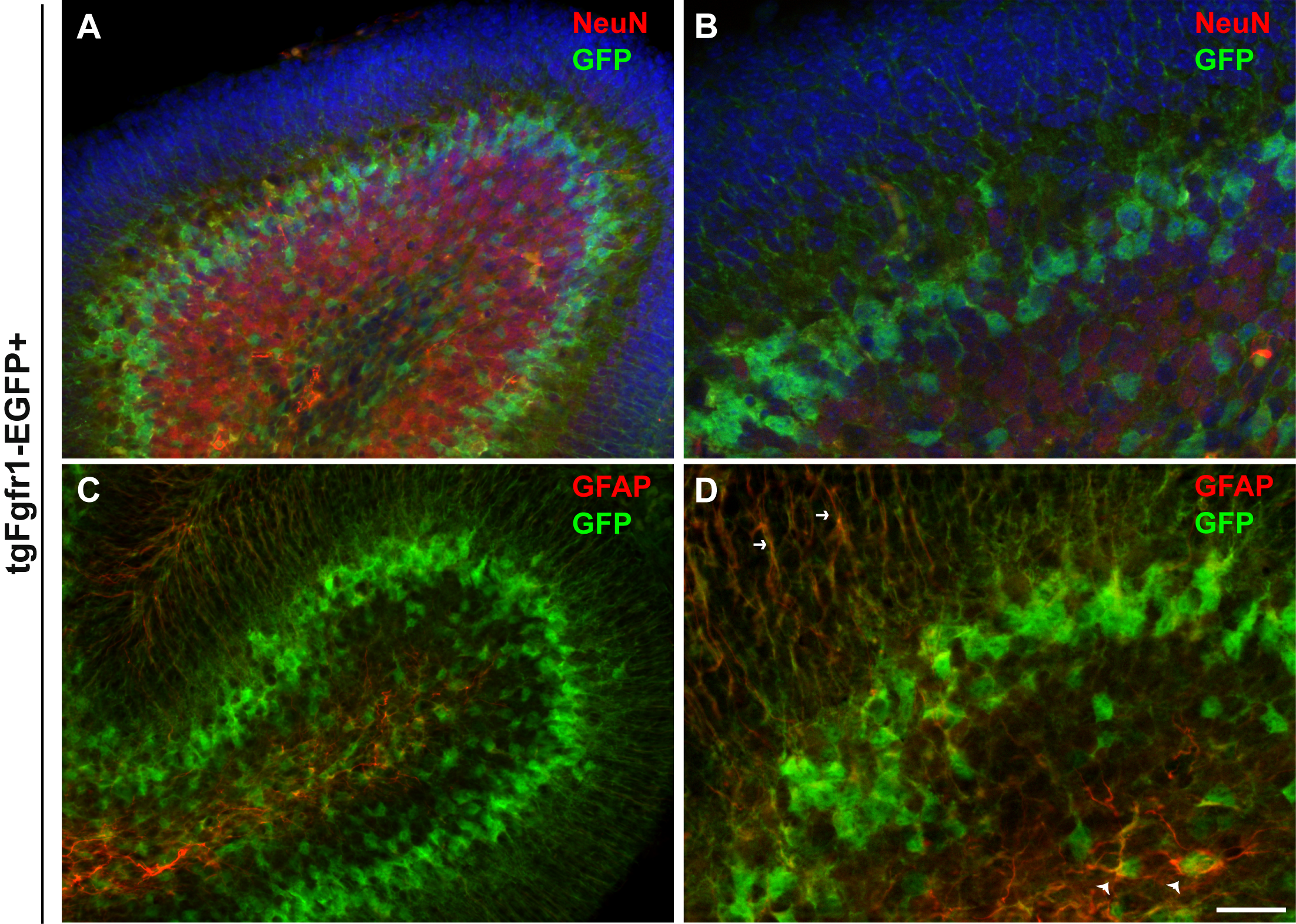
NeuN and GFAP staining in the cerebellum at P7 of tgFgfr1-EGFP+ mice
NeuN+ granule cell layer neurons did not colocalize with GFP at P7 (A, low magnification, B, high magnification). GFAP+ glia of the cerebellum do colocalize with GFP at P7 (C, low magnification, D, high magnification). Arrow heads denote double labeled cells while small arrows denote GFAP+/GFP+ glial fibers. Scale bar is 50µm in A, C, and 25µm in B, D.
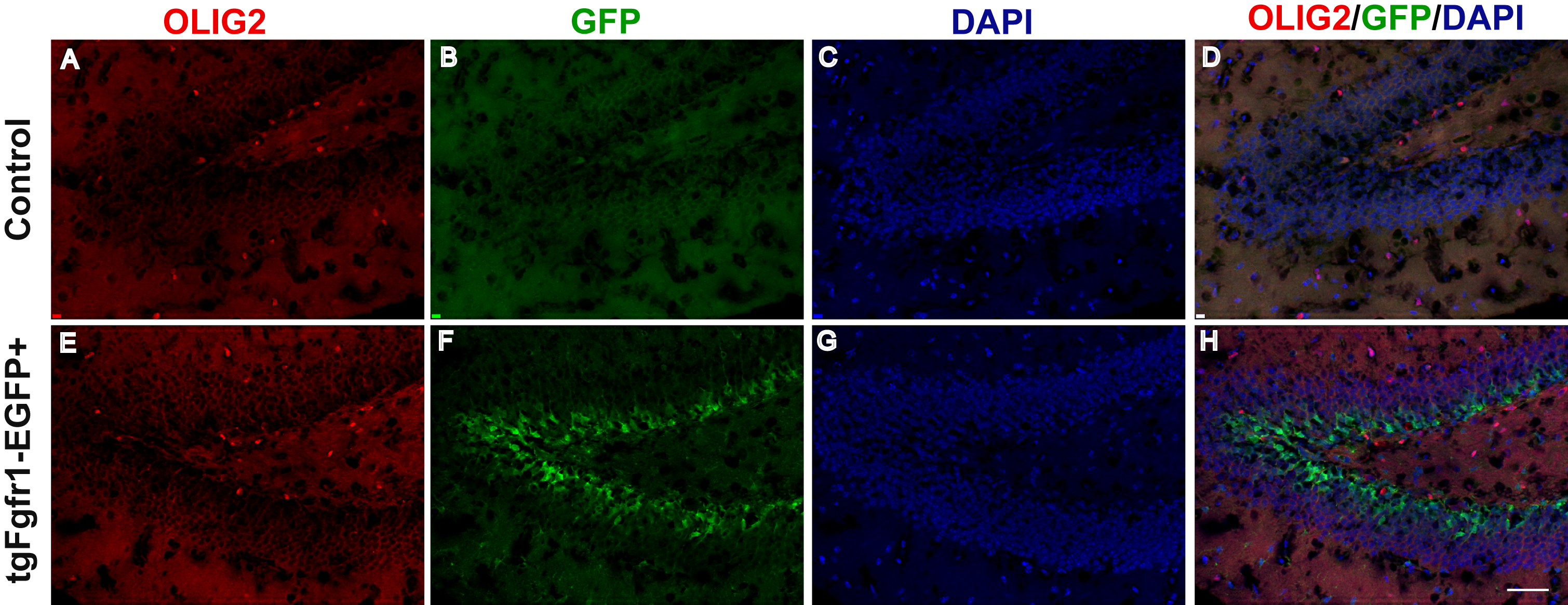
Fgfr1 promoter driven GFP does not colocalize with Olig2+ cells in the hippocampus
Olig2 staining in the hippocampus of control tgfgfr1-EGFP- mice ( A-D) and tgfgfr1-EGFP+ mice (E-H). Scale bar is 50µm.
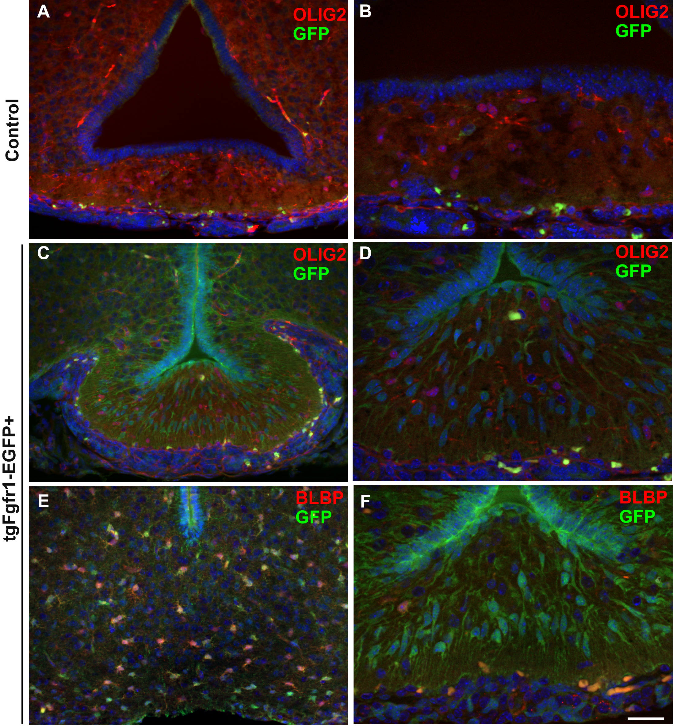
Fgfr1 expression in the hypothalamus does not colocalize with OLIG2 and does have some colocalization with BLBP
Fgfr1 promoter driven GFP in the hypothalamus of control tgfgfr1-EGFP- mice (A,B) and tgfgfr1-EGFP+ mice (C-F). OLIG2+ cells in the hypothalamus do not colocalize with Fgfr1 promoter driven GFP+ (C, D). BLBP+ cells near the third ventricle do colocolize with Fgfr1 promoter driven GFP+ (E), but there were not many of these colocalized cells in the medial eminence (F). Scale bar is 50µm in A, C, E and 25µm in B, D, F.