Abstracts
Purpose:
This study aimed to investigate the effect of quercetin on apoptotic cell death induced by ischemia-reperfusion (I/R) injury in the rat retina.
Methods:
Twenty-four rats were divided into four equal groups: control, ischemic, solvent, and quercetin. I/R injury was achieved by elevating the intraocular pressure above the perfusion pressure. Intraperitoneal injections of 20 mg/kg of quercetin and dimethyl sulfoxide (DMSO) were performed in the quercetin and solvent groups, respectively, immediately prior to I/R injury, and the researchers allowed for the retinas to be reperfused. Forty-eight hours after injury, the thicknesses of the retinal ganglion cell layer (RGCL), inner nuclear layer (INL), inner plexiform layer (IPL), outer plexiform layer (OPL), and outer nuclear layer (ONL) were measured in all groups. Moreover, the numbers of terminal deoxynucleotidyl transferase dUTP nick-end-labeled [TUNEL (+)] cells and caspase-3 (+) cells in both INL and ONL were evaluated in all groups.
Results:
The administration of quercetin was found to reduce the thinning of all retinal layers. The mean thickness of INL in the quercetin and ischemic groups was 21 ± 5.6 µm and 16 ± 6.4 µm, respectively (P<0.05). Similarly, the mean thickness of ONL in the quercetin and ischemic groups was 50 ± 12.8 µm and 40 ± 8.7 µm, respectively (P<0.05). The antiapoptotic effect of quercetin in terms of reducing the numbers of both TUNEL (+) cells and caspase-3 (+) cells was significant in INL. The mean number of TUNEL (+) cells in INL in the ischemic and quercetin groups was 476.8 ± 45.6/mm2 and 238.72 ± 251/mm2, respectively (P<0.005). The mean number of caspase-3 (+) cells in INL of ischemic and quercetin groups was 633.6 ± 38.7/mm2 and 342.4 ± 36.1/mm2, respectively (P<0.001).
Conclusion:
The use of quercetin may be beneficial in the treatment of retinal I/R injury because of its antiapoptotic effect on the retinal layers, particularly in INL.
Quercetin; Retina/drug effects; Reperfusion injury; Apoptosis; Ischemia; Disease models; Rats
Objetivo:
O objetivo deste estudo é investigar o efeito da quercetina, contra a morte celular por apoptose induzida por lesão consequente à isquemia-reperfusão (I/R) na retina de ratos.
Método:
Vinte e quatro ratos foram divididos em quatro grupos iguais: controle, isquêmico, solvente e quercetina. O modelo lesão por I/R foi realizado por meio da elevação da pressão intraocular acima da pressão de perfusão, em todos os grupos. Injecções intraperitoneais de 20 mg/kg de quercetina ou sulfóxido de dimetilo (DMSO) foram realizadas nos grupos quercetina e solvente, respectivamente, imediatamente antes da lesão por I/R, permitindo que as retinas fossem reperfundidas. Quarenta e oito horas após a lesão, as espessuras de camada de células ganglionares da retina (RGCL), camada nuclear interna (INL), camada plexiforme interna (IPL), camada plexiforme externa (OPL), e a camada nuclear externa (ONL) foram medidas em todos os grupos. Além disso, o número de células TUNEL (+) e caspase-3 (+) tanto na camada nuclear interna quanto na camada nuclear externa foi avaliada em todos os grupos.
Resultados:
A administração de quercetina diminuiu o afinamento de todas as camadas da retina em comparação com o grupo isquêmico. A espessura média da camada nuclear interna nos grupos quercetina e isquêmico foi de 21 ± 5,6 µm e 16 ± 6,4 µm, respectivamente (p<0,05). A espessura média da camada nuclear externa no grupo quercetina e isquêmico foi 50 ± 12,8 µm e 40 ± 8,7 µm, respectivamente (p<0,05). O efeito anti-apoptótico de quercetina em termos de redução do número de células TUNEL (+) e caspase-3 (+) foi significativa na INL. O número médio de células TUNEL (+) da camada nuclear interna no grupo isquêmico e quercetina foi 476,8 ± 45,6/mm2 e 238,72 ± 251/mm2, respectivamente (p<0,005). O médio número de células de caspase-3 (+) na INL do grupo isquêmico e quercetina foi 633,6 ± 38,7/mm2 e 342,4 ± 36,1/mm2, respectivamente (p<0,001).
Conclusão:
A utilização de quercetina pode ser benéfica no tratamento de lesão da retina por I/R devido ao seu efeito anti-apoptótico nas camadas da retina, particularmente na camada nuclear interna.
Quercetina; Retina/efeitos de drogas; Traumatismo por reperfusão; Apoptose; Isquemia; Modelos animais de doenças; Ratos
Introduction
Ischemia-reperfusion (I/R) injury occurs because of temporary disruption and subsequent improvement of the blood flow. This type of injury may become serious because significant amounts of reactive oxygen species (ROS), which are very toxic to cells, are produced as a result of tissue reoxygenation. Both direct and indirect mechanisms may be involved in ROS-induced cellular toxicity. In the direct mechanism, because both mitocondria and DNA are surrounded by membranes, ROS may lead to disruption of mitocondrial function and alterations in the structure of DNA, as a result of peroxidation of polyunsaturated fatty acids present in these membranes(11 Zimmerman BJ, Granger DN. Oxygen free radicals and the gastrointestinal tract: role in ischemia-reperfusion injury. Hepatogastroenterology 1994;41(1):337-42.). In the indirect mechanism, ROS induce hypersecrection of excitatory amino acids such as glutamate and aspartate(22 Bresnick GH. Excitotoxins: a possible new mechanism for the pathogenesis of ischemic retinal damage. Arch Ophthalmol.1989;107(3):339-41.). Hypersecrection of excitatory amino acids leads to an influx of excessive free calcium by stimulation of the N-methyl-D-aspartate (NMDA) receptor-operated channels, and increased levels of intracellular free calcium may subsequently trigger apoptosis of neuronal cells by a process called excitotoxicity(22 Bresnick GH. Excitotoxins: a possible new mechanism for the pathogenesis of ischemic retinal damage. Arch Ophthalmol.1989;107(3):339-41.,33 Qu J, Wang D, Grosskreutz CL. Mechanisms of retinal ganglion cell injury and defense in glaucoma. Exp Eye Res. 2010;91(1):48-53.). Neurotrophin, deprivation, glial activation, ischemia, glutamate excitotoxicity, and oxidative stress lead to induction of apoptotic cell death of retinal ganglion cells(33 Qu J, Wang D, Grosskreutz CL. Mechanisms of retinal ganglion cell injury and defense in glaucoma. Exp Eye Res. 2010;91(1):48-53.).
Retinal cell death is mostly due to apoptosis rather than necrosis, and this provides an important advantage to the retina because apoptosis does not result in an extreme inflammatory response; thus, limiting cell loss(44 Ryan SJ (ed). Retina. 5th ed. Philadelphia (PA): Elsevier/Mosby; 2013. Vol. 1, p.537). However, in the event of excessive or deficient apoptosis, such as in I/R injury, increased loss of retinal neurons may lead to visual impairment if the macula is affected(55 Büchi ER. Cell death in the rat retina after a pressure-induced ischaemia-reperfusion insult: an electron microscopic study. I. Ganglion cell layer and inner nuclear layer. Exp Eye Res. 1992;55(4):605-13.,66 Levin LA, Louhab A. Apoptosis of retinal ganglion cells in anterior ischemic optic neuropathy. Arch Ophthalmol. 1996;114(4):488-91.). I/R injury is the main pathogenetic mechanism underlying several clinically important diseases such as myocardial infarction, stroke, spinal injury, and shock, as well as some sight-threatening disorders such as diabetic retinopathy, retinal vascular occlusion, and glaucoma(77 Osborne NN, Casson RJ, Wood JP, Chidlow G, Graham M, Melena J. Retinal ischemia: mechanisms of damage and potential therapeutic strategies. Prog Retin Eye Res. 2004;23(1):91-147.). Thus, there may be a potential benefit from the use of medications, vitamins, and substances possessing both antioxidative and antiapoptotic effect as adjuvants in the treatment of eye diseases associated with I/R injury.
Flavonoids are plant-derived substances possessing several antioxidative properties. Among the flavonoids, quercetin is known as a powerful antioxidant and free radical-scavenging substance(88 Chen WJ, Sun SF, Cao W, Liang Y, Song JR. Antioxidant property of quercetin-Cr(III) complex: The role of Cr(III) ion. J Mol Struct. 2009;918:194-7.). Furthermore, quercetin also posseses antiapoptotic effects(99 Chao CL, Hou YC, Chao PD, Weng CS, Ho FM. The antioxidant effects of quercetin metabolites on the prevention of high glucose-induced apoptosis of human umbilical vein endothelial cells. Br J Nutr. 2009;101(8):1165-70.). Some studies have evaluated the antiapoptotic effect of quercetin; however, little data is available regarding the antiapoptotic effect of quercetin on retinal I/R injury(1010 Chang HC, Yang YR, Wang PS, Wang RY. Quercetin enhances exercise-mediated neuroprotective effects in brain ischemic rats. Med Sci Sports Exerc. 2014;46(10):1908-16.,1111 Sekaran S, Kandaswamy S, Gunasekaran K, Perumal E, Afsar Basha FY, Madhan Mohan BJ, et al. Protective role of quercetin on polychlorinated biphenyls (Aroclor-1254) induced oxidative stress and apoptosis in liver of adult male rats. J Biochem Mol Toxicol. 2012;26(12):522-32.).
In the present study, we aimed to evaluate whether quercetin decreases apoptotic cell loss in a rat retinal I/R injury model. Because the intensity of apoptosis occuring in the different layers of the retina may differ because of differences in the susceptibility of the retinal layers to I/R injury, we separately evaluated the antiapoptotic effect of quercetin on both the inner and outer nuclear layers of the retina.
METHODS
Twenty-four male Winstar Albino rats weighing 250-300 g, which were obtained from Saki Yenilli Research Animal Laboratory, were included in the study. After obtaining approval from Canakkale Onsekiz Mart University Ethics Committee of Experimental Animals, the rats were housed in cages with free access to standard food and drinking water, and they were maintained under controlled conditions, including a 12-h light/dark cycle (08:00-20:00, light; 20:00-08:00, dark), an ambient temperature of 23-25°C, and a humidity in the range of 55%-60% in the Canakkale Onsekiz Mart University Laboratory of Animal Research Center.
All operations were performed under aseptic conditions. For anesthetizing the rats, a single dose of 80 mg/kg ketamine (Ketasol, Richter Pharma Ag, Wels, Austria) and 5 mg/kg xylazine (Rompun, Bayer, İstanbul) was intraperitonally injected. To maintain body temperature within the physiological range, the rats were moved to special containers to ensure sufficient covering of their bodies. After instilling one or two drops of 0.5% proparakain HCl (Alcaine; Alcon, USA) for corneal analgesia, 10% povidone iodine was administered into the conjonctival sac to ensure desinfection of the ocular surface.
Twenty-four rats were divided into four groups of six according to weight and were used as control, ischemic, solvent, and quercetin treatment groups, respectively. In the control group, the right anterior chamber of each animal was penetrated without drug administration.
In the ischemic group, the I/R injury model was developed using the method previously described by Buchi et al.(1212 Büchi ER, Suivaizdis I, Fu J. Pressure-induced retinal ischemia in rats: an experimental model for quantitative study. Ophthalmologica. 1991;203(3):138-47.). According to this method, to establish retinal ischemia, the right anterior chamber was cannulated with a 30-gauge needle attached to an infusion set, and its reservoir was placed approximately 200 cm above the animal. After opening the reservoir of the infusion set containing physiological saline, the intraocular pressure (IOP) was raised above 50 mmHg, until whitening of the iris and loss of the red reflex of the fundus were observed, which are signs of retinal ischemia. All procedures and clinical examinations in the present study were performed using a portable biomicroscope (Shin Nippon, Japan). After 60 min of retinal ischemia, IOP was reverted to normal pressure by removing the infusion needle from the anterior chamber to allow the retinas to be reperfused. In the solvent group, after inducing retinal ischemia in the right eyes, di-methyl sulfoxide (DMSO) was intraperitoneally injected immediately before allowing the retinas to be reperfused. In the quercetin group, after inducing retinal ischemia in the right eyes, a dose of 20 mg/kg quercetin (Sigma-Aldrich Chemical Co., United Kingdom) dissolved in DMSO was intraperitoneally injected immediately before allowing the retinas to be reperfused. After 48 h, the right eye of each animal was enucleated to perform histopathological and immunohistochemical evaluations of the retina, and the experimental animals were sacrified.
Histopathological and immunohistochemical evaluations
Eye tissue samples, fixed in 10% buffered formaldehyde fixative solution and embedded in paraffin 48 h after acute retinal ischemia, were examined with hematoxylin and eosin (H&E) to evaluate histopathology. Sections that were 5 µm thick were obtained using a microtome (Leica, RM2245) and stained with H&E. Retinal thickness was calculated in three separate regions: peripheral (100-150 µm from the ora serrata), central (100-150 µm from the optic nerve), and midperipheral (halfway between the central and peripheral). Two representative sections were randomly selected from the same three positions for each eye, and their mean values were calculated(1313 Woo TT, Li SY, Lai WW, Wong D, Lo AC. Neuroprotective effects of lutein in a rat model of retinal detachment. Graefes Arch Clin Exp Ophthalmol. 2013;251(1):41-51.). The sections were also incubated with polyclonal anti-Caspase-3 antibody (Abcam, ab4051; dilution 1:100) at room temperature for 90 min. Sections were washed three times with PBS and incubated with biotinylated secondary antibody (Ultra Vision Detection System-HRP kit, Thermo, Fremont, USA), and subsequently streptavidin peroxidase (Ultra Vision Detection System-HRP kit, Lab Vision, Fremont, USA) was added and sections were maintained at room temperature for 20 min.
Terminal deoxynucleotidyl transferase dUTP nick-end labeling (TUNEL) assay
Enucleated eyes were immersed in 10% formaldehyde fixative solution for 48 h at room temperature and embedded in paraffin; 5-µm sections were obtained, and the slides were air dried. Apoptotic cells were identified using terminal deoxynucleotidyl transferase dUTP nick-end labeling (TUNEL; Calbiochem, San Diego, CA, USA), as previously reported(1414 Sayhan MB, Kanter M, Oguz S, Erboga M. Protective effect of Urtica dioica L. on renal ischemia/reperfusion injury in rat. J Mol Hist. 2012;43(6):691-8.). Nuclear counterstaining was performed using 3,3’-diaminobenzidine (DAB), and sections were counterstained with hematoxylin.
Statistical analysis
Data were expressed as mean ± standard deviation (SE). The Bartlett test was used to determine whether the data were heterogeneous or homogeneous. Bonferroni multiple comparison test was applied to identify differences between means. Differences were considered statistically significant at probability (P) levels of <0.05.
RESULTS
The mean thicknesses of RGCL, IPL, OPL, INL, and ONL were 19 ± 2.7 µm, 60 ± 12.1 µm, 11 ± 3.0 µm, 27 ± 9.9 µm, and 55 ± 13.3 µm, respectively, in the control group; 8 ± 1.4 µm; 22 ± 6.5 µm, 7 ± 1.9 µm, 16 ± 6.4 µm, and 40 ± 8.7 µm in the ischemic group; 9 ± 0.9 µm, 22 ± 7.4 µm, 7.4 ± 1.6 µm, 17 ± 5.2, and 41 ± 10.2 µm in the solvent group; and 10 ± 3.8 µm, 25 ± 8.2 µm, 9 ± 2.4 µm, 21 ± 5.6 µm, and 50 ± 12.8 µm, respectively, in the quercetin group (Figures 1 and 2).
Values indicating the mean thicknesses of RGCL, IPL, and OPL measured in the quercetin group were statistically significantly greater those measured in the ischemic and the solvent group (P<0.05 and P<0.05 respectively). However, among all of the retinal layers, the reduction in the thicknesses of both ONL and INL was statistically significant, particularly in the quercetin group; ONL mean thickness in the quercetin and ischemic groups were 50 ± 12.8 µm and 40 ± 8.7 µm, respectively (P<0.05), and INL mean thickness in the quercetin and ischemic groups were 21 ± 5.6 µm and 16 ± 6.4 µm, respectively (P<0.05).
Representative photographs showing retinal thickness detected in the rat retina. A) Normal appearance of the retina (control group). B and C) Retinal thickness in the ischemic group and solvent group, respectively; in both of B and C, the thicknesses of the retina was found to be significantly reduced after ischemia. D) Retinal thickness in the quercetin group. Total retinal thickness in the quercetin group was significantly less reduced compared with the ischemic group at end of the experiment. H&E staining (×400).
The effect of quercetin on the number of TUNEL (+) cells in INL and ONL
The mean number of TUNEL (+) cells counted in INL was 10.88 ± 3.5/mm2, 476.8 ± 45.6/mm2, 336 ± 34.9/mm2, and 238.72 ± 251/mm2 in the control, ischemic, solvent, and quercetin groups, respectively. The mean number of TUNEL (+) cells in INL was significantly lower in the quercetin group than in the ischemic group (P<0.005). Moreover, the mean number of TUNEL (+) cells counted in ONL was 197.76 ± 18.4/mm2, 176 ± 16.8/mm2, and 71.68 ± 11.8/mm2 in the ischemic, solvent, and quercetin groups, respectively. The mean number of TUNEL (+) cells in ONL was also significantly lower in the quercetin group than in the ischemic group (P<0.05; Figures 3 and 4).
Number of TUNEL (+) cells detected in the inner nuclear layer (INL) and outer nuclear layer (ONL) in each group.
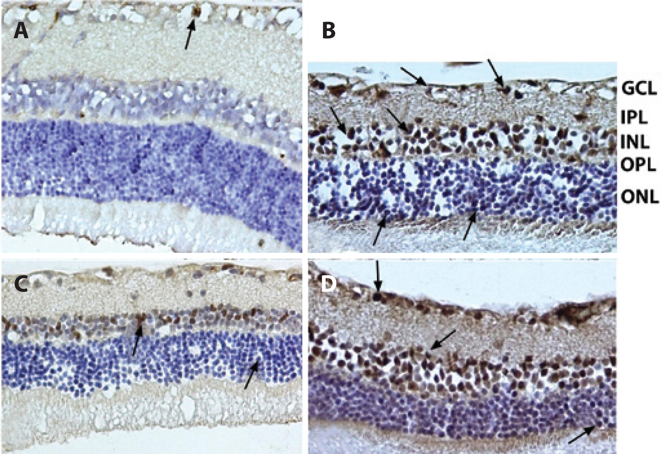
Representative photographs of TUNEL staining of rat retinas in control (A), ischemic (B), solvent (C), and quercetin (D) groups. The number of cells positive by TUNEL staining was increased in ischemic rats. However, treatment with quercetin markedly reduced retinal cell apoptosis. Arrows: TUNEL (+) cells. (×400).
Changes in the number of caspase-3 (+) cells in INL and ONL
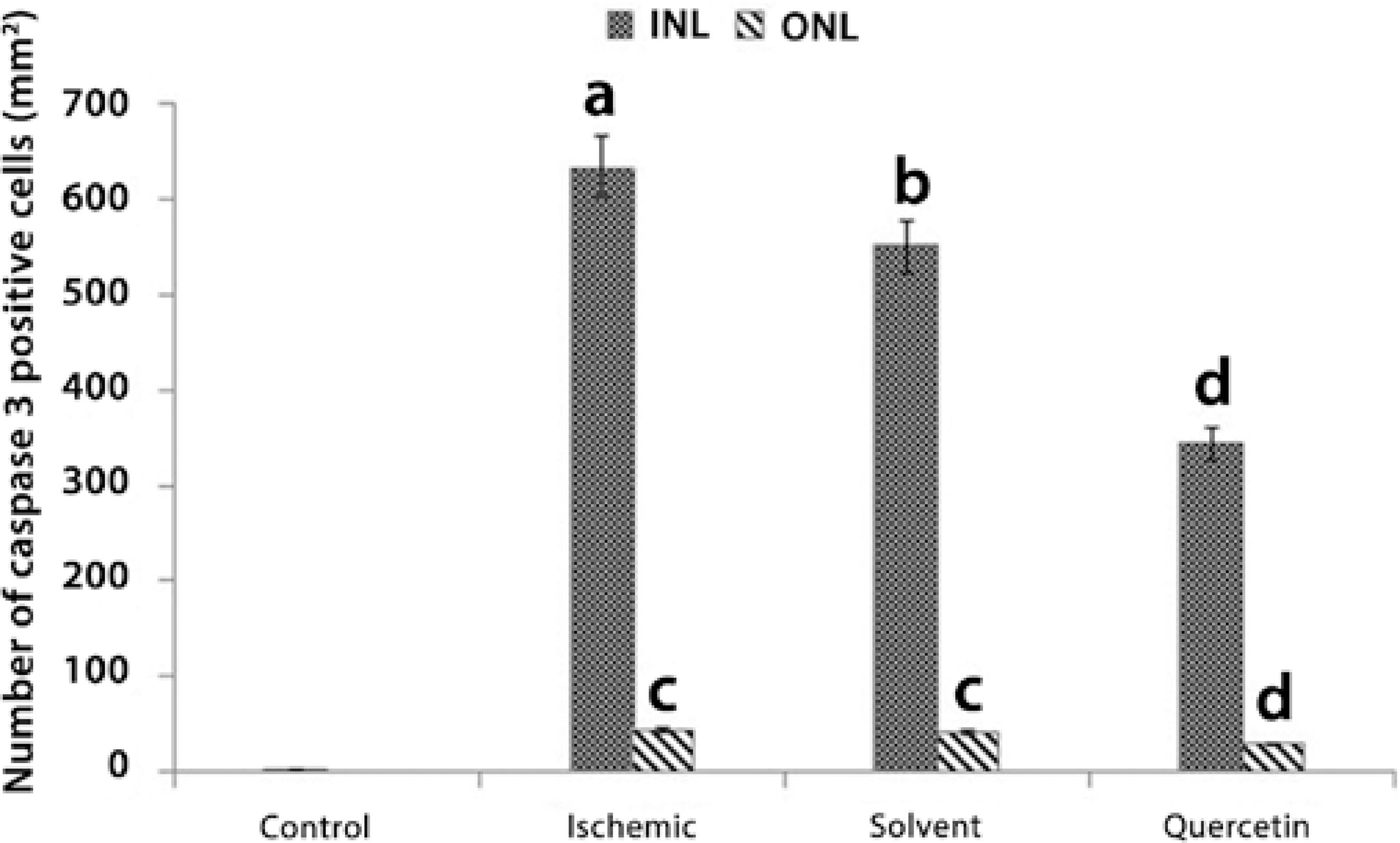
The mean number of caspase-3 (+) cells detected in INL was 62.56 ± 0.2/mm2, 633.6 ± 38.7/mm2, 550 ± 34.2/mm2, and 342.4 ± 36.1/mm2 in the control, ischemic, solvent, and quercetin groups, respectively. Treatment with quercetin was found to significantly decrease the number of caspase-3 (+) cells in INL by a ratio of nearly 46% when compared with the ischemic group (P<0.05). Moreover, the mean number of caspase-3 (+) cells detected in ONL was 43.6 ± 9.4/mm2, 41.2 ± 6.7/mm2, and 29,7 ± 8.4/mm2 in the ischemic, solvent, and quercetin groups, respectively. Quercetin treatment was found to significantly decrease the number of caspase-3 (+) cells in ONL by a ratio of nearly 32% when compared with the ischemic group (P<0.05; Figures 5 and 6).
The number of caspase-3 (+) cells in the inner nuclear layer (INL) and outer nucler layer (ONL) in each group.
Representative photographs of caspase-3 (+) cells. A) Control group; B) Ischemic group; C) Solvent group, and D) Quercetin group. The number of caspase-3 (+) cells observed among the quercetin-treated retinal cells was significantly lower than that among the ischemic retinal cells. Arrows: Caspase-3 (+) cells. Immunoperoxidase, hematoxylin counterstain (×400).
DISCUSSION
The antioxidant, free radical-scavenging, and neuroprotective effects of quercetin is
well known(1515 Rodriguez RJ, Miranda CL, Stevens JF, Deinzer ML, Buhler DR. Influence
of phenylated and non-phenylated flavonoids on liver microsomal lipid peroxidation
and oxidative injury in rat hepatocytes. Food Chem Toxicol.
2001;39(5):437-45.
16 Boots AW, Haenen GR, Bast A. Health effects of quercetin: from
antioxidant to nutraceutical. Eur J Pharmacol.
2008;13;585(2-3):325-37.-1717 Rogerio AP, Dora CL, Andrade EL, Chaves JS, Silva LF, Lemos-Senna E, et
al. Anti-inflammatory effect of quercetin-loaded microemulsion in the airways
allergic inflammatory model in mice. Pharmacol Res.
2010;61(4):288-97.). Besides these important benefits of
quercetin, it also has a protective effect against apoptotic cell damage. Some
experimental studies have revealed the antiapoptotic effect of quercetin on I/R injury
in various organs(1818 Ghosh A, Sarkar S, Mandal AK, Das N. Neuroprotective role of
nanoencapsulated quercetin in combating ischemia-reperfusion induced neuronal damage
in young and aged rats. PLoS One 2013;19;8(4):e57735.,1919 Chen YW, Chou HC, Lin ST, Chen YH, Chang YJ, Chen L, et al.
Cardioprotective effects of quercetin in cardiomyocyte under ischemia/reperfusion
injury. Evid Based Complement Alternat Med. 2013;2013:364519.); however, we have not been able to
find any experimental studies evaluating the benefit of quercetin in treatment of I/R
injury of the retina.
Because apoptosis is the mechanism predominantly involved in retinal cell death, antiapoptotic drugs or substances are considered particularly beneficial in the treatment and prevention of retinal diseases that are associated with the formation of excessive apoptosis, such as I/R injury.
The exact mechanism of cell death due to retinal ischemia is incompletely known;
however, it was previously demonstrated that in conditions of oxidative stress, retinal
ganglion cells are damaged as a result of increased intracellular ROS and calcium
influx(2020 Maher P, Hanneken A. Flavonoids protect retinal ganglion cells from
oxidative stress-induced death. Invest Ophthalmol Vis Sci.
2005;46(12):4796-803.). During I/R,
overproduction of ROS leads to excessive apoptosis by inducing the secretion of
glutamate and calcium influx(2121 Xu H, Chen M, Forrester JV. Para-inflammation in the aging retina. Prog
Retin Eye Res. 2009;28(5):348-68.
22 Jarrett SG, Lin H, Godley BF, Boulton ME. Mitochondrial DNA damage and
its potential role in retinal degeneration. Prog Retin Eye Res.
2008;27(6):596-607.-2323 Meldrum B, Garthwaite J. Excitatory amino acids in neurotoxicity and
neurodegenerative disease. Trends Pharmacol Sci. 1990;11(9):379-87.). Apart
from inducing apoptosis, increased intracellular calcium levels may also cause cellular
damage by hindering mitocondrial functions, decreasing the level of intracellular
adenosine triphosphate (ATP), inducing ROS production, and stimulating cellular
proteases and nitric oxide synthase(2424 Osborne NN, Ugarte M, Chao M, Childlow G, Wood JP, et al.
Neuroprotection in relation to retinal ischemia and relevance to glaucoma. Surv
Ophthalmol. 1999;43 Suppl 1:S102-28.). Therefore, it is conceivable that the maintainance of
intracellular free calcium levels at an optimal concentration may be critical for
treating I/R injury.
Flavonoids, such as quercetin, contain a flavonol nucleus that is surrounded by functional groups that have the ability to maintain the intracellular free calcium concentration at low levels by influencing glutathione metabolism, particularly in conditions of excessive ROS production(2525 Heim KE, Tagliaferro AR, Bobilya DJ. Flavonoid antioxidants: chemistry, metabolism and structure-activity relationships. J Nutr Biochem. 2002;13(10):572-84.,2626 Ishige K, Schubert D, Sagara Y. Flavonoids protect neuronal cells from oxidative stress by three distinct mechanisms. Free Radic Biol Med. 2001;30(4):433-46.). Therefore, it may be reasonable to use quercetin in the treatment of I/R injury because of its beneficial biochemical properties.
A supressor effect of quercetin against the activities of caspase-3 and calpain, which are the representitive proteases in apoptotic and necrotic pathways, was demonstrated in purified primary rat retinal ganglion cell cultures and in human retinal pigment epithelium cell cultures(2727 Nakayama M, Aihara M, Chen YN, Araie M, Tomita-Yokotani K, Iwashina T. Neuroprotective effects of flavonoids on hypoxia-, glutamate-, and oxidative stress-induced retinal ganglion cell death. Mol Vis. 2011;17:1784-93.,2828 Kook D, Wolf AH, Yu AL, Neubauer AS, Priglinger SG, Kampik A, et al. The protective effect of quercetin against oxidative stress in the human RPE in vitro. Invest Ophthalmol Vis Sci. 2008;49(4):1712-20.). Moreover, an antiapoptotic effect of quercetin in terms of reducing caspase-3 activity has also been demonstrated in a myocardial I/R model(2929 Wang Y, Zhang ZZ, Wu Y, Ke JJ, He XH, Wang YL. Quercetin postconditioning attenuates myocardial ischemia/reperfusion injury in rats through the PI3K/Akt pathway. Braz J Med Biol Res. 2013;46(10):861-7.). Because quercetin was found to induce cardioprotection in the myocardial I/R model, we thought that it may also be beneficial for retinal protection in the ocular I/R model; hence, we aimed to investigate this relationship in the present study.
To develop the ocular I/R model, we applied a widely used method, which involves increasing the intraocular pressure above the ocular perfusion pressure for nearly 60 min(55 Büchi ER. Cell death in the rat retina after a pressure-induced ischaemia-reperfusion insult: an electron microscopic study. I. Ganglion cell layer and inner nuclear layer. Exp Eye Res. 1992;55(4):605-13.). In the histological examination of the retina, this procedure was found to cause retinal damage through different types of cell death mechanisms such as apoptosis, necrosis, and nonlysosomal vesiculate(55 Büchi ER. Cell death in the rat retina after a pressure-induced ischaemia-reperfusion insult: an electron microscopic study. I. Ganglion cell layer and inner nuclear layer. Exp Eye Res. 1992;55(4):605-13.). Hughes demonstrated increased apoptosis and INL thinning in a tissue model of acute ischemic injury(3030 Hughes WF. Quantation of ischemic damage in the rat retina. Exp Eye Res. 1991;53(5):573-82.). In addition, a number of investigations have shown that a peak in apoptosis occurs 6 to 18 h after I/R, returning to control levels after 48 h(3131 Zhao Y, Niu YJ, Zhou ZY, Gao YX, Wang HY. Expression of Fas/Fas L and the apoptosis in rat ischemia/reperfusion-induced retinal injury and effects of bFGF. Int J Opthalmol. 2008;1:226-9.,3232 Zheng GY, Zhang C, Li ZG. Early activation of caspase-1 after retinal ischemia and reperfusion injury in mice. Chin Med J (Engl). 2004;117(5):717-21.). Therefore, in the present study, we administered quercetin immediately after the induction of ischemia but before the begining of reperfusion, i.e., before the peak of apoptosis. Forty-eight hours later, we found that the thickness of the retinal layers in the quercetin group was significantly less decreased than that in the ischemic and solvent groups.
Moreover, a significant decrease in the number of TUNEL (+) and caspase-3 (+) cells was detected in both INL and ONL in the quercetin group in comparison with the ischemic group. Because TUNEL (+) and caspase-3 (+) cells are important markers in the determination of apoptosis, obtaining a reduction in the number of such markers and a decrease in the thinning of retinal layers clearly demonstrates the antiapoptotic effect of quercetin on I/R-induced retinal injury. Although the decrease in the thinning of INL and ONL was found to be almost similar, the antiapoptotic effect of quercetin appeared much more evident in INL than in ONL. We believe that this disparity may arise from a difference in susceptibility of INL and ONL to ischemic damage. In a previous study, the intensity of apoptotic cell loss was found to be more significant in INL than in ONL in the earlier phases of I/R injury(3333 Ju WK, Kim KY, Hofmann HD, Kim IB, Lee MY, Oh SJ, et al. Selective neuronal survival and upregulation of PCNA in the rat inner retina following transient ischemia. J Neuropathol Exp Neurol. 2000;59(3):241-50.). Consistent with this finding, we also observed that the number of both TUNEL (+) and caspase-3 (+) cells was much higher in INL than in ONL.
In the present study, the reason for the significantly increased degree of apoptosis in INL may result from excitotoxicity, which is coupled to hypersecretion of excitatory amino acids such as glutamate. This idea is supported by the findings of both Massey et al. and Brandstätter et al. who showed that expression of glutamate receptors is confined to neurons of the ganglion cell layer and INL(3434 Massey SC. Cell types using glutamate as a neurotransmitter in the vertebrate retina. In: Osborne NN, Chader GJ, editors. Progress in retinal research, Oxford: Pergamon Press; 1994. p.399-426.,3535 Brandstätter JH, Koulen P, Wässle H. Diversity of glutamate receptors in the mammalian retina. Vision Res. 1998;38(10):1385-97.). This supports the hypothesis that quercetin may achieve its antiapoptotic effect depending on the intensity of the apoptosis, and it may be beneficial, particulary in the presense of increased apoptosis. On the other hand, because the number of both TUNEL (+)and caspase-3 cells was much lower in ONL in all the groups, it is speculated that the antiapoptotic effect of quercetin may not be prominent in ONL because of decereased induction of apoptosis. However, in an animal model of I/R injury, an increased rate of apoptosis in ONL was reported to be associated with extended postischemic survival time(3636 Kim IB, Kim KY, Joo CK, Lee MY, Oh SJ, Chung JW, et al. Reaction of Müller cells after increased intraocular pressure in the rat retina. Exp Brain Res. 1998;121(4):419-24.). In our study, upon evaluation of the early phases of I/R injury, we speculate that a significant decrease in the thickness of ONL in the ischemic group may also be due to a different cell death mechanism such as necrosis in addition to apoptosis. Therefore, the assessment of the intensity of apoptosis in ONL should be also elucidated in the later phases of the I/R injury model, to gain additional insight into the antiapoptotic effects of quercetin in all the layers of the retina.
Caspase-3 is an important marker of apoptosis, and it is capable of decreasing the thickness of retinal layers as a result of activating the proteolytic cascade(3737 Abu-El-Asrar AM, Dralands L, Missotten L, Al-Jadaan IA, Geboes K. Expression of apoptosis markers in the retinas of human subjects with diabetes. Invest Ophthalmol Vis Sci. 2004;45(8):2760-6.). Hence in the current study, the possible mechanism of the effect of quercetin on change in retinal thickness over a relatively short time may arise from its inhibitory effect on caspase-3 cells. In a previous study, a dose of 20 mg/kg i.p. quercetin was found to be sufficient for achieving cerebral protection in a cerebral I/R injury model; we also used quercetin at a dose of 20 mg/kg i.p. in our retinal I/R injury model(3838 Mansoorali KP, Prakash T, Kotresha D, Prabhu K, Rama Rao N. Cerebroprotective effect of Eclipta alba against global model of cerebral ischemia induced oxidative stress in rats. Phytomedicine 2012;15;19(12):1108-16.). Although, we could not assess the penetrance of quercetin into the retinal layers, we believe that when systemically used, quercetin may accumulate in the retinal layers at adequate concentrations during ischemic injury because of the possibly broken blood-retinal barrier. On the other hand, an antiapoptotic effect of quercetin was also previously demonstrated in the retinal layers of the streptozotocin-induced diabetic rats even after oral administration(3939 Kumar B, Gupta SK, Nag TC, Srivastava S, Saxena R, Jha KA, et al. Retinal neuroprotective effects of quercetin in streptozotocin-induced diabetic rats. Exp Eye Res. 2014;125:193-202.). This result may indicate that quercetin has a good oral bioavailability; therefore, if the therapeutic dose is adjusted for humans in future studies, oral use of quercetin may be beneficial in the treatment of various retinal diseases associated with I/R injury.
Toxicity studies of quercetin and electrophysiological tests of the retina could not be performed in the present study because of inadequate laboratory equipment; we view this as a major limitation of this study. Because we histologically examined the retinal tissues 48 h after I/R injury, the antiapoptotic effect of quercetin in the later periods of the injury could not be properly evaluated, and this is another major limitation of this study. However, we used an additional number of rats to create a control group. However, we believe that if we had used the left eyes of either the ischemic group or the solvent group as a control group, we would not have been able to perform accurate comparisons between the control group and the other experimental groups because of the influence of possibly increased inflammatory cytokines produced in the eye with I/R injury, as in the case of sympathetic ophthalmia.
CONCLUSION
To the best of our knowledge, this study is the first to demonstrate the neuroprotective effect of quercetin on apoptotic cell injury in a retinal I/R model. Because a significant antiapoptotic effect of quercetin was obtained when it was used even after the begining of ischemia, the present study could be valuable with respect to revealing the therapeutic effects of quercetin on retinal diseases, for which the pathophysiological mechanism involves I/R injury.
-
Funding: No specific financial support was available for this study.
-
Approved by the following research ethics committee: Canakkale Onsekiz Mart University Ethics Committee of Experimental Animals (2011/09-11).
REFERENCES
-
1Zimmerman BJ, Granger DN. Oxygen free radicals and the gastrointestinal tract: role in ischemia-reperfusion injury. Hepatogastroenterology 1994;41(1):337-42.
-
2Bresnick GH. Excitotoxins: a possible new mechanism for the pathogenesis of ischemic retinal damage. Arch Ophthalmol.1989;107(3):339-41.
-
3Qu J, Wang D, Grosskreutz CL. Mechanisms of retinal ganglion cell injury and defense in glaucoma. Exp Eye Res. 2010;91(1):48-53.
-
4Ryan SJ (ed). Retina. 5th ed. Philadelphia (PA): Elsevier/Mosby; 2013. Vol. 1, p.537
-
5Büchi ER. Cell death in the rat retina after a pressure-induced ischaemia-reperfusion insult: an electron microscopic study. I. Ganglion cell layer and inner nuclear layer. Exp Eye Res. 1992;55(4):605-13.
-
6Levin LA, Louhab A. Apoptosis of retinal ganglion cells in anterior ischemic optic neuropathy. Arch Ophthalmol. 1996;114(4):488-91.
-
7Osborne NN, Casson RJ, Wood JP, Chidlow G, Graham M, Melena J. Retinal ischemia: mechanisms of damage and potential therapeutic strategies. Prog Retin Eye Res. 2004;23(1):91-147.
-
8Chen WJ, Sun SF, Cao W, Liang Y, Song JR. Antioxidant property of quercetin-Cr(III) complex: The role of Cr(III) ion. J Mol Struct. 2009;918:194-7.
-
9Chao CL, Hou YC, Chao PD, Weng CS, Ho FM. The antioxidant effects of quercetin metabolites on the prevention of high glucose-induced apoptosis of human umbilical vein endothelial cells. Br J Nutr. 2009;101(8):1165-70.
-
10Chang HC, Yang YR, Wang PS, Wang RY. Quercetin enhances exercise-mediated neuroprotective effects in brain ischemic rats. Med Sci Sports Exerc. 2014;46(10):1908-16.
-
11Sekaran S, Kandaswamy S, Gunasekaran K, Perumal E, Afsar Basha FY, Madhan Mohan BJ, et al. Protective role of quercetin on polychlorinated biphenyls (Aroclor-1254) induced oxidative stress and apoptosis in liver of adult male rats. J Biochem Mol Toxicol. 2012;26(12):522-32.
-
12Büchi ER, Suivaizdis I, Fu J. Pressure-induced retinal ischemia in rats: an experimental model for quantitative study. Ophthalmologica. 1991;203(3):138-47.
-
13Woo TT, Li SY, Lai WW, Wong D, Lo AC. Neuroprotective effects of lutein in a rat model of retinal detachment. Graefes Arch Clin Exp Ophthalmol. 2013;251(1):41-51.
-
14Sayhan MB, Kanter M, Oguz S, Erboga M. Protective effect of Urtica dioica L. on renal ischemia/reperfusion injury in rat. J Mol Hist. 2012;43(6):691-8.
-
15Rodriguez RJ, Miranda CL, Stevens JF, Deinzer ML, Buhler DR. Influence of phenylated and non-phenylated flavonoids on liver microsomal lipid peroxidation and oxidative injury in rat hepatocytes. Food Chem Toxicol. 2001;39(5):437-45.
-
16Boots AW, Haenen GR, Bast A. Health effects of quercetin: from antioxidant to nutraceutical. Eur J Pharmacol. 2008;13;585(2-3):325-37.
-
17Rogerio AP, Dora CL, Andrade EL, Chaves JS, Silva LF, Lemos-Senna E, et al. Anti-inflammatory effect of quercetin-loaded microemulsion in the airways allergic inflammatory model in mice. Pharmacol Res. 2010;61(4):288-97.
-
18Ghosh A, Sarkar S, Mandal AK, Das N. Neuroprotective role of nanoencapsulated quercetin in combating ischemia-reperfusion induced neuronal damage in young and aged rats. PLoS One 2013;19;8(4):e57735.
-
19Chen YW, Chou HC, Lin ST, Chen YH, Chang YJ, Chen L, et al. Cardioprotective effects of quercetin in cardiomyocyte under ischemia/reperfusion injury. Evid Based Complement Alternat Med. 2013;2013:364519.
-
20Maher P, Hanneken A. Flavonoids protect retinal ganglion cells from oxidative stress-induced death. Invest Ophthalmol Vis Sci. 2005;46(12):4796-803.
-
21Xu H, Chen M, Forrester JV. Para-inflammation in the aging retina. Prog Retin Eye Res. 2009;28(5):348-68.
-
22Jarrett SG, Lin H, Godley BF, Boulton ME. Mitochondrial DNA damage and its potential role in retinal degeneration. Prog Retin Eye Res. 2008;27(6):596-607.
-
23Meldrum B, Garthwaite J. Excitatory amino acids in neurotoxicity and neurodegenerative disease. Trends Pharmacol Sci. 1990;11(9):379-87.
-
24Osborne NN, Ugarte M, Chao M, Childlow G, Wood JP, et al. Neuroprotection in relation to retinal ischemia and relevance to glaucoma. Surv Ophthalmol. 1999;43 Suppl 1:S102-28.
-
25Heim KE, Tagliaferro AR, Bobilya DJ. Flavonoid antioxidants: chemistry, metabolism and structure-activity relationships. J Nutr Biochem. 2002;13(10):572-84.
-
26Ishige K, Schubert D, Sagara Y. Flavonoids protect neuronal cells from oxidative stress by three distinct mechanisms. Free Radic Biol Med. 2001;30(4):433-46.
-
27Nakayama M, Aihara M, Chen YN, Araie M, Tomita-Yokotani K, Iwashina T. Neuroprotective effects of flavonoids on hypoxia-, glutamate-, and oxidative stress-induced retinal ganglion cell death. Mol Vis. 2011;17:1784-93.
-
28Kook D, Wolf AH, Yu AL, Neubauer AS, Priglinger SG, Kampik A, et al. The protective effect of quercetin against oxidative stress in the human RPE in vitro. Invest Ophthalmol Vis Sci. 2008;49(4):1712-20.
-
29Wang Y, Zhang ZZ, Wu Y, Ke JJ, He XH, Wang YL. Quercetin postconditioning attenuates myocardial ischemia/reperfusion injury in rats through the PI3K/Akt pathway. Braz J Med Biol Res. 2013;46(10):861-7.
-
30Hughes WF. Quantation of ischemic damage in the rat retina. Exp Eye Res. 1991;53(5):573-82.
-
31Zhao Y, Niu YJ, Zhou ZY, Gao YX, Wang HY. Expression of Fas/Fas L and the apoptosis in rat ischemia/reperfusion-induced retinal injury and effects of bFGF. Int J Opthalmol. 2008;1:226-9.
-
32Zheng GY, Zhang C, Li ZG. Early activation of caspase-1 after retinal ischemia and reperfusion injury in mice. Chin Med J (Engl). 2004;117(5):717-21.
-
33Ju WK, Kim KY, Hofmann HD, Kim IB, Lee MY, Oh SJ, et al. Selective neuronal survival and upregulation of PCNA in the rat inner retina following transient ischemia. J Neuropathol Exp Neurol. 2000;59(3):241-50.
-
34Massey SC. Cell types using glutamate as a neurotransmitter in the vertebrate retina. In: Osborne NN, Chader GJ, editors. Progress in retinal research, Oxford: Pergamon Press; 1994. p.399-426.
-
35Brandstätter JH, Koulen P, Wässle H. Diversity of glutamate receptors in the mammalian retina. Vision Res. 1998;38(10):1385-97.
-
36Kim IB, Kim KY, Joo CK, Lee MY, Oh SJ, Chung JW, et al. Reaction of Müller cells after increased intraocular pressure in the rat retina. Exp Brain Res. 1998;121(4):419-24.
-
37Abu-El-Asrar AM, Dralands L, Missotten L, Al-Jadaan IA, Geboes K. Expression of apoptosis markers in the retinas of human subjects with diabetes. Invest Ophthalmol Vis Sci. 2004;45(8):2760-6.
-
38Mansoorali KP, Prakash T, Kotresha D, Prabhu K, Rama Rao N. Cerebroprotective effect of Eclipta alba against global model of cerebral ischemia induced oxidative stress in rats. Phytomedicine 2012;15;19(12):1108-16.
-
39Kumar B, Gupta SK, Nag TC, Srivastava S, Saxena R, Jha KA, et al. Retinal neuroprotective effects of quercetin in streptozotocin-induced diabetic rats. Exp Eye Res. 2014;125:193-202.
Publication Dates
-
Publication in this collection
Mar-Apr 2015
History
-
Received
28 Oct 2014 -
Accepted
09 Feb 2015

 * P<0.05 compared with the control group.¥ P<0.05 compared with the control, ischemic, and solvent
groups.
* P<0.05 compared with the control group.¥ P<0.05 compared with the control, ischemic, and solvent
groups.

 a= P<0.001 compared with the control group.b= P<0.005 compared with the control and ischemic
groups.c= P<0.05 compared with the control, ischemic, and
solvent groups.d= P<0.05 compared with the control group.
a= P<0.001 compared with the control group.b= P<0.005 compared with the control and ischemic
groups.c= P<0.05 compared with the control, ischemic, and
solvent groups.d= P<0.05 compared with the control group.

 a= P<0.0001 compared with the control groupb= P<0.001 compared with the control and ischemic
groupsc= P<0.005 compared with the control and quercetin
groupsd= P<0.05 compared with the control, ischemic, and
solvent groups
a= P<0.0001 compared with the control groupb= P<0.001 compared with the control and ischemic
groupsc= P<0.005 compared with the control and quercetin
groupsd= P<0.05 compared with the control, ischemic, and
solvent groups