Abstract
Background:
Urethrocutaneous fistula (UCF) is the most prevalent complication after hypospadias repair surgery. Many methods have been developed for UCF correction, and the best technique for UCF repair is determined based on the size, location, and number of fistulas, as well as the status of the surrounding skin.Objectives:
In this study, we introduced and evaluated a simple method for UCF correction after tubularized incised plate (TIP) repair.Methods:
This clinical study was conducted on children with UCFs ≤ 4 mm that developed after TIP surgery for hypospadias repair. The skin was incised around the fistula and the tract was released from the surrounding tissues and the dartos fascia, then ligated with 5 - 0 polydioxanone (PDS) sutures. The dartos fascia, as the second layer, was covered on the fistula tract with PDS thread (gauge 5 - 0) by the continuous suture method. The skin was closed with 6 - 0 Vicryl sutures. After six months of follow-up, surgical outcomes were evaluated based on fistula relapse and other complications.Results:
After six months, relapse occurred in only one patient, a six-year-old boy with a single 4-mm distal opening, who had undergone no previous fistula repairs. Therefore, in 97.5% of the cases, relapse was non-existent. Other complications, such as urethral stenosis, intraurethral obstruction, and epidermal inclusion cysts, were not seen in the other patients during the six-month follow-up period.Conclusions:
This repair method, which is simple, rapid, and easily learned, is highly applicable, with a high success rate for the closure of UCFs measuring up to 4 mm in any location.Keywords
Urethrocutaneous Fistula Hypospadias Tubularized Incised Plate Repair
1. Background
Hypospadias is the second most prevalent congenital anomaly, affecting approximately 3.1 per 1,000 male live births (1, 2). The most common complications after hypospadias repair include urethrocutaneous fistula (UCF), narrow neourethra, meatal stenosis, urethral strictures, and residual chordee (3). UCF, as the most common unfavorable complication of hypospadias surgery, has been found to be associated with the type and location of hypospadias, as well as with the type of hypospadias repair surgery (4). Based on the severity of hypospadias, surgical technique, and experience of the operating surgeon, the incidence of UCF ranges from 5% to 10% (5).
Soft-tissue availability for multilayer repair; the time duration after hypospadias repair; the patient’s age; the fistula number, location, and size; previous fistula repairs; the type of suture material; and the operating surgeon’s skill are the key determinants of successful UCF repairs (6, 7). Many methods have been developed for UCF correction, and the best technique for fistula repair is determined based on the size, location, and number of fistulas, as well as the status of the surrounding skin. Smaller fistulas can be easily managed with simple closures; however, a local skin flap is necessary if the fistula is larger and surrounded by vascularized skin (8).
Among all of the available procedures, the most acceptable method for UCF closure is multilayered repair with a waterproofing layer, as these results in fewer recurrences and complications compared to other methods. However, although the multilayer closure method has a low recurrence rate, it is time-consuming and difficult to learn, and the UCF may become larger during the tract excision. Thus, many attempts have been made to develop alternative methods using a simple procedure, with a lower risk of tract enlargement by tract excision and greater cosmetic acceptability.
2. Objectives
In this study, we introduced a simple method for UCF repair, which can be described as release of the fistula from the surrounding tissues, with subsequent closure in two layers.
3. Methods
This clinical study was conducted during 2004 - 2014 at Nemazi and Shahid Faghihi Hospitals (Shiraz, Iran) on boys diagnosed with proximal, mid-shaft, or distal UCFs with a caliber of ≤ 4 mm, which had developed after primary TIP repair for hypospadias. Our exclusion criteria were a fistula size of > 4 mm, meatal stenosis, urethral diverticulum and stricture, and multiple urethral fistulas requiring total breakdown for the repair. This study was approved by the ethics committee of Shiraz University of Medical Sciences. The patients’ parents were informed about the surgical process, with clarification of the potential benefits and risks. The parents were required to provide informed written consent prior to their children’s participation in the study.
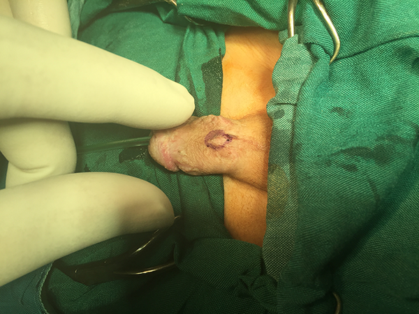
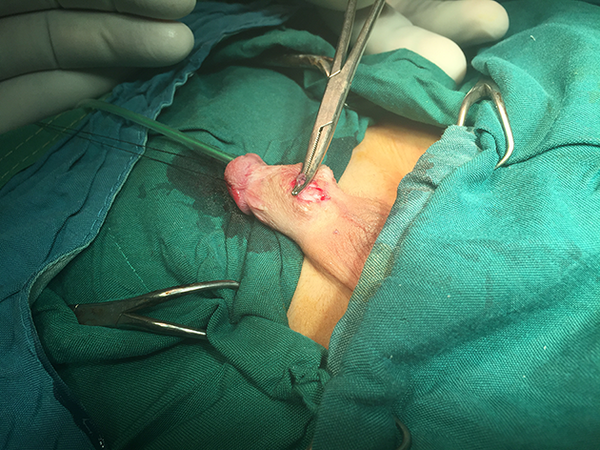
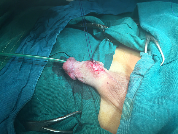
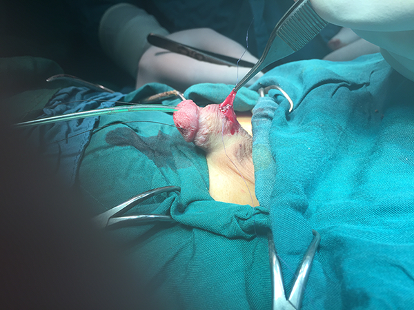
The patients were prepared and draped, then placed under general anesthesia in the supine position. For confirmation and identification of the fistula location and for the exclusion of any additional fistulas, sterile ophthalmic gentamycin ointment was injected into the urethra with simultaneous pressure on the bulbous urethra. The size of each fistula was determined with calipers. After insertion of a urethral catheter (French 6 or 8), the skin was incised around the site of the fistula. After the fistula was released from the surrounding tissue and the dartos fascia, it was ligated with 5 - 0 PDS sutures. Closure of the dartos fascia, as a second layer, was performed with 5 - 0 PDS thread, using the continuous suture method. To confirm the complete closure of the fistula tract, after suturing of the dartos fascia, a sterile saline irrigation solution was injected into the urethra with concurrent pressure on the bulbous urethra. For the last step, the skin was closed with 6 - 0 Vicryl sutures. Images of the surgical procedure are shown in Figures 1 - 4. All patients underwent the same anesthesia and surgical procedures, which were performed by the same urological surgeon. None of the patients faced unexpected problems or failures during the procedure. The surgical site was covered with light dressings postoperatively.
The Marker Around the Fistula Tract

The Fistula Tract Held in a Clamp After Dissection

The Dissected Fistula Tract, Ligated With 5 - 0 PDS Sutures

Closure of the dartos fascia, as a Second Layer, Performed with 5 - 0 PDS Thread

The outcomes were evaluated after six months and are described below. The success of the surgery was determined based on fistula relapse or the occurrence of other complications during the follow-up interval.
4. Results
The mean age of the patients was 5.7 (± 3.10) years (range 2 - 13, median 5). The characterization of the UCFs is shown in Table 1. Thirty-seven patients had a single fistula, two patients had two fistulas, and one patient had three fistulas. Five patients had a history of previous fistula repair, one with a proximal fistula and the remaining four with distal fistulas. The mean interval between primary TIP urethroplasty and the present attempt at fistula repair was approximately 7.9 (± 2.99) months (median 8 months).
Characterization of Urethrocutaneous Fistulas After TIP Urethroplasty
| Characteristic | Number |
|---|---|
| Number of fistulas | |
| Single | 37 |
| Two | 2 |
| Three | 1 |
| Total fistula count | 44 |
| Fistula size | |
| Small (2 mm or less) | 20 |
| Large (2 - 4 mm) | 24 |
| Fistula location | |
| Distal | 34 |
| Mid-shaft | 8 |
| Proximal | 2 |
The mean duration of the operation was 30.25 (± 5.18) minutes (range 25 - 40; median 30). The average length of hospital stay was 1.28 (± 0.45) days (range 1 - 2; median 1). After six months, relapse had occurred in only one patient, a six-year-old boy with a single 4-mm distal fistula, who had had no previous fistula repairs. Therefore, in 97.5% of the cases, relapse was non-existent. Other complications, such as urethral stenosis, intraurethral obstruction, urinary retention, difficulty voiding, and epidermal inclusion cysts, were not seen in the other patients during the six-month follow-up period.
5. Discussion
The results indicated that this simple technique has a high success rate for repairing UCFs measuring < 4 mm in any location. Attempts to achieve a higher success rate for closure of UCFs have led to the development of various surgical techniques, including simple closure, the trapdoor technique, an envelope-like closure, multilayered repair, and TIP urethroplasty. A previous study described some principles for the ideal repair technique: gentle tissue-handling, urethral mucosa inversion after excision of the epithelialized fistula tract, multilayer repair with a waterproofing layer, non-overlapping sutures, the use of absorbable and thin suture materials, a tension-free closure, the use of optical magnification, and needle-point cautery for coagulation (9).
Srivastava et al. studied the effects of the size, location, and number of fistulas and the surrounding tissue on selecting the procedure and its outcome. First, they excised the fistula tract, and smaller fistulas were repaired using the simple closure technique and subcutaneous tissue flaps. For larger fistulas with good surrounding skin, following simple closure of the fistula site, the skin was closed with a layered closure. Larger multiple or recurrent fistulas with surrounding scar tissue were closed with an additional waterproofing flap. A comparison of the success rate of the different fistula repair methods revealed that closure with a waterproofing layer was the most successful (100%), followed by layered (89%) and simple (77%) closures. These rates decreased on the second attempts at simple closure (33%), but there was 100% success for closure with a waterproofing layer. The significantly improved success rate with the addition of a waterproofing layer suggests that the use of this interposition layer can prevent the recurrence of UCFs (8).
Some studies have shown that simple closure of UCFs has a high failure rate compared to other techniques. In a study by Guan et al., modified prepuce-degloving repair for UCFs, compared to simple closure or Y-V plasty, was shown to have a significantly lower long-term recurrence rate, supporting its wide clinical application (10).
Another study evaluating the success rate of the prepuce-degloving method versus simple closure or Y-V plasty concluded that the former (96.2% success) compared to the latter (78.7% success) was a good choice for fistula repair after hypospadias surgery (11).
Some authors have reported that multilayer closure with an intermediate waterproofing layer between the neourethra and the skin plays a major role in decreasing the incidence of UCF recurrence, regardless of the surgical repair method. Various vascularized flaps, such as tunica vaginalis, dartos fascia, de-epithelialized skin, and Buck’s fascia, can be applied as the intermediate layer (12-14). On the other hand, some studies have shown that simple closure of the UCF followed by a second-layer closure has a high success rate compared to other methods.
Nerli et al. suggested the PATIO technique (which preserves the tract and turns it inside-out) for managing solitary UCFs following primary hypospadias repair. This repairs the fistula by inversion of the fistula tract to the urethra, and this method is effective in treating solitary UCFs measuring < 4 mm (15). In contrast to the PATIO method, we did not invert the fistula tract, in order to reduce the risk of urethral stenosis and stricture.
In a method used by Jamal et al. for the management of primary small UCFs (< 2 mm), after dissection of the tract, it was ligated tightly with 5 - 0 Vicryl and then the epithelium of the dissected tract was fulgurated by the diathermy. In order to prevent inclusion cyst formation, the local soft tissue was secured over the ligated fistula as a second layer. They concluded that this method was effective and successful in treating small-caliber UCFs (16). In the present study, although we did not fulgurate the dissected tract, no inclusion cysts formed. Another difference between our method and Jamal’s was that we operated on fistulas measuring > 2 mm and included three boys with recurrent fistulas.
Mohamed et al. used the midline relaxing incision for large fistulas, then covered it with a vascularized dartos-based or tunica vaginalis flap. This method demonstrated a high success rate for repairing midline and proximal urethral fistulas (17).
In the posterior urethral incision technique, after dissection of the skin edge around the urethral fistula and trimming the fistula edge, a posterior midline incision was made on the urethral wall; the edge was then closed in two layers, the urethral edge and the dartos fascia. The success rate was 90.6% for small- and large-size fistulas (18).
Although we did not incise the UCFs, no recurrences occurred in our cases after six months of follow-up, the surgical sites showed no tension, and there were good cosmetic results. The results showed that our simple method had a high success rate (97.5%) for repairing small (≤ 2 mm) and large (2 - 4 mm) UCFs. Our method is equal to previous methods in terms of success rate, and it involves a short operating time and an easy learning curve.
We suggest conducting a large, randomized, controlled clinical trial with a longer follow-up period to confirm the results of the present study. Further studies should also be conducted on the success rate in patients with fistulas larger than 4 mm or who have undergone another type of primary hypospadias repair surgery.
5.1. Conclusions
The present method, which is simple, rapid, and easily learned, has a high success rate in small (≤ 2 mm) and large (2 - 4 mm) UCFs that develop after TIP repair of hypospadias in any part of the penis. Relapses and complications were almost non-existent during six months of follow-up.
Acknowledgements
References
-
1.
Karabulut R, Turkyilmaz Z, Sonmez K, Kumas G, Ergun S, Ergun M, et al. Twenty-Four Genes are Upregulated in Patients with Hypospadias. Balkan J Med Genet. 2013;16(2):39-44. [PubMed ID: 24778562]. https://doi.org/10.2478/bjmg-2013-0030.
-
2.
Snodgrass W, Bush N. Recent advances in understanding/management of hypospadias. F1000Prime Rep. 2014;6:101. [PubMed ID: 25580255]. https://doi.org/10.12703/P6-101.
-
3.
Agrawal K, Misra A. Unfavourable results in hypospadias. Indian J Plast Surg. 2013;46(2):419-27. [PubMed ID: 24501477]. https://doi.org/10.4103/0970-0358.118623.
-
4.
Chung JW, Choi SH, Kim BS, Chung SK. Risk factors for the development of urethrocutaneous fistula after hypospadias repair: a retrospective study. Korean J Urol. 2012;53(10):711-5. [PubMed ID: 23136632]. https://doi.org/10.4111/kju.2012.53.10.711.
-
5.
Huang LQ, Ge Z, Tian J, Ma G, Lu RG, Deng YJ, et al. Retrospective analysis of individual risk factors for urethrocutaneous fistula after onlay hypospadias repair in pediatric patients. Ital J Pediatr. 2015;41:35. [PubMed ID: 25903765]. https://doi.org/10.1186/s13052-015-0140-8.
-
6.
Ikuerowo SO, Bioku MJ, Omisanjo OA, Esho JO. Urethrocutaneous fistula complicating circumcision in children. Niger J Clin Pract. 2014;17(2):145-8. [PubMed ID: 24553021]. https://doi.org/10.4103/1119-3077.127422.
-
7.
Shirazi M, Noorafshan A, Serhan A. Effects of different suture materials used for the repair of hypospadias: a stereological study in a rat model. Urol Int. 2012;89(4):395-401. [PubMed ID: 23108502]. https://doi.org/10.1159/000343423.
-
8.
Srivastava RK, Tandale MS, Panse N, Gupta A, Sahane P. Management of urethrocutaneous fistula after hypospadias surgery - An experience of thirty-five cases. Indian J Plast Surg. 2011;44(1):98-103. [PubMed ID: 21713169]. https://doi.org/10.4103/0970-0358.81456.
-
9.
Soni A, Sheoran S. Repair of large urethrocutaneous fistula with dartos-based flip flap: A study of 23 cases. Indian J Plastic Surg. 2007;40(1):34.
-
10.
Guan ZL, Wang SQ, Liu JL, Huang Y, Cheng G, Liu BJ, et al. [Modified prepuce-degloving repair for urethrocutaneous fistula following hypospadias surgery]. Zhonghua Nan Ke Xue. 2013;19(12):1091-4. [PubMed ID: 24432620].
-
11.
Qin C, Wang W, Wang SQ, Cao Q, Wang ZJ, Li PC, et al. A modified method for the treatment of urethral fistula after hypospadias repair. Asian J Androl. 2012;14(6):900-2. [PubMed ID: 23064690]. https://doi.org/10.1038/aja.2012.85.
-
12.
Elsaket H, Habib EM. Systematic Approach for the Management of Urethrocutaneous Fistulae after Hypospadias Repair. Egypt J Plast Reconstr Surg.
-
13.
Kadian YS, Rattan KN, Singh J, Singh M, Kajal P, Parihar D. Tunica vaginalis: an aid in hypospadias fistula repair: our experience of 14 cases. Afr J Paediatr Surg. 2011;8(2):164-7. [PubMed ID: 22005357]. https://doi.org/10.4103/0189-6725.86054.
-
14.
Sharma N, Bajpai M, Panda SS, Singh A. Tunica vaginalis flap cover in repair of recurrent proximal urethrocutaneous fistula: a final solution. Afr J Paediatr Surg. 2013;10(4):311-4. [PubMed ID: 24469479].
-
15.
Nerli RB, Metgud T, Bindu S, Guntaka A, Patil S, Neelgund SE, et al. Solitary urethrocutaneous fistula managed by the PATIO repair. J Pediatr Urol. 2011;7(2):166-9. [PubMed ID: 20541979]. https://doi.org/10.1016/j.jpurol.2010.04.016.
-
16.
Jamal YS, Kurdi MO, Moshref SS. Management of Small Urethrocutaneous Fistula by Tight Ligation with Fulguration of the External Epithelium of the Tract. Ann Pediatr Surg.
-
17.
Mohamed S, Mohamed N, Esmael T, Khaled S. A simple procedure for management of urethrocutaneous fistulas; post-hypospadias repair. Afr J Paediatr Surg. 2010;7(2):124-8. [PubMed ID: 20431229]. https://doi.org/10.4103/0189-6725.62844.
-
18.
Awad MS. A simple novel technique [PUIT] for closure of urethrocutaneous fistula after hypospadias repair: Preliminary results. Indian J Plastic Surg. 2005;38(2):114.
reply
reply