IMAGE IN CARDIOLOGY

DOI: 10.4244/EIJV11I10A227

Acute LAD occlusion by calcified plaque embolisation from the ascending aorta after contralateral LCA angiography

Masashi Amano, MD; Toshihiro Tamura*, MD; Yukiko Hayama, MD; Naoaki Onishi, MD; Yodo Tamaki, MD; Soichiro Enomoto, MD; Makoto Miyake, MD; Hirokazu Kondo, MD; Kazuaki Kaitani, MD; Chisato Izumi, MD; Yoshihisa Nakagawa, MD

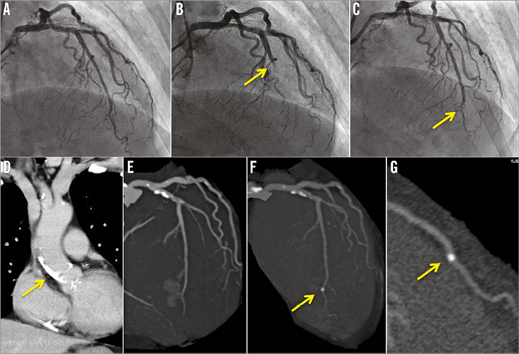
A 65-year-old female was admitted to our hospital for percutaneous coronary intervention (PCI) for chronic total occlusion (CTO) of the right coronary artery (RCA). Coronary angiography showed a CTO lesion in the mid RCA with good collateral circulation supplied from the left coronary artery (LCA) without significant stenosis or calcification (Panel A, Moving image 1). However, after simultaneous angiography, the patient had sudden chest pain with ST-elevation in leads V1-4. LCA angiography showed total occlusion in the mid left anterior descending coronary artery (LAD) (Panel B, Moving image 2). Emergent PCI of the LAD was then undertaken. While thrombus aspiration was tried repeatedly, no thrombus was aspirated, but the occluded site moved from mid to distal LAD (Moving image 3). After dilation with a 1.25 mm balloon, the occluded lesion improved to mild stenosis (Panel C, Moving image 4) and the patient’s symptoms disappeared. Computed tomography (CT) before PCI revealed severe calcification at the ascending aorta and no calcified finding at the distal LAD (Panel D, Panel E). Moreover, coronary CT angiography after PCI revealed a new calcified finding in accordance with the mild stenotic lesion of the distal LAD (Panel F, Panel G). We speculate that the calcified plaque of the ascending aorta embolised at the mid LAD during the catheter manipulation in the LCA.

Conflict of interest statement

The authors have no conflicts of interest to declare.

Supplementary data

Moving image 1. The initial LCA angiography showed no significant stenosis.

Moving image 2. The mid LAD was totally occluded after simultaneous angiography.

Moving image 3. After aspiration was tried repeatedly, the occluded site moved from mid to distal LAD.

Moving image 4. Finally, the mild stenosis remained at the distal LAD.

Supplementary data

To read the full content of this article, please download the PDF.

Moving image 1. The initial LCA angiography showed no significant stenosis.

Moving image 2. The mid LAD was totally occluded after simultaneous angiography.

Moving image 3. After aspiration was tried repeatedly, the occluded site moved from mid to distal LAD.

Moving image 4. Finally, the mild stenosis remained at the distal LAD.

Volume 11 Number 10
Feb 19, 2016
Volume 11 Number 10
View full issue


Key metrics

On the same subject

Image – Interventional flashlight

10.4244/EIJ-D-18-00915 Aug 7, 2020
Percutaneous revascularisation of a chronic total occlusion in a surgically reconstructed vessel
Goktekin O et al
free

IMAGE IN CARDIOLOGY

10.4244/EIJV11I13A291 Apr 20, 2016
A disaster never comes alone: total ostial occlusion of the left main coronary artery with an anomalous origin
Rodrigues P et al
free

INTERVENTIONAL FLASHLIGHT

10.4244/EIJ-D-17-00532 Jun 20, 2018
Single guiding catheter retrograde CTO recanalisation facilitated by trapping technique: introducing the modified rendezvous technique
Konstantinidis N and Sianos G
free

Image – Interventional flashlight

10.4244/EIJ-D-19-00175 Dec 6, 2019
Intravascular lithotripsy in chronic total occlusion percutaneous coronary intervention
Azzalini L et al
free

10.4244/EIJV12I11A218 Dec 9, 2016
Conquering CTO revascularisation: the summit is near with 90% of the ascent behind us
Di Mario C and Dini C
free
Trending articles
338.03

State-of-the-Art Review

10.4244/EIJ-D-21-00904 Apr 1, 2022
Antiplatelet therapy after percutaneous coronary intervention
Angiolillo D et al
free
284.93

State-of-the-Art Review

10.4244/EIJ-D-21-00695 Nov 19, 2021
Transcatheter treatment for tricuspid valve disease
Praz F et al
free
226.03

State-of-the-Art Review

10.4244/EIJ-D-21-00426 Dec 3, 2021
Myocardial infarction with non-obstructive coronary artery disease
Lindahl B et al
free
209.5

State-of-the-Art Review

10.4244/EIJ-D-21-01034 Jun 3, 2022
Management of in-stent restenosis
Alfonso F et al
free
168.4

Expert review

10.4244/EIJ-D-21-00690 May 15, 2022
Crush techniques for percutaneous coronary intervention of bifurcation lesions
Moroni F et al
free
150.28

State-of-the-Art

10.4244/EIJ-D-22-00776 Apr 3, 2023
Computed tomographic angiography in coronary artery disease
Serruys PW et al
free
118

Translational research

10.4244/EIJ-D-22-00718 Jun 5, 2023
Preclinical evaluation of the degradation kinetics of third-generation resorbable magnesium scaffolds
Seguchi M et al
X

The Official Journal of EuroPCR and the European Association of Percutaneous Cardiovascular Interventions (EAPCI)

EuroPCR EAPCI
PCR ESC
Impact factor: 6.2
2022 Journal Citation Reports®
Science Edition (Clarivate Analytics, 2023)
Online ISSN 1969-6213 - Print ISSN 1774-024X
© 2005-2024 Europa Group - All rights reserved