The Experience of Mammography Based on the Memoirs of Examinees ()

1. Introduction
In Japan, there has been a rapid increase in the incidence of breast cancer while the increase in the rate of cancer screening continues to lag sluggishly behind. In particular, the rate of mammography screening in Japan was only 23.8% in 2009. This was an increase of only 1.3% during the past 10 years (22.5% in 2000), which is low in comparison to rates in other countries [1] .
In recent years, screening systems used in the general population have been developed according to reports on screening rates from various regions and medical institutions [2] -[7] . However, there have been few reports on the individual’s experience that might discourage screening. In a study on the factors affecting a patient’s decision to undergo mammography, examinees reportedly took into account a variety of factors, such as costs, means of transportation and the extent of pain [8] -[17] . In the present study, the authors believe that it is necessary to similarly focus on the various factors influencing the mammography examinees’ decisions and to understand what occurs during the procedure to influence the examinee’s view.
In order to understand the effect of mammography on examinees, this study intended to understand the examinees’ experiences in the course of mammography based on their published memoirs.
2. Methods
2.1. Data Collection
Based on memoirs of breast cancer patients which have been published in booklet form in Japan, all sentences that described the episodes of mammography were extracted as data. These memoirs were believed to be an effective method of gathering data while avoiding privacy issues involved in the study and avoiding excess stress on the examinees.
2.2. Analytical Framework
A qualitative analytical approach was selected in this study. Method was adopted framework of qualitative study of society for structural constructivism [18] . In order to draw on a diversity of individual experiences, it is important to use the data without reference to time. The focus was only on how certain events were experienced. Therefore, the time axis was defined in the process.
The structure of the study was organized based on the steps of the mammography process: “start”, “positioning and breast compression”, “imaging” and “completion”. With regard to the mammography procedure itself, the process shown in the mammography guidelines was used for reference [19] .
2.3. Analytical Procedure
The extracted data that focused on the mammography experience were transcribed as understandable concepts. The data were classified and similar parts were combined as specific examples and given a concept name. The concept name, its definition, examples and viewpoints of analysis were written down in the theoretical notes, which were created in the form of one worksheet per concept. Further, several concepts were encompassed into a category. The concepts which were not contained within a category were treated as independent concepts. A structured model was then created in which the relationships between categories and concepts were demonstrated.
2.4. Ethical Considerations
Prior to conducting this study, the research plan was submitted to and approved by the research ethics committee at Graduate school of Health Sciences, Niigata University (approval number 82).
3. Results
3.1. Overview of Examinees’ Memoirs
The memoirs describing patients’ experiences of mammography that have been published in booklet form in Japan were identified by searching the National Diet Library Online Public Access Catalog (NDL-OPAC) and using major bookstores’ software [20] . Six memoirs were found to be relevant to our study. One additional memoir was found using other search software. Therefore, a total of seven memoirs were found to be potentially relevant to our study. All of these were obtained and checked as to whether or not they contained a description of the mammography episode. As a result, a description of the mammography episode was confirmed in four memoirs, and these were used for the study. As shown in Table 1.
3.2. Overview of Subjects
All memoirs concerned the experience of examination and treatment for breast cancer. The authors’ ages were around 30 years old (one person’s age was unknown). The mammography experience described was the first or second time for the examinee. The reason for undergoing mammography was as a result of a consultation for an abnormal feeling in the breast in three patients.
3.3. Structure of the Examinee’s Experience
The structure of the examinee’s experience could be described in the form of a story line as follows. As shown in Figure 1.

Table 1. Memoirs list and contents of the excerpt.
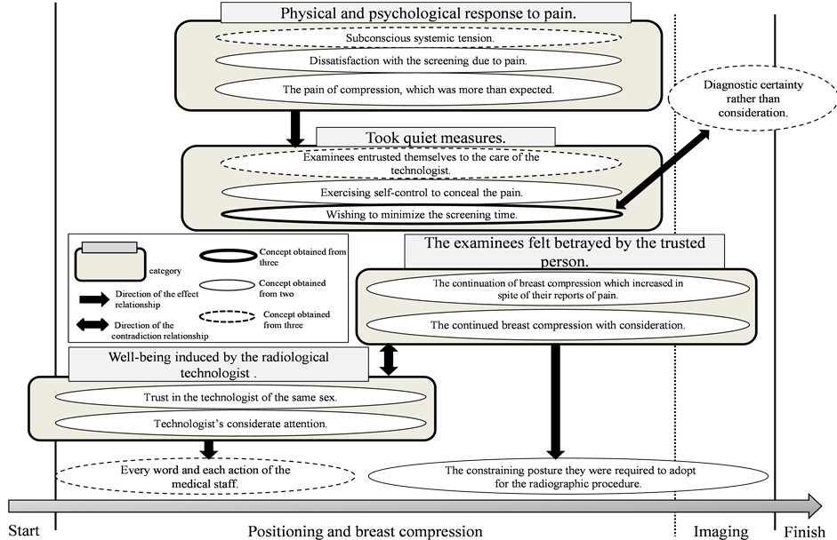
Figure 1. Experience structured model of mammography examinees.
From before imaging to during the imaging procedure, examinees had relied on a sense of well-being induced by the radiological technologist based on trust in the technologist of the same sex and a sense of security brought on by the technologist’s considerate attention. Even while relying on the radiological technologist, the examinees paid close attention to every word and each action of the medical staff, were interested in the disease state and held expectations of considerate treatment from the medical staff. However, as breast compression progressed, the examinees felt betrayed by the trusted person because of the continuation of breast compression which increased in spite of their reports of pain or discomfort, ignoring the situation, or the continued breast compression with consideration. The examinee’s sense of betrayal was increased due to the constraining posture they were required to adopt for the radiographic procedure. Further, due to the pain of compression, which was more than expected, and to subconscious systemic tension, examinees experienced dissatisfaction with the screening due to pain or their physical and mental reaction to it. Examinees entrusted themselves to the care of the radiological technologist and meanwhile took quiet measures such as earnestly desiring to minimize the screening time and exercising self-control to conceal the pain. While wishing to minimize the screening time on the one hand, the examinees desired, on the other hand, diagnostic certainty rather than consideration, which meant enduring the pain to ensure the quality of the imaging.
4. Discussion
Mammography is performed by compressing the breast with the X-ray transmissive plate, which is then pressed down further to make the thickness of the breast as thin as possible in order to obtain an accurate diagnosis and minimize exposure to radiation. This is a useful imaging technique which reduces risk for the examinees. During the imaging procedure, the examinees experience psychological and mental tension due to the pain and embarrassment associated with breast compression. The memoirs also revealed that they were sensitively attuned to capture the actions and words of the medical staff. Meanwhile, the support and considerate words of the radiological technologist provided some relief from the pain experienced by the examinee.
On the other hand, in spite of the perceived supportive attitude of the radiological technologist, when the examinees mentioned their pain, the breast compression increased, and this caused the examinees’ feelings to a feeling of betrayal by the trusted person.
Examinees experienced subconscious physical tension because of their feelings of embarrassment and anticipation of pain and consequently entrusted themselves to the care of the radiological technologist. Such long lasting tension is believed to cause more pain than necessary at the time of breast compression and thus increase the psychological burden. In addition, the fact that the pain of compression was more than expected and the dissatisfaction with the screening experience due to the pain arose because of the lack of information about the mammography procedure itself. Because of this, examinees were confused and grew anxious and increasingly distrustful of the screening procedure.
From the above, it appears that examinees experienced a psychological and physical burden during the mammography process and that the contributing factors were intertwined. Currently, the patient’s point of view is becoming an important issue in the support of cancer patients. These findings provide suggestions for improved intervention in the psychological and physical burden of examinees undergoing the screening process.
5. Conclusion
Thirteen concepts were extracted from the mammography experiences which could be classified into four categories and three independent concepts. Examinees experienced a psychological and physical burden during the mammography procedure and the contributing factors were intertwined. This study suggests that, in mammography, there is a need to adjust the environment and provide increased support to ease the psychological and physical tension and thus the burden on the examinee. This study is limited because it is the analysis of small memoirs. We believe that quantitative assessment during mammography would contribute to the improvement of burden-reduction program in mammography.
Acknowledgements
This work was supported by JSPS KAKENHI Grant Number 23593285.