Published online Jun 15, 1998. doi: 10.3748/wjg.v4.i3.260
Revised: April 20, 1998
Accepted: May 12, 1998
Published online: June 15, 1998
AIM: To confirm the therapeutic effect of Zijin capsule on liver fibrosis in rat model.
METHODS: Model group: Bovine serum albumin (BSA) Freund’s incomplete adjuvant 0.5 mL was injected subdermally at d1 d15 d22 d29 and d36 for primary sensitization. Seven days after the fifth injection, BSA antibody in the serum was detected by double agar diffusion method. Normal saline of 0.4 mL was injected through cauda vein to BSA antibody-positive rat twice a week for fifteen times. Traditional Chinese medicine (TCM) decoction group and Zijin capsule group: In the attack injection period, Chinese medicinal decoction or Zijin capsule was given ig, the others were the same as in the model group. NS was used in the control group. The collagen content of rat liver was determined by Bergman’s method and expressed as x-±s. The liver pathological changes were divided into four grades and expressed as the avarage of the total rank sum.
RESULTS: The collagen content (mg/g) of the liver in the control group (7.2 ± 1.9) was significantly lower than that in the other groups; it was higher in the model group (31.7 ± 16.6) than that in the two therapeutic groups; and lower in Zijin capsule group (9.7 ± 2.8) than that in the TCM decoction group (11.5 ± 5.3). The pathological changes were more aggravated in the model group (37.4) than those in the two therapeutic groups; and more severe in the TCM decoction group (30.2) than in the Zijin capsule group (22.9).
CONCLUSION: The therapeutic effect of Zijin capsule on the model was confirmed.
- Citation: Cai DY, Zhao G, Chen JC, Ye GM, Bing FH, Fan BW. Therapeutic effect of Zijin capsule in liver fibrosis in rats. World J Gastroenterol 1998; 4(3): 260-263
- URL: https://www.wjgnet.com/1007-9327/full/v4/i3/260.htm
- DOI: https://dx.doi.org/10.3748/wjg.v4.i3.260
The pathological changes of the rat liver fibrosis induced by bovine serum albumin (BSA) injections are similar to those in human portal cirrhosis. Compared with the TCM decoction proved effective in clinic the therapeutic effect of Zijin capsule in rat liver fibrosis was observed.
Wistar rats (female 30, male 30, 150 g-210 g) were purchased from Hubei Medical Institute. BSA was product of Shanghai Medical Testing Agent Plant, prepared as 18 g/L in normal saline, clean from bacteria through filtration, and stored at 4 °C. Freund’s incomplete adjuvant: One gm lipid from sheep hair (CP Third Lipid Company in Shanghai) was mixed with 2 gm liquid paraffin (Fushan Chemical Industry), sterilized in a steam autoclave and stored at 4 °C. L-hydroxyproline, standard sample, was from Biochemistry Institute of the Chinese Academy of Sciences. Zijin capsule. prepared mainly from herba swertiae puniceae and endothelium corneum gigeriae galli in Hubei College of Traditional Chinese Medicine. The concentration of crude drugs was 5 g/1 mL. The TCM decoction provided by the Department of Infectious Diseases, the Afficiated Hospital of Hubei TCM College, containing mainly Radix astragali, Radix condonopsis pilosulae, Rhizoma atractylodis macrocephalae, Rhizoma polygoniti, fructus lycii, fructus corni, radix rehmanniae and Radix rehmanniae preparata.
Wistar rats with free access to water were randomly divided into four groups.
Control group (n = 12). Normal saline was used for immunological primary (sensilization) and second (attack) injection instead of BSA, the others were the same as those in the model group.
Model group (n = 12). BSA Freund’ incomplete adjuvant 0.5 mL was injected subdermally at d1 d15 d22 d29 and d36 for primary sensitization. Seven days after the fifth injection, BSA antibody in rats’ serum was detected by double agar diffusion method. BSA of 0.4 mL in normal saline was administered once through cauda vein for attack injection in BSA antibody-positive rats, twice a week for fifteen times, the concentration of BSA was 5.00, 5.25, 5.50, 5.75, 6.00, 6.25, 6.50, 6.75, 7.00, 7.25, 7.50, 8.50, 9.00, 9.50 and 10.00 g/L. The rats were given 10 mL·kg·d N.S. ig at the same period. All animals were killed with decollation nine days after the last injection. The livers were taken for biochemistry detection and morphological observation. The whole period was 95 d.
TCM decoction group (n = 18). In the attack injection period, the Chinese medicinal decoction equivalent to 36 g crude drugs/kg·d was givenig, the others were the same as those in the model group. Zijin capsule group (n = 18). In the attack injection period, Zijin capsule, equivalent to 12.8 g crude drugs kg·d, was given ig, the others were the same as those in the model group.
The collagen content in rat liver was determined by Bergman’s method[1], and expressed as x-±s and analyzed by t test . Value of P < 0.05 was considered at a significant level. The rat livers were embedded with paraffin and stained with HE. The pathological changes were classified into four grades according to the quality and quantity of the liver histological features, expressed as the avarage of the total rank sum in each group and analyzed by H-test. Value of P < 0.05 wasc onsidered at a significant level.
The indexes are shown in Table 1. The collagen content was significantly lower in the control group than that in the model group (P < 0.01), the TCM decoction group (P < 0.05) and Zijin capsule group (P < 0.05); it was higher in the model group than that in the TCM decoction group (P < 0.01) or Zijin capsule group (P < 0.01); and not significantly lower in Zijin capsule group than that in the TCM decoction group (P > 0.05).
After a comprehensive observation on the pathologic changes, a distinct grading standard was defined as follows:
Grade 0 (normal rat liver) Liver capsula was a thin connective tissue. The parenchyma consisted of hepatic lobules and portal areas. The hepatic lobules were similar to round balls, their central veins, laminae of hepatocytes (radiated arrange) and siunsoids were normal and clear. The hepatic lobule boards were hepatic cell layers as limiting laminae. There was hepatic sinusoid between the hepatic laminae, their endothelia were distributed regularly, Kupffer’s cells can be seen obviously. The ratio of the hepatic laminae to the hepatic sinusoids was 3 to 2. The hepatocytes are polygonal cells, cytoplasm was normal acidophil, the hepatic nuclears in round shape, the chromatin distributed rarefactionally along the nuclear membrane with nucleole. The portal area was mainly connective tissues, including the interlobular hepatic artery, the portal vein branches and interlobular bile ducts. Some lymphocytes and plasma cells had infiltrated into the portal areas. The necrosis within a few hepatocytes and their lymphocyte or plasma cell infiltration (spotty necrosis) were occasionally observed in one hepatic lobule or two. There were many red-dyed microgranules (slight cloudy swelling) in some hepatocytes.
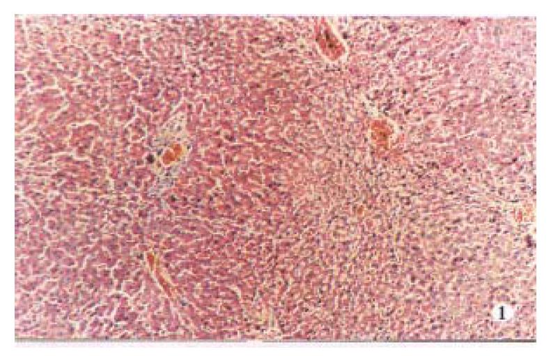
Grade 1 (liver injury change). Serious and extensive changes were present as the degeneration and necrosis of hepatocytes and the congestion or bleeding in the hepatic sinusoid. Cloudy swelling of hepatocytes: enlarged volume, round shape, many small red-dyed granules in hepatic cytoplasm were observed; hepatic laminae became wider, and hepatic sinusoid were pressed to ischemia. Thin hepatic cytoplasm: the hepatocyte volume became larger, red-dyed cytoplasm thinner and less homogeneous; the nuclears stained dim; and the ratio of hepatic laminae to the hepatic sinusoids became higher. Ballooning degeneration of hepatocytes: the volume of hepatocytes became extremely large, round shaped; the cytoplasm appeared empty (light transparency) like a balloon; nuclear not located at the cell centre. Lytic necrosis of hepatocytes: the ballooning degeneration of hepatocytes further developed to karyopyknosis, karyorrhexis and karyolysis; the whole structure of the hepatocyte body even disappeared, only the network of the reticular fibers remained; however, the lymphocytes infiltration in necrotic focus was not obvious; and the necrosis of hepatocytes did not occur at the limiting laminae in the hepatic lobules. The congestion and bleeding in the hepatic sinusoid: the hepatic sinusoids were expanded and filled with blood. The bleeding in Disse’s cavity was extensive. Two kinds of congestion and bleeding in hepatic lobules were observed in these cases, one distribution was limited around central veins, which was seldomly observed, the other mainly appeared near the limiting laminae of hepatic lobules, which were predominant. At the same time, there were light hyperemia, edema and fibrous proliferation in the portal areas (Figure 1).
Grade 2 (liver prefibrosis). The necrosis within a few hepatocytes occurred at the limiting laminae (piecemeal necrosis); and the necrosis range may be expanded to a large region of hepatic necrosis which connected the central veins or portal areas (bridging necrosis). Serious congestion and bleeding near the limiting laminae in the hepatic lobules were common. The hepatic laminae became thinner in the same region accompanied by hepatocyte atrophy, cloudy swelling and fatty degeneration and the hepatocytes even disappear in the same region. The congestion may be connected with the central veins or portal areas (bridgingco ngestion). In those cases, the Kupffer’s cells in the hepatic sinusoid enlarged and proliferated obviously with more processes. In the portal areas, fibroblasts proliferated obviously and collagen increased, which made the boundary in the normal hepatic lobules more clear, but they were not streching into the hepatic lobules through the limiting laminae.
Grade 3 (liver fibrosis). The hepatocyte injury (degeneration and necrosis) at the limiting laminae has advanced obviously, while the proliferated collagen at portal areas has invaded into the hepatic lobules along with the injuried limiting laminae. It was called as liver fibrosis while the proliferated collagen has not been completely contacted each other and absolutely separated the hepatic lobules. It was the cirrhosis when the proliferated collagen has been contacted each other completely, and absolutely separated the hepatic lobules, leading to the formation of pseudolobules (many round islands of hepatocytes). Their hepatic laminae have not radiated regularly, the central vein dystopy (asymmetry, disappear or several veins), the structure of portal areas was located in the pseudolobules, there were some atrophy, fatty degeneration and necrosis of the hepatocytes in the pseudolobules; they were separated continuously with the proliferated collagen, the cholestatic bile capillaries appeared and their bile thrombus formed. Among the pseudolobules, the fibrous bands were continuous, homogeneous and delicate; and there are more infiltrated lymphocytes. The collagenous fibers were stained bright-red (Figure 2).
According to the grading, H test was used to analyse statistically the liver pathological changes among those groups. The pathological changes are more moderate in the control group than those in the other three groups (P < 0.01, Table 2). They were more severe in the model group than those in the two therapeutic groups; and more aggravated in the TCM decoction group than those in the Zijin capsule group.
Rat liver fibrosis induced by BSA injections After BSA sensitization injection (sc), the rats were intravenously injected with BSA as attack injection, the CIC (the circulating immune complex formed with BSA antigens and their antibodies) or the remained antigens (BSA) has deposited in some tissues of the rats, leading to classical type III or/and type II of the allergic reactions, subsequently local tissue injuries. The administration of antigen, including the route, time and dose of the injected antigen decides the location and features of injury lesions. Based on the references about the experimental methods, BSA injected with various dosages, at different times and dates produced serious injuries (37/37) of livers and even obvious fibrosis or cirrhosis (8/37). The locations and features of the pathologic changes have suggested the pathogenesis of cirrhosis, i.e., the pathological changes were advanced near the limiting laminae in the hepatic lobules, because the CIC deposited earlier and more seriously at the location according to the circulative dynamics in the hepatic lobules. The features of the pseudolobules (such as shape, size, laminae arrange, cellular injuries) and the fibrous bands (delicate, homogeneous) were similar to the pathological changes in human poral cirrhosis. The hepatocytes did not regenerate to form pseudobobules obviously. However, the liver structures were normal (P < 0.01) and the collagen content was low (P < 0.05) in the control group. The results indicate that the rat cirrhosis models induced by BSA injections according to the immunological principles are succesful[1,2].
In contrast to the model group, the collagen was low (P < 0.01), and the pathologic changes were slight (P < 0.05 or P < 0.01) in Zijin capsule group as well as the TCM decoction group. It confirms the effect of Zijin capsule to treat the rat liver fibrosis. Compared with that in the TCM decoctiongroup with confirmed therapeutic effect in human liver fibrosis[3], the collagen content remained the same level (P > 0.05) in Zijin capsule group. The main active components[4] of Zijin capsule can increase the permeability of capillary, lead to the CIC depositing in different locations, enhance the ability of the mononuclear phagocyte system (it may promote the elimination of the CIC from the circulating blood), activate the hepatocyte metabolism to normalize their biology, and protect the hepatocytes from injuries. All these may be associated with the therapeutic effect of Zijin capsule in the rat liver fibrosis induced by BSA injections.
Project supported by the key Project Fund of Scientific Committee of Hubei Province.
| 1. | Zhu QG, Fang BW, Wu HS, Lan SL, Fu QL. The study of the immunity liver fibrosis animal model induced by bovine serum albumin. Chin J Pathol. 1993;22:121-122. [Cited in This Article: ] |
| 2. | Wang BE. The study of the experimental model in immunity liver fibrosis. Chin Med J. 1989;69:503-506. [Cited in This Article: ] |
| 3. | Fang BW, Zhu QG, Zhu JE, Wu HS, Lan SL, Fu QL. The experimental study of the drugs for supplementing Qi and activating blood circulation in the liver fibrosis animal model by bovine serum albumin. J Chin Traditio West Med. 1992;12:738-739. [Cited in This Article: ] |
| 4. | Chen JC, Huang XS. The chemical component analysis of Swertia Punicea Plant in Hubei Province. Chin Materia Med. 1990;13:29-31. [Cited in This Article: ] |


