Article

Transient Increases in Inflammation and Proapoptotic Potential Are Associated with the HESN Phenotype Observed in a Subgroup of Kenyan Female Sex Workers

1
Department of Biology, University of Winnipeg, Winnipeg, MB R3B 2R9, Canada
2
Yangquan Municipal Center for Disease Prevention and Control, Yangquan 045000, China
3
National HIV & Retrovirology Laboratories, Public Health Agency of Canada, Winnipeg, MB R3E 3L5, Canada
4
Department of Immunology, University of Manitoba, Winnipeg, MB R3E 0T5, Canada
5
Medical Microbiology & Infectious Diseases, University of Manitoba, Winnipeg, MB R3E 0J9, Canada
6
Department of Medical Microbiology, University of Nairobi, Nairobi 00202, Kenya
*
Author to whom correspondence should be addressed.
These authors contributed equally to this work.
Viruses 2022, 14(3), 471; https://doi.org/10.3390/v14030471
Submission received: 31 December 2021 / Revised: 13 February 2022 / Accepted: 21 February 2022 / Published: 25 February 2022
(This article belongs to the Special Issue Correlates of Immune Protection against HIV Infection)

Abstract

:
Interferon (IFN) -stimulated genes (ISGs) are critical effectors of IFN response to viral infection, but whether ISG expression is a correlate of protection against HIV infection remains elusive. A well-characterized subcohort of Kenyan female sex workers, who, despite being repeatedly exposed to HIV-1 remain seronegative (HESN), exhibit reduced baseline systemic and mucosal immune activation. This study tested the hypothesis that regulation of ISGs in the cells of HESN potentiates a robust antiviral response against HIV. Transcriptional profile of a panel of ISGs with antiviral function in PBMC and isolated CD4+ T cells from HESN and non-HESN sex worker controls were defined following exogenous IFN-stimulation using relative RT-qPCR. This study identified a unique profile of proinflammatory and proapoptotic ISGs with robust but transient responses to exogenous IFN-γ and IFN-α2 in HESN cells. In contrast, the non-HESN cells had a strong and prolonged proinflammatory ISG profile at baseline and following IFN challenge. Potential mechanisms may include augmented bystander apoptosis due to increased TRAIL expression (16-fold), in non-HESN cells. The study also identified two negative regulators of ISG induction associated with the HESN phenotype. Robust upregulation of SOCS-1 and IRF-1, in addition to HDM2, could contribute to the strict regulation of proinflammatory and proapoptotic ISGs in HESN cells. As reducing IRF-1 in the non-HESN cells resulted in the identified HESN ISG profile, and decreased HIV susceptibility, the unique HESN ISG profile could be a correlate of protection against HIV infection.

1. Introduction

Despite the tremendous efforts put forth, the number of new HIV infections remained stable at around 1.3 million per year for the last 3 years [1,2,3]. The success of preventative measures, such as increased condom use, pre-exposure prophylaxis (PrEP), and treatment as prevention (TasP) remain suboptimal and dependent on the level of adherence [4,5,6,7,8,9]. There is an ever-urgent need for novel preventative tools, which require better understanding of the protective mechanisms against HIV-1 infection.
Fortunately, not all individuals exposed to HIV-1 become infected. Several studies reported a subset of around 5–15% of HIV-exposed individuals who remain seronegative (HIV-exposed seronegative (HESN)) even after repeated exposure [10]. These HESN individuals have been found in several geographic locations with diverse ethnic backgrounds and routes of exposure. Studies have been performed to identify genetic and immunologic mechanism(s) of protection in the HESN subsets [11,12,13,14,15,16,17,18,19,20,21,22,23,24,25,26,27,28,29,30,31,32,33,34,35,36]. The mechanism(s) reported to be associated with seeming resistance to HIV infection in HESN are most likely multifactorial [16,26,36,37]. Host factors that are associated with delayed seroconversion or reduced susceptibility to HIV infection include CCR5, CCR2b, ERAP2, MX2, TIM3, TLR3, APOBEC3 family, TRIM family, HLA (DQB1*03:02, HLA-B*57 and HLA-B*35, HLA-E, HLA-F, CD1a-e, MR1), Vitamin D receptor (VDR), and IRF-1. In addition to these genetic factors, a generalized phenotype of immune quiescence (IQ) has also been observed in HESN [26]. IQ is characterized by reduced baseline expression of T-cell activation markers, low baseline levels of pro-inflammatory cytokine and chemokine production in the peripheral and genital mucosal of the HESN, and increased baseline numbers of systematic regulatory T cells (Treg), which are known to suppress T-cell activation [38]. Several genetic factors also support the IQ phenotype in HESN; for example, reduced baseline interferon (IFN) regulatory factor-1 (IRF-1) expression and increased baseline VDR expression, which are associated with reduced immune activation in HESN [14,25,39,40,41]. Our earlier studies characterized IRF-1 in a highly HIV-exposed commercial sex worker cohort in Nairobi, Kenya. IRF-1, an IFN-stimulated gene (ISG) is a functional correlate of protection against HIV-1 acquisition [14,41,42,43] and its expression is regulated by both genetic and epigenetic mechanisms in HESN. This study further investigated the potential role of IRF-1 in supporting the IQ phenotype in HESN, perhaps contributing to reduced HIV susceptibility.
The IFN signaling pathway protects cells from invading viral pathogens by trans-activating a variety of interferon-stimulated genes (ISGs) [44,45,46,47]. Some ISGs encode innate detectors of viral molecules, while some encode effectors with varied antiviral functions, such as regulators of the apoptotic pathway, and IFN-signaling molecules [45,48,49]. Our studies and others suggest that a “fine-tuned” ISG response is critical for suppressing undesired, exaggerated viral responses, including HIV, that induce prolonged immune activation, and for preventing the consequential pathogenic host damages, while maintaining some innate antiviral activity [41,43,44,46]. Persistent type-I interferon responses have been associated with pathogenic outcomes of HIV-1 and SIV infections [46,50,51,52]. Our earlier work found that CD4+ T cells from Kenyan HESN female sex workers (FSWs) exhibit a lower basal level of IRF-1 but have the capacity for normal and transient IRF-1 response to HIV-1 infection. In vitro studies that moderate reduction of cellular IRF-1 expression via small-interfering RNA (siRNA) by 30–60%, decreased in vitro susceptibility to HIV-1 infection by >95% [14,41,43,53,54,55]. Although IRF-1 is an ISG itself, it is also a key transcription regulator for the ISG transcriptional program [56,57,58]. Thus, IRF-1 is a key regulator of late antiviral ISGs. Together, these support the crucial role of modulating ISG-mediated host defenses/inflammation in determining the outcome of infection.
This study focused on examining the expression and regulation of a subset of ISGs that have the described antiviral function. It tested the hypothesis that antiviral ISGs are uniquely regulated in immune cells from HESN individuals and that strict regulation of these ISGs is strongly associated with protection against acquisition of HIV-1 infection observed in HESN.
Furthermore, expression analysis of ISGs revealed that during both acute and chronic viral infection, most differentially expressed ISGs are induced by both type-I and type-II IFNs [59,60]. In addition, these data suggest that IFN-γ strongly contributes to shaping ISG upregulation, in addition to type-I IFNs. This study thus also compared the response of key ISGs to IFN-γ and IFN-α2 stimulation in the PBMC from HESN and non-HESN individuals. A unique set of proinflammatory antiviral ISGs and proapoptotic ISGs, which mounted normal but brief transcriptional responses, were found to have close association with the low susceptibility to HIV infection observed in HESN FSWs.

2. Materials and Methods

2.1. Ethics

The ethical review boards from the Kenyatta National Hospital/University of Nairobi (#P211/09/2006, renewed and amended annually since 2006), the University of Manitoba (HS15280-B2012:043, regularly renewed since 2012), and the Regional Ethical Review Board in Stockholm (2014/959–31, amended in 2018: 2018/1306–31) approved the study. Informed written consents were obtained from the participants in this study, following the declaration of Helsinki protocols.

2.2. Study Subjects

Participants with natural resistance to HIV infection (HESN) and HIV-susceptible seronegative participants (non-HESN) were chosen semi-randomly from subsets (age ranging 21–46 years at the time of sample collection, >7 or <3 years in the sex trade, active in sex work the week prior to sample collection and having no known infections, seronegative for HIV-1) of a well-characterized cohort of FSWs at the Majengo Clinic, Pumwani district of Nairobi, Kenya during the years of 2006–2010 (Table 1). These women maintained high-risk sexual behavior (0–70 clients the week prior to sample collection) with known HIV-infected clients (ranging between 0–3 clients). HESN and non-HESN FSWs were HIV negative (tested by ELISA and DNA/RNA testing) for >7 years and <3 years of follow-up, respectively. The non-HESN participants had been active in the sex trade for <3 years and were seronegative when the blood samples were collected. The definition of HESN and non-HESN is based on available epidemiological data of this cohort [26,61,62]. All FSWs participating in the Majengo cohorts were followed up twice a year during resurveys, which ended in 2013. By 2013, all HESN participants in this study remained seronegative for HIV-1, despite unprotected sexual behaviors. By 2013, 11 of the non-HESN participants (42%) seroconverted; 8 of the non-HESN participants (31%) did not return for follow-ups after 2009/2010. The other non-HESN FSWs (n = 7) remained seronegative up to the 2011/2012 resurvey. Despite an earlier study of this cohort demonstrating a ~85% seroconversion rate among the FSWs attending the Majengo clinic during a 13–14-year follow-up period [61], the HIV incidence has greatly dropped in Nairobi [63], perhaps due to the huge rollout of antiretroviral treatments (ART) in 2009 and active educational campaigns for safe sex and condom use. However, with the relatively small biological variations and significant differences observed within each group in the parameters examined in this study, the potential caveat pertaining to the epidemiological definition of HESN and non-HESN is deemed acceptable for this study.

2.3. Cell Culture

PBMCs were isolated by Ficoll–Hypaque density gradient centrifugation, frozen, and shipped to University of Manitoba where experiments were performed. Upon thawing (cell viability > 90%), PBMCs were cultured immediately in RPMI 1640 (supplemented with 10% fetal bovine serum, 100 unit/mL of penicillin, and 100 μg/mL of streptomycin) for 3 h at 37 °C and 5% CO2. Resting CD4+ T cells were enriched using StemSep® Human Naïve CD4+ T Cell Enrichment Kit (STEMCELL Technologies, Vancouver, BC, Canada), following the manufacturer’s protocol. The enriched population was CD4+ when assessed on a flow-cytometer (>95% CD3+CD4+). The PBMCs and CD4+ T cells (after an additional 3 h of resting, to minimize in vitro cell activation as a result of the enrichment process) were then stimulated with IFN-γ (10 ng/mL) or IFN-α2 (10 ng/mL; Sigma, Oakville, ON, Canada) for the indicated time.

2.4. Synthesis and Purification of CD4-Binding Aptamer-siRNA Chimeras (CD4-AsiCs)

Chimeric RNAs, composed of an siRNA (specific of IRF-1 or a scrambled negative control) fused to a CD4-aptamer, a structured RNA selected to bind to surface CD4 molecules with high affinity, were used to knock down the expression of IRF-1 in CD4+ T cells, since CD4+ T cells were the main target cells for HIV-1 infection. CD4-AsiCs were generated following the protocol established in Dr. Lieberman’s laboratory [64]. Briefly, CD4-AsiCs were designed with a CD4 aptamer at the 5′ end ligated to the sense (inactive) siRNA strand. The construct was cloned into an expression plasmid and was in vitro transcribed, using the 2′-fluoropyrimidines to enhance stability in culture and reduce stimulation of cellular immune sensors that detect foreign nucleic acid. CD4-AsiCs transcripts were eluted from denaturing SDS-PAGE gels and analyzed by native SDS-PAGE gels, before annealing the antisense (active) siRNA strand. IRF-1 siRNA (3′ IRF-1 siRNA: 5′ p- aag gaU gcc UgU UUg UUc cgg dTdT -3′) and scrambled siRNA (3′ scrambled siRNA: 5′ p- aaU UcU ccg aac gUc Uca cgU dTdT -3′) were ordered from Integrated DNA Technologies (IDT). CD4+ T-cell-enriched cultures were incubated with 5μM of IRF-1-specific or scrambled-control CD4-AsiCs for 16 h at 37 °C, 5% CO2. The knockdown of IRF-1 expression was quantitated using intracellular staining and flow cytometry with antibodies specific for IRF-1 (ab243895, Abcam). Cell cultures with confirmed IRF-1 knockdown (30–60%) were then treated with media or IFN-γ (10 ng/mL) as described in the text.

2.5. Quantitative RT–PCR

Cells were lysed in buffer LTR (Qiagen, Venlo, The Netherlands) with β-mercaptoethanol and total RNA was isolated from lysates as described previously [41], using Trizol (Sigma, Oakville, ON, Canada) and RNeasy MinElute Cleanup Kit (Qiagen, Toronto, ON, Canada). RNA was treated with RNase-free DNase I prior to reverse transcription (Qiagen, Toronto, ON, Canada). Resulting cDNA was evaluated in qPCR with specific primer sets for listed ISGs (Supplementary Table S1), and 18S rRNA (sequences available upon request). Annealing temperature for all primer sets was 60 °C. All qPCR were performed with SYBR-Green qPCR Master Mix (Qiagen, Toronto, ON, Canada). All primer sets used in the study were tested for amplification efficiencies and the results were similar (Supplementary Table S1). Average threshold cycle (Ct) from duplicate wells (with covariance less than 10%) was determined and standardized with the 18S rRNA internal control (input control) for relative expression level and normalized to untreated, culture media (CM) -alone culture conditions (as a reference) using a comparative ∆∆Ct program (LightCycler 480 Real-Time PCR System; Roche Applied Science, Laval, QC, Canada) for fold-changes in transcript level.

2.6. Data Analysis

Statistical analyses were performed with Graph Pad Prism 9.0 (San Diego, CA, USA). Normality tests were performed for each sample set. Data sets assuming Gaussian distribution were analyzed using parametric statistical tests; non-Gaussian distributed sample sets were analyzed using nonparametric statistical tests. The unpaired t-test was used to determine whether mean/median values differed significantly between two groups of sample sets. One-way analysis of variance was used when more than two groups of data sets were involved in the analysis.

3. Results

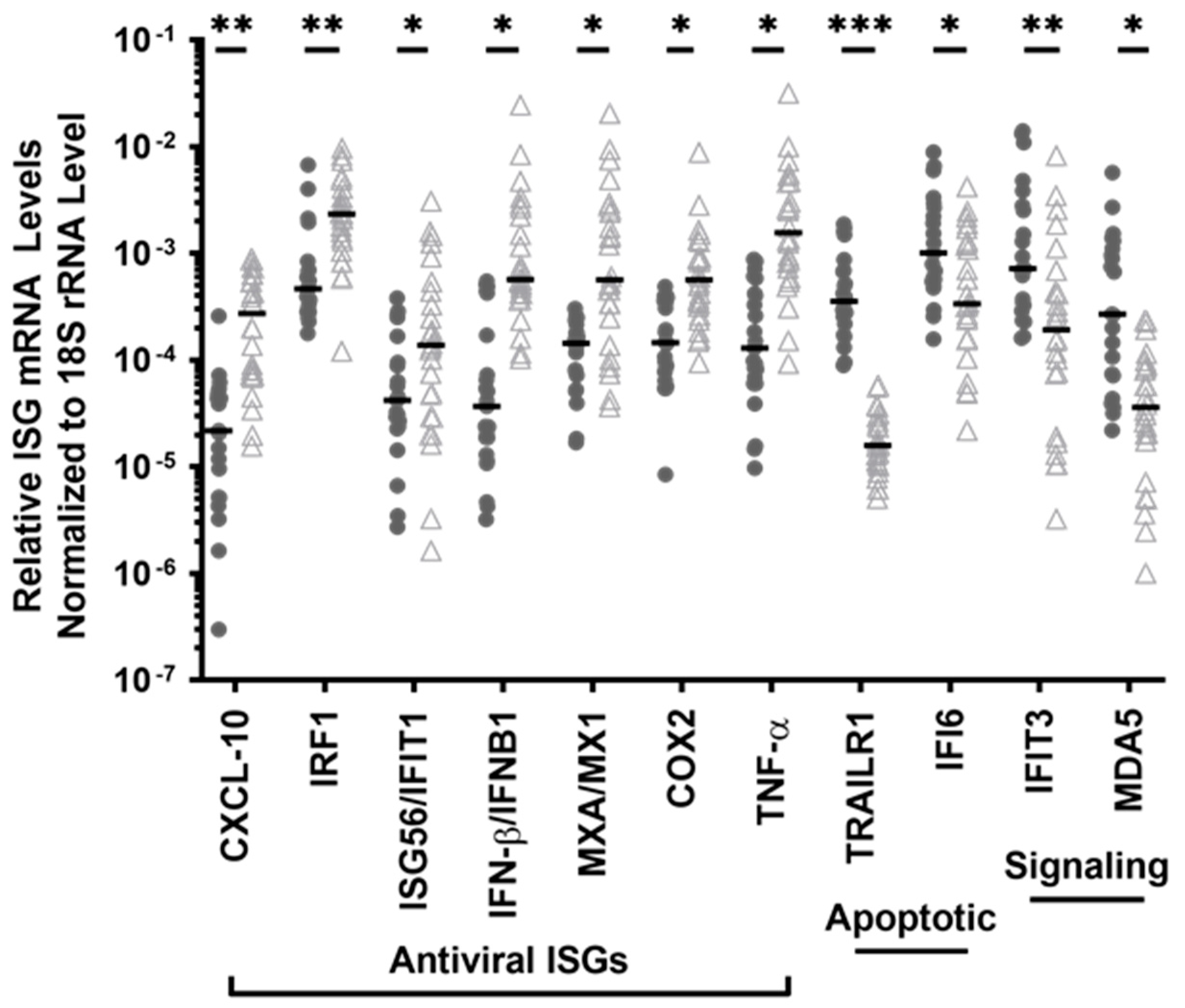
3.1. The PBMC of HESN FSWs Have Low Levels of ISG Transcripts Encoding Proinflammatory Signalling Pathway Proteins but Increased Transcripts of 2 ISGs, Key to the Detection of RNA and DNA Viruses

To determine whether HESN FSWs have constitutively stronger basal antiviral defenses, transcripts of the genes encoding ISGs with antiviral function were assessed using relatively quantitative RT-PCR. A total of 76 ISGs that have been shown to play crucial roles in antiviral immunity [45,47,56,58,65,66] were examined in the peripheral blood mononuclear cells (PBMC) from HESN women and non-HESN FSWs (Table 2, Supplementary Table S1). Only 35 of the 76 ISGs examined had detectable RNA transcripts in ex vivo, PBMC (n = 16).
The other 41 ISGs, although not detected via RT-qPCR in unstimulated PBMC, might still be differentially regulated in HESN PBMCs upon viral infection or challenge with exogenous stimuli. This possibility was investigated in a pilot project. These 41 ISGs were examined in small subsets of PBMC (n = 4, HESN and n = 5 non-HESN), then subjected to a 3-h stimulation by exogenous interferon-γ (IFN-γ, 10 ng/mL) and IFN-α2 (10 ng/mL). Similar increases in the transcripts of 36 ISGs (out of 41) were found in both HESN and non-HESN groups (data not shown, Table 2: bolded ISGs). Among the 36 responsive ISGs, 8 ISGs (CASP8, FLIP, GADD45, IRF4, IRF5, MxB, p53, TAP1) showed trending differences between the 2 groups (12–38% difference in transcript levels, p-values ranged 0.065 to 0.09). Five ISGs remained undetected following stimulation with IFN-γ and IFN-α2 (Table 2: underlined ISGs). These 41 ISGs did not seem to be differentially regulated in HESN PMBCs.
The study focused on profiling the 35 ISGs listed in Table 3. Transcripts of all 35 ISGs were detected in the PBMC from non-HESN control (n = 26) but only 25 ISG transcripts (at varying concentrations) were detectable in the HESN PBMC (n = 23), suggesting potentially reduced basal expression of these 10 ISGs in the HESN FSWs (Table 3). When examining the properties of the 10 ISGs that were constitutively expressed only in the non-HESN controls, these ISGs were enriched in the signaling pathways of apoptosis, and pathogenic hyper-cytokinemia/-chemokinemia (ingenuity pathway analysis, p-values <10−5), potentiating a more proinflammatory milieu in the non-HESN FSWs. Contrarily, HESN PBMCs had less expression of most ISGs with antiviral properties, supporting a less inflammatory microenvironment.
Among the 25 ISGs that were constitutively expressed in the PBMC of both HESN and non-HESN controls, 11 ISGs had differential expression levels between the two groups (Table 3, bolded), while the other 14 ISGs were equivalent. The transcript levels of CXCL10, IRF-1, IFIT1, IFNB1, MX1, COX2, and TNF were significantly lower by 61–94% in the PBMC of the HESN FSWs, compared to that in the non-HESN controls (i.e., 1.5- to 16.6-fold more transcripts in the non-HESN controls) (Figure 1). These ISGs were enriched in the pathways of antiviral response and the replication of virus RNA in immune cells (ingenuity pathway analysis, p-values < 105). Reduced expression of these ISGs reflected a weak basal antiviral readiness in the HESN PBMC. However, the transcripts of two key signaling molecules of proinflammatory antiviral response were significantly higher in HESN PBMC. The levels of IFIT3 and IFIH1/MDA5 transcripts were 2.3-fold (p = 0.04) and 11.3-fold (p = 0.003) higher in HESN PBMC. Both IFIT3 and MDA5 have RNA-binding properties [67,68,69], and are involved in responses against DNA and RNA virus infection, suggesting that HESN PBMCs are more ready to detect RNA or DNA viral infection.

3.2. The PBMC of HESN FSWs Had Reduced Transcript Levels of Antiapoptotic ISGs and Elevated Proapoptotic TRAILR1 Transcripts

In addition to antiviral function, some ISGs also play a crucial role in regulating cellular apoptosis [45,47,65]. The HESN PBMCs had no detectable level of BCL2 or BCL2L.1 transcripts, encoding crucial anti-apoptotic molecules at baseline (Table 3). In addition, the HESN PBMCs had a significantly higher RNA transcript level of TNFRSF10A/TRAILR1, a critical inducer of apoptosis, compared to the HESN controls (by 26-fold, p < 4 × 10⁻⁶, Figure 1). Although it seems that the HESN PBMCs have greater pro-apoptotic potential, the transcript level of IFI6, an anti-apoptotic regulator and immune modulator was doubled in the HESN PBMC, versus the non-HESN controls (p =0.01, Figure 1), suggesting a mechanism of balance.

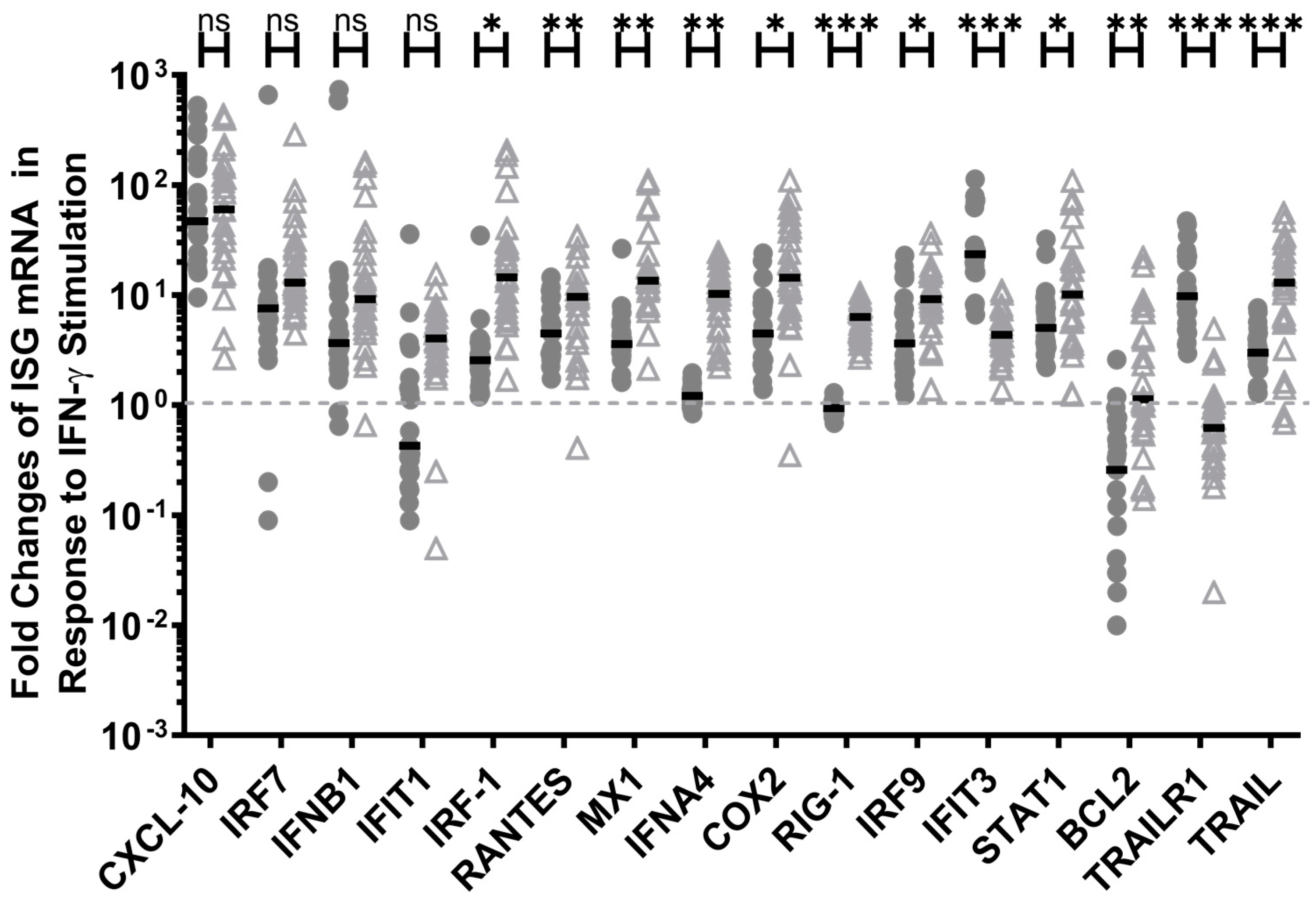
3.3. IFN-γ Induced the Expression of Pro-Inflammatory ISGs in HESN PBMC, Although to a Lesser Extent than in the Non-HESN PBMC, but Greatly Enhanced the Expression of Pro-Apoptosis ISGs

The transcriptional response of the 35 detectable ISGs to exogenous IFN-γ treatment was evaluated in HESN and non-HESN PBMCs. Not all 35 ISGs responded following the 3-h stimulation with exogenous IFN-γ; 19 ISGs had only a modest 12–25% increase or decrease in transcript level in either the HESN or the non-HESN PBMCs (data not shown). Among these 19 ISGs, 7 ISGs (ACTB, E2F1, FCGR2B, IL-1B, PKR, MYD88, TNF) did not mount a statistically significant response in either group when compared to the mock stimulation (p > 0.05). IFN-γ induced changes in the other 12 ISG transcript levels (ATM, BAX, BclXL, CDK4, CDKN2A, DPP4, HDM2, IFI6, IFIH1, IFNA2, ISG15, NOXA) were similar between the HESN and non-HESN PBMCs (p-values > 0.05, data not shown).
The other 16 ISGs responded with either a >4-fold increase or a >25% decrease in transcripts in either the HESN PBMCs or the non-HESN controls (Figure 2). IFN-γ up regulated the expression of 11 ISGs (CXCL10, IRF-7, IFNB1, IRF-1, RANTES, MX1, COX2, IRF-9, IFIT3, STAT1A, and TRAIL) in both the HESN and non-HESN PBMC samples, ranging from 113-fold to 4-fold (Figure 2, HESN, p-values <0.0001; non-HESN, p-values range from 0.0001 to 0.02). The increases in the transcript levels of CXCL10, IRF-7, and IFNB1 were similar in both groups (p-values >0.05), while the increases in IRF-1 (31-fold in non-HESN vs. 4-fold in HESN), RANTES (10- vs. 6-fold), MX1 (22- vs. 5-fold), COX2 (24- vs. 7-fold), IRF-9 (11- vs. 7-fold), STAT1A (12- vs. 7-fold), and TRAIL (16- vs. 4-fold) were notably greater in the non-HESN PBMCs. The increase in IFIT3 transcripts, encoding for a sensor of RNA virus was much higher (p < 0.0001) in the HESN PBMC (by 34-fold) versus that in the non-HESN controls (by 5-fold), boosting the antiviral potential in HESN. In addition, IFN-γ up regulated ISG56 (4-fold), IFNA4 (10-fold), and RIG-1 (6-fold) transcripts only in the non-HESN PBMCs, without affecting the expression of ISG56, IFNA4, and RIG-1 in the HESN PBMCs (Figure 2), suggesting a less pro-inflammatory response in HESN. Together, IFN-γ stimulation seemed to further enhance the differences in the baseline ISG transcript levels between the HESN and the non-HESN controls (Table 3: bolded ISGs and non-HESN only ISGs), showing reduced inflammatory potential in HESN PBMCs when responding to IFN-γ but enhanced antiviral readiness via increased IFIT3.
Similarly, IFN-γ seemed to enhance expression of pro-apoptotic potential in HESN PBMC, while boosting anti-apoptotic transcripts in non-HESN PBMCs. IFN-γ stimulation down regulated the expression of anti-apoptotic ISG, BCL2 by 74%, (vs. mock-stimulation, p = 0.002) and up-regulated the initiator of the apoptotic pathway, TRAILR1, by 10-fold (vs. mock-stimulation, p < 0.0001) in the HESN PBMCs. IFN-γ stimulation almost had the opposite effects on the ISGs regulating apoptosis in the non-HESN PBMC. IFN-γ down regulated TRAILR1 expression by 38%, (vs. mock-stimulation, p =0.02) but had no effect on the BCL2 expression (p > 0.05) in the non-HESN PBMCs, reducing the overall apoptotic potential of the non-HESN PBMC. It is noteworthy that IFN-γ up regulated TRAIL expression in both groups, supporting the initiation of apoptosis but did so with a significantly greater impact on the non-HESN PBMC (13-fold increases, p < 0.0001) and a relatively less propensity for apoptosis for the HESN PBMC (4-fold, Figure 2).

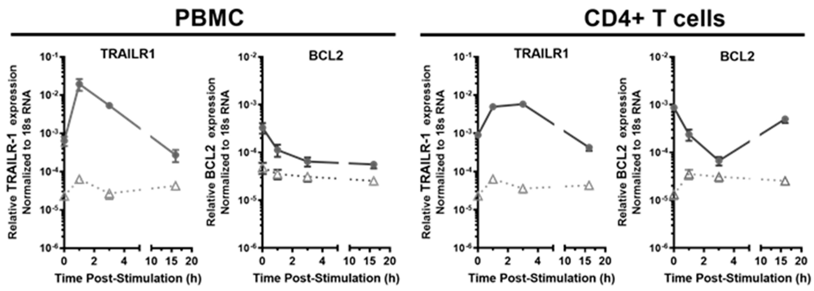
3.4. The Transcriptional Response of Antiviral Effector ISGs Was Transient in the HESN PBMC and CD4+ T Cells, Compared to the Prolonged Response in the Non-HESN Controls

A prolonged state of immune activation or inflammation and/or resistance to apoptosis that is induced by interferons or viral infection has been associated with pathogenic outcomes [51,52,70,71]. The kinetics of ISG transcriptional responses to IFN-γ were studied at 1 h, 3 h, and 16 h following the initial exposure to exogenous IFN-γ (10 ng/mL) to determine the duration of ISG response. The antiviral effector ISGs, including IRF-1, CXCL10, IFNB1, IRF-9, and IFIT3 were examined to profile the changes of ISG transcripts over time in both groups (Figure 3). IRF-1 was included as a positive control, as our earlier work showed transient IRF-1 transcript increases in the HESN [41,43]. Expression of CXCL10 and IFNB1 can be regulated by IRF-1 [56,57,58] while expression of IRF-9 and IFIT3/ISG60 is not dependent on IRF-1. Similar to what we observed for IRF-1, the transcript level of the other antiviral effector ISGs increased drastically within an hour in both the HESN and non-HESN PBMCs (Figure 3A). However, 3-h post IFN-γ stimulation, transcripts levels dropped in HESN PBMC but continued to increase or plateau in the non-HESN PBMCs. At 16 h post IFN-γ stimulation, ISG transcripts in the HESN PBMC returned to the baseline level while ISG transcripts in the non-HESN PBMC remained elevated. Significant differences in the ISG transcript levels of IFNB1, CXCL10, IRF-1, and IRF-9 were observed between the two groups at 16 h (Figure 3A). Interestingly, the IFIT3 transcript level in non-HESN PBMC plateaued at the 16th hour poststimulation, reaching the same level as the IFIT3 transcripts in the HESN PBMC, suggesting that, when activated, IFIT3 responds with a strong transient ISG upregulation, rather than a sustained, slow response pattern.
While assessing ISG expression and response in PBMC enabled the evaluation of systematic ISG regulation, representing a general antiviral response, assessing ISG regulation in the CD4+ T cells allowed the evaluation of immune activation of the main target for HIV infections. This is of importance because HIV-1 replicates in activated CD4+ T cells [72,73]. The study determined whether ISG responses to IFN-γ in the CD4+ T cells followed similar kinetics as to bulk PBMC (Figure 3B). Similar patterns of ISGs responses were observed in CD4+ T cells in both HESN and non-HESN groups. Surprisingly, elevated IRF-9 transcripts in the HESN CD4+ T cells did not return to the baseline level, as in the HESN PBMC. However, at 16 h poststimulation, the IRF-9 transcript level in the HESN CD4+ T cells was still lower than that in the non-HESN CD4+ T cells, suggesting a slightly prolonged but controlled IRF-9 mediated activation of IFN-responsive genes in the CD4+ T cells, compared to other cell types in the PBMC.

3.5. IFN-γ Transiently Upregulated Proapoptotic Pathway ISG (TRAILR1 and TRAIL) and Downregulated Antiapoptotic ISG, BCL2 in HESN CD4+ T Cells

We also characterized the kinetics of the transcriptional responses of two ISGs encoding proteins regulating cellular apoptosis, TRAILR1 and BCL2 in the PBMC and CD4+ T cells of both the HESN and the non-HESN FSWs (Figure 4). The baseline expression of these two ISGs were significantly different in the HESN and non-HESN controls (Figure 1 and Figure 4). Following the IFN-γ challenge, the transcripts of the proapoptotic receptor gene, TRAILR1 were increased within 1 h in PBMC (HESN: 31-fold vs. non-HESN 3-fold) and the CD4+ T cells (HESN: 5.5-fold vs. non-HESN 2.7-fold) of both the HESN and the non-HESN controls. The TRAILR1 transcript level was quickly reduced at 3-h poststimulation in the PBMC and CD4+ T cells of both groups. At the 16th hour poststimulation, the TRAILR1 transcript returned to the baseline level in the PBMC and CD4+ T cells of both groups. Although the kinetics were similar for the TRAILR1 in both the HESN and the non-HESN controls, the magnitude of changes in the TRAILR1 transcripts was significantly larger in the HESN group, suggesting a very tight regulation of TRAILR1 expression in the HESN cells (Figure 4).
In contrast to its effects on TRAILR1, IFN-γ reduced the level of BCL2 transcripts, an anti-apoptotic/pro-survival ISG in the PBMC of both the HESN (by 64%) and the non-HESN controls (by 27%) within the first hour of stimulation. BCL2 transcript levels remained low in the PBMC of both groups (Figure 4), up to 16 h post-IFN-γ exposure. The effects of IFN-γ on BCL2 transcripts in the CD4+ T cells, however, did not mirror those in the PBMC. Similar reduction of BCL2 transcripts in HESN CD4+ T cells were observed during the first 3 h of IFN-γ challenge, but at 16 h poststimulation, the BCL2 transcripts returned to baseline level, instead of remaining reduced, as observed in the HESN PBMC. It is not clear whether there is a functional gain for restoring BCL2 transcript level in CD4+ T cells and whether that contributes to the HESN phenotype.
Furthermore, IFN-γ treatment of the non-HESN CD4+ T cells increased BCL2 transcripts (2.7-fold); together with the reduced TRAILR1 expression, IFN-γ protected against apoptosis in the non-HESN CD4+ T cells. In contrast, IFN-γ stimulation transiently primed the HESN CD4+ T cells to initiate apoptosis with increased TRAILR1 and decreased BCL2 transcripts.

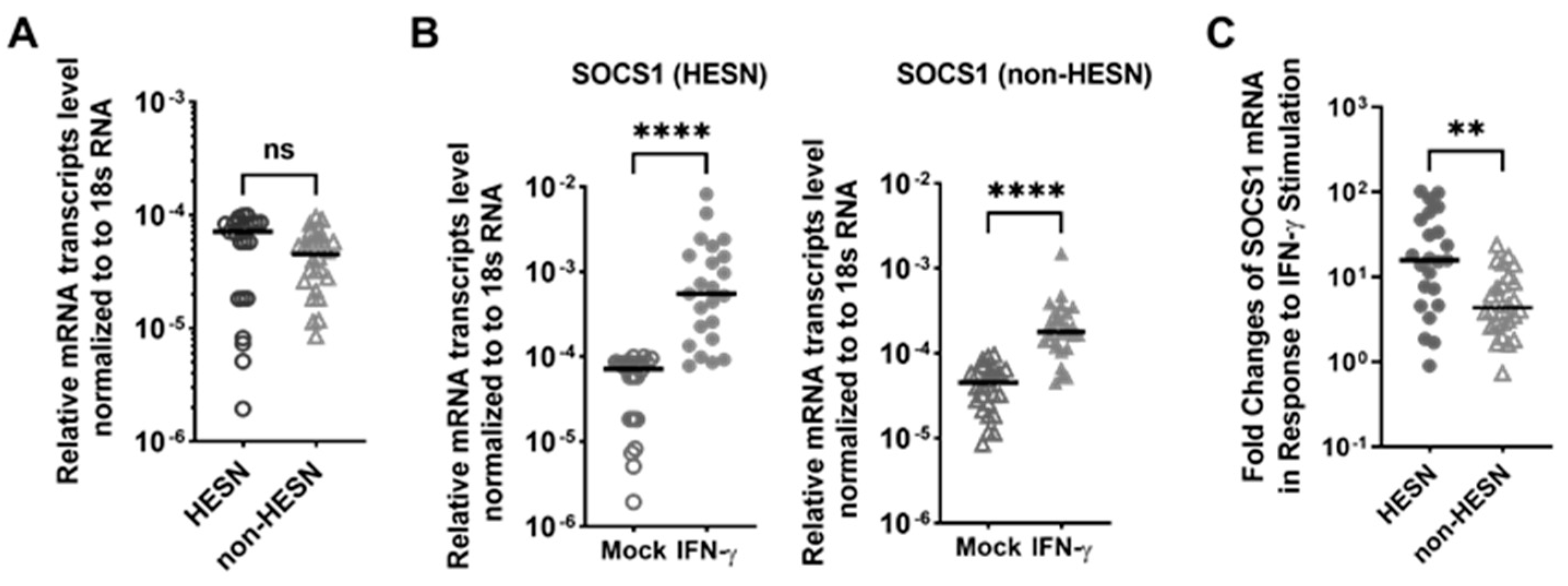
3.6. IFN-γ Induced a Stronger Negative Feedback Regulatory Response in the HESN PBMC, When Compared to Non-HESN Controls

SOCS proteins have been shown to be induced by interferons as part of a negative feedback loop to limit the extent and duration of IFN response [74]. This study examined whether there is a differential regulation of suppressor of cytokine signaling-1 (SOCS1) expression in the HESN versus the non-HESN controls. (Figure 5). Basal level of SOCS1 transcripts in HESN PBMC was not different from the SOCS1 transcript level in the non-HESN PBMC (Figure 5A). At 3-h following IFN-γ stimulation, the SOCS1 transcript levels were significantly upregulated in both the HESN and the non-HESN PBMC (Figure 5B). Furthermore, in comparing the extent of SOCS1 upregulation in the HESN cells with that in the non-HESN cells, SOCS1 transcripts were increased by approximately 16-fold in the HESN PBMC, versus a 4-fold increase in the non-HESN PBMC (p-value = 0.002, Figure 5C). Together, these data showed that IFN-γ induced a much stronger negative feedback signal in the HESN PBMCs, when compared to the non-HESN controls, suggesting a tighter control of the IFN-γ induced ISGs in the HESN.

3.7. The Transcriptional Response of Antiviral Effector ISGs and Antiapoptotic ISGs to Exogenous IFN-α2 Stimulation Was Similarly Transient in HESN PBMC and CD4+ T Cells

Similar to IFN-γ, the type-I IFN, IFN-α, is also a strong inducer of antiviral ISGs [49,60,66,75] and uncontrolled IFN-α responses have been associated with increased pathogenesis [51,52,70,71]. This study examined whether the kinetics of ISG responses to IFN-α treatment differed from that by IFN-γ in PBMCs (Figure 6). IFN-α2, the prototypical IFN-α and a key part of the innate immune response with potent antiviral, antiproliferative, and immunomodulatory properties was used. The expression of IRF-1, IRF-9/ISGF3G, FcGR1A, and HDM2 transcripts dramatically increased within an hour following IFN-α2-treatment in the PBMC of both the HESN and the non-HESN controls (Figure 6(Ai–Aiii,Av,Avi), p-values < 0.0001). Similar to the results of IFN-γ stimulation, a reduction in IRF-1, IRF-7, and IRF-9 transcript levels was observed in HESN PBMC at 3-h post exposure to IFN-α2; The transcript levels of IRF-1, IRF-7, and IRF-9 seemed to plateau in non-HESN PBMC at the 16th hour post-treatment (Figure 6(Ai–Aiii)). Unlike the responses to IFN-γ, the IRF-9 transcripts only started to decrease at 16-h post-treatment (p = 0.03) and the IRF-1 and IRF-9 transcripts were not restored to the baseline level in the HESN PBMCs. Overall, HESN PBMC responded to IFN-α2 stimulation with transient increases in the antiviral effector ISGs (Figure 6(Ai–Aiii)). These transient increases of IRF-1, IRF-7, and IRF-9 were also observed in the HESN CD4+ T-cell populations (Figure 6(Bi–Biii)). Interestingly, the maximum IRF-7 transactivation response to IFN-α2 in the HESN PBMC (64-fold (HESN) vs. 4-fold (non-HESN)) or CD4+ T cells (142-fold (HESN) vs. 18-fold (non-HESN)) were much more robust than that in the non-HESN cells, suggesting a potentially strong but transient inflammatory response to IFN-α2 in the HESN CD4+ T-cell populations.
When we examined the pro-apoptotic marker BCL2, similar to the kinetics of BCL2 response, elicited by IFN-γ (Figure 4), BCL2 transcript levels dropped in the PBMC of both HESN and non-HESN during the first 3 h of IFN-α2 treatment (Figure 6(Aiv)). However, instead of remaining at low levels, as observed during IFN-γ stimulation (Figure 4), the BCL2 transcript level began to increase at the 16th hour post-treatment in the PBMC of both HESN and non-HESN (Figure 6(Aiv)). In CD4+ T cells, the kinetics of BCL2 response to the IFN-α2 (Figure 6(Aiv)) was the same as the BCL2 response to IFN-γ (Figure 4). In HESN CD4+ T cells, the drop of BCL2 transcripts was transient (by 38–84%) compared to non-HESN, where IFN-α2 treatment increased the BCL2 transcripts by 1–2 fold (Figure 6(Biv)). This suggests regulation of apoptosis in the CD4+ T cells of HESN, is altered compared to non-HESN. By dampening the pro-survival ISG, BCL2 expression to allow apoptosis was quite exact during both IFN-γ and IFN-α2 stimulation.

3.8. Not All Antiviral ISGs in the HESN Cells Exhibited Transient Response to IFN-α2 Treatment

ISG15 is a ubiquitin-like protein that becomes conjugated to many cellular proteins upon activation by type-I IFNs [76], while HDM2 gene product is critical for the negative feedback of ISG activation via dampening the expression and function of IRFs in antiviral response [77,78]. The baseline level of ISG15 and HMD2 transcripts were very low in both the PBMC and CD4+ T cells of both groups (Figure 6(Av,Avi,Bv,Bvi)); ISG15 transcripts were not detected in HESN cells at baseline (Table 3, Figure 6(Av,Bv)). The relative level of transcripts detected were close to the water control blank in the RT-qPCR (ct-value < 37). However, following stimulation with IFNα2, ISG15 and HMD2 transcripts increased steadily in both HESN PBMC and CD4+ T cells, and reached plateaus in the non-HESN PBMCs. The fact that not all ISGs appear to be alternatively regulated in HESN suggests a specific pattern of ISG expression may be associated with protection from HIV infection. Steady increases in the expression of these two ISGs might be critical for regulating antiviral response.

3.9. Reducing IRF-1 Expression in Non-HESN CD4+ Cells Altered the Non-HESN ISG Profile to the ISG Profile, Similar to What Was Observed in the HESN Cells

This next step was to determine if a moderate reduction of baseline IRF-1 in isolated CD4+ T cells, like that observed in the HESN FSWs (by 30–60%), would mimic the HESN ISGs profile. IRF-1 expression was knocked down with IRF-1 specific siRNA, delivered by CD4-specific RNA aptamer chimera (CD4-AsiCs, n = 6 per group). CD4-AsiCs with scrambled siRNA were used as negative control. Greater than 95% of CD4+ T cells were shown to have IRF-1 protein level reduced by 30–60% (data not shown, [41]). Due to the limitation in cell numbers, only the expression of key antiviral ISGs shown to be regulated by IRF-1 were examined at 18 h (Figure 7). The baseline level of IFN-γ, TNF-α, STAT1α, RIG-1, and IFIH1 transcripts were not affected by reduced IRF-1 expression, suggesting that the basal expression of these genes is independent of the IRF-1 expression level. IRF-1-specific CD4-AsiC reduced cellular IRF-1 transcripts by 99.7%, reflecting a 30–60% decrease in IRF-1 protein level at 18-h. The transcript levels of two proinflammatory ISGs, MX1 (by 96%) and IRF-7 (by 88%) were greatly reduced (Figure 7) while transcripts of the RNA-sensing ISG, IFIT3, were increased by 283-fold. The resulting MX1, IRF-7, and IFIT3 transcript levels were similar to the low MX1 (Figure 1) and IRF-7 (Figure 6(Biii)) and high IFIT3 (Figure 3B) levels observed in HESN (solid circles). In addition, the resulting transcript levels of the proapoptotic ISG, TRAILR1, and antiapoptotic regulator, IFI6, were increased by 22-fold and 62-fold, respectively (Figure 7), similar to the TRAILR1 and IFI6 transcript levels observed in HESN (Figure 1 and Figure 4). Together, reducing IRF-1 expression in non-HESN CD4+ T cells altered the transcript levels of MX1, IRF-7, TRAILR1, IFIT3, and IFI6 to similar levels found in HESN. A high IRF-1 level seemed to negatively regulate IFIT3, TRAILR1, and IFI6, but positively promote the expression of MX1 and IFIH1.

4. Discussion

Antiviral ISGs are a critical response to viral infection; in vitro loss of function studies showed that knockout of antiviral ISGs resulted in increased replication of several viruses [48,49,59,66,75]. This is the first study exploring ISG expression as a correlate of protection against HIV-1 infection. This study identified a unique profile of antiviral ISGs in HESN FSWs that is characterized by (1) a lower level of ISG transcripts that potentiate a proinflammatory response, (2) a balance of proapoptotic ISGs and prosurvival ISGs, and (3) transient increases of proinflammatory ISGs and ISGs with apoptotic potential. This study further showed that reduced IRF-1 expression, observed in the HESN cells may be one of the key regulators of this HESN ISG profile.
Although increased inflammation has been associated with increased risk to HIV infection [79,80,81,82,83,84] and baseline immune quiescence (IQ) was observed in these Kenyan HESN FSWs [26], little is known of the mechanism(s) involved in the maintenance of IQ. IQ refers to a state of low baseline immune activation, which was proposed to protect against infection by limiting activated HIV target cells [26,62] and was associated with increased regulatory T cells [38]. Baseline IQ is not to be mistaken as “immune deficiency”. Even though the baseline level of several antiviral ISG transcripts in the HESN cells were significantly less than that in the non-HESN cells, the two groups mounted equivalent magnitudes of ISG responses to exogenous IFNs (Figure 2, Figure 3 and Figure 6). This is especially important because IFN is one of the primary innate immune responses against viral infection [51,52,59]. HIV-1 infection elicits type-I, type-II and type-III IFN responses, which activate the transcription of ISGs [46].
Uniquely to the HESN cells, the proinflammatory and proapoptotic ISG-transcription response was transient and appeared to be promptly downregulated after 3 h. In comparison, the ISG mRNA transcript level continued to increase or reach a plateau in the non-HESN cells, which may result in prolonged inflammation. Activation of immune cells, especially CD4+ T cells, is essential for HIV-1 replication and the establishment of infection [80,85,86,87]. Previous studies suggest that sustained elevation of type-I IFN expression is associated with persistent immune activation or inflammation, which promotes HIV-1 replication and transmission. The persistently high expression of ISGs has been shown to be a major contributor of inflammation and the resultant establishment of infection [51,52,59] and disease progression during persistent HIV-1 and SIV infections [51,52,70,88,89,90]. Hence, the regulation of these ISGs, as observed in HESN cells, may be critical for effective antiviral response with minimum pathogenesis.
Negative regulators of immune activation are critical for resolving the IFN-induced antiviral state and facilitating the return to immune homeostasis. In addition to reduced IRF-1 expression, this study found rapid upregulation of SOCS-1, a negative regulator of IFN-signaling in HESN individuals. The HESN cells had four-times-higher levels of SOCS-1 than the non-HESN cells, following IFN-γ stimulation. SOCS-1 has also been shown to negatively regulate IRF-1 expression [91]. While the baseline level of SOCS-1 transcripts, which are similar in both the HESN and the non-HESN groups could not explain the reduced IRF-1 basal expression, the increased SOCS-1 transcripts following IFN-γ stimulation may contribute to the transient IRF-1 response. Furthermore, SOCS-1 polymorphisms have been associated with a rapid HIV progression rate [92]. Recently, in in vivo genome-wide CRISPR screens, SOCS1 was identified as “a major non-redundant checkpoint imposing a brake on CD4+ T-cell proliferation,” which promotes HIV-1 replication in infected cells [93]. This is in agreement with the brief, robust, but transient ISG response observed in the HESN cells, suggesting that SOCS-1 may be another key molecule enabling the HESN phenotype.
The regulatory mechanism(s) underlying reduced baseline IRF-1 expression and increased SOCS-1 expression following IFN-γ stimulation in the HESN cells need further research. Thus far, our earlier study showed that the IRF-1 genetic polymorphisms, found in approximately 72% of the HESN FSWs are associated with reduced IRF-1 expression, delayed IRF-1 response to IFN-γ and delayed establishment of HIV-1 infection [14]. However, the IRF-1 polymorphism alone could not explain the reduced IRF-1 expression in the HESN cells. The HESN FSWs without the protective IRF-1 polymorphism also exhibited reduced baseline IRF-1 levels. Our work suggested that epigenetic mechanism(s), which are influenced by environment and diets, may contribute to the regulation of IRF-1. On the note of environmental influences, the HESN FSWs were in the sex trade for a significantly longer period of time and had been exposed to more clients; it is highly possible that the HESN phenotype was driven by high exposure to foreign antigens, which induced immune tolerance to avoid inflammation at the mucosal site [94]. However, the mechanism(s) of such remained to be sought. IRF-1 regulates the differentiation of Treg, via suppressing FOXP3 transactivation [95]; reduced IRF-1 expression, perhaps affected by levels of metabolites (e.g., short-chain fatty acids) may induce immune tolerance via increasing differentiation of Treg.
Interestingly, while SOCS1 can regulate IRF-1 [91], SOCS1 was shown to be regulated by IRF-1 as well [74,96]. The SOCS1 promoter consists of IRF element (IRF-E); IRF-1 binding to IRF-E is necessary to transactivate the expression of SOCS1 [96]. This study highlights the importance to define the inter-regulatory relationship between IRF-1 and SOCS1 to gain a better understanding of the HESN phenotype.
In addition to inflammation, an increased potential in eliciting an apoptotic pathway may also contribute to the HESN phenotype. Programmed cell death (i.e., apoptosis) of infected cells is critical to limit the spread of pathogens and inappropriate immune activation [45,47,66,97]. However, when the surrounding tissue is also severely damaged, via bystander effects, the damaged tissue would enhance the proinflammatory response and disease severity is often augmented. The regulation of pro-apoptotic and pro-survival ISGs was found to be unique in the HESN cells. IFN stimulation induced proapoptotic ISGs (TRAILR1, TRAIL) but reduced the expression of pro-survival ISG, BCL2 (Figure 2 and Figure 4). TRAIL (TNF-related apoptosis-inducing ligand) is induced in several cell types including immune cells such as macrophages and T cells [47,59] and its expression has been associated with induction of apoptosis of the TRAIL-expressing cells and the surrounding bystander cells and further shown to affect disease outcomes. This study found that HESN cells had a 4-fold increase in TRAIL mRNA transcripts following IFN-γ stimulation, compared to a 16-fold increase observed in the non-HESN cells (Figure 3). Contrary to that, the HESN cells had a 15-fold increase in the expression of TRAIL-receptor, TRAILR1 but the non-HESN cells have no significant change in TRAILR1 expression following IFN-γ challenge. Together, these data suggest a proapoptotic milieu promoting cell death of bystander cells in non-HESN; but in HESN individuals, perhaps a transient proapoptotic state may be more protective against immune activation, and bystander cell/tissue death and damage. In support, the pro-survival ISG, BCL2 was also transiently reduced in the HESN cells. The HESN cells seem to have a strict, fine control of these pro- and anti-apoptotic ISGs to quickly maximize the death of affected cells and minimize damages to surrounding cells.
Lastly, our earlier work and findings from this study support IRF-1 as playing a key role in the maintenance of IQ in HESN FSWs. IRF-1 is not only a trans-activator of several pro-inflammatory ISGs; it is also a repressor of FOXP3 and, thus, an inhibitor of the differentiation of regulatory T cells (Treg). With reduced IRF-1 protein (30–60%), as found in the HESN FSWs [42,98], the expression of IRF-1-dependent ISGs were decreased (Figure 1 and Figure 7), and the expression of FOXP3 and TRAILR1 were increased, perhaps leading to increased Treg differentiation and apoptotic potential, respectively. IRF-1′s crucial role in HIV-1 susceptibility was further supported by its upregulation by HIV-1 infection and the consequence of knocking down its expression using siRNA. IRF-1 also binds to the HIV-1 promoter; together with NF-κB, IRF-1 is required for the trans-activation of viral genes. IRF-1 knockdown resulted in significant reduction and delay of HIV-1 replication [41]. Together, these support the notion that reduced baseline IRF-1 expression is key to avoiding the establishment of HIV infection in HESN.
Limitations/caveats: There are several limitations in the design of the study. As this was a retrospective analysis, we were unable to select large numbers of cryopreserved samples to best fit the grouping criteria due to limitations of the bio-repository. In addition, we had to limit analysis to a limited number of cell samples with the viability >90% after thawing. Furthermore, as mentioned in the methodology, the definition criteria for HESN and non-HESN was based on the epidemiological data. To control for the demonstrable effects of sex work on immune activation, we used FSWs who have not been in the sex trade long enough to meet the HESN definition. Over the years of follow-up, there were significant numbers of seroconversions among non-HESN FSWs and there was loss to follow-up as several of the participants did not return for the final resurvey in this cohort, which ended in 2013. Furthermore, this study focused on a subset of antiviral ISGs that have been implicated in several studies of innate responses to viral infection [45,47,56,58,65,66], instead of unbiased analysis of all known ISGs. There are more than a thousand ISGs; hence, it is possible that several potential ISGs that are critical for the HESN phenotype were omitted in this study and that data from this study is only a tip of an iceberg. On a separate study, RNA-seq was performed with four PBMC samples from each of the HESN and the non-HESN group (data not shown); only a few ISG were found to be differentially expressed between the two groups, and most of those have been included in this study. This study also focused on CD4+ T cells; future study should focus on RNA from different cell subsets. The expression of ISGs is tightly regulated and specific to cell types; hence, levels of ISG transcripts may be diluted in the RNA pool from PBMC. This study avoided using ISG super-arrays and RNA-seq to include more samples in the analyses due to sample limitation and to include the kinetic study and to allow presentation of validated data, as only validated primer sets were used. The data is interpreted with caution so as not to emphasize that these ISGs are the only molecules contributing to the HESN phenotype. Lastly, although there are significant discrepancies between the cells in the blood and the cells found at the mucosal compartments [99], findings from studies of PBMCs are highly valuable in guiding research at the vaginal site.

5. Conclusions

The regulation of IFN signaling and ISG expression are unique in HESN cells and may be part of the molecular mechanisms underlying the reduced susceptibility of HESN FSWs to HIV-1 infection. The transient ISG response observed in the HESN cells may be mediated by the drastic increases of SOCS1, short-lived upregulation of IRF-1 and reinforced by increasing HDM2 and subsequent regulation. Together with the coordinated regulation of proapoptotic and prosurvival ISGs, IRF-1-regulated ISGs founded the basis for the HESN phenotype observed in the Kenyan FSWs.

Supplementary Materials

The following supporting information can be downloaded at: https://www.mdpi.com/article/10.3390/v14030471/s1, Table S1: Validated ISG Primer Pairs.

Author Contributions

Conceptualization, M.G., X.Y. and R.-C.S.; methodology, M.G., B.A. and R.N.D.; validation, X.Y.; Data analysis, M.G., X.Y. and R.-C.S.; clinical resources, T.B.B. and J.K.; epidemiological data curation, S.Y.; writing—original draft preparation, M.G. and R.-C.S.; writing—review and editing, R.-C.S. and T.B.B.; visualization, R.-C.S.; supervision, R.-C.S. and T.B.B.; project administration, R.-C.S.; funding acquisition, T.B.B. and R.-C.S. All authors have read and agreed to the published version of the manuscript.

Funding

This research was funded by grants from the Canadian Institutes of Health Research (CIHR), grant number 154043 (to T.B.B. & R.-C.S.). M.G. is an honor thesis project student from the University of Winnipeg and X.Y. is a visiting scientist from the Yangquan Municipal Center for Disease Prevention and Control, China.

Institutional Review Board Statement

The study was conducted according to the guidelines of the Declaration of Helsinki. The ethical review boards from the Kenyatta National Hospital/University of Nairobi (#P211/09/2006, renewed and amended annually since 2006), the University of Manitoba (HS15280 (B2012:043, regularly renewed since 2012), and the Regional Ethical Review Board in Stockholm (2014/959-31, amended in 2018: 2018/1306-31) approved the study.

Informed Consent Statement

Informed consent was obtained from all subjects involved in the study.

Data Availability Statement

All data supporting our findings are found in this manuscript.

Acknowledgments

The authors would like to thank the study participants and the staff at the Sex Worker Outreach Program and Partners for Health and Development in Africa, especially the staff of the Majengo/Pumwani clinics. A special thanks to Makobu Kimani, for performing sampling and their help with participant recruitment and follow-up. The authors would also like to thank Judy Lieberman and Lee Adam Wheeler for sharing their protocol in designing and synthesizing CD4-AsiCs.

Conflicts of Interest

The authors declare no conflict of interest.

References

  1. UNAIDS Data; HIV/AIDS (Ed.) UNAIDS: Geneva, Switzerland, 2018; Volume 2018, Available online: https://www.unaids.org/sites/default/files/media_asset/unaids-data-2018_en.pdf (accessed on 30 December 2021).
  2. UNAIDS Data; HIV/AIDS (Ed.) UNAIDS: Geneva, Switzerland, 2019; Volume 2019, Available online: https://www.unaids.org/sites/default/files/media_asset/2019-UNAIDS-data_en.pdf (accessed on 30 December 2021).
  3. UNAIDS. GLOBAL HIV STATISTICS; UNAIDS: Geneva, Switzerland, 2020; Volume 2020, Available online: https://www.unaids.org/sites/default/files/media_asset/2020_aids-data-book_en.pdf (accessed on 30 December 2021).
  4. Izulla, P.; McKinnon, L.R.; Munyao, J.; Karanja, S.; Koima, W.; Parmeres, J.; Kamuti, S.; Kioko, R.; Nagelkerke, N.; Gakii, G.; et al. HIV Postexposure Prophylaxis in an Urban Population of Female Sex Workers in Nairobi, Kenya. JAIDS J. Acquir. Immune Defic. Syndr. 2013, 62, 220–225. [Google Scholar] [CrossRef]
  5. McKinnon, L.; Gakii, G.; Juno, J.A.; Izulla, P.; Munyao, J.; Ireri, N.; Kariuki, C.W.; Shaw, S.Y.; Nagelkerke, N.J.D.; Gelmon, L.; et al. High HIV risk in a cohort of male sex workers from Nairobi, Kenya. Sex. Transm. Infect. 2013, 90, 237–242. [Google Scholar] [CrossRef] [PubMed] [Green Version]
  6. Chou, R.; Evans, C.; Hoverman, A.; Sun, C.; Dana, T.; Bougatsos, C.; Grusing, S.; Korthuis, P.T. Preexposure Prophylaxis for the Prevention of HIV Infection: Evidence Report and Systematic Review for the US Preventive Services Task Force. JAMA 2019, 321, 2214–2230. [Google Scholar] [CrossRef] [Green Version]
  7. Bavinton, B.R.; Rodger, A.J. Undetectable viral load and HIV transmission dynamics on an individual and population level: Where next in the global HIV response? Curr. Opin. Infect. Dis. 2020, 33, 20–27. [Google Scholar] [CrossRef]
  8. Bor, J.; Musakwa, N.; Onoya, D.; Evans, D. Perceived efficacy of HIV treatment-as-prevention among university students in Johannesburg, South Africa. Sex. Transm. Infect. 2021, 97, 596–600. [Google Scholar] [CrossRef]
  9. Leis, M.; McDermott, M.; Koziarz, A.; Szadkowski, L.; Kariri, A.; Beattie, T.S.; Kaul, R.; Kimani, J. Intimate partner and client-perpetrated violence are associated with reduced HIV pre-exposure prophylaxis (PrEP) uptake, depression and generalized anxiety in a cross-sectional study of female sex workers from Nairobi, Kenya. J. Int. AIDS Soc. 2021, 24, e25711. [Google Scholar] [CrossRef] [PubMed]
  10. Shearer, G.; Clerici, M. Historical Perspective on HIV?Exposed Seronegative Individuals: Has Nature Done the Experiment for Us? J. Infect. Dis. 2010, 202 (Suppl. S3), S329–S332. [Google Scholar] [CrossRef] [PubMed] [Green Version]
  11. Su, B.; Jin, L.; Hu, F.; Xiao, J.; Luo, J.; Lu, D.; Zhang, W.; Chu, J.; Du, R.; Geng, Z.; et al. Distribution of Two HIV-1–Resistant Polymorphisms (SDF1-3′A and CCR2-64I) in East Asian and World Populations and Its Implication in AIDS Epidemiology. Am. J. Hum. Genet. 1999, 65, 1047–1053. [Google Scholar] [CrossRef] [Green Version]
  12. Fowke, K.R.; Kaul, R.; Rosenthal, K.L.; Oyugi, J.; Kimani, J.; Rutherford, W.J.; Nagelkerke, N.J.; Ball, T.B.; Bwayo, J.J.; Simonsen, J.N.; et al. HIV-1-specific cellular immune responses among HIV-1-resistant sex workers. Immunol. Cell Biol. 2000, 78, 586–595. [Google Scholar] [CrossRef] [Green Version]
  13. Iqbal, S.M.; Ball, T.B.; Kimani, J.; Kiama, P.; Thottingal, P.; Embree, J.E.; Fowke, K.; Plummer, F.A. Elevated T Cell Counts and RANTES Expression in the Genital Mucosa of HIV-1–Resistant Kenyan Commercial Sex Workers. J. Infect. Dis. 2005, 192, 728–738. [Google Scholar] [CrossRef] [Green Version]
  14. Ball, T.B.; Ji, H.; Kimani, J.; McLaren, P.; Marlin, C.; Hill, A.V.; Plummer, F.A. Polymorphisms in IRF-1 associated with resistance to HIV-1 infection in highly exposed uninfected Kenyan sex workers. AIDS 2007, 21, 1091–1101. [Google Scholar] [CrossRef] [PubMed]
  15. Burgener, A.; Boutilier, J.; Wachihi, C.; Kimani, J.; Carpenter, M.; Westmacott, G.; Cheng, K.; Ball, T.B.; Plummer, F. Identification of Differentially Expressed Proteins in the Cervical Mucosa of HIV-1-Resistant Sex Workers. J. Proteome Res. 2008, 7, 4446–4454. [Google Scholar] [CrossRef] [PubMed]
  16. Horton, R.E.; McLaren, P.J.; Fowke, K.; Kimani, J.; Ball, T.B. Cohorts for the Study of HIV-1–Exposed but Uninfected Individuals: Benefits and Limitations. J. Infect. Dis. 2010, 202 (Suppl. S3), S377–S381. [Google Scholar] [CrossRef] [PubMed] [Green Version]
  17. Guerini, F.R.; Lo Caputo, S.; Gori, A.; Bandera, A.; Mazzotta, F.; Uglietti, A.; Zanzottera, M.; Maserati, R.; Clerici, M. Under representation of the inhibitory KIR3DL1 molecule and the KIR3DL1+/BW4+ complex in HIV exposed seronegative individuals. J. Infect. Dis. 2011, 203, 1235–1239. [Google Scholar] [CrossRef] [Green Version]
  18. Malhotra, R.; Hu, L.; Song, W.; Brill, I.; Mulenga, J.; Allen, S.; Hunter, E.; Shrestha, S.; Tang, J.; Kaslow, R.A. Association of chemokine receptor gene (CCR2-CCR5) haplotypes with acquisition and control of HIV-1 infection in Zambians. Retrovirology 2011, 8, 22. [Google Scholar] [CrossRef] [Green Version]
  19. Tomescu, C.; Abdulhaqq, S.; Montaner, L.J. Evidence for the innate immune response as a correlate of protection in human immunodeficiency virus (HIV)-1 highly exposed seronegative subjects (HESN). Clin. Exp. Immunol. 2011, 164, 158–169. [Google Scholar] [CrossRef]
  20. Wilen, C.B.; Wang, J.; Tilton, J.C.; Miller, J.C.; Kim, K.A.; Rebar, E.J.; Sherrill-Mix, S.A.; Patro, S.C.; Secreto, A.J.; Jordan, A.P.O.; et al. Engineering HIV-Resistant Human CD4+ T Cells with CXCR4-Specific Zinc-Finger Nucleases. PLoS Pathog. 2011, 7, e1002020. [Google Scholar] [CrossRef]
  21. Ghadially, H.; Keynan, Y.; Kimani, J.; Kimani, M.; Ball, T.B.; Plummer, F.A.; Mandelboim, O.; Meyers, A.F. Altered dendritic cell–natural killer interaction in Kenyan sex workers resistant to HIV-1 infection. AIDS 2012, 26, 429–436. [Google Scholar] [CrossRef]
  22. Lajoie, J.; Juno, J.; Burgener, A.; Rahman, S.; Mogk, K.; Wachihi, C.; Mwanjewe, J.; Plummer, F.A.; Kimani, J.; Ball, T.B.; et al. A distinct cytokine and chemokine profile at the genital mucosa is associated with HIV-1 protection among HIV-exposed seronegative commercial sex workers. Mucosal Immunol. 2012, 5, 277–287. [Google Scholar] [CrossRef]
  23. Sironi, M.; Biasin, M.; Cagliani, R.; Forni, D.; De Luca, M.; Saulle, I.; Caputo, S.L.; Mazzotta, F.; Macías, J.; Pineda, J.A.; et al. A Common Polymorphism in TLR3 Confers Natural Resistance to HIV-1 Infection. J. Immunol. 2011, 188, 818–823. [Google Scholar] [CrossRef] [Green Version]
  24. Songok, E.M.; Luo, M.; Liang, B.; Mclaren, P.; Kaefer, N.; Apidi, W.; Boucher, G.; Kimani, J.; Wachihi, C.; Sekaly, R.; et al. Microarray Analysis of HIV Resistant Female Sex Workers Reveal a Gene Expression Signature Pattern Reminiscent of a Lowered Immune Activation State. PLoS ONE 2012, 7, e30048. [Google Scholar] [CrossRef] [PubMed] [Green Version]
  25. Jimenez, W.A.; Zapata, W.; Caruz, A.; Rugeles, M.T. High Transcript Levels of Vitamin D Receptor Are Correlated with Higher mRNA Expression of Human Beta Defensins and IL-10 in Mucosa of HIV-1-Exposed Seronegative Individuals. PLoS ONE 2013, 8, e82717. [Google Scholar]
  26. Card, C.M.; Ball, T.B.; Fowke, K.R. Immune Quiescence: A model of protection against HIV infection. Retrovirology 2013, 10, 141. [Google Scholar] [CrossRef] [PubMed] [Green Version]
  27. de Sorrentino, A.H.; Sinchi, J.L.; Marinic, K.; López, R.; Iliovich, E. KIR-HLA-A and B alleles of the Bw4 epitope against HIV infection in discordant heterosexual couples in Chaco Argentina. Immunology 2013, 140, 273–279. [Google Scholar] [CrossRef] [PubMed]
  28. Pattacini, L.; Murnane, P.M.; Kahle, E.M.; Bolton, M.J.; Delrow, J.J.; Lingappa, J.R.; Katabira, E.; Donnell, D.; McElrath, M.J.; Baeten, J.M.; et al. Differential Regulatory T Cell Activity in HIV Type 1-Exposed Seronegative Individuals. AIDS Res. Hum. Retrovir. 2013, 29, 1321–1329. [Google Scholar] [CrossRef] [Green Version]
  29. Zapata, W.; Jimenez, W.A.; Trujillo, N.P.; Rojas, W.; Estrada, H.; Rugeles, M.T. Influence ofCCR5andCCR2Genetic Variants in the Resistance/Susceptibility to HIV in Serodiscordant Couples from Colombia. AIDS Res. Hum. Retrovir. 2013, 29, 1594–1603. [Google Scholar] [CrossRef] [Green Version]
  30. Li, H.; Liu, T.-J.; Hong, Z.-H. Gene polymorphisms in CCR5, CCR2, SDF1 and RANTES among Chinese Han population with HIV-1 infection. Infect. Genet. Evol. 2014, 24, 99–104. [Google Scholar] [CrossRef]
  31. Lima, J.F.; Oliveira, L.M.S.; Pereira, N.Z.; Mitsunari, G.E.; Duarte, A.J.S.; Sato, M.N. Distinct Natural Killer Cells in HIV-Exposed Seronegative Subjects with Effector Cytotoxic CD56dim and CD56bright Cells and Memory-Like CD57+NKG2C+CD56dim Cells. JAIDS J. Acquir. Immune Defic. Syndr. 2014, 67, 463–471. [Google Scholar] [CrossRef]
  32. Shen, R.; Smith, P.D. Mucosal Correlates of Protection in HIV-1-Exposed Sero-negative Persons. Am. J. Reprod. Immunol. 2014, 72, 219–227. [Google Scholar] [CrossRef] [Green Version]
  33. Sironi, M.; Biasin, M.; Gnudi, F.; Cagliani, R.; Saulle, I.; Forni, D.; Rainone, V.; Trabattoni, D.; Garziano, M.; Mazzotta, F.; et al. A Regulatory Polymorphism in HAVCR2 Modulates Susceptibility to HIV-1 Infection. PLoS ONE 2014, 9, e106442. [Google Scholar] [CrossRef]
  34. Van Raemdonck, G.; Zegels, G.; Coen, E.; Vuylsteke, B.; Jennes, W.; Van Ostade, X. Increased Serpin A5 levels in the cervicovaginal fluid of HIV-1 exposed seronegatives suggest that a subtle balance between serine proteases and their inhibitors may determine susceptibility to HIV-1 infection. Virology 2014, 458–459, 11–21. [Google Scholar] [CrossRef] [PubMed] [Green Version]
  35. Ruiz-Riol, M.; Llano, A.; Ibarrondo, J.; Zamarreño, J.; Yusim, K.; Bach, V.; Mothe, B.; Perez-Alvarez, S.; Fernandez, M.A.; Requena, G.; et al. Alternative effector-function profiling identifies broad HIV-specific T-cell responses in highly HIV-exposed individuals who remain uninfected. J. Infect. Dis. 2014, 211, 936–946. [Google Scholar] [CrossRef] [PubMed] [Green Version]
  36. Fenizia, C.; Saulle, I.; Clerici, M.; Biasin, M. Genetic and epigenetic regulation of natural resistance to HIV-1 infection: New approaches to unveil the HESN secret. Expert Rev. Clin. Immunol. 2020, 16, 429–445. [Google Scholar] [CrossRef]
  37. Clerici, M. A role for mucosal immunity in resistance to HIV infection. Immunol. Lett. 1999, 66, 21–25. [Google Scholar] [CrossRef]
  38. Card, C.M.; McLaren, P.J.; Wachihi, C.; Kimani, J.; Plummer, F.A.; Fowke, K.R. Decreased Immune Activation in Resistance to HIV-1 Infection Is Associated with an Elevated Frequency of CD4+CD25+FOXP3+Regulatory T Cells. J. Infect. Dis. 2009, 199, 1318–1322. [Google Scholar] [CrossRef] [Green Version]
  39. Sánchez de la Torre, M.; Torres, C.; Nieto, G.; Vergara, S.; Carrero, A.J.; Macías, J.; Pineda, J.A.; Caruz, A.; Fibla, J. Vitamin D receptor gene haplotypes and susceptibility to HIV-1 infection in injection drug users. J. Infect. Dis. 2008, 197, 405–410. [Google Scholar] [CrossRef] [Green Version]
  40. Torres, C.; Sanchez-De-La-Torre, M.; Garcia-Moruja, C.; Carrero, A.J.; Trujillo, M.D.M.; Fibla, J.; Caruz, A. Immunophenotype of Vitamin D Receptor Polymorphism Associated to Risk of HIV-1 Infection and Rate of Disease Progression. Curr. HIV Res. 2010, 8, 487–492. [Google Scholar] [CrossRef]
  41. Su, R.-C.; Plesniarski, A.; Ao, Z.; Kimani, J.; Sivro, A.; Jaoko, W.; Plummer, F.A.; Yao, X.; Ball, T.B. Reducing IRF-1 to Levels Observed in HESN Subjects Limits HIV Replication, But Not the Extent of Host Immune Activation. Mol. Ther.-Nucleic Acids 2015, 4, e259. [Google Scholar] [CrossRef]
  42. Ji, H.; Ball, T.B.; Ao, Z.; Kimani, J.; Yao, X. Reduced HIV-1 long terminal repeat transcription in subjects with protective interferon regulatory factor-1 genotype: A potential mechanism mediating resistance to infection by HIV-1. Scand. J. Infect. Dis. 2010, 42, 389–394. [Google Scholar] [CrossRef]
  43. Su, R.-C.; Sivro, A.; Kimani, J.; Jaoko, W.; Plummer, F.A.; Ball, T.B. Epigenetic control of IRF1 responses in HIV-exposed seronegative versus HIV-susceptible individuals. Blood 2011, 117, 2649–2657. [Google Scholar] [CrossRef] [Green Version]
  44. Sivro, A.; Su, R.-C.; Plummer, F.A.; Ball, T.B. HIV and Interferon Regulatory Factor 1: A Story of Manipulation and Control. AIDS Res. Hum. Retrovir. 2013, 29, 1428–1433. [Google Scholar] [CrossRef] [PubMed]
  45. Schneider, W.M.; Chevillotte, M.D.; Rice, C.M. Interferon-Stimulated Genes: A Complex Web of Host Defenses. Annu. Rev. Immunol. 2014, 32, 513–545. [Google Scholar] [CrossRef] [PubMed] [Green Version]
  46. Sivro, A.; Su, R.-C.; Plummer, F.A.; Ball, T.B. Interferon responses in HIV infection: From protection to disease. Aids Rev. 2014, 16, 43–51. [Google Scholar] [PubMed]
  47. JSchoggins, J.W. Interferon-Stimulated Genes: What Do They All Do? Annu. Rev. Virol. 2019, 6, 567–584. [Google Scholar] [CrossRef]
  48. TNguyen, T.A.; Pang, K.; Masters, S.L. Intercellular communication for innate immunity. Mol. Immunol. 2017, 86, 16–22. [Google Scholar]
  49. Walker, F.C.; Sridhar, P.R.; Baldridge, M.T. Differential roles of interferons in innate responses to mucosal viral infections. Trends Immunol. 2021, 42, 1009–1023. [Google Scholar] [CrossRef]
  50. Lederer, S.; Favre, D.; Walters, K.-A.; Proll, S.; Kanwar, B.; Kasakow, Z.; Baskin, C.R.; Palermo, R.; McCune, J.M.; Katze, M.G. Transcriptional Profiling in Pathogenic and Non-Pathogenic SIV Infections Reveals Significant Distinctions in Kinetics and Tissue Compartmentalization. PLoS Pathog. 2009, 5, e1000296. [Google Scholar] [CrossRef] [Green Version]
  51. Sandler, N.G.; Bosinger, S.E.; Estes, J.D.; Zhu, R.T.R.; Tharp, G.K.; Boritz, E.; Levin, D.; Wijeyesinghe, S.; Makamdop, K.N.; Del Prete, G.Q.; et al. Type I interferon responses in rhesus macaques prevent SIV infection and slow disease progression. Nature 2014, 511, 601–605. [Google Scholar] [CrossRef] [Green Version]
  52. Dagenais-Lussier, X.; Loucif, H.; Murira, A.; Laulhé, X.; Stäger, S.; Lamarre, A.; Van Grevenynghe, J. Sustained IFN-I Expression during Established Persistent Viral Infection: A “Bad Seed” for Protective Immunity. Viruses 2017, 10, 12. [Google Scholar] [CrossRef] [Green Version]
  53. Marsili, G.; Remoli, A.L.; Sgarbanti, M.; Battistini, A. Role of Acetylases and Deacetylase Inhibitors in IRF-1-Mediated HIV-1 Long Terminal Repeat Transcription. Ann. N. Y. Acad. Sci. 2004, 1030, 636–643. [Google Scholar] [CrossRef]
  54. Remoli, A.L.; Marsili, G.; Perrotti, E.; Gallerani, E.; Ilari, R.; Nappi, F.; Cafaro, A.; Ensoli, B.; Gavioli, R.; Battistini, A. Intracellular HIV-1 Tat protein represses constitutive LMP2 transcription increasing proteasome activity by interfering with the binding of IRF-1 to STAT1. Biochem. J. 2006, 396, 371–380. [Google Scholar] [CrossRef] [PubMed]
  55. Sgarbanti, M.; Remoli, A.L.; Marsili, G.; Ridolfi, B.; Borsetti, A.; Perrotti, E.; Orsatti, R.; Ilari, R.; Sernicola, L.; Stellacci, E.; et al. IRF-1 is required for full NF-kappaB transcriptional activity at the human immunodeficiency virus type 1 long terminal repeat enhancer. J. Virol. 2008, 82, 3632–3641. [Google Scholar] [CrossRef] [PubMed] [Green Version]
  56. Panda, D.; Gjinaj, E.; Bachu, M.; Squire, E.; Novatt, H.; Ozato, K.; Rabin, R.L. IRF1 Maintains Optimal Constitutive Expression of Antiviral Genes and Regulates the Early Antiviral Response. Front. Immunol. 2019, 10, 1019. [Google Scholar] [CrossRef] [PubMed]
  57. Feng, H.; Zhang, Y.-B.; Gui, J.-F.; Lemon, S.M.; Yamane, D. Interferon regulatory factor 1 (IRF1) and anti-pathogen innate immune responses. PLoS Pathog. 2021, 17, e1009220. [Google Scholar] [CrossRef] [PubMed]
  58. Song, R.; Gao, Y.; Dozmorov, I.; Malladi, V.; Saha, I.; McDaniel, M.M.; Parameswaran, S.; Liang, C.; Arana, C.; Zhang, B.; et al. IRF1 governs the differential interferon-stimulated gene responses in human monocytes and macrophages by regulating chromatin accessibility. Cell Rep. 2021, 34, 108891. [Google Scholar] [CrossRef]
  59. Peteranderl, C.; Herold, S. The Impact of the Interferon/TNF-Related Apoptosis-Inducing Ligand Signaling Axis on Disease Progression in Respiratory Viral Infection and Beyond. Front. Immunol. 2017, 8, 313. [Google Scholar] [CrossRef] [Green Version]
  60. Echebli, N.; Tchitchek, N.; Dupuy, S.; Bruel, T.; Peireira Bittencourt Passaes, C.; Bosquet, N.; Le Grand, R.; Bourgeois, C.; Favier, B.; Cheynier, R.; et al. Stage-specific IFN-induced and IFN gene expression reveal convergence of type I and type II IFN and highlight their role in both acute and chronic stage of pathogenic SIV infection. PLoS ONE 2018, 13, e0190334. [Google Scholar]
  61. Fowke, K.; Nagelkerke, N.J.; Kimani, J.; Simonsen, J.N.; Anzala, A.O.; Bwayo, J.J.; MacDonald, K.S.; Ngugi, E.N.; Plummer, F.A. Resistance to HIV-1 infection among persistently seronegative prostitutes in Nairobi, Kenya. Lancet 1996, 348, 1347–1351. [Google Scholar] [CrossRef]
  62. Cohen, J. AIDS vaccine research. HIV natural resistance field finally overcomes resistance. Science 2009, 326, 1476–1477. [Google Scholar] [CrossRef]
  63. Tago, A.; McKinnon, L.R.; Wanjiru, T.; Muriuki, F.; Munyao, J.; Gakii, G.; Akolo, M.; Kariri, A.; Reed, N.; Shaw, S.Y.; et al. Declines in HIV prevalence in female sex workers accessing an HIV treatment and prevention programme in Nairobi, Kenya over a 10-year period. AIDS 2021, 35, 317–324. [Google Scholar] [CrossRef]
  64. Wheeler, L.A.; Trifonova, R.; Vrbanac, V.; Basar, E.; McKernan, S.; Xu, Z.; Seung, E.; Deruaz, M.; Dudek, T.; Einarsson, J.I.; et al. Inhibition of HIV transmission in human cervicovaginal explants and humanized mice using CD4 aptamer-siRNA chimeras. J. Clin. Investig. 2011, 121, 2401–2412. [Google Scholar] [CrossRef] [PubMed] [Green Version]
  65. Kane, M.; Zang, T.M.; Rihn, S.J.; Zhang, F.; Kueck, T.; Alim, M.; Schoggins, J.; Rice, C.M.; Wilson, S.J.; Bieniasz, P.D. Identification of Interferon-Stimulated Genes with Antiretroviral Activity. Cell Host Microbe 2016, 20, 392–405. [Google Scholar] [CrossRef] [PubMed] [Green Version]
  66. Yang, E.; Li, M.M.H. All About the RNA: Interferon-Stimulated Genes That Interfere with Viral RNA Processes. Front. Immunol. 2020, 11, 605024. [Google Scholar] [CrossRef] [PubMed]
  67. Funabiki, M.; Kato, H.; Miyachi, Y.; Toki, H.; Motegi, H.; Inoue, M.; Minowa, O.; Yoshida, A.; Deguchi, K.; Sato, H.; et al. Autoimmune Disorders Associated with Gain of Function of the Intracellular Sensor MDA5. Immunity 2014, 40, 199–212. [Google Scholar] [CrossRef] [PubMed] [Green Version]
  68. Lincez, P.J.; Shanina, I.; Horwitz, M.S. Reduced Expression of the MDA5 Gene IFIH1 Prevents Autoimmune Diabetes. Diabetes 2015, 64, 2184–2193. [Google Scholar] [CrossRef] [Green Version]
  69. Johnson, B.; VanBlargan, L.A.; Xu, W.; White, J.P.; Shan, C.; Shi, P.-Y.; Zhang, R.; Adhikari, J.; Gross, M.L.; Leung, D.W.; et al. Human IFIT3 Modulates IFIT1 RNA Binding Specificity and Protein Stability. Immunity 2018, 48, 487–499.e5. [Google Scholar] [CrossRef] [Green Version]
  70. Jacquelin, B.; Mayau, V.; Targat, B.; Liovat, A.-S.; Kunkel, D.; Petitjean, G.; Dillies, M.-A.; Roques, P.; Butor, C.; Silvestri, G.; et al. Nonpathogenic SIV infection of African green monkeys induces a strong but rapidly controlled type I IFN response. J. Clin. Investig. 2009, 119, 3544–3555. [Google Scholar] [CrossRef] [Green Version]
  71. Honke, N.; Shaabani, N.; Merches, K.; Gassa, A.; Kraft, A.; Ehrhardt, K.; Häussinger, D.; Löhning, M.; Dittmer, U.; Hengel, H.; et al. Immunoactivation induced by chronic viral infection inhibits viral replication and drives immunosuppression through sustained IFN-I responses. Eur. J. Immunol. 2015, 46, 372–380. [Google Scholar] [CrossRef]
  72. BBurke, B.; Brown, H.J.; Marsden, M.D.; Bristol, G.; Vatakis, D.N.; Zack, J.A. Primary Cell Model for Activation-Inducible Human Immunodeficiency Virus. J. Virol. 2007, 81, 7424–7434. [Google Scholar] [CrossRef] [Green Version]
  73. Vatakis, D.N.; Nixon, C.C.; Bristol, G.; Zack, J.A. Differentially stimulated CD4+ T cells display altered human immunodeficiency virus infection kinetics: Implications for the efficacy of antiviral agents. J. Virol. 2009, 83, 3374–3378. [Google Scholar] [CrossRef] [Green Version]
  74. Liau, N.P.D.; Laktyushin, A.; Lucet, I.S.; Murphy, J.M.; Yao, S.; Whitlock, E.; Callaghan, K.; Nicola, N.A.; Kershaw, N.J.; Babon, J.J. The molecular basis of JAK/STAT inhibition by SOCS1. Nat. Commun. 2018, 9, 1558. [Google Scholar] [CrossRef] [PubMed]
  75. Mazewski, C.; Perez, R.E.; Fish, E.N.; Platanias, L.C. Type I Interferon (IFN)-Regulated Activation of Canonical and Non-Canonical Signaling Pathways. Front. Immunol. 2020, 11, 606456. [Google Scholar] [CrossRef]
  76. Zhao, C.; Denison, C.; Huibregtse, J.M.; Gygi, S.; Krug, R.M. Human ISG15 conjugation targets both IFN-induced and constitutively expressed proteins functioning in diverse cellular pathways. Proc. Natl. Acad. Sci. USA 2005, 102, 10200–10205. [Google Scholar] [CrossRef] [PubMed] [Green Version]
  77. Landré, V.; Pion, E.; Narayan, V.; Xirodimas, D.P.; Ball, K.L. DNA-binding regulates site-specific ubiquitination of IRF-1. Biochem. J. 2013, 449, 707–717. [Google Scholar] [CrossRef] [PubMed] [Green Version]
  78. Remoli, A.L.; Marsili, G.; Perrotti, E.; Acchioni, C.; Sgarbanti, M.; Borsetti, A.; Hiscott, J.; Battistini, A. HIV-1 Tat Recruits HDM2 E3 Ligase To Target IRF-1 for Ubiquitination and Proteasomal Degradation. mBio 2016, 7, e01528-16. [Google Scholar] [CrossRef] [Green Version]
  79. Galvin, S.R.; Cohen, M.S. The role of sexually transmitted diseases in HIV transmission. Nat. Rev. Genet. 2004, 2, 33–42. [Google Scholar] [CrossRef] [PubMed]
  80. Bebell, L.M.; Passmore, J.; Williamson, C.; Mlisana, K.; Iriogbe, I.; Van Loggerenberg, F.; Karim, Q.A.; Karim, S.A. Relationship between Levels of Inflammatory Cytokines in the Genital Tract and CD4+ Cell Counts in Women with Acute HIV-1 Infection. J. Infect. Dis. 2008, 198, 710–714. [Google Scholar] [CrossRef] [PubMed] [Green Version]
  81. Chaudhuri, A.; Yang, B.; Gendelman, H.E.; Persidsky, Y.; Kanmogne, G.D. STAT1 signaling modulates HIV-1-induced inflammatory responses and leukocyte transmigration across the blood-brain barrier. Blood 2008, 111, 2062–2072. [Google Scholar] [CrossRef] [PubMed]
  82. Kaul, R.; Prodger, J.; Joag, V.; Shannon, B.; Yegorov, S.; Galiwango, R.; McKinnon, L. Inflammation and HIV Transmission in Sub-Saharan Africa. Curr. HIV/AIDS Rep. 2015, 12, 216–222. [Google Scholar] [CrossRef]
  83. McKinnon, L.R.; Nyanga, B.; Kim, C.J.; Izulla, P.; Kwatampora, J.; Kimani, M.; Shahabi, K.; Mugo, N.; Smith, J.S.; Anzala, A.O.; et al. Early HIV-1 Infection Is Associated with Reduced Frequencies of Cervical Th17 Cells. JAIDS J. Acquir. Immune Defic. Syndr. 2015, 68, 6–12. [Google Scholar] [CrossRef] [Green Version]
  84. Mwatelah, R.; McKinnon, L.R.; Baxter, C.; Abdool Karim, Q.; Abdool Karim, S.S. Mechanisms of sexually transmitted infection-induced inflammation in women: Implications for HIV risk. J. Int. AIDS Soc. 2019, 22 (Suppl. S6), e25346. [Google Scholar] [CrossRef] [PubMed] [Green Version]
  85. Haase, A.T. Population Biology of HIV-1 Infection: Viral and CD4+ T Cell Demographics and Dynamics in Lymphatic Tissues. Annu. Rev. Immunol. 1999, 17, 625–656. [Google Scholar] [CrossRef] [PubMed]
  86. Koning, F.A.; Otto, S.A.; Hazenberg, M.D.; Dekker, L.; Prins, M.; Miedema, F.; Schuitemaker, H. Low-Level CD4+ T Cell Activation Is Associated with Low Susceptibility to HIV-1 Infection. J. Immunol. 2005, 175, 6117–6122. [Google Scholar] [CrossRef] [PubMed] [Green Version]
  87. Salgado, M.; Swanson, M.D.; Pohlmeyer, C.W.; Buckheit, R.W.; Wu, J.; Archin, N.M.; Williams, T.M.; Margolis, D.M.; Siliciano, R.F.; Garcia, J.V.; et al. HLA-B*57 Elite Suppressor and Chronic Progressor HIV-1 Isolates Replicate Vigorously and Cause CD4+ T Cell Depletion in Humanized BLT Mice. J. Virol. 2014, 88, 3340–3352. [Google Scholar] [CrossRef] [PubMed] [Green Version]
  88. Bosinger, S.E.; Li, Q.; Gordon, S.N.; Klatt, N.R.; Duan, L.; Xu, L.; Francella, N.; Sidahmed, A.; Smith, A.J.; Cramer, E.M.; et al. Global genomic analysis reveals rapid control of a robust innate response in SIV-infected sooty mangabeys. J. Clin. Investig. 2009, 119, 3556–3572. [Google Scholar] [CrossRef]
  89. Cheng, L.; Yu, H.; Li, G.; Li, F.; Ma, J.; Li, J.; Chi, L.; Zhang, L.; Su, L. Type I interferons suppress viral replication but contribute to T cell depletion and dysfunction during chronic HIV-1 infection. JCI Insight 2017, 2, e94366. [Google Scholar] [CrossRef] [PubMed] [Green Version]
  90. Wang, B.; Kang, W.; Zuo, J.; Kang, W.; Sun, Y. The Significance of Type-I Interferons in the Pathogenesis and Therapy of Human Immunodeficiency Virus 1 Infection. Front. Immunol. 2017, 8, 1431. [Google Scholar] [CrossRef] [Green Version]
  91. Alexander, W.S.; Starr, R.; Fenner, J.E.; Scott, C.L.; Handman, E.; Sprigg, N.S.; Corbin, J.E.; Cornish, A.L.; Darwiche, R.; Owczarek, C.M.; et al. SOCS1 is a critical inhibitor of interferon gamma signaling and prevents the potentially fatal neonatal actions of this cytokine. Cell 1999, 98, 597–608. [Google Scholar] [CrossRef] [Green Version]
  92. Hersberger, M.; Schlaepfer, E.; Bühler, M.; Bochud, P.-Y.; Vernazza, P.; Marti-Jaun, J.; Nemeth, J.; Zwahlen, M.; Schmidlin, K.; Speck, R.F.; et al. Polymorphisms of SOCS-1 Are Associated with a Rapid HIV Progression Rate. JAIDS J. Acquir. Immune Defic. Syndr. 2020, 84, 189–195. [Google Scholar] [CrossRef] [PubMed]
  93. Sutra Del Galy, A.; Menegatti, S.; Fuentealba, J.; Lucibello, F.; Perrin, L.; Helft, J.; Darbois, A.; Saitakis, M.; Tosello, J.; Rookhuizen, D.; et al. In vivo genome-wide CRISPR screens identify SOCS1 as intrinsic checkpoint of CD4+ TH1 cell response. Sci. Immunol. 2021, 6, eabe8219. [Google Scholar] [CrossRef]
  94. McKinnon, L.R.; Izulla, P.; Nagelkerke, N.; Munyao, J.; Wanjiru, T.; Shaw, S.Y.; Gichuki, R.; Kariuki, C.; Muriuki, F.; Musyoki, H.; et al. Risk Factors for HIV Acquisition in a Prospective Nairobi-Based Female Sex Worker Cohort. AIDS Behav. 2015, 19, 2204–2213. [Google Scholar] [CrossRef] [PubMed]
  95. Fragale, A.; Gabriele, L.; Stellacci, E.; Borghi, P.; Perrotti, E.; Ilari, R.; Lanciotti, A.; Remoli, A.L.; Venditti, M.; Belardelli, F.; et al. IFN Regulatory Factor-1 Negatively Regulates CD4+CD25+ Regulatory T Cell Differentiation by Repressing Foxp3 Expression. J. Immunol. 2008, 181, 1673–1682. [Google Scholar] [CrossRef] [PubMed] [Green Version]
  96. Mostecki, J.; Showalter, B.M.; Rothman, P.B. Early Growth Response-1 Regulates Lipopolysaccharide-induced Suppressor of Cytokine Signaling-1 Transcription. J. Biol. Chem. 2005, 280, 2596–2605. [Google Scholar] [CrossRef] [PubMed] [Green Version]
  97. Kotredes, K.P.; Gamero, A.M. Interferons as Inducers of Apoptosis in Malignant Cells. J. Interferon Cytokine Res. 2013, 33, 162–170. [Google Scholar] [CrossRef] [Green Version]
  98. Ji, H.; Ball, T.B.; Kimani, J.; Plummer, F.A. Novel interferon regulatory factor-1 polymorphisms in a Kenyan population revealed by complete gene sequencing. J. Hum. Genet. 2004, 49, 528–535. [Google Scholar] [CrossRef] [PubMed]
  99. Horton, R.E.; Kaefer, N.; Songok, E.; Guijon, F.B.; Kettaf, N.; Boucher, G.; Sekaly, R.-P.; Ball, T.B.; Plummer, F.A. A Comparative Analysis of Gene Expression Patterns and Cell Phenotypes between Cervical and Peripheral Blood Mononuclear Cells. PLoS ONE 2009, 4, e8293. [Google Scholar] [CrossRef] [PubMed] [Green Version]
Figure 1. Basal expression of 11 ISGs in the ex vivo PBMC differed between the HESN (•) and the non-HESN controls (△). Cellular RNA transcript levels were quantitated using RT-PCR, and total transcript levels were normalized to 18S rRNA. Median values (-) were shown for each group. Multiple t-tests between the HESN and non-HESN groups were performed (Prism 9.0); n = 23 HESN, and n = 26 non-HESN controls. * p-value < 0.05; ** p-value < 0.01, *** p-value < 0.001.
Viruses 14 00471 g001
Figure 2. Changes in a subset of antiviral ISG gene expression in ex vivo PBMC, in response to exogenous IFN-γ stimulation. Three hours post-stimulation, cellular RNA transcript levels were quantitated using RT-qPCR, and total transcript levels were normalized to 18S rRNA. Multiple t-tests between the HESN (•) and the non-HESN (△) groups were performed (Prism 9.0); n = 23 HESN, and n = 26 non-HESN controls. Median values (-) were shown for each group. * p-value < 0.05; ** p-value < 0.01, *** p-value < 0.001; n.s. denotes ‘not statistically significant’.
Viruses 14 00471 g002
Figure 3. Kinetics of transcriptional response to exogenous IFN-γ stimulation in PBMC and CD4+ T cells. (A) PBMC and (B) enriched CD4+ T cells from the HESN (• n = 12) and the non-HESN (△, n = 10) study participants were stimulated with IFN-γ (10 ng/mL) for the indicated time (hour, h). Levels of nascent transcripts of antiviral effector ISGs, including IRF-1, CXCL-10, IFNB1, IRF-9, and IFIT3 were assessed using RT-qPCR and normalized to 18S rRNA levels. We performed t-tests or the transcript levels at each time point between the two study groups.
Viruses 14 00471 g003
Figure 4. The kinetics of the transcription of apoptotic genes, TRAILR-1 and BCL2 in PBMC and CD4+ T cells in response to exogenous IFN-γ. PBMC and enriched CD4+ T cells from the HESN (• n = 12) and the non-HESN (△, n = 10) study participants were stimulated with IFN-γ (10 ng/mL). Total RNA was isolated at the indicated time, following stimulation. Levels of nascent transcripts were assessed using RT-qPCR and normalized to 18S rRNA levels.
Viruses 14 00471 g004
Figure 5. Expression of SOCS1 mRNA in HESN and non-HESN PBMC. Relative levels of SOCS1 RNA transcripts in HESN and non-HESN PBMC were evaluated (A) before and (B) 3 h after IFN-γ stimulation (10 ng/mL). Levels of SOCS1 transcripts were normalized to 18S rRNA levels. (C) Fold changes in SOCS1 transcript levels, included by IFN-γ were calculated. t-tests were used to compare the differences between the two groups. Median values (-) were shown for each group. ** p-value < 0.01, **** p-value < 0.0001; n.s. denotes ‘not statistically significant’.
Viruses 14 00471 g005
Figure 6. The kinetics of the transcription of ISGs in PBMC and CD4+ T cells in response to exogenous IFN-a2. (A panel) PBMC and (B panel) enriched CD4+ T cells from the HESN (• n = 12) and the non-HESN (△, n = 10) study participants were stimulated with IFN-a2 (10 ng/mL). Total RNA was isolated at the indicated time, following stimulation. Levels of nascent transcripts of (Ai,Bi) IRF-1, (Aii,Bii) IRF-9, (Aiii,Biii) IRF-7, (Aiv,Biv) BCL2, (Av,Bv) ISG15, and (Avi,Bvi) HMD2 were assessed using RT-qPCR and normalized to 18S rRNA levels.
Viruses 14 00471 g006
Figure 7. A modest reduction in IRF-1 expression altered the ISG expression profile in unstimulated primary CD4+ T cells from the non-HESN controls. Ex vivo CD4+ T cells were treated with IRF-1 specific (•), or control siRNA (△) aptamer chimera (8 nM) that enters cells via binding to cell surface CD4 molecule. At 24 h, the efficiency of RNA knockdown was assessed using flow cytometry and ISG RNA transcript levels in the CD4+ T cells were quantitated using RT-qPCR. All relative expression values were normalized to the corresponding 18S rRNA. (n = 6 per group). Median values (-) were shown for each group. * p-value < 0.05; ** p-value < 0.01, *** p-value < 0.001; n.s. denotes ‘not statistically significant’.
Viruses 14 00471 g007
Table 1. Clinical characterization of study subjects at enrolment.
ParameterHESN (n = 23)Non-HESN (n = 26)p-Value a
Median or Number (Range or %)
Age (years)37 (26–45)31 (21–38)<0.001
Weight (Kg)77 (52–98)71 (42–96)n.s.
Active in sex work (years)12 (7–24)2 (0–3)<10−6
No. clients last week9 (0–50)12 (0–70)0.50
Self-reported HIV+ clients last week1 (0–3)1 (0–3)0.52
Unprotected sex acts in the last 7 days2 (0–5)2 (0–12)0.55
BV b status
negative (n, %)
23, 100%26, 100%n.s.
Yeast infection
negative (n, %)
0, 0%0, 0%n.s.
Vaginal douching (n, %) c23, 100%26, 100%n.s.
a n.s.: not significant (p < 0.05), unpaired t test with Welch correction, two-stage step-up (Benjamin, Krieger, and Yekutieli); b BV: bacterial vaginosis with Nugent score ≥ 7. c Any douching performed by inserting water, or water and soap, in the vagina in the last 7 days.
Table 2. List of ISGs expressed in the unstimulated ex vivo PBMC.
35 ISGs Were Detected in Unstimulated Ex Vivo PBMC a
ACTB/B-ACTIN, ATM, BAX, BCL2, BclXL/BCL2L1, CCL5/RANTES, CDK4, CDKN2A/p16/p14, CXCL10/IP-10, DPP4/CD26, E2F1, FCGR2B, EIF2AK2/PKR, HDM2, IFI6/G1P3, IFIH1/MDA5, IFIT1/ISG56, IFIT3/ISG60, IFNA2, IFNA4, IFNB1, IL1B, IRF-1, IRF-7, IRF-9, ISG15, MXA, MYD88, PMAIP1/NOXA, PTGS2/COX2, RIGI/DDX58, STAT1, TNF/TNFA, TNFSF10/TRAIL, TNFRSF10A/TRAILR1
41 ISGs Were NOT Detected in Unstimulated Ex Vivo PBMC a
5LO, ANKRD1, BRCA1, C5, CAR, CASP8, CASP9, CDC25B, CDC25A, CDK1, CDK6, CDK2, CDK4A, CDK7, CIAP2, FAS, FASL, FLIP, FLAP, GADD45, GPT2, IFNa113, IRF4, IRF5, LMP7, LMP10, MCL1, MX2, OAS1, P21cip1, p53/Tp53, p8/NUPR1, PELI1, PSME2, PUMA, RB1, RELA, RSAD2/Viperin TAP1, XIAP, ZFP36L2
a Cellular RNA transcript levels were quantitated using RT-PCR, and total transcript levels were normalized to 18S rRNA. Expression of ISGs were positive when the threshold cycle (Ct) is 2Ct > the negative controls (n = 16). Underlined ISGs were not detected in PBMCs stimulated with IFN-γ/IFN-α2; the induced ISGs (bolded) were trending but not statistically different between the two subgroups.
Table 3. Expression of ISGs in unstimulated HESN versus non-HESN PBMC a.
HESN Only (0)HESN & Non-HESN (25)Non-HESN Only (10)
ACTB/B-ACTIN, BAX, CXCL10/IP-10, CCL5/RANTES, CDKN2A/p16/p14, DPP4/CD26, EIF2AK2/PKR, E2F1, HDM2, IFI6/G1P3, IFIH1/MDA5, IFIT1/ISG56, IFIT3/ISG60, IFNA4, IFNB1, IRF-1, IRF-7, IRF-9, MX1, MYD88, PTGS2/COX2, TNF/TNFA, TNFSF10/TRAIL1, TNFRSF10A/TRAILR1, STAT1ATM, BCL2, BclXL/BCL2L1, CDK4, FCGR2B, IFNA2, IL-1B, ISG15, PMAIP1/NOXA, RIG-1/DDX58
a Cellular RNA transcript levels were quantitated using RT-PCR, and total transcript levels were normalized to 18S rRNA. Expression of ISGs were positive when the threshold cycle (Ct) is 2Ct > the negative controls (n = 7 in HESN group, n = 9 in the non-HESN control group).
Publisher’s Note: MDPI stays neutral with regard to jurisdictional claims in published maps and institutional affiliations.

