Antiviral Activity of Ribosome-Inactivating Proteins


Department of Biochemistry and Molecular Biology and Physiology, Faculty of Sciences, University of Valladolid, E-47011 Valladolid, Spain
*
Author to whom correspondence should be addressed.
†
Authors contributed equally to this work.
Toxins 2021, 13(2), 80; https://doi.org/10.3390/toxins13020080
Submission received: 22 December 2020
/
Revised: 14 January 2021
/
Accepted: 20 January 2021
/
Published: 22 January 2021
(This article belongs to the Special Issue Biological Activities of Ribosome-Inactivating Proteins)
Abstract
:Ribosome-inactivating proteins (RIPs) are rRNA N-glycosylases from plants (EC 3.2.2.22) that inactivate ribosomes thus inhibiting protein synthesis. The antiviral properties of RIPs have been investigated for more than four decades. However, interest in these proteins is rising due to the emergence of infectious diseases caused by new viruses and the difficulty in treating viral infections. On the other hand, there is a growing need to control crop diseases without resorting to the use of phytosanitary products which are very harmful to the environment and in this respect, RIPs have been shown as a promising tool that can be used to obtain transgenic plants resistant to viruses. The way in which RIPs exert their antiviral effect continues to be the subject of intense research and several mechanisms of action have been proposed. The purpose of this review is to examine the research studies that deal with this matter, placing special emphasis on the most recent findings.
Keywords:
adenine polynucleotide glycosylase; antiviral therapy; human virus; immunotoxin; ribosome-inactivating protein (RIP); rRNA glycosylase (EC 3.2.2.22); virus-resistant transgenic plant (VRTP)Key Contribution: Ribosome-inactivating proteins might help in the fight against human and plant viruses.
1. Introduction
One of the main efforts of virologists and molecular biologists is the search for antivirals that can help in the fight against viruses causing diseases in animals and especially in humans. Strategies are also being searched to tackle the challenge of plant viruses causing significant crop losses. This has led to the discovery of a number of antivirals with different chemical nature or proteins with different enzymatic activities [1,2]. The search for more effective and safer antivirals continues to be a field of intense investigation and plants are one of the most used sources, since they have evolved a variety of protein-based defense mechanisms to tackle viral infections [3]. Regarding ribosome-inactivating proteins (RIPs), it is worth noting the fact that one of the first RIPs to be purified was PAP (pokeweed antiviral protein) and although many RIPs have been purified as protein synthesis inhibitors, many others have been isolated as powerful antivirals. For many years, RIPs have been studied as potent inhibitors of protein synthesis that can be used for the construction of immunotoxins [4]. Since linked to a monoclonal antibody or a protein that specifically binds to a receptor, they can be used to specifically kill tumor cells [4,5]. RIPs have initially been studied as a family of proteins widely distributed among angiosperms although they have also been found in other taxons [6,7]. They irreversibly inactivate ribosomes inhibiting protein synthesis and thus causing cell death [6,7]. The first RIPs to be isolated, the extremely potent toxins ricin and abrin, were purified at the end of the nineteenth century and it was believed that their red cell agglutinating activity was the reason for the toxic effect [8,9]. In the early 1970s, it was reported that abrin, ricin, and PAP strongly inhibited protein synthesis in a cell-free rabbit reticulocyte system [8,9,10]; and Barbieri and Stirpe classified these and other related proteins as type 1 RIPs (a single polypeptide chain, such as PAP) and type 2 RIPs (two chains, an A chain similar to type 1 RIPs, and a B chain that possesses lectin activity, such as abrin and ricin) [4]. The enzymatic activity of ricin was discovered by Endo and colleagues, that is, RIPs are considered as 28S rRNA N-glycosylases (EC 3.2.2.22) that cleave the N-glycosidic bond between the adenine No. 4324 and its ribose in the 60S subunit of rat ribosomes [11] or the equivalent one in sensitive ribosomes from other organisms [12]. This adenine is located in the sarcin-ricin loop (SRL) that is crucial for anchoring the elongation factors EFG and EF2 on the ribosome during mRNA-tRNA translocation in prokaryotes and eukaryotes, respectively. This loop is also the target of ribotoxins such as α-sarcin, enzymes with rRNA endonuclease activity (EC 3.1.27.10) [13]. However, some RIPs are also able to remove more than an adenine from the rRNA [14] and many of them are able to deadenylate not only rRNA but also other polynucleotide substrates such as DNA, poly(A), mRNA, tRNA, and viral RNA [15], and because of this, the name of adenine polynucleotide glycosylase (or polynucleotide: adenosine glycosidase) was proposed for RIPs [15]. Additionally, other activities have been reported for RIPs, just as shown in Table 1.
A convincing picture of the role played by these proteins in plants is not yet available. They seem to play different roles in different species, so antiviral, antifungal, plant defense, storage, programmed senescence, antifeedant, stress protection, and development regulation roles have been proposed for RIPs [7].
The need to find new antivirals has encouraged researchers to study the antiviral activity of RIPs. On the other hand, much research is underway, focused on the use of these proteins to obtain crops with resistance to viral pathogens. The aim of this review is to compile the advances that have been made within this field, placing special emphasis on the most recent findings.
2. Activity on Animal (Human) Viruses
Global health threats such as the emergence of human viruses resistant to commonly used antiviral drugs, has prompted the study of RIPs as possible tools for fighting these agents. Antiviral activity of RIPs against different animal viruses has been reported (Table 2).
RIPs with antiviral activity belong to the main types of RIPs found in angiosperms [7]: monocot type 1 RIPs (Poaceae), dicot type 1 RIPs (Euphorbiaceae, Caryophyllaceae, Phytolaccaceae), type 2 RIPs (ricin, Euphorbiaceae), and type 1 RIPs derived from type 2 RIPs (Cucurbitaceae); which suggests that all these proteins could have, to a greater or lesser extent, antiviral activity and that their main biological role could be precisely the defense of the plant against viruses. However, researchers have focused on the study of proteins obtained from species of the families Phytolaccaceae, Cucurbitaceae, Caryophyllaceae, and Euphorbiaceae; and the most studied RIPs are pokeweed antiviral protein (PAP), trichosanthin (TCS) and Momordica antiviral protein (MAP30), which have been the subject of recent reviews [10,35,36,38,58]. It is noteworthy that RIPs have shown to be active against viruses of very different nature: double-stranded (ds) DNA viruses (hepatitis B virus, HBV; human gammaherpesvirus, HHV; human poliovirus, HPV; herpes simplex virus, HSV), retroviruses (human immunodeficiency virus, HIV; human T-cell leukemia virus, HTLV; simian–human immunodeficiency virus, SHIV), positive-sense single-stranded (ss) RNA viruses (Japanese encephalitis virus, JEV; dengue virus, DENV; chikungunya virus, CHIKV), and negative-sense (ss) RNA viruses (human influenza virus, FLUV; lymphocytic choriomeningitis virus, LCMV; Pichinde virus, PICV). Most of the viruses studied are enveloped viruses that infect humans, with the exceptions of the simian–human immunodeficiency virus (SHIV), the Pichinde virus (PICV), and the non-enveloped human poliovirus. This virus was the first in which activity against an animal virus was reported [59]. Results obtained with HEp-2 cells infected with human poliovirus or herpes simplex virus (HSV) showed that gelonin, momordin, dianthin 32, and PAP-S impaired viral replication by inhibiting protein synthesis in virus-infected cells, in which presumably they enter more easily than in uninfected cells [30], suggesting that antiviral activity could be a general property of RIPs.
2.1. Activity on Human Immunodeficiency Virus
The most studied virus is the human immunodeficiency virus (HIV). The lack of effective antivirals against this virus and its rapid spread around the world prompted studies on the activity of RIPs against this virus since 1989 [60]. At least 20 RIPs have shown activity against HIV (Table 2). Thus, several RIPs obtained from Euphorbiaceae and Caryophylaceae, but mostly from Cucurbitaceae and Phytolocaceae, inhibit the replication of HIV in vitro [35]. It has also been reported that maize RIP transiently reduces viral load in SHIV infected Chinese rhesus macaques [27]. The results obtained with RIPs promoted their use in clinical trials [61]. Although the development of specific HIV antivirals such as reverse-transcriptase and protease inhibitors have directed AIDS therapy to other treatments, these studies demonstrated the potential of RIPs for the treatment of virus-related diseases.
2.2. Activity on Herpes Simplex Virus
Another virus that has been targeted by RIPs is the herpes simplex virus (HSV). Currently, there is no treatment that completely eliminates HSV infection from the body, because once the virus enters an organism, it remains dormant until reactivated. This has encouraged researchers to study RIPs as candidates for HSV therapy. Gelonin, trichosanthin, dianthin 32, PAP, PAP-S, and several RIPs obtained from Momordica charantia have shown anti-HSV activity in vitro (Table 2).
2.3. Activity on Other Animal Viruses
Exposure of HepG2.2.15 cells to MAP30 [44], PAP-S [56], α-momorcharin [41], and an eukaryotic expression plasmid encoding PAP [56] inhibits the production of hepatitis B virus (HBV). Additionally, an extract from Radix Trichosanthis had a stronger inhibitive effect on expression of HBsAg and HBeAg in HepG2.2.15, and trichosanthin has been proposed as the main component of the aqueous extract responsible for the anti-hepatitis B viral effect [62].
On the other hand, it has also been reported that PAP inhibits replication of human T-cell leukemia (HTLV), human influenza, chikungunya (CHIKV), Japanese encephalitis (JEV), and lymphocytic choriomeningitis (LCMV) viruses, gelonin inhibits Pichinde virus replication, and MAP30 inhibits human gammaherpesvirus 8 (HHV8) and dengue virus [10,31,35,42,52,53,54,55].
2.4. Citotoxicity of RIPs
An important aspect to consider when working with antivirals is their cytotoxicity. In this sense, type 1 RIPs and type 2 RIPs can be distinguished. Type 1 RIPs consist of a polypeptide chain with rRNA N-glycosylase activity, while type 2 RIPs are constituted by two chains linked by a disulfide bond: The A chain (active) is equivalent to a type 1 RIP and the B chain (binding) is a lectin able to bind to membrane glycoproteins and glycolipids allowing endocytosis of RIP by cells. This is why RIPs such as ricin and abrin are extremely toxic showing IC50 (concentration that inhibits protein synthesis by 50%) values of 0.67–8 pM in cell cultures [63]. There are type 2 RIPs such as those from Sambucus which are much less toxic to cultured cells with IC50 values of 27–64 nM [64]. Type 1 RIPs are much less toxic and have highly variable IC50 values (0.2–10 μM) [63]. Due to the high cytotoxicity of type 2 RIPs, only type 1 RIPs or the ricin A-chain (which has a cytotoxicity similar to that of type 1 RIPs) [63] have been used as antiviral agents.
A good antiviral should display a substantial difference between the antiviral concentration and the cytotoxic concentration. Due to the diverse toxicities of type 1 RIPs, there are also differences in this regard, but the most commonly used proteins such as PAP, MAP30, or trichosanthin always show a substantial difference between toxic concentrations for cells (3–30 μM) [63,65,66] and concentrations that have antiviral activity (around 30 nM) [35].
3. Activity against Plant Viruses
To date, 39 RIPs have been described that display some type of activity against plant viruses (Table 3).
These RIPs have been found in 26 plant species belonging to one family of monocotyledons and ten families of dicotyledons, that are distributed throughout the phylogenetic tree of angiosperms in a similar way to the RIP-containing plants [7], thus suggesting that most RIPs could be active against plant viruses. As a matter of fact, only two type 2 RIPs from Sambucus nigra (SNAI and SNLRP) have been reported to fail to protect transgenic plants against viral infection [76].
Despite the fact that these antiviral proteins are distributed in a great variety of families, most of them (thirty one) belong to the orders Caryophyllales and Lamiales (families Caryophyllaceae, Amaranthaceae, Phytolaccaceae, Nyctaginaceae, Basellaceae, Lamiaceae), which are RIPs with well-defined structural and phylogenetic characteristics [7].
RIPs seem to be active against a wide range of viruses (Table 3), all of them belonging to different families of positive-sense single-stranded (ss) RNA viruses. The exception is the geminivirus ACMV (African cassava mosaic virus), which contains a single-stranded circular DNA genome. They seem to protect all kinds of plants and, although the most commonly used plant for testing has been Nicotiana tabacum L., RIPs have also shown ability to protect other species of the genus Nicotiana (N. benthamiana Domin and N. glutinosa L.) as well as other species commonly used in research or crops such as Brassica rapa L. (=B. parachinensis L.H.Bailey) (choy sum), Cyamopsis tetragonoloba (L.) Taub. (guar), Crotalaria juncea L. (sunn hemp). Phaseolus vulgaris L. (common bean), Momordica charantia L. (bitter melon), Beta vulgaris L. (sugar beet), Cucurbita pepo L. (squash), Solanum tuberosum L. (potato), Carica papaya L. (papaya), Chenopodium quinoa Willd. (quinoa), or Lycopersicon esculentum Mill. (tomato).
It is difficult to compare the antiviral activity of the different RIPs because different criteria have been used to evaluate their antiviral capacity. In some cases, the putative antiviral character is based on their N-glycosylase activity on the virus genome [105]; all RIPs are able to release adenines from any kind of RNA or DNA, including viral genomes [4]. This adenine polynucleotide glycosylase activity has been detected by electrophoresis [87], or HPLC [103,105]. In many cases, the test has involved applying a RIP solution on the leaf surface of the plant together with the virus and comparing the result with the control that does not contain RIP. In some cases, the virus is applied simultaneously [86,92,113] and in others, sometime after the application of the RIP [90,115]. The evaluation of antiviral activity has been done by counting the number of lesions [88,93], the time of onset of symptoms [77,79], the number of infected plants [105], or the severity of the infection symptoms [78,115]. Virus levels have also been estimated by ELISA [99], Western blotting analysis [81], RT-PCR analysis [101], quantitative real-time PCR analysis [81,82], electron microscopy [92], or by determining the infection capacity of an extract from the infected plant [92]. Another approach has been the construction of virus-resistant transgenic plants [80,102]. The virus has been inoculated mechanically or by aphids [102] and the resistance has been determined by one of the methods listed above.
Other studies link RIPs to the defense of plants against viruses, especially studies of induction of RIPs through signaling compounds such as salicylic acid, hydrogen peroxide, or jasmonic acid, which are involved in the systemic acquired resistance (SAR) of plants against viruses and other pathogens. Thus, it has been reported that artichoke mottled crinkle virus (AMCV), salicylic acid, and hydrogen peroxide induce the expression of BE27 in both treated and untreated leaves of sugar beet plant [86,117]. On the other hand, it has been reported that alpha-momorcharin induces the generation of salicylic acid, jasmonic acid, and reactive oxygen species, which improve tobacco mosaic virus (TMV) tolerance [118]. Additionally, alpha-momorcharin induces the expression of the N gene [118], which encodes the N protein that recognizes the TMV replicase fragment and triggers signal transduction cascades, initiating a hypersensitive response (HR) and inhibiting the spread of TMV [118]. Other RIPs in which some type of elicitor activity has been reported are pokeweed antiviral protein II (PAPII) [104], CIP-29 [111], and CA-SRI [113,115]. By contrast, the antiviral activity of SNAI’ [116], IRIP and IRAb [77], and nigrin b [76] is not accompanied by an induction of pathogenesis-related proteins. All this suggests that some, but not all RIPs, could be part of the SAR or/and HR to defend the plant against viral infections.
4. Antiviral Mechanisms of RIPs
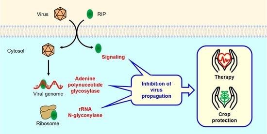
RIPs have long been recognized as antiviral proteins in both plants and animals, but the mechanism responsible for this activity continues to be the subject of intense research today. The mechanism that triggers protection against viruses could have both common and different elements in plants and animals (Figure 1).
4.1. Antiviral Mechanisms of RIPs in Plants
4.1.1. Protein Synthesis Inhibition (rRNA N-glycosylase)
It has long been known that RIPs can inhibit protein synthesis in plants [119,120,121,122]. The mechanism is the same as that described for inhibition of protein synthesis in animals, i.e., RIPs act as N-glycosylases of the major rRNA by removing a specific adenine from the sarcin-ricin loop (SRL), which is highly conserved in animals and plants [120]. Moreover, it has been shown that some RIPs can inhibit protein synthesis carried out by ribosomes of the same plants that produce them [123] and in addition, in the case of some RIPs, a positive correlation between rRNA N-glycosylase activity on tobacco ribosomes and antiviral activity against TMV has been reported [124].
The fact that RIPs do not cause cell death in the absence of the virus and allow plant growth is due to the fact that, at least for type 1 RIPs from dicots, they are synthesized as preproteins with a leader peptide that directs them into the apoplastic space [125]. Viral infection is supposed to facilitate the entry of the RIP, which inactivates cell ribosomes, causing cell death and preventing the virus from using the cellular machinery to replicate and spread [125]. So far, the mechanism by which the virus facilitates the entry of RIPs has not been shown, although the ability of viruses to modify plasma membrane permeability is well-known [126].
4.1.2. Adenine Polynucleotide Glycosylase Activity
However, although some type 1 RIPs can inactivate ribosomes of some plants, they do not do so with those of others and usually act at much higher concentrations than in animal ribosomes [127]. In addition, mutants have been obtained from PAP that do not depurinate tobacco or reticulocyte lysate ribosomes but inhibit translation of brome mosaic virus (BMV) and potato virus X (PVX) [128].
The specificity of RIPs is highly variable, therefore some RIPs can act on other adenines in both animal [14] and plant [120,129] ribosomes. In addition, all RIPs release adenines from eukaryotic DNA and many of them also release adenines from other RNAs, including viral RNAs [15,22,87]. It has also been reported that some RIPs may have DNA nicking, DNase or RNase activities (Table 1). This can alter the life cycle of the virus, both its replication and transcription [130], translation [91], and assembly [131].
The adenine polynucleotide glycosylase activity on viral RNAs might be more specific. Thus, it has been reported that some RIPs can inhibit the translation of capped RNA by binding to the cap of viral RNAs and depurinating these RNAs downstream of the cap structure. For these RIPs, viral RNA depurination could be the main mechanism of their antiviral activity [51]. On the other hand, one of them (PAP) can also bind to translation initiation factors, allowing it to depurinate preferentially uncapped viral RNAs [103]. Viral capped RNA sequestration has also been proposed as an antiviral mechanism for MbRIP-1, a RIP from Momordica balsamina [132]. All this suggests that the antiviral mechanism of RIPs could be more complex than a simple and direct depurination of viral RNA.
4.1.3. Antiviral Protection through Signaling Pathways
The other proposed mechanism involves signaling molecules that defend the plant from viral infection. However, different results have been obtained depending on the RIP studied and the approach used. Thus, it has been reported that α-momorcharin (α-MMC), in N. benthamiana plants sprayed with a solution of the RIP, up-regulates the expression of reactive oxygen species (ROS) scavenging-related genes, modulating ROS homeostasis and conferring resistance to TMV, ChiVMV, and CMV infection [81,133]. Additionally, this RIP also up-regulates some salicylic acid-responsive defence-related genes [81]. By contrast, the same RIP sprayed in M. charantia plants increases plant resistance to CMV but by increasing jasmonic acid biosynthesis and inducing ROS without a relevant increase in salicylic acid [82]. It has also been reported that α-momorcharin induces an increase of both jasmonic acid and salicylic acid in tobacco plants, enhancing TMV resistance [118]. On the other hand, it has been postulated that PAP generates a signal that leads to the overexpression of pathogenesis-related proteins rendering transgenic tobacco plants resistant to virus infection in the absence of an increase in the salicylic acid levels [129,134,135]. Finally, it has been reported that the expression of IRAb and IRIP in transgenic tobacco plants provides a strong local protection against TMV and TEV but without induction of pathogenesis-related proteins [77]. The relationship between the enzymatic activity of RIPs and their ability to induce production of signaling molecules in plants has not been studied. In animals, the enzymes that exert their cytotoxic function through modification of the sarcin-ricin loop (SRL), such as ricin, α-sarcin, or Shiga toxin, strongly activate signaling pathways through the mitogen-activated protein kinases (MAPKs) p38 and JNK [136]. The trichothecenes deoxynivalenol (DON) and T-2 toxin inhibit protein synthesis and have been shown to induce activation of ERK1/2 and p38 MAP kinase in several animal and human cell lines followed by increased cytokine production [137]. This ribosome mediated activation of MAPKs is termed ‘ribotoxic stress response’ [137]. In Arabidopsis, DON and T-2 toxin led to the expression of MPK3 and MPK6 MAP kinases, implicated as positive regulators of the hypersensitive response via ethylene signaling and ROS [137]. Therefore, it would be possible that the generation of signaling compounds by plants was a response to ribotoxic stress produced by RIPs.
4.2. Antiviral Mechanisms of RIPs in Animals
4.2.1. Protein Synthesis Inhibition (rRNA N-glycosylase)
Early studies on the mechanism of antiviral action of RIPs in animal cells focused on their ability to inhibit protein synthesis [30]. Several type 1 RIPs (gelonin, Momordica charantia inhibitor, dianthin 32, and PAP-S) reduced viral production and plaque formation in HEp-2 cells infected with Herpes simplex virus-1 (HSV-1) or poliovirus I. In addition, the four RIPs inhibited protein synthesis more efficiently in cells infected with one of the two viruses than in uninfected cells, suggesting that RIPs inhibited viral replication by inhibiting protein synthesis of infected cells, presumably because they entered infected cells more easily than uninfected cells [30]. Although the mechanism by which viruses can facilitate the entry of RIPs is not established, it is known that type 1 RIPs can enter cells through pinocytosis or receptor-mediated endocytosis [138,139] and that both processes are stimulated by viruses [140,141].
4.2.2. Adenine Polynucleotide Glycosylase Activity
However, RIPs can inhibit virus replication without apparently inactivating ribosomes [34,52,142,143]. The adenine polynucleotide glycosylase activity on viral RNA [57] or DNA [33] is able to inactivate the viral genome and explains inhibition of virus replication [37,142,143]. In addition, RIPs can also depurinate viral mRNAs, thus avoiding the synthesis of proteins that are vital for its functions [52,144,145]. In the case of HIV, a strong inhibition of the integration of viral DNA into the host genome [32,45,50], caused by the adenine polynucleotide glycosylase activity on LTRs (long-terminal repeats) [33,146,147] and the nicking activity on the supercoiled DNA [148,149] of the virus, has been reported. Trichosanthin is also able to enter viral particles during budding, resulting in virions unable to infect other cells [150,151].
4.2.3. Antiviral Protection through Signaling Pathways
Finally, it has also been proposed that the antiviral activity of RIPs can be carried out through signaling pathways. Thus, it has been reported that RIPs promote p53 and c-Jun N-terminal kinase (JNK) activity [152,153] and block the activation of KF-κB, p38MAPK, and Bcl-2 [152,154,155] during viral infection. The modulation of these pathways would lead to the death of infected cells, thus preventing the spread of the virus. Cell DNA damage [152] or ribotoxic stress [153] caused by RIPs could trigger some of these signaling pathways. Ribotoxic stress response (RSR) is a response of cells to a variety of agents that affect the functions of ribosome, such as some antibiotics, alkaloids, mycotoxins, RIPs, ribotoxins, or ultraviolet radiation [136]. Ribotoxic stress is sensed by the MAP3K ZAKα that transduces the signal from ribosomes to activate MAP2K that in turn activates SAPKs. There are two SAPKs (stress-activated protein kinases) families in mammals: p38 and c-Jun N-terminal kinase (JNK). Activation of p38 induces cell-cycle arrest whereas activation of JNK promotes apoptosis [156], inducing both pro-survival and pro-apoptotic signaling. Additionally, mRNA damage by the adenine polynucleotide glycosylase activity of RIPs could trigger RSR as has been reported for ultraviolet radiation [156]. However, much research is still required to clarify how RIPs protect cells from viral infection through these pathways.
Therefore, RIPs can exert their antiviral effect through different mechanisms that could originate from their activity on the different nucleic acids from both the virus and the infected cell. Depending on the type of RIP, virus and infected cell, some mechanisms could predominate over others and more research is required to determine in each case which are the predominant ones.
5. Experimental Therapy
Because of its strong antiviral activity, RIPs have been used in experimental therapy, especially to treat the acquired immune deficiency syndrome (AIDS), but also against hepatitis, chikungunya, dengue, and lymphomas caused by the Epstein–Barr virus. Additionally, they have also been tested in vivo against viruses that infect animals, such as the murine cytomegalovirus, the Pichinde virus, or the simian–human immunodeficiency virus (Table 4).
5.1. RIPs and PEGylated RIPs
Trichosanthin (GLQ223) was used alone [61,157] or in combination with zidovudine (azidothymidine, AZT) [158] in clinical trials with AIDS patients. Trichosanthin infusions were safe and relatively well tolerated [157]. In patients, a decrease in serum p24 antigen [61] and an increase in CD4+ and CD8+ T cells [157,158] were observed. Recently, it has also been reported that maize RIP reduces the viral load of an HIV-related virus, the simian–human immunodeficiency virus in Chinese rhesus macaques [27].
Despite its potential as therapeutic agents, the strong immunogenicity, allergic reaction, and short half-life are the biggest barriers to their application as therapeutic agents. Polyethylene glycol (PEG) conjugation (PEGylation) can confer on these proteins, increasing plasma half-life, decreasing toxicity, and reducing immunogenicity and antigenicity. PEGylated alpha-momorcharin and MAP30 showed about 60%–70% antivirus activities against HSV-1, and at the same time decreased 50%–70% immunogenicity when compared with the non-PEGylated proteins [40].
5.2. Immunotoxins and Other Conjugates
RIPs have been used in medicine mainly as the toxic part of immunotoxins, that is, chimeric proteins consisting of an antibody specifically directed against a target, linked to a toxin of plant or bacterial origin. The design of immunotoxins has been improved over the past 40 years to minimize the off-target toxicity and immunogenicity [159,160]. Several types of antiviral immunotoxins have been constructed using either bacterial toxins (or their fragments) such as pseudomonas exotoxin A or diphteria toxin [161], and RIPs from plants (Table 4). The most commonly used RIP has been the ricin A-chain and the most studied virus the HIV. Viral proteins (gp41, gp120, or gp 160) or proteins from infected cells (CD4, CD25, or CD45RO) have been selected as targets. Despite the success of highly active antiretroviral therapy (HAART), antiviral immunotoxins continue to be developed in order to deplete persisting HIV-infected cell reservoirs [162]. Immunotoxins have also shown to be active in vitro against Epstein–Barr [163,164] and Pichinde [31] viruses and in vivo (in combination with the synthetic analogue of 2′-deoxy-guanosine ganciclovir) against the murine cytomegalovirus [165].
5.3. Designed Antiviral Proteins and Nanocapsules
RIPs have also been used to design antiviral proteins. One of these engineered proteins contains an internal sequence that is recognized by the HIV protease and that is blocking the N-glycosylase activity of the RIP. This protein is activated in infected cells and has shown antiviral activity [28]. Similarly, variants of the ricin A-chain with the sequence recognized by the HIV protease in the C-terminus are activated in infected cells and show antiviral activity [29].
Another approach is to fuse the sequences of RIPs with antimicrobial peptides such as latarcin, thanatin, protegrin-1, and plectasin that are able to inhibit viral replication inside the infected cells, viral entry and replication, dengue NS2B-NS3 serine protease, and virus replication, respectively [42,53]. The aim is to target different stages of the viral life cycle. Thus, the peptide-fusion proteins Latarcin-PAP1-Thanatin and Protegrin1-MAP30-Plectasin inhibit virus replication in vitro and protect the virus-infected mice from chikungunya and dengue viruses, respectively [42,53]. Another fusion protein containing ricin A-chain and PAP-S displays antiviral activity in vitro against hepatitis B virus suggesting a synergistic activity of both proteins [167]. This has encouraged its authors to propose it as an anti-SARS-CoV-2 agent [75].
The latest approach is the use of nanocapsules to deliver RIPs to virus-infected cells. Nanocapsules are vesicular objects in which the encapsulated compound is confined in an internal cavity surrounded by an outer membrane [182,183]. Nanocapsules containing MAP30 [180] or ricin A-chain [181] have shown antiviral activity in vitro against HIV. In the latter case, targeting has been achieved by using peptide crosslinkers that are sensitive to cleavage by HIV-1 protease [181].
5.4. Side Effects of RIP Therapy
Although trichosanthin was, in general, well tolerated in clinical trials when used in AIDS patients [157], some side effects were reported [61,157,158]. Clinical trials using RIPs as antivirals are scarce, but there are many clinical trials that have used RIPs as part of immunotoxins for the treatment of malignancies [9,64,184]. Side effects that may be mild or moderate like fever, nausea, vomiting, diarrhea, myalgia, edema, and hypoalbuminemia have been reported in these trials. Other effects are severe, such as immunogenicity and vascular leak syndrome (VLS), and could limit the therapeutic use of immunotoxins [64,184]. Immunogenicity may be the result of the formation of human anti-mouse antibodies (HAMA) or human anti-toxin antibodies (HATA). These antibodies can prevent repeated treatment cycles. The development of immunotoxins containing humanized antibodies or the use of part of antibodies containing only the variable domains can solve this problem [64,184]. To address the problem of the immunogenicity of RIPs, PEGylation [40,184] and elimination of epitopes through genetic manipulation have been used [184]. Vascular leak syndrome, characterized by increased vascular permeability, is caused by the nonspecific binding of RIP to vascular endothelial cells. The identification and elimination of some peptides present in RIPs, nonessentials for RIP activity and responsible for this unspecific binding, have allowed the obtaining of less toxic recombinant RIPs [184].
6. Genetically Engineered Virus-Resistant Plants
Viruses cause epidemics in all major crops, representing a significant restriction on the yield and quality of agricultural production. As strict intracellular pathogens, they cannot be chemically controlled and prophylactic measures consist mainly in the destruction of infected plants and biocide applications to limit the population of vector organisms (arthropods, nematodes, and plasmodiophorids). A powerful alternative often used in agriculture is based on the use of crop genetic resistances, an approach that depends on mechanisms governing plant-virus interactions [185]. Several transgenic plants carrying virus resistance genes have been obtained by transferring virus-derived genes, including viral coat proteins, replicases, movement proteins, defective interfering RNAs, non-coding RNA sequences and proteases into susceptible plants, or non-viral genes including R genes, microRNAs, RIPs, protease inhibitors, dsRNAses, RNA modifying enzymes, and scFvs [186]. In recent years, transgenic plants carrying RIP genes that are resistant to fungi, insects and, above all, viruses have been reported. Thus, transgenic plants bearing RIP genes have been obtained that are resistant to a wide variety of viruses (Table 5).
Most of the times, tobacco has been transformed (Nicotiana tabacum L. and N. benthamiana Domin) but also potato (Solanum tuberosum L.) and tomato (Lycopersicon esculentum Mill.). Agrobacterium tumefaciens containing the plant transformation vectors has been used to transform either tobacco by the leaf disc co-cultivation method or potato (S. tuberosum) by the stem or tuber section co-cultivation method. The CaMV 35S promoter has always been used to express the RIPs, except in the case of dianthin 30 [84]. In the case of trichosanthin, tissue-specific promoters have also been used [80]. The CaMV 35S promoter is the most studied and most widely used plant promoter for transgenic expression [189], it is a very strong constitutive promoter that facilitates a high level of RNA transcription in a wide variety of plant species. For effective protection against viruses, it is preferable to achieve high levels of RIP expression since there is a direct correlation between expression level and resistance to viruses [78]. So, for example, in lines expressing small amounts of curcin 2, symptoms of TMV infection begin to appear after about 7 days, while lines that accumulate the highest level of curcin 2 (about 1.45 µg/mg) begin to develop symptoms after about 18 days.
Using the promoter CaMV 35S, plants with a RIP content of up to 2.7% of the total soluble protein have been obtained [80]. However, a high expression of RIP results in plants with an aberrant phenotype, which usually includes leaf mottling, extreme leaf discoloration, stunted leaf growth and/or excessive curvature, slow rooting and growth rates, and high plant mortality rates [80,188]. This could be because some RIPs can kill plant cells by inactivating their ribosomes [120,121,122]. Several approaches have been used to overcome this problem. One strategy might be to introduce the gene encoding for the preprotein [80], this allows the RIP to accumulate in the apoplasma instead of the cytosol, thus preventing access to the ribosomes. Transgenic tobacco plants expressing the preprotein of trichosanthin exhibited resistance to cucumber mosaic virus (CMV) and tobacco mosaic virus (TMV) but did not show an abnormal phenotype [80]. In the case of PAP, despite being the most widely used, it inhibits protein synthesis and is toxic to plant cells, but transgenic plants have been obtained with mutants that are not toxic to the plant maintaining the antiviral activity [188]. The lack of toxicity of these mutants has been attributed to a change in the location of the protein preventing contact with ribosomes [188]. PAP (PAPI) has also been replaced by PAPII in order to obtain virus-resistant plants [104]. The protein sequence of PAPII shows only 41% identity to PAPI. PAPII expressed in transgenic tobacco was correctly processed to the mature form and accumulated to at least 10-fold higher levels than wild-type PAP (up to 250 ng/mg PAPII). PAPII is less toxic than PAP and symptomless transgenic lines expressing PAPII were resistant to TMV and PVX [104]. Another approach is to use a promoter that is induced by viral infection, thus, the gene that encodes for dianthin 30 was introduced into N. benthamiana and expressed from the promoter ACMV virion-sense [84]. This promoter is induced specifically by the ACMV infection and transgenic plants displayed a normal phenotype and were resistant to ACMV [84].
7. Conclusions
After decades of research, RIPs continue to be a topic of interest and a useful tool in many research fields. The new advances in plant molecular biology, virology, immunotherapy, and nanotechnology open new possibilities in the use of RIPs in medicine and agriculture in order to find solutions to the continuous challenge posed by viruses to human health and crop yields.
Author Contributions
Conceptualization, L.C. and J.M.F.; writing—original draft preparation, R.I. and J.M.F.; writing—review and editing, L.C. and R.I.; funding acquisition, J.M.F. All authors have read and agreed to the published version of the manuscript.
Funding
This research was funded by Consejería de Educación (Junta de Castilla y León) to the GIR ProtIBio, grant number VA033G19.
Institutional Review Board Statement
Not applicable.
Informed Consent Statement
Not applicable.
Data Availability Statement
Data are available upon request. Please, contact the contributing authors.
Conflicts of Interest
The authors declare no conflict of interest.
References
- De Clercq, E. Looking Back in 2009 at the Dawning of Antiviral Therapy Now 50 Years Ago: An Historical Perspective. In Advances in Virus Research; Maramorosch, K., Shatkin, A.J., Murphy, F.A., Eds.; Elsevier Academic Press Inc.: San Diego, CA, USA, 2009; Volume 73, pp. 1–53. [Google Scholar]
- Ng, T.; Cheung, R.C.F.; Wong, J.H.; Chan, W.-Y. Proteins, peptides, polysaccharides, and nucleotides with inhibitory activity on human immunodeficiency virus and its enzymes. Appl. Microbiol. Biotechnol. 2015, 99, 10399–10414. [Google Scholar] [CrossRef] [PubMed]
- Musidlak, O.; Nawrot, R.; Goździcka-Józefiak, A. Which Plant Proteins Are Involved in Antiviral Defense? Review on In Vivo and In Vitro Activities of Selected Plant Proteins against Viruses. Int. J. Mol. Sci. 2017, 18, 2300. [Google Scholar] [CrossRef] [PubMed] [Green Version]
- Bolognesi, A.; Bortolotti, M.; Maiello, S.; Battelli, M.G.; Polito, L. Ribosome-Inactivating Proteins from Plants: A Historical Overview. Molecules 2016, 21, 1627. [Google Scholar] [CrossRef] [PubMed]
- Ferreras, J.M.; Citores, L.; Iglesias, R.; Jiménez, P.; Girbés, T. Use of Ribosome-Inactivating Proteins from Sambucus for the Construction of Immunotoxins and Conjugates for Cancer Therapy. Toxins 2011, 3, 420–441. [Google Scholar] [CrossRef] [PubMed] [Green Version]
- Stirpe, F. Ribosome-inactivating proteins: From toxins to useful proteins. Toxicon 2013, 67, 12–16. [Google Scholar] [CrossRef] [PubMed]
- Di Maro, A.; Citores, L.; Russo, R.; Iglesias, R.; Ferreras, J.M. Sequence comparison and phylogenetic analysis by the Maximum Likelihood method of ribosome-inactivating proteins from angiosperms. Plant Mol. Biol. 2014, 85, 575–588. [Google Scholar] [CrossRef] [PubMed]
- Olsnes, S. The history of ricin, abrin and related toxins. Toxicon 2004, 44, 361–370. [Google Scholar] [CrossRef]
- Polito, L.; Bortolotti, M.; Battelli, M.G.; Calafato, G.; Bolognesi, A. Ricin: An Ancient Story for a Timeless Plant Toxin. Toxins 2019, 11, 324. [Google Scholar] [CrossRef] [Green Version]
- Domashevskiy, A.V.; Goss, D.J. Pokeweed Antiviral Protein, a Ribosome Inactivating Protein: Activity, Inhibition and Prospects. Toxins 2015, 7, 274–298. [Google Scholar] [CrossRef] [Green Version]
- Endo, Y.; Tsurugi, K. The RNA N-glycosidase activity of ricin A-chain. The characteristics of the enzymatic activity of ricin A-chain with ribosomes and with rRNA. J. Biol. Chem. 1988, 263, 8735–8739. [Google Scholar] [CrossRef]
- Iglesias, R.; Citores, L.; Ferreras, J.M. Ribosomal RNA N-glycosylase Activity Assay of Ribosome-inactivating Proteins. Bio-Protocol 2017, 7, e2180. [Google Scholar] [CrossRef]
- Citores, L.; Ragucci, S.; Ferreras, J.M.; Di Maro, A.; Iglesias, R. Ageritin, a Ribotoxin from Poplar Mushroom (Agrocybe aegerita) with Defensive and Antiproliferative Activities. ACS Chem. Biol. 2019, 14, 1319–1327. [Google Scholar] [CrossRef] [PubMed]
- Barbieri, L.; Ferreras, J.M.; Barraco, A.; Ricci, P.; Stirpe, F. Some ribosome-inactivating proteins depurinate ribosomal RNA at multiple sites. Biochem. J. 1992, 286, 1–4. [Google Scholar] [CrossRef] [Green Version]
- Barbieri, L.; Valbonesi, P.; Bonora, E.; Gorini, P.; Bolognesi, A.; Stirpe, F. Polynucleotide: Adenosine glycosidase activity of ribosome-inactivating proteins: Effect on DNA, RNA and poly(A). Nucleic Acids Res. 1997, 25, 518–522. [Google Scholar] [CrossRef]
- Ogasawara, T.; Sawasaki, T.; Morishita, R.; Ozawa, A.; Madin, K.; Endo, Y. A new class of enzyme acting on damaged ribosomes: Ribosomal RNA apurinic site specific lyase found in wheat germ. EMBO J. 1999, 18, 6522–6531. [Google Scholar] [CrossRef] [Green Version]
- Choudhary, N.L.; Yadav, O.P.; Lodha, M.L. Ribonuclease, deoxyribonuclease, and antiviral activity of Escherichia coli-expressed Bougainvillea xbuttiana antiviral protein-1. Biochemistry 2008, 73, 273–277. [Google Scholar] [CrossRef] [PubMed]
- Chen, H.; Wang, Y.; Yan, M.; Yu, M.; Yao, Q. 5’-AMP Phosphatase activity on trichosanthin and other single chain ribosome inactivating proteins. Chin. Biochem. J. 1996, 12, 125–130. [Google Scholar]
- Li, X.; Chen, W.-F.; Liu, W.-Y.; Wang, G.-H. Large-Scale Preparation of Two New Ribosome-Inactivating Proteins—Cinnamomin and Camphorin from the Seeds of Cinnamomum camphora. Protein Expr. Purif. 1997, 10, 27–31. [Google Scholar] [CrossRef] [PubMed]
- Helmy, M.; Lombard, S.; Piéroni, G. Ricin RCA60: Evidence of Its Phospholipase Activity. Biochem. Biophys. Res. Commun. 1999, 258, 252–255. [Google Scholar] [CrossRef]
- Shih, N.; McDonald, K.A.; Jackman, A.P.; Girbés, T.; Iglesias, R. Bifunctional plant defence enzymes with chitinase and ribosome inactivating activities from Trichosanthes kirilowii cell cultures. Plant Sci. 1997, 130, 145–150. [Google Scholar] [CrossRef]
- Iglesias, R.; Citores, L.; Di Maro, A.; Ferreras, J.M. Biological activities of the antiviral protein BE27 from sugar beet (Beta vulgaris L.). Planta 2014, 241, 421–433. [Google Scholar] [CrossRef] [PubMed]
- Polito, L.; Bortolotti, M.; Pedrazzi, M.; Mercatelli, D.; Battelli, M.G.; Bolognesi, A. Apoptosis and necroptosis induced by stenodactylin in neuroblastoma cells can be completely prevented through caspase inhibition plus catalase or necrostatin-1. Phytomedicine 2016, 23, 32–41. [Google Scholar] [CrossRef] [PubMed] [Green Version]
- Panda, P.K.; Behera, B.; Meher, B.R.; Das, D.N.; Mukhopadhyay, S.; Sinha, N.; Naik, P.P.; Roy, B.; Das, J.; Paul, S.; et al. Abrus Agglutinin, a type II ribosome inactivating protein inhibits Akt/PH domain to induce endoplasmic reticulum stress mediated autophagy-dependent cell death. Mol. Carcinog. 2016, 56, 389–401. [Google Scholar] [CrossRef] [PubMed]
- Rustgi, S.; Pollmann, S.; Buhr, F.; Springer, A.; Reinbothe, C.; Von Wettstein, D.; Reinbothe, S. JIP60-mediated, jasmonate- and senescence-induced molecular switch in translation toward stress and defense protein synthesis. Proc. Natl. Acad. Sci. USA 2014, 111, 14181–14186. [Google Scholar] [CrossRef] [Green Version]
- Przydacz, M.; Jones, R.; Pennington, H.G.; Belmans, G.; Bruderer, M.; Greenhill, R.; Salter, T.; Wellham, P.A.; Cota, E.; Spanu, P.D. Mode of Action of the Catalytic Site in the N-Terminal Ribosome-Inactivating Domain of JIP60. Plant Physiol. 2020, 183, 385–398. [Google Scholar] [CrossRef]
- Wang, R.-R.; Au, K.-Y.; Zheng, H.-Y.; Gao, L.-M.; Zhang, X.; Luo, R.-H.; Law, S.K.-Y.; Mak, A.N.-S.; Wong, K.-B.; Zhang, M.-X.; et al. The Recombinant Maize Ribosome-Inactivating Protein Transiently Reduces Viral Load in SHIV89.6 Infected Chinese Rhesus Macaques. Toxins 2015, 7, 156–169. [Google Scholar] [CrossRef] [Green Version]
- Law, S.K.-Y.; Wang, R.-R.; Mak, A.N.-S.; Wong, K.-B.; Zheng, Y.-T.; Shaw, P.C. A switch-on mechanism to activate maize ribosome-inactivating protein for targeting HIV-infected cells. Nucleic Acids Res. 2010, 38, 6803–6812. [Google Scholar] [CrossRef] [Green Version]
- Au, K.-Y.; Wang, R.-R.; Wong, Y.-T.; Wong, K.-B.; Zheng, Y.-T.; Shaw, P.C. Engineering a switch-on peptide to ricin A chain for increasing its specificity towards HIV-infected cells. Biochim. Biophys. Acta Gen. Subj. 2014, 1840, 958–963. [Google Scholar] [CrossRef]
- Foà-Tomasi, L.; Campadelli-Fiume, G.; Barbieri, L.; Stirpe, F. Effect of ribosome-inactivating proteins on virus-infected cells. Inhibition of virus multiplication and of protein synthesis. Arch. Virol. 1982, 71, 323–332. [Google Scholar] [CrossRef]
- Barnett, B.B.; Burns, N.J.; Park, K.J.; Dawson, M.I.; Kende, M.; Sidwell, R.W. Antiviral immunotoxins: Antibody-mediated delivery of gelonin inhibits Pichinde virus replication in vitro. Antivir. Res. 1991, 15, 125–138. [Google Scholar] [CrossRef]
- Au, T.K.; Collins, R.A.; Lam, T.L.; Ng, T.B.; Fong, W.P.; Wan, D. The plant ribosome inactivating proteins luffin and saporin are potent inhibitors of HIV-1 integrase. FEBS Lett. 2000, 471, 169–172. [Google Scholar] [CrossRef] [Green Version]
- Li, H.-G.; Huang, P.L.; Zhang, D.; Sun, Y.; Chen, H.-C.; Zhang, J.; Huang, P.L.; Kong, X.-P.; Lee-Huang, S. A new activity of anti-HIV and anti-tumor protein GAP31: DNA adenosine glycosidase – Structural and modeling insight into its functions. Biochem. Biophys. Res. Commun. 2010, 391, 340–345. [Google Scholar] [CrossRef] [PubMed]
- Lee-Huang, S.; Kung, H.-F.; Huang, P.L.; Huang, P.L.; Li, B.-Q.; Huang, P.; Huang, H.I.; Chen, H.-C. A new class of anti-HIV agents: GAP31, DAPs 30 and 32. FEBS Lett. 1991, 291, 139–144. [Google Scholar] [CrossRef] [Green Version]
- Kaur, I.; Gupta, R.C.; Puri, M. Ribosome inactivating proteins from plants inhibiting viruses. Virol. Sin. 2011, 26, 357–365. [Google Scholar] [CrossRef]
- Fang, E.F.; Ng, T.B.; Shaw, P.C.; Wong, R.N.S. Recent Progress in Medicinal Investigations on Trichosanthin and other Ribo-some Inactivating Proteins from the Plant Genus Trichosanthes. Curr. Med. Chem. 2011, 18, 4410–4417. [Google Scholar] [CrossRef]
- He, D.-X.; Tam, S.-C. Trichosanthin affects HSV-1 replication in Hep-2 cells. Biochem. Biophys. Res. Commun. 2010, 402, 670–675. [Google Scholar] [CrossRef]
- Shi, W.-W.; Wong, K.-B.; Shaw, P.C. Structural and Functional Investigation and Pharmacological Mechanism of Trichosanthin, a Type 1 Ribosome-Inactivating Protein. Toxins 2018, 10, 335. [Google Scholar] [CrossRef] [PubMed] [Green Version]
- Zheng, Y.T.; Ben, K.L.; Jin, S.W. Anti-HIV-1 activity of trichobitacin, a novel ribosome-inactivating protein. Acta Pharmacol. Sin. 2000, 21, 179–182. [Google Scholar]
- Meng, Y.; Liu, S.; Li, J.; Zhao, X. Preparation of an antitumor and antivirus agent: Chemical modification of α-MMC and MAP30 from Momordica Charantia L. with covalent conjugation of polyethyelene glycol. Int. J. Nanomed. 2012, 7, 3133–3142. [Google Scholar] [CrossRef] [Green Version]
- Yao, X.; Li, J.; Deng, N.; Wang, S.; Meng, Y.; Shen, F. Immunoaffinity purification of α-momorcharin from bitter melon seeds (Momordica charantia). J. Sep. Sci. 2011, 34, 3092–3098. [Google Scholar] [CrossRef]
- Rothan, H.A.; Bahrani, H.; Mohamed, Z.; Rahman, N.A.; Yusof, R. Fusion of Protegrin-1 and Plectasin to MAP30 Shows Significant Inhibition Activity against Dengue Virus Replication. PLoS ONE 2014, 9, e94561. [Google Scholar] [CrossRef] [Green Version]
- Sun, Y.; Huang, P.L.; Li, J.J.; Huang, Y.Q.; Zhang, L.; Huang, P.L.; Lee-Huang, S. Anti-HIV Agent MAP30 Modulates the Expression Profile of Viral and Cellular Genes for Proliferation and Apoptosis in AIDS-Related Lymphoma Cells Infected with Kaposi’s Sarcoma-Associated Virus. Biochem. Biophys. Res. Commun. 2001, 287, 983–994. [Google Scholar] [CrossRef]
- Fan, J.M.; Zhang, Q.; Xu, J.; Zhu, S.; Ke, T.; Gao, D.F.; Xu, Y.B. Inhibition on Hepatitis B virus in vitro of recombinant MAP30 from bitter melon. Mol. Biol. Rep. 2007, 36, 381–388. [Google Scholar] [CrossRef]
- Lee-Huang, S.; Huang, P.L.; Bourinbaiar, A.S.; Chen, H.C.; Kung, H.F. Inhibition of the integrase of human immunodeficiency virus (HIV) type 1 by anti-HIV plant proteins MAP30 and GAP31. Proc. Natl. Acad. Sci. USA 1995, 92, 8818–8822. [Google Scholar] [CrossRef] [Green Version]
- Bourinbaiar, A.S.; Lee-Huang, S. The Activity of Plant-Derived Antiretroviral Proteins MAP30 and GAP31 against Herpes Simplex Virus Infectionin Vitro. Biochem. Biophys. Res. Commun. 1996, 219, 923–929. [Google Scholar] [CrossRef]
- Kaur, I.; Puri, M.; Ahmed, Z.; Blanchet, F.P.; Mangeat, B.; Piguet, V. Inhibition of HIV-1 Replication by Balsamin, a Ribosome Inactivating Protein of Momordica balsamina. PLoS ONE 2013, 8, e73780. [Google Scholar] [CrossRef]
- Wachinger, M.; Samtleben, R.; Gerhauser, C.; Wagner, H.; Erfle, V. Bryodin, a single-chain ribosome-inactivating protein, selectively inhibits the growth of HIV-1-infected cells and reduces HIV-1 production. Res. Exp. Med. 1993, 193, 1–12. [Google Scholar] [CrossRef]
- Leitman, E.M.; Palmer, C.D.; Buus, S.; Chen, F.; Riddell, L.; Sims, S.; Klenerman, P.; Saez-Cirion, A.; Walker, B.D.; Hess, P.R.; et al. Saporin-conjugated tetramers identify efficacious anti-HIV CD8+ T-cell specificities. PLoS ONE 2017, 12, e0184496. [Google Scholar] [CrossRef] [Green Version]
- Yadav, S.K.; Batra, J.K. Mechanism of Anti-HIV Activity of Ribosome Inactivating Protein, Saporin. Protein Pept. Lett. 2015, 22, 497–503. [Google Scholar] [CrossRef]
- Vivanco, J.M.; Tumer, N.E. Translation Inhibition of Capped and Uncapped Viral RNAs Mediated by Ribosome-Inactivating Proteins. Phytopathology 2003, 93, 588–595. [Google Scholar] [CrossRef] [PubMed] [Green Version]
- Mansouri, S.; Choudhary, G.; Sarzala, P.M.; Ratner, L.; Hudak, K.A. Suppression of Human T-cell Leukemia Virus I Gene Expression by Pokeweed Antiviral Protein. J. Biol. Chem. 2009, 284, 31453–31462. [Google Scholar] [CrossRef] [Green Version]
- Rothan, H.A.; Bahrani, H.; Shankar, E.M.; Rahman, N.A.; Yusof, R. Inhibitory effects of a peptide-fusion protein (Latarcin–PAP1–Thanatin) against chikungunya virus. Antivir. Res. 2014, 108, 173–180. [Google Scholar] [CrossRef]
- Ishag, H.Z.A.; Li, C.; Huang, L.; Sun, M.-X.; Ni, B.; Guo, C.-X.; Mao, X. Inhibition of Japanese encephalitis virus infection in vitro and in vivo by pokeweed antiviral protein. Virus Res. 2013, 171, 89–96. [Google Scholar] [CrossRef]
- Uckun, F.M.; Rustamova, L.; Vassilev, A.O.; Tibbles, H.E.; Petkevich, A.S. CNS activity of Pokeweed Anti-viral Protein (PAP) in mice infected with Lymphocytic Choriomeningitis Virus (LCMV). BMC Infect. Dis. 2005, 5, 9. [Google Scholar] [CrossRef] [Green Version]
- He, Y.-W.; Guo, C.-X.; Pan, Y.-F.; Peng, C.; Weng, Z.-H. Inhibition of hepatitis B virus replication by pokeweed antiviral protein in vitro. World J. Gastroenterol. 2008, 14, 1592–1597. [Google Scholar] [CrossRef]
- Rajamohan, F.; Venkatachalam, T.K.; Irvin, J.D.; Uckun, F.M. Pokeweed Antiviral Protein Isoforms PAP-I, PAP-II, and PAP-III Depurinate RNA of Human Immunodeficiency Virus (HIV)-1. Biochem. Biophys. Res. Commun. 1999, 260, 453–458. [Google Scholar] [CrossRef]
- Di, R.; Tumer, N.E. Pokeweed Antiviral Protein: Its Cytotoxicity Mechanism and Applications in Plant Disease Resistance. Toxins 2015, 7, 755–772. [Google Scholar] [CrossRef] [Green Version]
- Ussery, M.A.; Irvin, J.D.; Hardesty, B. Inhibition of Poliovirus Replication by A Plant Antiviral Peptide. Ann. N. Y. Acad. Sci. 1977, 284, 431–440. [Google Scholar] [CrossRef]
- McGrath, M.S.; Hwang, K.M.; Caldwell, S.E.; Gaston, I.; Luk, K.C.; Wu, P.; Ng, V.L.; Crowe, S.; Daniels, J.; Marsh, J. GLQ223: An inhibitor of human immunodeficiency virus replication in acutely and chronically infected cells of lymphocyte and mononuclear phagocyte lineage. Proc. Natl. Acad. Sci. USA 1989, 86, 2844–2848. [Google Scholar] [CrossRef] [Green Version]
- Byers, V.S.; Levin, A.S.; Waites, L.A.; Starrett, B.A.; Mayer, R.A.; Clegg, J.A.; Price, M.R.; Robins, R.A.; Delaney, M.; Baldwin, R.W. A phase I/II study of trichosanthin treatment of HIV disease. AIDS 1990, 4, 1189–1196. [Google Scholar] [CrossRef]
- Wen, D.; Wang, J.; Yan, H.; Chen, J.; Xia, K.; Liu, J.; Zhang, A. Effect of Radix Trichosanthis and Trichosanthin on Hepatitis B Virus in HepG2.2.15 Cells. J. Nanosci. Nanotechnol. 2015, 15, 2094–2098. [Google Scholar] [CrossRef] [PubMed]
- Barbieri, L.; Battelli, M.G.; Stirpe, F. Ribosome-inactivating proteins from plants. Biochim. Biophys. Acta Rev. Biomembr. 1993, 1154, 237–282. [Google Scholar] [CrossRef]
- Citores, L.; Iglesias, R.; Ferreras, J.M. Ribosome Inactivating Proteins from Plants: Biological Properties and their Use in Experimental Therapy. In Antitumor Potential and Other Emerging Medicinal Properties of Natural Compounds; Fang, E.F., Ng, T.B., Eds.; Springer: Dordrecht, The Netherlands, 2013; pp. 127–143. [Google Scholar]
- Lv, Q.; Yang, X.-Z.; Fu, L.-Y.; Lu, Y.-T.; Lu, Y.-H.; Zhao, J.; Wang, F.-J. Recombinant expression and purification of a MAP30-cell penetrating peptide fusion protein with higher anti-tumor bioactivity. Protein Expr. Purif. 2015, 111, 9–17. [Google Scholar] [CrossRef] [PubMed]
- Lin, B.; Yang, X.-Z.; Cao, X.-W.; Zhang, T.-Z.; Wang, F.-J.; Zhao, J. A novel trichosanthin fusion protein with increased cytotoxicity to tumor cells. Biotechnol. Lett. 2016, 39, 71–78. [Google Scholar] [CrossRef] [PubMed]
- Ferens, W.A.; Hovde, C.J. Antiviral Activity of Shiga Toxin 1: Suppression of Bovine Leukemia Virus-Related Spontaneous Lymphocyte Proliferation. Infect. Immun. 2000, 68, 4462–4469. [Google Scholar] [CrossRef] [PubMed] [Green Version]
- Shi, P.L.; Binnington, B.; Sakac, D.; Katsman, Y.; Ramkumar, S.; Gariépy, J.; Kim, M.; Branch, D.R.; Lingwood, C. Verotoxin A Subunit Protects Lymphocytes and T Cell Lines against X4 HIV Infection in Vitro. Toxins 2012, 4, 1517–1534. [Google Scholar] [CrossRef] [Green Version]
- Yadav, S.K.; Batra, J.K. Ribotoxin restrictocin manifests anti-HIV-1 activity through its specific ribonuclease activity. Int. J. Biol. Macromol. 2015, 76, 58–62. [Google Scholar] [CrossRef]
- Wong, J.H.; Wang, H.X.; Ng, T.B. Marmorin, a new ribosome inactivating protein with antiproliferative and HIV-1 reverse transcriptase inhibitory activities from the mushroom Hypsizigus marmoreus. Appl. Microbiol. Biotechnol. 2008, 81, 669–674. [Google Scholar] [CrossRef]
- Wang, H.; Ng, T.B. Isolation and characterization of velutin, a novel low-molecular-weight ribosome-inactivating protein from winter mushroom (Flammulina velutipes) fruiting bodies. Life Sci. 2001, 68, 2151–2158. [Google Scholar] [CrossRef]
- Lam, S.; Ng, T. First Simultaneous Isolation of a Ribosome Inactivating Protein and an Antifungal Protein from a Mushroom (Lyophyllum shimeji) Together with Evidence for Synergism of their Antifungal Effects. Arch. Biochem. Biophys. 2001, 393, 271–280. [Google Scholar] [CrossRef]
- Yao, Q.Z.; Yu, M.M.; Ooi, L.S.; Ng, T.B.; Chang, S.T.; Sun, S.S.; Ooi, V.E. Isolation and Characterization of a Type 1 Ribosome-Inactivating Protein from Fruiting Bodies of the Edible Mushroom (Volvariella volvacea). J. Agric. Food Chem. 1998, 46, 788–792. [Google Scholar] [CrossRef]
- Arslan, I.; Akgul, H.; Kara, M. Saporin, a Polynucleotide–Adenosine Nucleosidase, May Be an Efficacious Therapeutic Agent for SARS-CoV-2 Infection. SLAS Discov. Adv. Life Sci. 2020. [Google Scholar] [CrossRef]
- Hassan, Y.; Ogg, S.; Ge, H. Novel Binding Mechanisms of Fusion Broad Range Anti-Infective Protein Ricin A Chain Mutant-Pokeweed Antiviral Protein 1 (RTAM-PAP1) against SARS-CoV-2 Key Proteins in Silico. Toxins 2020, 12, 602. [Google Scholar] [CrossRef]
- Vandenbussche, F.; Desmyter, S.; Ciani, M.; Proost, P.; Peumans, W.J.; Van Damme, E.J.M. Analysis of the in planta antiviral activity of elderberry ribosome-inactivating proteins. Eur. J. Biochem. 2004, 271, 1508–1515. [Google Scholar] [CrossRef]
- Vandenbussche, F.; Peumans, W.J.; Desmyter, S.; Proost, P.; Ciani, M.; Van Damme, E.J.M. The type-1 and type-2 ribosome-inactivating proteins from Iris confer transgenic tobacco plants local but not systemic protection against viruses. Planta 2004, 220, 211–221. [Google Scholar] [CrossRef]
- Huang, M.-X.; Hou, P.; Wei, Q.; Xu, Y.; Chen, F. A ribosome-inactivating protein (curcin 2) induced from Jatropha curcas can reduce viral and fungal infection in transgenic tobacco. Plant Growth Regul. 2007, 54, 115–123. [Google Scholar] [CrossRef]
- Lam, Y.-H.; Wong, Y.-S.; Wang, B.; Wong, R.N.-S.; Yeung, H.-W.; Shaw, P.-C. Use of trichosanthin to reduce infection by turnip mosaic virus. Plant Sci. 1996, 114, 111–117. [Google Scholar] [CrossRef]
- Krishnan, R.; McDonald, K.A.; Dandekar, A.M.; Jackman, A.P.; Falk, B. Expression of recombinant trichosanthin, a ribosome-inactivating protein, in transgenic tobacco. J. Biotechnol. 2002, 97, 69–88. [Google Scholar] [CrossRef]
- Zhu, F.; Zhang, P.; Meng, Y.-F.; Xu, F.; Zhang, D.-W.; Cheng, J.; Lin, H.-H.; Xi, D. Alpha-momorcharin, a RIP produced by bitter melon, enhances defense response in tobacco plants against diverse plant viruses and shows antifungal activity in vitro. Planta 2012, 237, 77–88. [Google Scholar] [CrossRef]
- Yang, T.; Meng, Y.; Chen, L.-J.; Lin, H.; Xi, D.-H. The Roles of Alpha-Momorcharin and Jasmonic Acid in Modulating the Response of Momordica charantia to Cucumber Mosaic Virus. Front. Microbiol. 2016, 7, 1796. [Google Scholar] [CrossRef] [Green Version]
- Ruan, X.-L.; Liu, L.-F.; Li, H. Transgenic tobacco plants with ribosome inactivating protein gene cassin from Cassia occidentalis and their resistance to tobacco mosaic virus. J. Plant Physiol. Mol. Biol. 2007, 33, 517–523. [Google Scholar]
- Hong, Y.; Saunders, K.; Hartley, M.R.; Stanley, J. Resistance to Geminivirus Infection by Virus-Induced Expression of Dianthin in Transgenic Plants. Virology 1996, 220, 119–127. [Google Scholar] [CrossRef] [PubMed] [Green Version]
- Stirpe, F.; Williams, D.G.; Onyon, L.J.; Legg, R.F.; Stevens, W.A. Dianthins, ribosome-damaging proteins with anti-viral properties from Dianthus caryophyllus L. (carnation). Biochem. J. 1981, 195, 399–405. [Google Scholar] [CrossRef] [Green Version]
- Iglesias, R.; Pérez, Y.; de Torre, C.; Ferreras, J.M.; Antolín, P.; Jiménez, P.; Ángeles Rojo, M.; Méndez, E.; Girbés, T. Molecular characterization and systemic induction of single-chain ribosome-inactivating proteins (RIPs) in sugar beet (Beta vulgaris) leaves. J. Exp. Bot. 2005, 56, 1675–1684. [Google Scholar] [CrossRef] [Green Version]
- Iglesias, R.; Citores, L.; Ragucci, S.; Russo, R.; Di Maro, A.; Ferreras, J.M. Biological and antipathogenic activities of ribosome-inactivating proteins from Phytolacca dioica L. Biochim. Biophys. Acta Gen. Subj. 2016, 1860, 1256–1264. [Google Scholar] [CrossRef]
- Roy, S.; Sadhana, P.; Begum, M.; Kumar, S.; Lodha, M.; Kapoor, H. Purification, characterization and cloning of antiviral/ribosome inactivating protein from Amaranthus tricolor leaves. Phytochemistry 2006, 67, 1865–1873. [Google Scholar] [CrossRef]
- Kwon, S.-Y.; An, C.S.; Liu, J.R.; Paek, K.-H. A Ribosome–Inactivating Protein fromAmaranthus viridis. Biosci. Biotechnol. Biochem. 1997, 61, 1613–1614. [Google Scholar] [CrossRef]
- Gholizadeh, A. Purification of a ribosome-inactivating protein with antioxidation and root developer potencies from Celosia plumosa. Physiol. Mol. Biol. Plants 2018, 25, 243–251. [Google Scholar] [CrossRef]
- Baranwal, V.K.; Tumer, N.E.; Kapoor, H.C. Depurination of ribosomal RNA and inhibition of viral RNA translation by an antiviral protein of Celosia cristata. Indian J. Exp. Biol. 2002, 40, 1195–1197. [Google Scholar]
- Balasubrahmanyam, A.; Baranwal, V.K.; Lodha, M.; Varma, A.; Kapoor, H. Purification and properties of growth stage-dependent antiviral proteins from the leaves of Celosia cristata. Plant Sci. 2000, 154, 13–21. [Google Scholar] [CrossRef]
- Begam, M.; Kumar, S.; Roy, S.; Campanella, J.J.; Kapoor, H. Molecular cloning and functional identification of a ribosome inactivating/antiviral protein from leaves of post-flowering stage of Celosia cristata and its expression in E. coli. Phytochemistry 2006, 67, 2441–2449. [Google Scholar] [CrossRef] [PubMed]
- Dutt, S.; Narwal, S.; Kapoor, H.C.; Lodha, M.L. Isolation and Characterization of Two Protein Isoforms with Antiviral Activity from Chenopodium album L Leaves. J. Plant Biochem. Biotechnol. 2003, 12, 117–122. [Google Scholar] [CrossRef]
- Park, J.-S.; Hwang, D.-J.; Lee, S.-M.; Kim, Y.-T.; Choi, S.-B.; Cho, K.-J. Ribosome-inactivating activity and cDNA cloning of antiviral protein isoforms of Chenopodium album. Mol. Cells 2004, 17, 73–80. [Google Scholar] [PubMed]
- Torky, Z.A. Isolation and characterization of antiviral protein from Salsola longifolia leaves expressing polynucleotide adenosine glycoside activity. TOJSAT 2012, 2, 52–58. [Google Scholar]
- Straub, P.; Adam, G.; Mundry, K.-W. Isolation and Characterization of a Virus Inhibitor from Spinach (Spinacia oleracea L.). J. Phytopathol. 1986, 115, 357–367. [Google Scholar] [CrossRef]
- Kawade, K.; Ishizaki, T.; Masuda, K. Differential expression of ribosome-inactivating protein genes during somatic embryogenesis in spinach (Spinacia oleracea). Physiol. Plant. 2008, 134, 270–281. [Google Scholar] [CrossRef]
- Moon, Y.H.; Song, S.K.; Choi, K.W.; Lee, J.S. Expression of a cDNA encoding Phytolacca insularis antiviral protein confers virus resistance on transgenic potato plants. Mol. Cells 1997, 7, 807–815. [Google Scholar]
- Bulgari, D.; Landi, N.; Ragucci, S.; Faoro, F.; Di Maro, A. Antiviral Activity of PD-L1 and PD-L4, Type 1 Ribosome Inactivating Proteins from Leaves of Phytolacca dioica L. in the Pathosystem Phaseolus vulgaris–Tobacco Necrosis Virus (TNV). Toxins 2020, 12, 524. [Google Scholar] [CrossRef]
- Sipahioglu, H.M.; Kaya, I.; Usta, M.; Ünal, M.; Ozcan, D.; Özer, M.; Güller, A.; Pallás, V. Pokeweed (Phytolacca americana L.) antiviral protein inhibits Zucchini yellow mosaic virus infection in a dose-dependent manner in squash plants. Turk. J. Agric. For. 2017, 41, 256–262. [Google Scholar] [CrossRef]
- Lodge, J.K.; Kaniewski, W.K.; Tumer, N.E. Broad-spectrum virus resistance in transgenic plants expressing pokeweed antiviral protein. Proc. Natl. Acad. Sci. USA 1993, 90, 7089–7093. [Google Scholar] [CrossRef] [Green Version]
- Domashevskiy, A.V.; Williams, S.; Kluge, C.; Cheng, S.-Y. Plant Translation Initiation Complex eIFiso4F Directs Pokeweed Antiviral Protein to Selectively Depurinate Uncapped Tobacco Etch Virus RNA. Biochemistry 2017, 56, 5980–5990. [Google Scholar] [CrossRef] [PubMed]
- Wang, P.; Zoubenko, O.; Tumer, N.E. Reduced toxicity and broad spectrum resistance to viral and fungal infection in transgenic plants expressing pokeweed antiviral protein II. Plant Mol. Biol. 1998, 38, 957–964. [Google Scholar] [CrossRef] [PubMed]
- Bolognesi, A.; Polito, L.; Olivieri, F.; Valbonesi, P.; Barbieri, L.; Battelli, M.G.; Carusi, M.V.; Benvenuto, E.; Blanco, F.D.V.; Di Maro, A.; et al. New ribosome-inactivating proteins with polynucleotide:adenosine glycosidase and antiviral activities from Basella rubra L. and Bougainvillea spectabilis Willd. Planta 1997, 203, 422–429. [Google Scholar] [CrossRef] [PubMed]
- Srivastava, S.; Verma, H.N.; Srivastava, A.; Prasad, V. BDP-30, a systemic resistance inducer from Boerhaavia diffusa L., suppresses TMV infection, and displays homology with ribosome-inactivating proteins. J. Biosci. 2015, 40, 125–135. [Google Scholar] [CrossRef] [PubMed]
- Bolognesi, A.; Polito, L.; Lubelli, C.; Barbieri, L.; Parente, A.; Stirpe, F. Ribosome-inactivating and Adenine Polynucleotide Glycosylase Activities in Mirabilis jalapa L. Tissues. J. Biol. Chem. 2002, 277, 13709–13716. [Google Scholar] [CrossRef] [PubMed] [Green Version]
- Güller, A.; Sipahioğlu, H.M.; Usta, M.; Durak, E.D. Antiviral and Antifungal Activity of Biologically Active Recombinant Bouganin Protein from Bougainvillea spectabilis Willd. J. Agric. Sci. 2018, 24, 227–237. [Google Scholar] [CrossRef] [Green Version]
- Narwal, S.; Balasubrahmanyam, A.; Lodha, M.L.; Kapoor, H.C. Purification and properties of antiviral proteins from the leaves of Bougainvillea xbuttiana. Indian J. Biochem. Biophys. 2001, 38, 342–347. [Google Scholar]
- Narwal, S.; Balasubrahmanyam, A.; Sadhna, P.; Kapoor, H.; Lodha, M.L. A systemic resistance inducing antiviral protein with N-glycosidase activity from Bougainvillea xbuttiana leaves. Indian J. Exp. Biol. 2001, 39, 600–603. [Google Scholar]
- Olivieri, F.; Prasad, V.; Valbonesi, P.; Srivastava, S.; Ghosal-Chowdhury, P.; Barbieri, L.; Bolognesi, A.; Stirpe, F. A systemic antiviral resistance-inducing protein isolated from Clerodendrum inerme Gaertn. is a polynucleotide:adenosine glycosidase (ribosome-inactivating protein). FEBS Lett. 1996, 396, 132–134. [Google Scholar] [CrossRef] [Green Version]
- Prasad, V.; Mishra, S.K.; Srivastava, S.; Srivastava, A. A virus inhibitory protein isolated from Cyamopsis tetragonoloba (L.) Taub. upon induction of systemic antiviral resistance shares partial amino acid sequence homology with a lectin. Plant Cell Rep. 2014, 33, 1467–1478. [Google Scholar] [CrossRef]
- Verma, H.; Srivastava, S.; Kumar, D. Induction of Systemic Resistance in Plants Against Viruses by a Basic Protein from Clerodendrum aculeatum Leaves. Phytopathology 1996, 86, 485–492. [Google Scholar] [CrossRef]
- Verma, H.N.; Tewari, K.K.; Kumar, D.; Tuteja, N. Cloning and characterisation of a gene encoding an antiviral protein from Clerodendrum aculeatum L. Plant Mol. Biol. 1997, 33, 745–751. [Google Scholar] [CrossRef]
- Srivastava, A.; Trivedi, S.; Krishna, S.K.; Verma, H.N.; Prasad, V. Suppression of Papaya ringspot virus infection in Carica papaya with CAP-34, a systemic antiviral resistance inducing protein from Clerodendrum aculeatum. Eur. J. Plant Pathol. 2008, 123, 241–246. [Google Scholar] [CrossRef]
- Chen, Y.; Peumans, W.J.; Van Damme, E.J.M. The Sambucus nigratype-2 ribosome-inactivating protein SNA-I’ exhibits in planta antiviral activity in transgenic tobacco. FEBS Lett. 2002, 516, 27–30. [Google Scholar] [CrossRef] [Green Version]
- Iglesias, R.; Pérez, Y.; Citores, L.; Ferreras, J.M.; Méndez, E.; Girbés, T. Elicitor-dependent expression of the ribosome-inactivating protein beetin is developmentally regulated. J. Exp. Bot. 2008, 59, 1215–1223. [Google Scholar] [CrossRef] [Green Version]
- Yang, T.; Zhu, L.-S.; Meng, Y.; Lv, R.; Zhou, Z.; Zhu, L.; Lin, H.-H.; Xi, D. Alpha-momorcharin enhances Tobacco mosaic virus resistance in tobacco NN by manipulating jasmonic acid-salicylic acid crosstalk. J. Plant Physiol. 2018, 223, 116–126. [Google Scholar] [CrossRef]
- Harley, S.M.; Beevers, H. Ricin inhibition of in vitro protein synthesis by plant ribosomes. Proc. Natl. Acad. Sci. USA 1982, 79, 5935–5938. [Google Scholar] [CrossRef] [Green Version]
- Iglesias, R.; Arias, F.; Ángeles Rojo, M.; Escarmis, C.; Ferreras, J.M.; Girbés, T. Molecular action of the type 1 ribosome-inactivating protein saporin 5 on Vicia sativa ribosomes. FEBS Lett. 1993, 325, 291–294. [Google Scholar] [CrossRef] [Green Version]
- Ángeles Rojo, M.; Arias, F.; Ferreras, J.M.; Iglesias, R.; Muñoz, R.; Girbés, T. Development of a cell-free translation system from Cucumis melo: Preparation, optimization and evaluation of sensitivity to some translational inhibitors. Plant Sci. 1993, 90, 127–134. [Google Scholar] [CrossRef]
- Ángeles Rojo, M.; Arias, F.; Iglesias, R.; Ferreras, J.M.; Munoz, R.; Girbés, T. A Cucumis sativus cell-free translation system: Preparation, optimization and sensitivity to some antibiotics and ribosome-inactivating proteins. Physiol. Plant. 1993, 88, 549–556. [Google Scholar] [CrossRef]
- Bonness, M.S.; Ready, M.P.; Irvin, J.D.; Mabry, T.J. Pokeweed antiviral protein inactivates pokeweed ribosomes; implications for the antiviral mechanism. Plant J. 1994, 5, 173–183. [Google Scholar] [CrossRef] [PubMed]
- Taylor, S.; Massiah, A.; Lomonossoff, G.; Roberts, L.M.; Lord, J.M.; Hartley, M. Correlation between the activities of five ribosome-inactivating proteins in depurination of tobacco ribosomes and inhibition of tobacco mosaic virus infection. Plant J. 1994, 5, 827–835. [Google Scholar] [CrossRef] [PubMed]
- Park, S.-W.; Vepachedu, R.; Sharma, N.; Vivanco, J.M. Ribosome-inactivating proteins in plant biology. Planta 2004, 219, 1093–1096. [Google Scholar] [CrossRef] [PubMed]
- Luvisi, A.; Panattoni, A.; Materazzi, A.; Rizzo, D.; De Bellis, L.; Aprile, A.; Sabella, E.; Rinaldelli, E. Early trans-plasma membrane responses to Tobacco mosaic virus infection. Acta Physiol. Plant. 2017, 39, 225. [Google Scholar] [CrossRef]
- Girbés, T.; Ferreras, J.M. Ribosome-inactivating proteins from plants. Recent Res. Dev. Agric. Biol. Chem. 1998, 2, 1–16. [Google Scholar]
- Hudak, K.A.; Wang, P.; Tumer, N.E. A novel mechanism for inhibition of translation by pokeweed antiviral protein: Depurination of the capped RNA template. RNA 2000, 6, 369–380. [Google Scholar] [CrossRef] [Green Version]
- Zoubenko, O.; Hudak, K.; Tumer, N.E. A non-toxic pokeweed antiviral protein mutant inhibits pathogen infection via a novel salicylic acid-independent pathway. Plant Mol. Biol. 2000, 44, 219–229. [Google Scholar] [CrossRef]
- Picard, D.; Kao, C.C.; Hudak, K.A. Pokeweed Antiviral Protein Inhibits Brome Mosaic Virus Replication in Plant Cells. J. Biol. Chem. 2005, 280, 20069–20075. [Google Scholar] [CrossRef] [Green Version]
- Karran, R.A.; Hudak, K.A. Depurination of Brome mosaic virus RNA3 inhibits its packaging into virus particles. Nucleic Acids Res. 2011, 39, 7209–7222. [Google Scholar] [CrossRef]
- Kushwaha, G.S.; Yamini, S.; Kumar, M.; Sinha, M.; Kaur, P.; Sharma, S.; Singh, T.P. First structural evidence of sequestration of mRNA cap structures by type 1 ribosome inactivating protein from Momordica balsamina. Proteins Struct. Funct. Bioinform. 2013, 81, 896–905. [Google Scholar] [CrossRef]
- Zhu, F.; Zhu, P.; Xu, F.; Che, Y.; Ma, Y.; Ji, Z.-L. Alpha-momorcharin enhances Nicotiana benthamiana resistance to tobacco mosaic virus infection through modulation of reactive oxygen species. Mol. Plant Pathol. 2020, 21, 1212–1226. [Google Scholar] [CrossRef] [PubMed]
- Smirnov, S.; Shulaev, V.; Tumer, N.E. Expression of Pokeweed Antiviral Protein in Transgenic Plants Induces Virus Resistance in Grafted Wild-Type Plants Independently of Salicylic Acid Accumulation and Pathogenesis-Related Protein Synthesis. Plant Physiol. 1997, 114, 1113–1121. [Google Scholar] [CrossRef] [PubMed] [Green Version]
- Zoubenko, O.; Uckun, F.; Hur, Y.; Chet, I.; Tumer, N. Plant resistance to fungal infection induced by nontoxic pokeweed antiviral protein mutants. Nat. Biotechnol. 1997, 15, 992–996. [Google Scholar] [CrossRef] [PubMed]
- Vind, A.C.; Genzor, A.V.; Bekker-Jensen, S. Ribosomal stress-surveillance:Three pathways is a magic number. Nucleic Acids Res. 2020, 48, 10648–10661. [Google Scholar] [CrossRef] [PubMed]
- Arunachalam, C.; Doohan, F.M. Trichothecene toxicity in eukaryotes: Cellular and molecular mechanisms in plants and animals. Toxicol. Lett. 2013, 217, 149–158. [Google Scholar] [CrossRef] [PubMed]
- Madan, S.; Ghosh, P.C. Interaction of gelonin with macrophages: Effect of lysosomotropic amines. Exp. Cell Res. 1992, 198, 52–58. [Google Scholar] [CrossRef]
- Chan, W.Y.; Huang, H.; Tam, S.C. Receptor-mediated endocytosis of trichosanthin in choriocarcinoma cells. Toxicology 2003, 186, 191–203. [Google Scholar] [CrossRef]
- Freeman, M.C.; Peek, C.T.; Becker, M.M.; Smith, E.C.; Denison, M.R. Coronaviruses Induce Entry-Independent, Continuous Macropinocytosis. MBio 2014, 5, e01340-14. [Google Scholar] [CrossRef] [Green Version]
- Schelhaas, M. Come in and take your coat off—How host cells provide endocytosis for virus entry. Cell. Microbiol. 2010, 12, 1378–1388. [Google Scholar] [CrossRef]
- Teltow, G.J.; Irvin, J.D.; Aron, G.M. Inhibition of herpes simplex virus DNA synthesis by pokeweed antiviral protein. Antimicrob. Agents Chemother. 1983, 23, 390–396. [Google Scholar] [CrossRef] [Green Version]
- Zarling, J.M.; Moran, P.A.; Haffar, O.; Sias, J.; Richman, D.D.; Spina, C.A.; Myers, D.E.; Kuebelbeck, V.; Ledbetter, J.A.; Uckun, F.M. Inhibition of HIV replication by pokeweed antiviral protein targeted to CD4+ cells by monoclonal antibodies. Nature 1990, 347, 92–95. [Google Scholar] [CrossRef] [PubMed]
- Krivdova, G.; Hudak, K.A. Pokeweed antiviral protein restores levels of cellular APOBEC3G during HIV-1 infection by depurinating Vif mRNA. Antivir. Res. 2015, 122, 51–54. [Google Scholar] [CrossRef] [PubMed]
- Zhabokritsky, A.; Mansouri, S.; Hudak, K.A. Pokeweed antiviral protein alters splicing of HIV-1 RNAs, resulting in reduced virus production. RNA 2014, 20, 1238–1247. [Google Scholar] [CrossRef] [PubMed] [Green Version]
- Wang, Y.-X.; Neamati, N.; Jacob, J.; Palmer, I.; Stahl, S.J.; Kaufman, J.D.; Huang, P.L.; Huang, P.L.; Winslow, H.E.; Pommier, Y.; et al. Solution Structure of Anti-HIV-1 and Anti-Tumor Protein MAP30: Structural insights into its multiple functions. Cell 1999, 99, 433–442. [Google Scholar] [CrossRef] [Green Version]
- Zhao, W.-L.; Feng, D.; Wu, J.; Sui, S.-F. Trichosanthin inhibits integration of human immunodeficiency virus type 1 through depurinating the long-terminal repeats. Mol. Biol. Rep. 2009, 37, 2093–2098. [Google Scholar] [CrossRef]
- Li, M.-X.; Yeung, H.-W.; Pan, L.-P.; Chan, S.I. Trichosanthin, a potent HIV-1 inhibitor, can cleave supercoiled DNA in vitro. Nucleic Acids Res. 1991, 19, 6309–6312. [Google Scholar] [CrossRef] [Green Version]
- Huang, P.L.; Chen, H.C.; Kung, H.F.; Huang, P.; Huang, H.I.; Lee-Huang, S. Anti-HIV plant proteins catalyze topological changes of DNA into inactive forms. BioFactors 1992, 4, 37–41. [Google Scholar]
- Zhao, W.-L.; Zhang, F.; Feng, D.; Wu, J.; Chen, S.; Sui, S.-F. A novel sorting strategy of trichosanthin for hijacking human immunodeficiency virus type 1. Biochem. Biophys. Res. Commun. 2009, 384, 347–351. [Google Scholar] [CrossRef]
- Zhao, W.; Feng, D.; Sun, S.; Han, T.; Sui, S. The anti-viral protein of trichosanthin penetrates into human immunodeficiency virus type 1. Acta Biochim. Biophys. Sin. 2009, 42, 91–97. [Google Scholar] [CrossRef] [Green Version]
- He, N.; Zheng, Y.; Tam, S.-C. The anti-herpetic activity of trichosanthin via the nuclear factor-κB and p53 pathways. Life Sci. 2012, 90, 673–681. [Google Scholar] [CrossRef]
- Ouyang, D.-Y.; Chan, H.; Wang, Y.-Y.; Huang, H.; Tam, S.-C.; Zheng, Y.-T. An inhibitor of c-Jun N-terminal kinases (CEP-11004) counteracts the anti-HIV-1 action of trichosanthin. Biochem. Biophys. Res. Commun. 2006, 339, 25–29. [Google Scholar] [CrossRef] [PubMed]
- He, D.; Yau, K.; He, X.-H.; Shi, H.; Zheng, Y.; Tam, S.-C. Conversion of trichosanthin-induced CD95 (Fas) type I into type II apoptotic signaling during Herpes simplex virus infection. Mol. Immunol. 2011, 48, 2000–2008. [Google Scholar] [CrossRef] [PubMed]
- Huang, H.; Chan, H.; Wang, Y.-Y.; Ouyang, D.-Y.; Zheng, Y.-T.; Tam, S.-C. Trichosanthin suppresses the elevation of p38 MAPK, and Bcl-2 induced by HSV-1 infection in Vero cells. Life Sci. 2006, 79, 1287–1292. [Google Scholar] [CrossRef] [PubMed]
- Wu, C.C.-C.; Peterson, A.; Zinshteyn, B.; Regot, S.; Green, R. Ribosome Collisions Trigger General Stress Responses to Regulate Cell Fate. Cell 2020, 182, 404–416. [Google Scholar] [CrossRef]
- Kahn, J.O.; Gorelick, K.J.; Gatti, G.; Arri, C.J.; Lifson, J.D.; Gambertoglio, J.G.; Boström, A.; Williams, R. Safety, activity, and pharmacokinetics of GLQ223 in patients with AIDS and AIDS-related complex. Antimicrob. Agents Chemother. 1994, 38, 260–267. [Google Scholar] [CrossRef] [Green Version]
- Byers, V.; Levin, A.; Malvino, A.; Waites, L.; Robins, R.; Baldwin, R. A Phase II Study of Effect of Addition of Trichosanthin to Zidovudine in Patients with HIV Disease and Failing Antiretroviral Agents. AIDS Res. Hum. Retrovir. 1994, 10, 413–420. [Google Scholar] [CrossRef]
- Kim, J.-S.; Jun, S.-Y.; Shin, T.-H. Critical Issues in the Development of Immunotoxins for Anticancer Therapy. J. Pharm. Sci. 2020, 109, 104–115. [Google Scholar] [CrossRef] [Green Version]
- Rust, A.; Partridge, L.J.; Davletov, B.; Hautbergue, G.M. The Use of Plant-Derived Ribosome Inactivating Proteins in Immunotoxin Development: Past, Present and Future Generations. Toxins 2017, 9, 344. [Google Scholar] [CrossRef] [Green Version]
- Spiess, K.; Jakobsen, M.H.; Kledal, T.N.; Rosenkilde, M.M. The future of antiviral immunotoxins. J. Leukoc. Biol. 2016, 99, 911–925. [Google Scholar] [CrossRef] [Green Version]
- Berger, E.A.; Pastan, I. Immunotoxin Complementation of HAART to Deplete Persisting HIV-Infected Cell Reservoirs. PLoS Pathog. 2010, 6, e1000803. [Google Scholar] [CrossRef] [Green Version]
- Tazzari, P.L.; De Totero, D.; Bolognesi, A.; Testoni, N.; Pileri, S.; Roncella, S.; Reato, G.; Stein, H.; Gobbi, M.; Stirpe, F. An Epstein-Barr virus-infected lymphoblastoid cell line (D430B) that grows in SCID-mice with the morphologic features of a CD30+ anaplastic large cell lymphoma, and is sensitive to anti-CD30 immunotoxins. Haematology 1999, 84, 988–995. [Google Scholar]
- Tedder, T.F.; Goldmacher, V.S.; Lambert, J.M.; Schlossman, S.F. Epstein Barr virus binding induces internalization of the C3d receptor: A novel immunotoxin delivery system. J. Immunol. 1986, 137, 1387–1391. [Google Scholar] [PubMed]
- Smee, D.F.; Sidwell, R.W.; Barnett, B.B. Combination of antiviral immunotoxin and ganciclovir or cidofovir for the treatment of murine cytomegalovirus infections. Antivir. Res. 1996, 32, 165–171. [Google Scholar] [CrossRef]
- Till, M.A.; Ghetie, V.; Gregory, T.; Patzer, E.J.; Porter, J.P.; Uhr, J.W.; Capon, D.J.; Vitetta, E.S. HIV-infected cells are killed by rCD4-ricin A chain. Science 1988, 242, 1166–1168. [Google Scholar] [CrossRef] [PubMed]
- Hassan, Y.; Ogg, S.; Ge, H. Expression of novel fusion antiviral proteins ricin a chain-pokeweed antiviral proteins (RTA-PAPs) in Escherichia coli and their inhibition of protein synthesis and of hepatitis B virus in vitro. BMC Biotechnol. 2018, 18, 47. [Google Scholar] [CrossRef] [PubMed] [Green Version]
- Matsushita, S.; Koito, A.; Maeda, Y.; Hattori, T.; Takatsuki, K. Selective Killing of HIV-Infected Cells by Anti-gp120 Immunotoxins. AIDS Res. Hum. Retrovir. 1990, 6, 193–203. [Google Scholar] [CrossRef]
- Kim, Y.W.; Fung, M.S.; Sun, N.C.; Sun, C.R.; Chang, N.T.; Chang, T.W. Immunoconjugates that neutralize HIV virions kill T cells infected with diverse strains of HIV-1. J. Immunol. 1990, 144, 1257–1262. [Google Scholar]
- Pincus, S.H.; McClure, J. Soluble CD4 enhances the efficacy of immunotoxins directed against gp41 of the human immunodeficiency virus. Proc. Natl. Acad. Sci. USA 1993, 90, 332–336. [Google Scholar] [CrossRef] [Green Version]
- Sadraeian, M.; Guimarães, F.E.G.; Araujo, A.P.U.; Worthylake, D.K.; Lecour, L.J.; Pincus, S.H. Selective cytotoxicity of a novel immunotoxin based on pulchellin A chain for cells expressing HIV envelope. Sci. Rep. 2017, 7, 1–12. [Google Scholar] [CrossRef] [Green Version]
- Pincus, S.H.; Song, K.; Maresh, G.A.; Hamer, D.H.; Dimitrov, D.S.; Chen, W.; Zhang, M.-Y.; Ghetie, V.F.; Chan-Hui, P.-Y.; Robinson, J.E.; et al. Identification of Human Anti-HIV gp160 Monoclonal Antibodies That Make Effective Immunotoxins. J. Virol. 2017, 91, e01955-16. [Google Scholar] [CrossRef] [Green Version]
- Pincus, S.H.; Cole, R.L.; Hersh, E.M.; Lake, D.; Masuho, Y.; Durda, P.J.; McClure, J. In vitro efficacy of anti-HIV immunotoxins targeted by various antibodies to the envelope protein. J. Immunol. 1991, 146, 4315–4324. [Google Scholar] [PubMed]
- Till, M.A.; Zolla-Pazner, S.; Gorny, M.K.; Patton, J.S.; Uhr, J.W.; Vitetta, E.S. Human immunodeficiency virus-infected T cells and monocytes are killed by monoclonal human anti-gp41 antibodies coupled to ricin A chain. Proc. Natl. Acad. Sci. USA 1989, 86, 1987–1991. [Google Scholar] [CrossRef] [PubMed] [Green Version]
- Pincus, S.H.; Song, K.; Maresh, G.A.; Frank, A.; Worthylake, D.; Chung, H.-K.; Polacino, P.; Hamer, D.H.; Coyne, C.P.; Rosenblum, M.G.; et al. Design and In Vivo Characterization of Immunoconjugates Targeting HIV gp160. J. Virol. 2017, 91, e01360-16. [Google Scholar] [CrossRef] [PubMed] [Green Version]
- McCoig, C.; Van Dyke, G.; Chou, C.-S.; Picker, L.J.; Ramilo, O.; Vitetta, E.S. An anti-CD45RO immunotoxin eliminates T cells latently infected with HIV-1 in vitro. Proc. Natl. Acad. Sci. USA 1999, 96, 11482–11485. [Google Scholar] [CrossRef] [Green Version]
- Erice, A.; Balfour, H.H.; Myers, D.E.; Leske, V.L.; Sannerud, K.J.; Kuebelbeck, V.; Irvin, J.D.; Uckun, F.M. Anti-human immunodeficiency virus type 1 activity of an anti-CD4 immunoconjugate containing pokeweed antiviral protein. Antimicrob. Agents Chemother. 1993, 37, 835–838. [Google Scholar] [CrossRef] [Green Version]
- Bell, K.D.; Ramilo, O.; Vitetta, E.S. Combined use of an immunotoxin and cyclosporine to prevent both activated and quiescent peripheral blood T cells from producing type 1 human immunodeficiency virus. Proc. Natl. Acad. Sci. USA 1993, 90, 1411–1415. [Google Scholar] [CrossRef] [Green Version]
- Barnett, B.B.; Smee, D.F.; Malek, S.M.; Sidwell, R.W. Selective cytotoxicity of ricin A chain immunotoxins towards murine cytomegalovirus-infected cells. Antimicrob. Agents Chemother. 1996, 40, 470–472. [Google Scholar] [CrossRef] [Green Version]
- Caizhen, G.; Yan, G.; Ronron, C.; Lirong, Y.; Panpan, C.; Xuemei, H.; Yuanbiao, Q.; Li, Q.S. Zirconium phosphatidylcholine-based nanocapsules as an in vivo degradable drug delivery system of MAP30, a momordica anti-HIV protein. Int. J. Pharm. 2015, 483, 188–199. [Google Scholar] [CrossRef]
- Wen, J.; Yan, M.; Liu, Y.; Li, J.; Xie, Y.; Lu, Y.; Kamata, M.; Chen, I.S.Y. Specific Elimination of Latently HIV-1 Infected Cells Using HIV-1 Protease-Sensitive Toxin Nanocapsules. PLoS ONE 2016, 11, e0151572. [Google Scholar] [CrossRef] [Green Version]
- Szafraniec-Szczęsny, J.; Janik-Hazuka, M.; Odrobińska, J.; Zapotoczny, S. Polymer Capsules with Hydrophobic Liquid Cores as Functional Nanocarriers. Polymers 2020, 12, 1999. [Google Scholar] [CrossRef]
- Pizzo, E.; Di Maro, A. A new age for biomedical applications of Ribosome Inactivating Proteins (RIPs): From bioconjugate to nanoconstructs. J. Biomed. Sci. 2016, 23, 1–8. [Google Scholar] [CrossRef] [PubMed] [Green Version]
- Giansanti, F.; Flavell, D.J.; Angelucci, F.; Fabbrini, M.S.; Ippoliti, R. Strategies to Improve the Clinical Utility of Saporin-Based Targeted Toxins. Toxins 2018, 10, 82. [Google Scholar] [CrossRef] [PubMed] [Green Version]
- Nicaise, V. Crop immunity against viruses: Outcomes and future challenges. Front. Plant Sci. 2014, 5, 660. [Google Scholar] [CrossRef] [PubMed]
- Reddy, D.; Sudarshana, M.; Fuchs, M.; Rao, N.; Thottappilly, G. Genetically Engineered Virus-Resistant Plants in Developing Countries: Current Status and Future Prospects. Adv. Virus Res. 2009, 75, 185–220. [Google Scholar] [CrossRef] [PubMed]
- Jiang, G.Y.; Jin, D.M.; Weng, M.L.; Guo, B.T.; Wang, B. Transformation and expression of trichosanthin gene in tomato. Acta Bot. Sin. 1999, 41, 334–336. [Google Scholar]
- Tumer, N.E.; Hwang, D.-J.; Bonness, M. C-terminal deletion mutant of pokeweed antiviral protein inhibits viral infection but does not depurinate host ribosomes. Proc. Natl. Acad. Sci. USA 1997, 94, 3866–3871. [Google Scholar] [CrossRef] [Green Version]
- Pauli, S.; Rothnie, H.M.; Chen, G.; He, X.; Hohn, T. The Cauliflower Mosaic Virus 35S Promoter Extends into the Transcribed Region. J. Virol. 2004, 78, 12120–12128. [Google Scholar] [CrossRef] [Green Version]
Figure 1.
Proposed mechanisms for the antiviral activity of RIPs against plant viruses (upper panel), animal viruses (lower panel), and retroviruses (lower panel including dashed square). (upper panel) In plants, viral infection promotes the passage of the RIP from the apoplast to the cytosol. In the cytosol, it can inactivate ribosomes (rRNA glycosylase activity), causing the death of infected cells and thus preventing the spread of the virus. The RIP can also depurinate the viral RNA (adenine polynucleotide glycosylase, APG, activity), inhibiting its replication, transcription, translation, and assembly. It can also trigger antiviral defense signaling pathways, causing an increase in the levels of salicylic acid, jasmonic acid, pathogenesis-related (PR) proteins, and both reactive oxygen species (ROS) and ROS scavenging enzymes. (lower panel) In animal cells, the RIP can enter by pinocytosis or receptor-mediated endocytosis. RIP can inactivate ribosomes (rRNA glycosylase activity), causing the death of infected cells or inactivate the viral genome, DNA, or RNA (APG activity), preventing their replication, transcription, and translation. Some RIPs depurinate specific sequences (APG activity), blocking critical functions for the virus life cycle. In the case of retroviruses, the RIP can also depurinate the long terminal repeats (LTRs) (APG activity) or cleave the circular DNA (APG activity) preventing its integration into the cell genome. It can also be introduced into virions during budding (viral membrane association), making them less infective. Ribotoxic stress (rRNA glycosylase activity or APG activity on mRNA) and DNA damage (APG activity) caused by RIPs can trigger the activation of signaling pathways that cause infected-cell death preventing virus spreading.


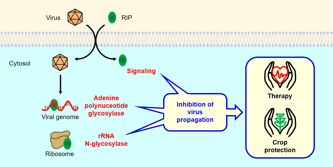
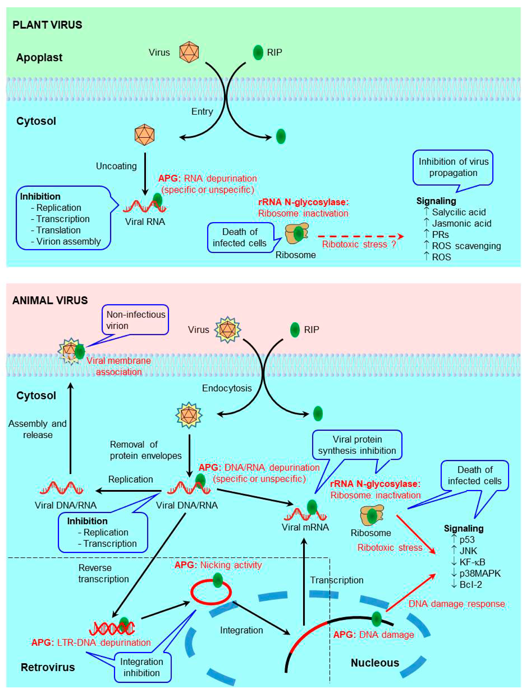
Table 1.
Proposed activities and other biological properties of ribosome-inactivating proteins (RIPs).
Table 1.
Proposed activities and other biological properties of ribosome-inactivating proteins (RIPs).
| Activity | Example of RIP | References |
|---|---|---|
| Agglutinin | Ricin | [8] |
| Antiviral | PAP | [10] |
| rRNA N-glycosylase | Ricin | [11] |
| Adenine polynucleotide glycosylase | Saporin-L1 | [15] |
| rRNA N-glycosylase/lyase | Gypsophilin/RALyase | [16] |
| RNase | BBAP1 | [17] |
| DNase | BBAP1 | [17] |
| Phosphatase | Trichosanthin | [18] |
| Superoxide dismutase | Camphorin | [19] |
| Phospholipase | Ricin | [20] |
| Chitinase | TKC 28-I | [21] |
| DNA nicking | BE27 | [22] |
| Apoptosis induction | Stenodactylin | [4,23] |
| Necroptosis induction | Stenodactylin | [4,23] |
| Autophagia induction | Abrus Agglutinin | [24] |
| Senescence induction | JIP60 | [25] |
| Plant tissue necrosis | JIP60 | [26] |
Table 2.
RIPs active against animal viruses. RIPs with antiviral activity, the families and species from which they have been obtained and the viruses in which this activity has been demonstrated are shown.
Table 2.
RIPs active against animal viruses. RIPs with antiviral activity, the families and species from which they have been obtained and the viruses in which this activity has been demonstrated are shown.
| Species and RIP | Virus | References |
|---|---|---|
| POACEAE | ||
| Zea mays L. | ||
| Maize RIP | HIV, SHIV | [27,28] |
| EUPHORBIACEAE | ||
| Ricinus communis L. | ||
| Ricin A chain | HIV | [29] |
| Suregada multiflora (A.Juss.) Baill. (=Gelonium multiflorum A.Juss.) | ||
| Gelonin | HIV, HPV, HSV, PICV, | [2,30,31,32] |
| GAP31 | HIV | [33,34] |
| CUCURBITACEAE | ||
| Trichosanthes kirilowii Maxim | ||
| Trichosanthin (TCS) | HBV, HIV, HSV | [32,35,36,37,38] |
| TAP29 | HIV | [36] |
| Trichobitacin | HIV | [36,39] |
| Momordica charantia L. | ||
| Momordin (M. charantia inhibitor) | HPV, HSV | [30] |
| Alpha-momorcharin (α-MMC) | HBV, HIV, HSV | [2,32,40,41] |
| Beta-momorcharin | HIV | [2,32] |
| Momordica antiviral protein (MAP30) | DENV-2, HHV8, HBV, HIV, HSV | [35,42,43,44,45,46] |
| Momordica balsamina L. | ||
| Balsamin | HIV | [47] |
| Luffa cylindrica (L.) M.Roem. | ||
| Luffin | HIV | [32] |
| Bryonia cretica subsp. dioica (Jacq.) Tutin (=Bryonia dioica Jacq.) | ||
| Bryodin | HIV | [48] |
| CARYOPHYLLACEAE | ||
| Saponaria officinalis L. | ||
| Saporin | HIV | [32,49,50] |
| Dianthus caryophyllus L. | ||
| Dianthin 32 (DAP32) | HIV, HPV, HSV | [30,34] |
| Dianthin 30 (DAP30) | HIV | [34] |
| Agrostemma githago L. | ||
| Agrostin | HIV | [2,32] |
| PHYTOLACCACEAE | ||
| Phytolacca americana L. | ||
| PAP (PAPI) | CHIKV, FLUV, HBV, HIV, HPV, | [10,35,51,52,53,54,55,56,57] |
| HSV, HTLV, JEV, LCMV | ||
| PAPII | HIV | [57] |
| PAPIII | HIV | [57] |
| PAP-S | HSV, HPV, HBV | [30,56] |
Virus name abbreviations: CHIKV (chikungunya virus), DENV (dengue virus), FLUV (human influenza virus), HBV (hepatitis B virus), HHV (human gammaherpesvirus), HIV (human immunodeficiency virus), HPV (human poliovirus), HSV (herpes simplex virus), HTLV (human T-cell leukemia virus), JEV (Japanese encephalitis virus), LCMV (lymphocytic choriomeningitis virus), PICV (Pichinde virus), SHIV (simian–human immunodeficiency virus).
Table 3.
RIPs active against plant viruses. RIPs with antiviral activity, the families and species from which they have been obtained and the viruses in which this activity has been demonstrated are shown.
Table 3.
RIPs active against plant viruses. RIPs with antiviral activity, the families and species from which they have been obtained and the viruses in which this activity has been demonstrated are shown.
| Species and RIP | Virus | References |
|---|---|---|
| IRIDACEAE | ||
| Iris x hollandica Tub. | ||
| IRIP | TMV, TEV | [77] |
| IRAb | TMV, TEV | [77] |
| EUPHORBIACEAE | ||
| Jatropha curcas L. | ||
| Curcin 2 | TMV | [78] |
| CUCURBITACEAE | ||
| Trichosanthes kirilowii Maxim | ||
| Trichosanthin | TuMV, CMV, TMV | [79,80] |
| Momordica charantia L. | ||
| α-Momorcharin | CMV, ChiVMV, TMV, TuMV | [81,82] |
| LEGUMINOSAE | ||
| Senna occidentalis (L.) Link (=Cassia occidentalis L.) | ||
| Cassin | TMV | [83] |
| CARYOPHYLLACEAE | ||
| Saponaria officinalis L. | ||
| Saporin | BMV, TMV, AMV | [51] |
| Dianthus caryophyllus L. | ||
| Dianthin 30 | ACMV, TMV | [84,85] |
| Dianthin 32 | TMV | [85] |
| AMARANTHACEAE | ||
| Beta vulgaris L. | ||
| BE27 | TMV, AMCV | [86,87] |
| Amaranthus tricolor L. | ||
| AAP-27 | SHMV | [88] |
| Amaranthus viridis L. | ||
| Amaranthin | TMV | [89] |
| Celosia argentea L. (=Celosia cristata L., = Celosia plumosa (Voss) Burv.) | ||
| CCP 25 | BMV, PMV, TMV, SHMV, ICRSV | [90,91,92] |
| CCP 27 | TMV, SHMV, ICRSV | [92,93] |
| Chenopodium album L. | ||
| CAP-I | TMV, SHMV | [94] |
| CAP-II | TMV, SHMV | [94] |
| CAP30 | TMV | [95] |
| Salsola longifolia Forssk. | ||
| SLP-32 | BYMV, TNV | [96] |
| Spinacia oleracea L. | ||
| VI (SoRIP2) | TMV | [97,98] |
| PHYTOLACCACEAE | ||
| Phytolacca insularis Nakai | ||
| PIP | TMV, CMV, PVY, PVX, PLRV | [99] |
| Phytolacca dioica L. | ||
| Dioicin 2 | TMV | [87] |
| PD-S2 | TMV | [87] |
| PD-L1 | TNV | [100] |
| PD-L4 | TMV, TNV | [87,100] |
| Phytolacca americana L. | ||
| PAP (PAPI) | BMV, TMV, AMV, TBSV, SPMV, ZYMV | [51,58,101,102,103,104,105] |
| CMV, PVY, PVX, TEV, SBMV | ||
| PAPII | TMV, PVX | [104] |
| PAP-S | AMCV | [105] |
| NYCTAGINACEAE | ||
| Boerhaavia diffusa L. | ||
| BDP-30 | TMV | [106] |
| Mirabilis expansa (Ruiz & Pav.) Standl. | ||
| ME1 | TMV, BMV | [51] |
| Mirabilis jalapa L. | ||
| MAP | TMV | [107] |
| Bougainvillea spectabilis Willd. | ||
| Bouganin | ZYMV, AMCV | [105,108] |
| Bougainvillea buttiana Holttum & Standl. | ||
| BBAP1 | SHMV | [17] |
| BBP-24 | TMV, SHMV | [109,110] |
| BBP-28 | TMV, SHMV | [109,110] |
| BASELLACEAE | ||
| Basella alba L. (=Basella rubra L.) | ||
| RIP2 | AMCV | [105] |
| LAMIACEAE | ||
| Volkameria inermis L. (=Clerodendrum inerme (L.) Gaertn.) | ||
| CIP-29 | TMV, PRSV, SHMV | [111,112] |
| Volkameria aculeata L. (=Clerodendrum aculeatum (L.) Schltdl.) | ||
| CA-SRI (CAP-34) | TMV, SHMV, PRSV | [113,114,115] |
| ADOXACEAE | ||
| Sambucus nigra L. | ||
| SNAI’ | TMV | [116] |
| Nigrin b (SNAV) | TMV | [76] |
Virus name abbreviations: ACMV (African cassava mosaic virus), AMCV (artichoke mottled crinkle virus), AMV (alfalfa mosaic virus), BMV (brome mosaic virus), BYMV (bean yellow mosaic virus), ChiVMV (Chilli veinal mottle virus), CMV (cucumber mosaic virus), ICRSV (Indian citrus ringspot virus = citrus ringspot virus, CRSV), PLRV (potato leafroll virus), PMV (pokeweed mosaic virus), PRSV (papaya ringspot virus), PVX (potato virus X), PVY (potato virus Y), SBMV (southern bean mosaic virus), SHMV (sunn-hemp mosaic virus = sunn-hemp rosette virus, SRV), SPMV (satellite panicum mosaic virus), TBSV (tomato bushy stunt virus), TEV (tobacco etch virus), TMV (tobacco mosaic virus), TNV (tobacco necrosis virus), TuMV (turnip mosaic virus), ZYMV (zucchini yellow mosaic virus).
Table 4.
Ribosome-inactivating proteins used in experimental antiviral therapy. RIPs have been used alone, PEGylated, or as part of immunotoxins, conjugates, engineered proteins, or nanocapsules.
Table 4.
Ribosome-inactivating proteins used in experimental antiviral therapy. RIPs have been used alone, PEGylated, or as part of immunotoxins, conjugates, engineered proteins, or nanocapsules.
| Virus | Target | RIP | References |
|---|---|---|---|
| RIPs alone | |||
| HIV | HIV infected cells | TCS | [61,157,158] |
| SHIV | SHIV infected cells | Maize RIP | [27] |
| PEGylated RIPs | |||
| HSV-1 | HIV infected cells | α-MMC | [40] |
| HIV infected cells | MAP30 | [40] | |
| Immunotoxins | |||
| HIV | gp 120 | RAC, PAP-S, PAC, Gelonin | [168,169,170,171,172] |
| gp 41 | RAC, PAC, Gelonin | [170,171,172,173,174,175] | |
| gp 160 | RAC | [173] | |
| CD45RO | RAC | [176] | |
| CD4 | PAP | [143,177] | |
| CD25 | RAC | [178] | |
| PICV | PICV | Gelonin | [31] |
| EBV | CD30 | Saporin 6 | [163] |
| EBV/C3d receptor | Gelonin | [164] | |
| MCMV | MCMV | RAC | [165,179] |
| Conjugates | |||
| HIV | gp 120 | RAC | [166] |
| CD8+ T-cells | Saporin | [49] | |
| Engineered proteins | |||
| HIV | HIV protease | RAC | [29] |
| HIV protease | Maize RIP | [28] | |
| CHIKV | Viral life cycle | PAP | [53] |
| DENV | Viral life cycle | MAP30 | [42] |
| HBV | HBV infected cells | RAC-PAP | [167] |
| Nanocapsules | |||
| HIV | HIV infected cells | MAP30 | [180] |
| HIV protease | RAC | [181] |
Virus name abbreviations: CHIKV (chikungunya virus), DENV (dengue virus), EBV (Epstein–Barr virus), HBV (hepatitis B virus), HIV (human immunodeficiency virus), HSV (herpes simplex virus), MCMV (murine cytomegalovirus), PICV (Pichinde virus), SHIV (simian–human immunodeficiency virus). RIP name abbreviations: MAP (Momordica antiviral protein), α-MMC (alpha-momorcharin), PAC (Pulchellin A-chain), PAP (pokeweed antiviral protein), RAC (ricin A-chain), TCS (trichosanthin).
Table 5.
Transgenic plants bearing RIP genes. The degree of protection achieved is indicated as the percentage reduction of lesions, infected plants or detected virus levels, or as the delay in the onset of symptoms.
Table 5.
Transgenic plants bearing RIP genes. The degree of protection achieved is indicated as the percentage reduction of lesions, infected plants or detected virus levels, or as the delay in the onset of symptoms.
| RIP | Host | Virus | Protection | Ref. |
|---|---|---|---|---|
| IRIP | Nicotiana tabacum | TMV, TEV | 73% L.L. | [77] |
| IRAb | Nicotiana tabacum | TMV, TEV | 54% L.L. | [77] |
| Curcin 2 | Nicotiana tabacum | TMV | 9 D.D. | [78] |
| Trichosanthin | Nicotiana tabacum | TuMV | 100% L.L. | [79] |
| Nicotiana tabacum | TMV, CMV | 14 D.D. | [80] | |
| Lycopersicon esculentum | TMV, CMV | 100% L.I.P. | [187] | |
| Cassin | Nicotiana tabacum | TMV | 13 D.D. | [83] |
| Dianthin 30 | Nicotiana benthamiana | ACMV | 100% L.L. | [84] |
| PIP | Solanum tuberosum | PVY, PYX, PLRV | 98% L.V.L | [99] |
| PAP | Nicotiana tabacum | PVY, PYX, CMV | 100% L.I.P. | [102,188] |
| Nicotiana benthamiana | PVY | 67% L.I.P. | [102] | |
| Solanum tuberosum | PVY, PYX | 84% L.I.P. | [102] | |
| PAPII | Nicotiana tabacum | TMV, PVX | 89% L.L. | [104] |
| SNAI’ | Nicotiana tabacum | TMV | 59% L.L. | [116] |
| Nigrin b (SNAV) | Nicotiana tabacum | TMV | 43% L.L. | [76] |
Virus name abbreviations: ACMV (African cassava mosaic virus), CMV (cucumber mosaic virus), PLRV (potato leafroll virus), PVX (potato virus X), PVY (potato virus Y), TEV (tobacco etch virus), TMV (tobacco mosaic virus), TuMV (turnip mosaic virus). Protection abbreviations: L.L. (less lesions), D.D. (days of delay), L.I.P. (less infected plants), L.V.L. (less virus level).
Publisher’s Note: MDPI stays neutral with regard to jurisdictional claims in published maps and institutional affiliations. |
© 2021 by the authors. Licensee MDPI, Basel, Switzerland. This article is an open access article distributed under the terms and conditions of the Creative Commons Attribution (CC BY) license (http://creativecommons.org/licenses/by/4.0/).
Share and Cite
MDPI and ACS Style
Citores, L.; Iglesias, R.; Ferreras, J.M. Antiviral Activity of Ribosome-Inactivating Proteins. Toxins 2021, 13, 80. https://doi.org/10.3390/toxins13020080
AMA Style
Citores L, Iglesias R, Ferreras JM. Antiviral Activity of Ribosome-Inactivating Proteins. Toxins. 2021; 13(2):80. https://doi.org/10.3390/toxins13020080
Chicago/Turabian StyleCitores, Lucía, Rosario Iglesias, and José M. Ferreras. 2021. "Antiviral Activity of Ribosome-Inactivating Proteins" Toxins 13, no. 2: 80. https://doi.org/10.3390/toxins13020080
Note that from the first issue of 2016, this journal uses article numbers instead of page numbers. See further details here.