Article

β-Hemolysis May Not Be a Reliable Indicator of Leukotoxicity of Mannheimia haemolytica Isolates

by
Jegarubee Bavananthasivam
,
Sudarvili Shanthalingam
,
Abirami Kugadas
,
Bindu Raghavan
,
Sai Batra
and
Subramaniam Srikumaran
*
Department of Veterinary Microbiology and Pathology, College of Veterinary Medicine, Washington State University, Pullman, WA 99164-7040, USA
*
Author to whom correspondence should be addressed.
Toxins 2018, 10(5), 173; https://doi.org/10.3390/toxins10050173
Submission received: 21 March 2018 / Revised: 20 April 2018 / Accepted: 23 April 2018 / Published: 25 April 2018
(This article belongs to the Special Issue Leukotoxins)

Abstract

:
Mannheimia (Pasteurella) haemolytica causes bronchopneumonia in domestic and wild ruminants. Leukotoxin is the critical virulence factor of M. haemolytica. Since β-hemolysis is caused by a large number of leukotoxin-positive M. haemolytica isolates, all β-hemolytic M. haemolytica isolates are considered to be leukotoxic as well. However, conflicting reports exist in literature as to the leukotoxic and hemolytic properties of M. haemolytica. One group of researchers reported their leukotoxin-deletion mutants to be hemolytic while another reported their mutants to be non-hemolytic. The objective of this study was to determine whether β-hemolysis is a reliable indicator of leukotoxicity of M. haemolytica isolates. Ninety-five isolates of M. haemolytica were first confirmed for presence of leukotoxin gene (lktA) by a leukotoxin-specific PCR assay. Culture supernatant fluids from these isolates were then tested for presence of leukotoxin protein by an ELISA, and for leukotoxic activity by a cytotoxicity assay. All isolates were tested for β-hemolysis by culture on blood agar plates. Sixty-two isolates (65%) produced leukotoxin protein while 33 isolates (35%) did not. Surprisingly, 18 of the 33 isolates (55%), that did not produce leukotoxin protein, were hemolytic. Of the 62 isolates that produced leukotoxin, 55 (89%) were leukotoxic while 7 (11%) were not. All except one of the 55 leukotoxic isolates (98%) were also hemolytic. All seven isolates that were not leukotoxic were hemolytic. Taken together, these results suggest that β-hemolysis may not be a reliable indicator of leukotoxicity of M. haemolytica isolates. Furthermore, all M. haemolytica isolates that possess lktA gene may not secrete active leukotoxin.
Key Contribution: Our results suggest that Mannheimia haemolytica isolates that induce β-hemolysis cannot be routinely assumed to be leukotoxic as well. Presence of lktA gene in an M. haemolytica isolate may not necessarily mean that the isolate secretes the leukotoxin protein.

1. Introduction

Mannheimia (Pasteurella) haemolytica is a Gram-negative coccobacillus that causes bronchopneumonia in domestic and wild ruminants [1,2,3]. The virulence factors of M. haemolytica include the capsule, outer membrane proteins, lipopolysaccharide and leukotoxin (Lkt) [4]. M. haemolytica Lkt belongs to the family of Gram-negative bacterial exotoxins, referred to as the RTX (repeats in toxin) toxins. It shares extensive homology with the exotoxins produced by Escherichia coli [5], Actinobacillus pleuropneumoniae [6], and Actinobacillus actinomycetemcomitans [7]. However, leukotoxic activity of M. haemolytica Lkt is specific for ruminant leukocytes [8,9]. Although all subsets of ruminant leukocytes are susceptible to Lkt-induced cytolysis, polymorphonuclear cells (PMNs) are the most susceptible subset [10]. Based on the finding that Lkt-deletion mutants cause no mortality [11] or reduced mortality and much milder lung lesions [12,13], Lkt has been widely accepted as the critical virulence factor of M. haemolytica. Lkt-induced lysis and degranulation of PMNs is primarily responsible for the acute inflammation and lung injury characteristic of this disease [14,15,16]. Since a large number of leukotoxic M. haemolytica isolates are β-hemolytic, β-hemolytic M. haemolytica isolates have been broadly assumed to be leukotoxic as well. However, there are conflicting reports in the literature as to the leukotoxic and hemolytic properties of M. haemolytica. One group of researchers reported their leukotoxin-deletion mutants to be hemolytic [17] while others reported their mutants to be non-hemolytic [18,19]. The objective of this study, therefore, was to determine whether β-hemolysis is a reliable indicator of leukotoxicity of M. haemolytica isolates.

2. Results and Discussion

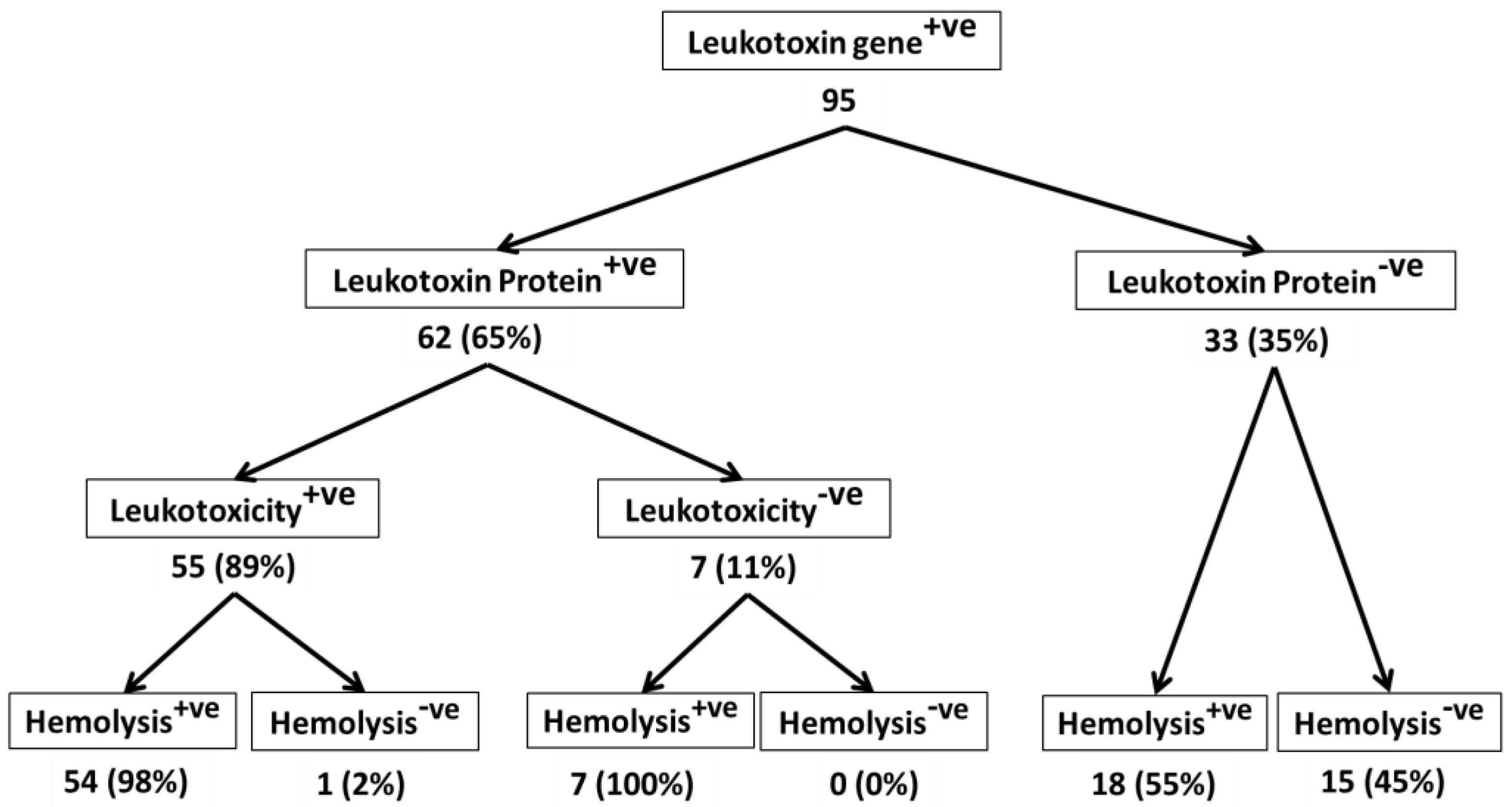
2.1. All M. haemolytica Isolates that Possess the lktA Gene May Not Produce Lkt Protein

The ELISA used to detect Lkt protein revealed that Lkt protein was not present in the culture supernatant fluids from 33 (35%) of the 95 isolates (Figure 1). Lkt is encoded by the lktCABD operon where lktA encodes the inactive protoxin, lktC encodes a trans-acylase that adds fatty acid chains to internal lysine residues in the protoxin, and lktB and lktD encoded components of a type 1 secretion system apparatus, along with the outer membrane protein TolC, secrete the toxin from the cell [5,20,21]. LktC-mediated acylation of LktA protoxin is not required for its expression or secretion [22]. It is plausible that in the isolates that were negative for Lkt protein, the secretion system apparatus was defective or totally absent. It is also possible that although the lktA gene was present, it was not functional. Further experiments are necessary to determine whether the Lkt protein was not synthesized, or the synthesized protein was not secreted, in these isolates whose culture supernatant fluids were negative for the Lkt protein by ELISA. These experiments are beyond the scope of this study.

2.2. Lkt-Negative M. haemolytica Isolates Can Induce Hemolysis

More importantly, 18 (55%) of the 33 isolates that did not have Lkt protein in the culture supernatant fluid were hemolytic (Figure 1), suggesting that not all hemolytic isolates cause leukotoxicity to target cells. Thus, hemolysis may not be a reliable indicator of leukotoxicity of M. haemolytica isolates. In this context, it is interesting to note that Miller et al. [27] showed that 16 isolates of the closely related bacterium Bibersteinia trehalosi that were reported to be hemolytic, were lktA gene negative.

2.3. Not All Lkt-Positive M. haemolytica Isolates May Cause Leukotoxicity

Of the 62 isolates that were positive for the presence of Lkt protein in the culture supernatant fluid, only 55 (89%) induced leukotoxicity to target cells, while 7 (11%) did not (Figure 1). As mentioned earlier, Lkt structural protein is encoded by lktA gene as an inactive protoxin, which is converted to biologically active Lkt by the lktC-encoded trans-acylase that adds fatty acid chains to internal lysine residues in the protoxin. It is likely that in some isolates, lktC gene is defective or totally absent. Reports of development of mutants that produce inactive Lkt by inactivation of lktC gene [28] supports this notion. However, further experiments are necessary to confirm this notion.

2.4. Majority of Leukotoxic M. haemolytica Isolates Are Hemolytic

Of the 55 isolates that were leukotoxic, 54 (98%) were hemolytic while one (2%) was not. These results suggest that leukotoxic isolates are for the most part hemolytic, but hemolytic isolates may not necessarily be leukotoxic as well. This notion is also supported by the fact that all 7 non-leukotoxic isolates were hemolytic (Figure 1).
Of the 95 isolates that were analyzed in this study, 65 were from bighorn sheep, and 15 each were from domestic sheep and cattle. Differences among the isolates from bighorn sheep, domestic sheep, and cattle are apparent when we consider the % of lktA gene-positive isolates that secreted Lkt protein, the % of Lkt protein-positive isolates that induced leukotoxicity, and the % of leukotoxic isolates that are hemolytic (Figure 2, Figure 3 and Figure 4). While only 49% of lktA gene-positive isolates from bighorn sheep secreted Lkt protein, 100% of the isolates from domestic sheep and cattle secreted Lkt protein. The difference among the three species in the % of isolates that were leukotoxic is small (84%, 87%, and 100% for bighorn sheep, domestic sheep and cattle, respectively). The % of leukotoxic isolates that were hemolytic was very similar in the three species (96%, 100%, and 100% for bighorn sheep, domestic sheep and cattle, respectively). Nevertheless, it should be noted that the number of isolates from domestic sheep and cattle (15 each) analyzed in this study is much smaller than the number of isolates from bighorn sheep (65). When larger number of isolates from domestic sheep and cattle are analyzed, it is very likely that similar results will be observed for the isolates from the different species. Nevertheless, the findings of this study suggest that there may be no correlation between hemolysis and leukotoxicity of M. haemolytica isolates.
Leukotoxin cytotoxicity assay is relatively more laborious than hemolysis assay. Probably because of this reason, most laboratories routinely perform only hemolysis assay on M. haemolytica isolates. Isolates that are β-hemolytic are considered to be leukotoxic as well. More recently, lktA PCR assay has been included as a means of identifying leukotoxic isolates of M. haemolytica. lktA gene-positive isolates are considered to be leukotoxic. The findings of this study suggest that hemolysis may not be a reliable indicator of leukotoxicity of M. haemolytica isolates. This study also suggests that lktA PCR assay may not be substituted for cytotoxicity assays for determination of leukotoxicity of M. haemolytica isolates.
The findings of this study raise the question as to what component of M. haemolytica causes hemolysis. Unfortunately, no information is available on M. haemolytica hemolysin protein or the gene that encodes it. We are hopeful that our findings will prompt studies aimed at the elucidation of the hemolytic activity of M. haemolytica.

3. Conclusions

1. Not all isolates of M. haemolytica that possess the lktA gene express the Lkt protein; 2. Not all secreted Lkt exhibit leukotoxic activity; 3. Majority of leukotoxic isolates of M. haemolytica are hemolytic, but not all hemolytic isolates of M. haemolytica are leukotoxic; 4. β-hemolysis may not be a reliable indicator of leukotoxicity of M. haemolytica isolates; 5. It is prudent to determine the leukotoxicity of the culture supernatant fluid by cytotoxicity assay with target cells before concluding an isolate of M. haemolytica to be leukotoxic.

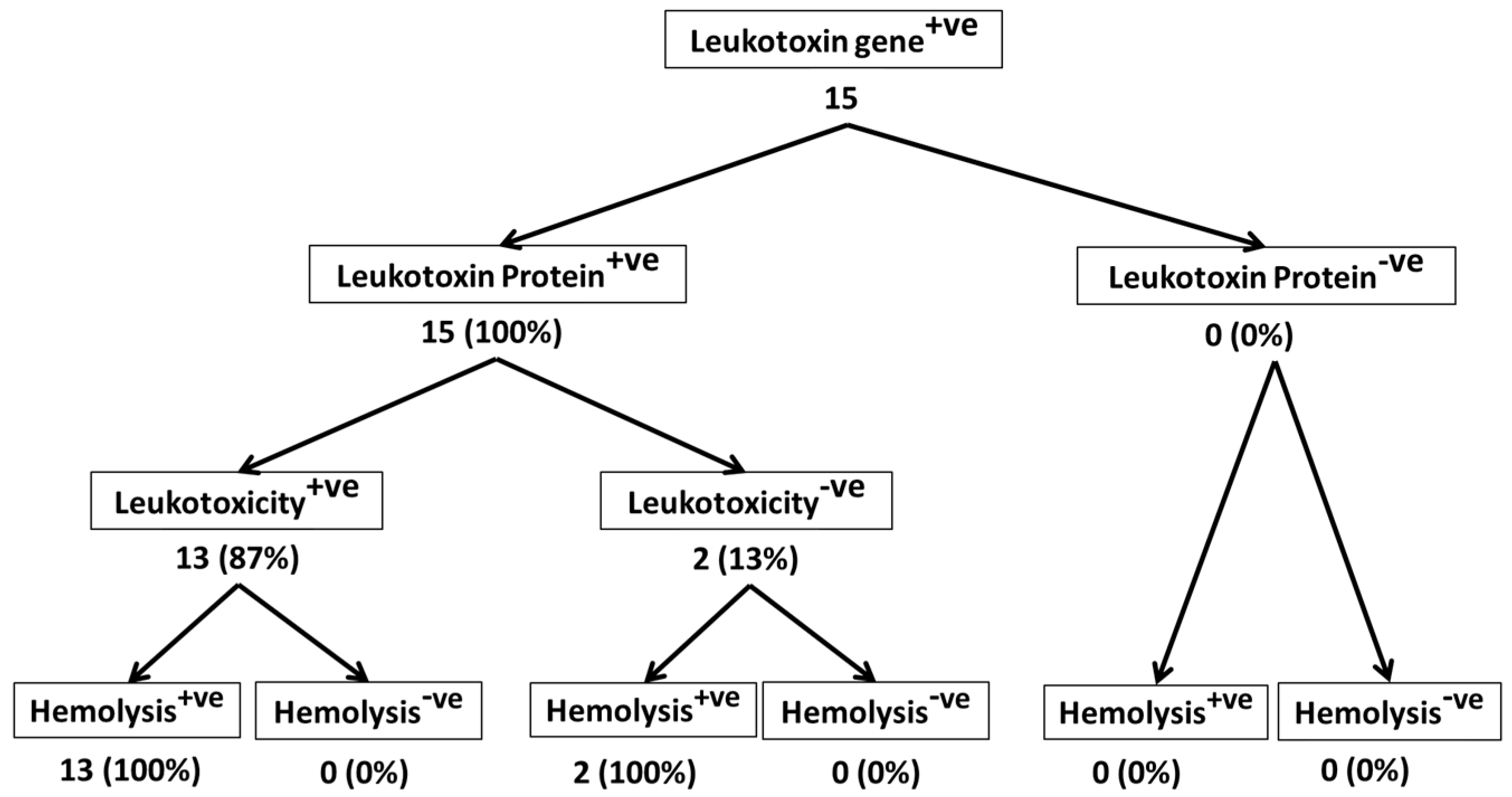
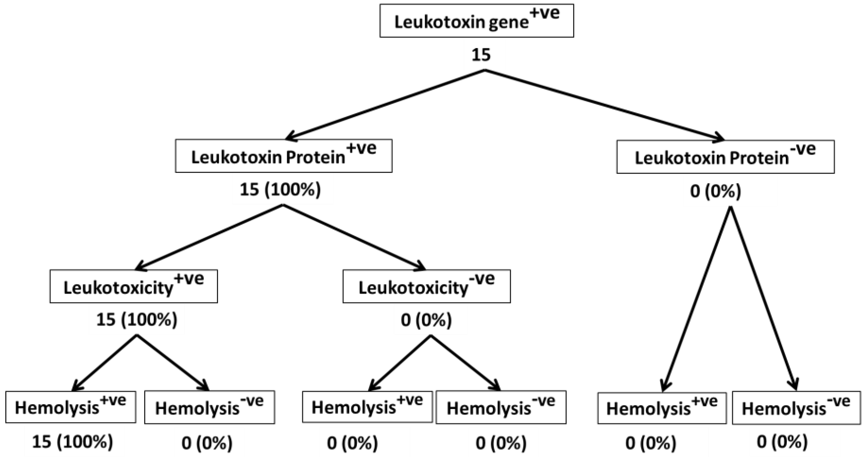
4. Materials and Methods

4.1. Experimental Design

Ninety-five field isolates of M. haemolytica [65 from bighorn sheep (Ovis canadensis) and 15 each from domestic sheep (Ovis aries) and cattle (Bos taurus)] were used in this study. All were revived from freezer stock on brain heart infusion (BHI) agar plates. Single colonies were sub-cultured. All isolates were first confirmed to be bona fide M. haemolytica isolates by a species-specific PCR assay [23]. Subsequently, they were tested for the presence of Lkt gene (lktA) by an Lkt-specific PCR assay. Culture supernatant fluids from these isolates were then tested for the presence of Lkt protein by an ELISA, and for leukotoxic activity by a cytotoxicity assay. All isolates were tested for β-hemolysis by culture on blood agar plates.

4.2. M. haemolytica and Lkt-Specific PCR Assays

The M. haemoltyica-specific PCR assay, and the Lkt-specific PCR assay were performed as previously described [23]. Briefly, the forward and reverse primers for the M. haemoltyica-specific PCR assay were TGG GCA ATA CGA ACT ACT CGG G, and CTT TAA TCG TAT TCG CAG, respectively. For the Lkt-specific PCR assay, the forward and reverse primers were CTT ACA TTT TAG CCC AAC GTG and TAA ATT CGC AAG ATA ACG GG, respectively. PCR assay consisted of an initial denaturation step at 95 °C for 5 min and a final extension at 72 °C for 10 min for both PCR assays. Cycling conditions used were 40 cycles of 95 °C for 30 s, 54 °C for 30 s, and 72 °C for 30 s for M. haemolytica PCR, and 95 °C for 30 s, 52 °C for 30 s, and 72 °C for 40 s for Lkt PCR.

4.3. Leukotoxin Production

Leukotoxin from M. haemolytica was produced as described previously [25]. Briefly, bacteria were grown to logarithmic phase in BHI broth (Remel, Lenexa, KS, USA) at 37 °C, harvested by centrifugation (13,500× g for 20 min at 4 °C), and resuspended in twice the original culture volume of colorless RPMI 1640 medium (Invitrogen, Carlsbad, CA, USA). After an additional 1–1.5 h of growth at 37 °C, the bacteria were pelleted by centrifugation, and the supernatant fluid was filter-sterilized using a 0.22 μm filter. This supernatant preparation containing Lkt was stored at −20 °C until needed.

4.4. ELISA for Detection of Lkt Protein

Presence of Lkt protein in bacterial culture supernatant fluids was determined by an indirect ELISA described previously [24], with minor modifications. Lkt in supernatant fluid immobilized on the ELISA plate wells was detected by the addition of a Lkt-specific mouse monoclonal antibody, MM601 [25]. Binding of the mouse monoclonal antibody was detected by the addition of HRP-conjugated sheep anti-mouse Ig antibodies followed by the substrate ABTS. The supernatant fluids that were negative by this ELISA with MM601 were re-confirmed to be negative by an ELISA using a bighorn sheep polyclonal serum known to detect Lkt.

4.5. Assay for Detection of Leukotoxic Activity

Leukotoxic activity in culture supernatant fluids was determined by the MTT [3-(4,5-dimethylthiazoyl-2-yl)-2,5-diphenyl tetrazolium bromide] dye-reduction cytotoxicity assay described previously [25]. Briefly, the supernatant fluid was incubated with PMNs from bighorn sheep, domestic sheep, or cattle, and subsequently the viability of the PMNs was determined by the addition of MTT dye which gets converted to a purple formazan precipitate by ER-resident enzymes in viable cells. The O.D of the acid isopropanol-dissolved formazan precipitate is directly proportional to the viability of the cells. The % cytotoxicity was calculated by the following formula: % cytotoxicity = [1 − (OD of toxin-treated cells/OD of toxin-untreated cells)] × 100.

4.6. Assay for Detection of β-Hemolysis

The hemolytic ability of M. haemolytica isolates was detected by the standard hemolysis assay [26]. M. haemolytica isolates were plated on Columbia blood agar plates containing 5% sheep blood (Carolina Biological Supply Company, Burlington, NC, USA) and incubated overnight. The following day, the plates were observed for β-hemolysis with transmitted light. Non-hemolyic isolates were incubated further for another 24 h and observed again for the hemolytic zone. Hemolysis of M. haemolytica isolates was determined by comparison with the hemolysis of Staphylococcus aureus and Streptococcus pneumoniae.

Author Contributions

S.Sr. conceived and designed the experiments. J.B. and S.Sh. designed and performed the experiments. A.K., B.R. and S.B. assisted in the performance of some experiments. J.B. and S.Sh. wrote the paper, guided by S.Sr.

Acknowledgments

This research was supported by funds from the Wild Sheep Foundation and its State Chapters, Rocky Mountain Bighorn Society, Nevada Bighorns Unlimited, and US Forest service.

Conflicts of Interest

The authors declare no conflict of interest.

References

  1. Ackermann, M.R.; Brogden, K.A. Response of the ruminant respiratory tract to Mannheimia (Pasteurella) haemolytica. Microb. Infect. 2000, 2, 1079–1088. [Google Scholar] [CrossRef]
  2. Miller, M.W. Pasteurellosis. In Infectious Diseases of Wild Mammals; Iowa State University Press: Iowa City, IA, USA, 2008; pp. 330–339. [Google Scholar]
  3. Mosier, D.A. Bacterial pneumonia. Vet. Clin. N. Am. Food Anim. Pract. 1997, 13, 483–493. [Google Scholar] [CrossRef]
  4. Confer, A.W.; Panciera, R.J.; Clinkenbeard, K.D.; Mosier, D.A. Molecular aspects of virulence of Pasteurella haemolytica. Can. J. Vet. Res. 1990, 54, S48–S52. [Google Scholar] [PubMed]
  5. Strathdee, C.A.; Lo, R.Y. Cloning, nucleotide sequence, and characterization of genes encoding the secretion function of the Pasteurella haemolytica leukotoxin determinant. J. Bacteriol. 1989, 171, 916–928. [Google Scholar] [CrossRef] [PubMed]
  6. Devenish, J.; Rosendal, S.; Johnson, R.; Hubler, S. Immunoserological comparison of 104-kilodalton proteins associated with hemolysis and cytolysis in Actinobacillus pleuropneumoniae, Actinobacillus suis, Pasteurella haemolytica, and Escherichia coli. Infect. Immun. 1989, 57, 3210–3213. [Google Scholar] [PubMed]
  7. Kolodrubetz, D.; Dailey, T.; Ebersole, J.; Kraig, E. Cloning and expression of the leukotoxin gene from Actinobacillus actinomycetemcomitans. Infect. Immun. 1989, 57, 1465–1469. [Google Scholar] [PubMed]
  8. Kaehler, K.L.; Markham, R.J.; Muscoplat, C.C.; Johnson, D.W. Evidence of species specificity in the cytocidal effects of Pasteurella haemolytica. Infect. Immun. 1980, 30, 615–616. [Google Scholar] [PubMed]
  9. Chang, Y.F.; Renshaw, H.W.; Martens, R.J.; Livingston, C.W., Jr. Pasteurella haemolytica leukotoxin: Chemiluminescent responses of peripheral blood leukocytes from several different mammalian species to leukotoxin- and opsonin-treated living and killed Pasteurella haemolytica and Staphylococcus aureus. Am. J. Vet. Res. 1986, 47, 67–74. [Google Scholar] [PubMed]
  10. Shewen, P.E.; Wilkie, B.N. Cytotoxin of Pasteurella haemolytica acting on bovine leukocytes. Infect. Immun. 1982, 35, 91–94. [Google Scholar] [PubMed]
  11. Dassanayake, R.P.; Shanthalingam, S.; Herndon, C.N.; Lawrence, P.K.; Frances Cassirer, F.E.; Potter, K.A.; Foreyt, W.J.; Clinkenbeard, K.D.; Srikumaran, S. Mannheimia haemolytica serotype A1 exhibits differential pathogenicity in two related species, Ovis canadensis and Ovis aries. Vet. Microbiol. 2009, 133, 366–371. [Google Scholar] [CrossRef] [PubMed]
  12. Petras, S.F.; Chidambaram, M.; Illyes, E.F.; Froshauer, S.; Weinstock, G.M.; Reese, C.P. Antigenic and Virulence properties of Pasteurella haemolytica leukotoxin mutants. Infect. Immun. 1995, 63, 1033–1039. [Google Scholar] [PubMed]
  13. Highlander, S.K.; Fedorova, N.D.; Dusek, D.M.; Panciera, R.; Alvarez, L.E.; Rinehart, C. Inactivation of Pasteurella (Mannheimia) haemolytica leukotoxin causes partial attenuation of virulence in a calf challenge model. Infect. Immun. 2000, 68, 3916–3922. [Google Scholar] [CrossRef] [PubMed]
  14. Slocombe, R.J.; Marark, J.; Ingersoll, R.; Derksen, F.J.; Robinson, N.E. Importance of neutrophils in the pathogenesis of acute pneumonic pasteurellosis in calves. Am. J. Vet. Res. 1985, 46, 2253–2258. [Google Scholar] [PubMed]
  15. Highlander, S.K. Molecular genetic analysis of virulence in Mannheimia (Pasteurella) haemolytica. Front. Biosci. 2001, 1, D1128–D1150. [Google Scholar]
  16. Jayaseelan, S.; Sreevatsan, S.; Maheswaran, S.K. Role of Mannheimia haemolytica leukotoxin in the pathogenesis of bovine pneumonic pasteurellosis. Anim. Health Res. Rev. 2002, 3, 69–82. [Google Scholar] [CrossRef]
  17. Chidambaram, M.; Sharma, B.; Petras, S.F.; Reese, C.P.; Froshauer, S.; Weinstock, G.M. Isolation of Pasteurella haemolytica leukotoxin mutants. Infect. Immun. 1995, 63, 1027–1032. [Google Scholar] [PubMed]
  18. Tatum, F.M.; Briggs, R.E.; Sreevatsan, S.S.; Zehr, E.S.; Ling Hsuan, S.; Whiteley, L.O.; Ames, T.R.; Maheswaran, S.K. Construction of an isogenic leukotoxin deletion mutant of Pasteurella haemolytica serotype 1: Characterization and virulence. Microb. Pathog. 1998, 24, 37–46. [Google Scholar] [CrossRef] [PubMed]
  19. Murphy, G.L.; Whitworth, L.C.; Clinkenbeard, K.D.; Clinkenbeard, P.A. Hemolytic activity of the Pasteurella haemolytica leukotoxin. Infect. Immun. 1995, 63, 3209–3212. [Google Scholar] [PubMed]
  20. Highlander, S.K.; Chidambaram, M.; Engler, M.J.; Weinstock, G.M. DNA sequence of the Pasteurella haemolytica leukotoxin gene cluster. DNA 1989, 8, 15–28. [Google Scholar] [CrossRef] [PubMed]
  21. Highlander, S.K.; Engler, M.J.; Weinstock, G.M. Secretion and expression of the Pasteurella haemolytica Leukotoxin. J. Bacteriol. 1990, 172, 2343–2350. [Google Scholar] [CrossRef] [PubMed]
  22. Thumbikat, P.; Briggs, R.E.; Kannan, M.S.; Maheswaran, S.K. Biological effects of two genetically defined leukotoxin mutants of Mannheimia haemolytica. Microb. Pathog. 2003, 34, 217–226. [Google Scholar] [CrossRef]
  23. Shanthalingam, S.; Goldy, A.; Bavananthasivam, J.; Subramaniam, R.; Batra, S.A.; Kugadas, A.; Raghavan, B.; Dassanayake, R.P.; Jennings-Gaines, J.E.; Killion, H.J.; et al. PCR assay detects Mannheimia haemolytica in culture-negative pneumonic lung tissues of bighorn sheep (Ovis canadensis) from outbreaks in the western USA, 2009–2010. J. Wildl. Dis. 2014, 50, 1–10. [Google Scholar] [CrossRef] [PubMed]
  24. Subramaniam, R.; Shanthalingam, S.; Bavananthasivam, J.; Kugadas, A.; Potter, K.A.; Foreyt, W.J.; Hodgins, D.C.; Shewen, P.; Barrington, G.M.; Knowles, D.P.; et al. A multivalent Mannheimia/Bibersteinia vaccine protects bighorn sheep against Mannheimia haemolytica challenge. Clin. Vaccine Immunol. 2011, 18, 1689–1694. [Google Scholar] [CrossRef] [PubMed]
  25. Gentry, M.J.; Srikumaran, S. Neutralizing monoclonal antibodies to Pasteurella haemolytica leukotoxin affinity-purify the toxin from crude culture supernatants. Microb. Pathog. 1991, 10, 411–417. [Google Scholar] [CrossRef]
  26. Smibert, R.M.; Krieg, N.R. Phenotypic Characterization. In Methods for General and Molecular Bacteriology; Gerhardt, P., Murray, R.G.E., Wood, W.A., Krieg, N.R., Eds.; American Society for Microbiology: Washington, DC, USA, 1994; pp. 607–654. [Google Scholar]
  27. Miller, M.W.; Hause, B.M.; Killion, H.J.; Fox, K.A.; Edwards, W.H.; Wolfe, L.L. Phylogenetic and epidemiologic relationships among Pasteurellaceae from Colorado bighorn sheep herds. J. Wildl. Dis. 2013, 49, 653–660. [Google Scholar] [CrossRef] [PubMed]
  28. Fedorova, N.D.; Highlander, S.K. Generation of targeted nonpolar gene insertions and operon fusions in Pasteurella haemolytica and creation of a strain that produces and secretes inactive leukotoxin. Infect. Immun. 1997, 65, 2593–2598. [Google Scholar] [PubMed]
Figure 1. Leukotoxic and hemolytic activity of M. haemolytica isolates from bighorn sheep, domestic sheep and cattle. A total of 95 M. haemolytica isolates (65 from bighorn sheep and 15 each from domestic sheep and cattle) were analyzed. Presence of leukotoxin gene in M. haemolytica isolates was determined by an lktA-specific PCR assay [23]. Presence of leukotoxin protein in the culture supernatant fluids was determined by an indirect ELISA [24]. Leukotoxicity of the isolates was determined by the MTT dye-reduction cytotoxicity assay [25] of culture supernatant fluids on PMNs from the respective species. β-hemolytic activity of the isolates was determined by the standard hemolysis assay [26]. Each isolate was tested three times by each of the assays.
Toxins 10 00173 g001
Figure 2. Leukotoxic and hemolytic activity of bighorn sheep isolates of M. haemolytica. This figure shows the leukotoxic and hemolytic activities of 65 bighorn sheep isolates of M. haemolytica that were part of the 95 isolates shown in Figure 1.
Toxins 10 00173 g002
Figure 3. Leukotoxic and hemolytic activity of domestic sheep isolates of M. haemolytica. This figure shows the leukotoxic and hemolytic activities of 15 domestic sheep isolates of M. haemolytica that were part of the 95 isolates shown in Figure 1.
Toxins 10 00173 g003
Figure 4. Leukotoxic and hemolytic activity of cattle isolates of M. haemolytica. This figure shows the leukotoxic and hemolytic activities of 15 cattle isolates of M. haemolytica that were part of the 95 isolates shown in Figure 1.
Toxins 10 00173 g004

