Case Report

Post-Radiation Angiosarcoma (PRA) of the Small Bowel: Report of a Case and Review of the Literature

1
Department of General Surgery, “A. Costa” Hospital—Alto Reno Terme, 40046 Bologna, Italy
2
Department of Surgical Sciences, La Sapienza University, 00161 Rome, Italy
3
Department of Pathological Anatomy, Maggiore “Carlo Alberto Pizzardi” Hospital, 40133 Bologna, Italy
4
Department of Gastroenterology, Maggiore “Carlo Alberto Pizzardi” Hospital, 40133 Bologna, Italy
*
Author to whom correspondence should be addressed.
Surgeries 2023, 4(3), 381-390; https://doi.org/10.3390/surgeries4030038
Submission received: 30 May 2023 / Revised: 7 July 2023 / Accepted: 14 July 2023 / Published: 19 July 2023

Abstract

:
Angiosarcoma is a rare and aggressive neoplasia of endothelial cells which represents only 2% of all soft-tissue tumors and frequently occurs in the skin and subcutaneous tissues. It is classified in two groups: the first is represented by primary angiosarcoma, which includes cutaneous and breast angiosarcoma; the second is constituted by secondary angiosarcoma, which is related to radiation therapy, lymphedema, exposure to some chemical toxins, and familiar syndromes. Post-radiation intestinal angiosarcoma is a special type of secondary angiosarcoma, and only a few cases have been reported in the literature. We present a case of radiation-induced small bowel angiosarcoma in an 88-year-old female patient who was admitted to our department for abdominal pain and signs of intestinal obstruction. Her clinical history included previous radiotherapy treatments after a hysterectomy for uterine fibroids, excision of the vaginal stump for squamous cell carcinoma, and the surgical removal of a left-leg cutaneous angiosarcoma. She underwent emergency surgery, and features of peritoneal carcinomatosis were detected. A histological examination showed the presence of a small intestinal angiosarcoma. At the histochemical analysis, MYC amplification was detected, suggesting that her small bowel angiosarcoma was related to past radiation treatments.

1. Introduction

Angiosarcoma (AS) is an aggressive soft-tissue tumor that originates from endothelial cells and represents only 2% of all soft-tissue sarcomas [1]. It is classified into primary angiosarcoma, which includes cutaneous and breast AS, and secondary angiosarcoma, which is related mainly to radiation therapy or lymphedema.
Secondary AS is mainly associated with risk factors such as chronic lymphedema, neurofibromatosis, xeroderma pigmentosum, retinoblastoma, previous radio-chemotherapy treatment, benign vascular lesions, arteriovenous fistulas, and exposure to environmental agents such as thorium dioxide, ultraviolet light, arsenic, anabolic steroids, and foreign bodies [2]. In addition, familiar syndromes have been associated with angiosarcoma, such as neurofibromatosis type 1, bilateral retinoblastoma, Maffucci syndrome, hemochromatosis, and Klippel–Trenaunay syndrome [2].
Cutaneous AS constitutes about 60% of all angiosarcomas and often presents with palpable masses that affect the skin and subcutaneous tissue of the extremities; the most frequent sites are the head, neck, and legs.
Cutaneous AS metastases occur in about 30% of patients and are due to hematogenous spread. The most common site is the lung, followed by the liver, bones, and lymph nodes.
Post-radiation angiosarcoma (PRA) is an unusual intestinal neoplasm related to radiation treatment for tumors such as breast, uterine, and skin cancer. Currently, no definition of post-radiation sarcomas is available, but there are generally accepted criteria, proposed by Cahan and modified from Arlen, namely: “radiation treatment at least 3 years from the appearance of the tumor, onset within the field of the irradiation and different histology by the primitive tumor and the post radiation one” [3].
The clinical diagnosis is difficult because most common symptoms (abdominal pain, vomiting, weakness, gastrointestinal bleeding, and anemia) are nonspecific and can be easily confused with other diseases. A clinical history of previous radiotherapy treatment may help in medical assessment and clinical suspicion.
The bowel localization in PRA is very uncommon and has a poor prognosis, especially in older patients that frequently present with diffuse diseases not amenable to radical excision [4]. Patients with bowel localizations of AS often present with nonspecific abdominal pain, melena, or hematochezia. In some rare cases, the clinical presentation is more aggressive, with intestinal obstruction or an acute abdomen, so that an urgent operation is needed [5]. According to published data, the overall 5-year survival, both for primary and secondary angiosarcoma, is 30–35%, but it is higher in patients with age < 60 years, tumor size < 5 cm, and in the absence of distant metastasis and nodal involvement [5].
For local diseases, radical surgery with complete resection and negative margins is the best treatment. Metastatic disease can be managed with chemotherapy based on paclitaxel, doxorubicin, and thalidomide; the use of combined agents has not shown a better outcome and has increased toxicity. Based on the literature, paclitaxel seems to have better results, with responder rates of about 60% [2]. There is poor evidence for the role of node dissection in the treatment of intestinal angiosarcoma; the presence of pathological nodes is proof of a widespread disease and should suggest an oncological assessment [5].
Herein, we present a case of post-radiation small bowel angiosarcoma in an 88-year-old female patient who was admitted to our department with intestinal obstruction.

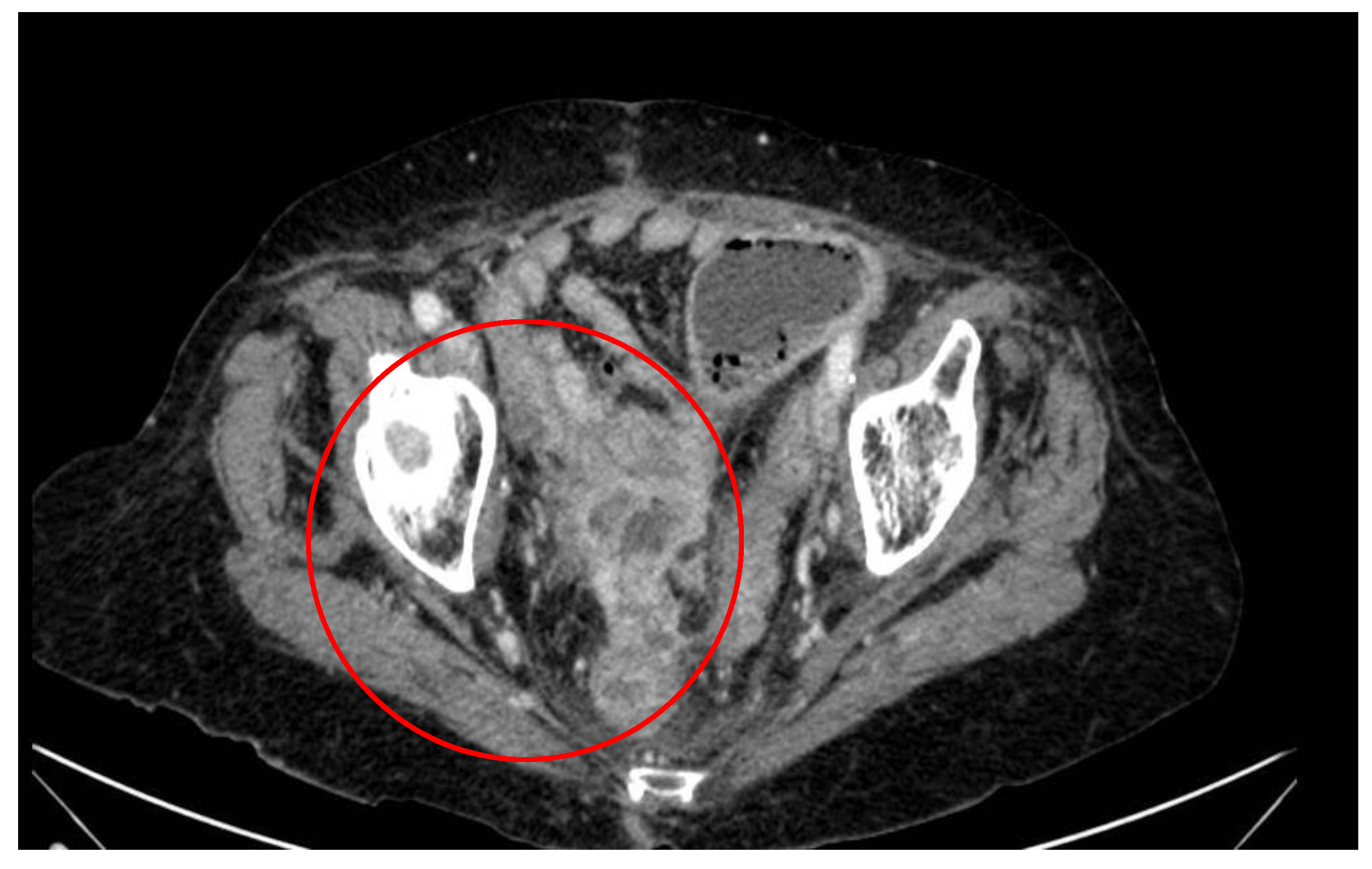
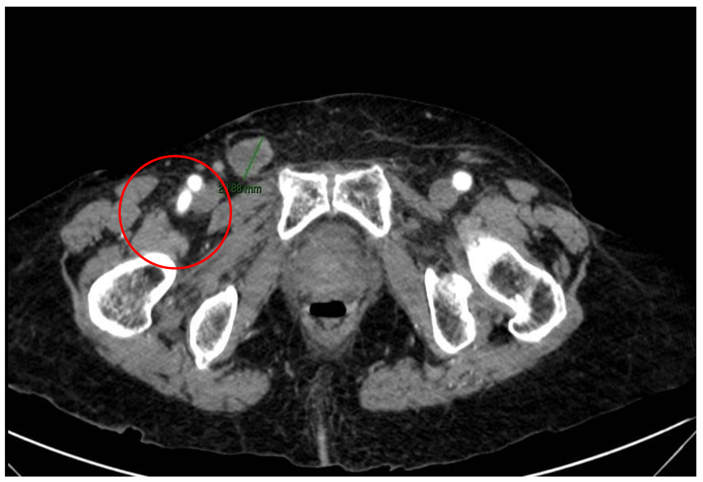
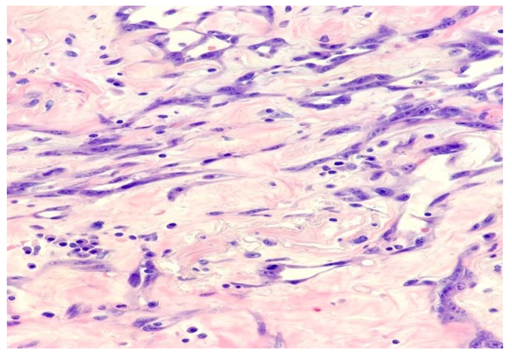
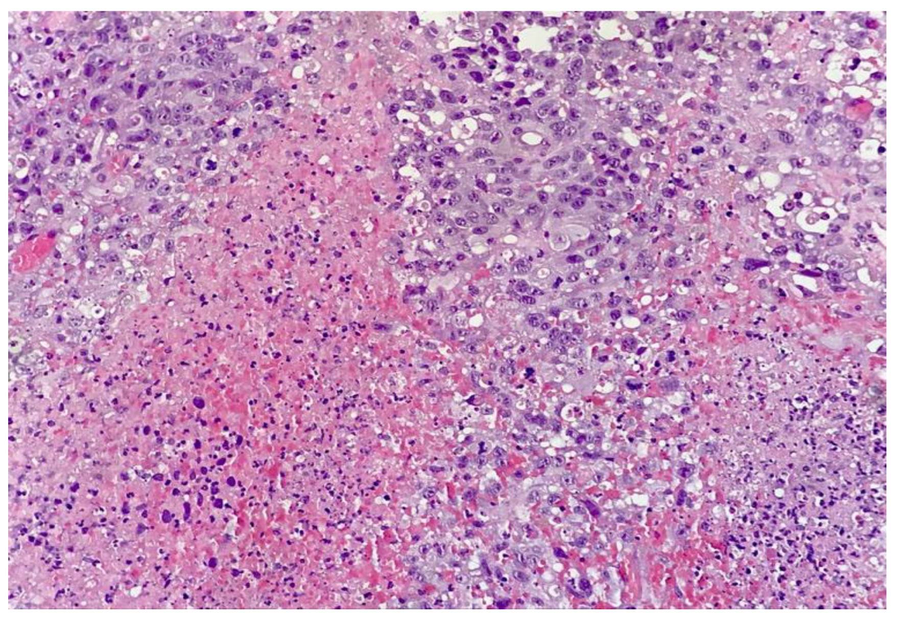
2. Case Report and Evolution

An 88-year-old female patient was admitted to our Emergency Department (ED) in ‘A. Costa Hospital’ Alto Reno Terme, Bologna, Italy, with abdominal pain and constipation. The patient was independent in her daily activities and had no allergies. Her primary medical comorbidities were hypertension and gastroesophageal reflux disease (GERD).
Her past surgical medical history was significant for many procedures, such as a previous partial hysterectomy, a squamous cell carcinoma of the vaginal stump managed with excision of the uterine cervix, pelvic lymph node dissection, removal of cutaneous angiosarcoma in the abdomen, and adjuvant radiotherapy of the lower abdomen; a further surgical operation was needed for the recurrence of a left-leg angiosarcoma.
At the hospital admission, an abdomen X-ray was performed with significant abdominal distension, so a nasogastric tube was inserted with drainage of abundant enteric fluid. A laboratory test reported white blood cells of 17 × 109/L, hemoglobin of 12.4 g/dL, creatinine of 2.1 mg/dL, and C-reactive protein of 8 mg/dL.
The CT scan showed a condition of intestinal obstruction caused by a conglomerate of intestinal loops with adhesion to the pelvic floor and initial signs of intestinal vascular distress with reduced contrast enhancement (Figure 1); a pathological node was also detected in the right inguinal side (Figure 2).
The patient underwent emergency surgery. A laparoscopic exploration of the abdomen was performed; an abnormal distension of the small bowel with a tract of restricted intestinal loops adherent to the pelvic floor and features of peritoneal carcinosis were found. After a difficult tumor dissection and recognition of intestinal loops afferent to the lesion, an intestinal resection was made with the subsequent reconstruction of the intestinal tract with an ileal–ileal, lateral–lateral anastomosis. Due to the pattern of peritoneal carcinosis and the risk of left colon involvement, a left colostomy was carried out. Abundant purulent fluid in the pelvis was collected for cytological and microbiological analysis. The postoperative stay was characterized by heart failure, so the patient was transferred to another medical department and treated with cardiological medical therapy. The patient was later discharged from the hospital on the tenth postoperative day in good health conditions, but unfortunately died within one month.
The histological examination showed angiosarcoma cells infiltrating both the small bowel and the pelvic wall with a solid growth pattern and foci of necrosis (Figure 3, Figure 4 and Figure 5). At high power, neoplastic vessels are lined by pleomorphic endothelial cells; neoplastic cells lining a vascular structure are represented in Figure 6 and Figure 7.
The abdominal fluid revealed the presence of neoplastic cells positive for angiosarcoma, corroborating the finding of peritoneal carcinosis.
In the immunohistochemical analysis, neoplastic cells expressed ERG, CD31, and vimentin but were negative for CKws, CK7, HHV8, S100, and MART. MYC amplification analysis was positive for diffuse expression of the C-MYC antibody, suggesting it was associated with radiation treatment.

3. Review of the Literature

We performed research on PubMed with the following key words: ”radiation,” ”bowel,” and “angiosarcoma”. All works inherent to PRA have been evaluated. No temporal filter was adopted.
The main characteristics of small bowel angiosarcoma (primary and PRA) are summarized in Table 1.
Though there are some cases of small bowel angiosarcoma, only a few are related to radiation therapy [Table 2 and Table 3]. The age of presentation of the tumor ranges from 47 to 80 years, with an average of 64, and the ratio of female to male is 6:1. The most frequent clinical presentation is abdominal pain, followed by vomiting and weight loss, while gastrointestinal bleeding (GI) is less common. Sometimes, the clinical presentation is more aggressive, with intestinal obstruction or perforation.
Most of the patients were irradiated for gynecological tumors such as endometrial adenocarcinoma and squamous cell carcinoma of the uterine cervix. In the reported cases, the interval between radiation exposure and tumor onset ranged from 3 to 24 years, with an average of 13 years. The majority of intestinal AS are localized in the terminal ileum, followed by the jejunum, making the diagnosis difficult because of the lack of endoscopic tools able to detect this part of the intestine. We also found that tumors of the breast and skin are common reasons for irradiation. Li et al. [6], in their retrospective analysis, reported 66 cases of small intestine angiosarcoma between 1970 and 2017 [2], of which only 17 cases were detected as post-radiation angiosarcomas. In their study, they found some important differences between the two groups: patients with primary small bowel AS and those with post-radiation AS. In particular, primary angiosarcoma was more frequent in male patients than in female ones, while in the second group there was a slight prevalence in women. GI bleeding was the most common symptom in patients with primary angiosarcoma, having a negative impact on survival with a median survival of 8.5 weeks compared to 50.4 weeks in patients without. The interval between radiation exposure and tumor onset ranged from 3 to 20 years, with an average of 10 years [6]. Intestinal AS is often misdiagnosed with other diseases such as GIST, lymphoma, and intestinal adenocarcinoma, both for the low incidence and the atypical clinical manifestations [4]. According to these features, the correct diagnosis is usually made after surgical treatment and prior to the histological exam.
Table 1. Main features of small bowel angiosarcoma.
SiteSymptomsGender
Prevalence
Markers for Angiosarcoma Diagnosis
Primary [7]Duodenum, jejunum, terminal ileumAbdominal pain, vomiting, anemia, gastrointestinal bleeding, weaknessMaleCD31, vWF, CD34, ERG, vimentin
Secondary
(Included PRA) [4]
Duodenum, jejunum, terminal ileumAbdominal pain, gastrointestinal bleeding intestinal obstruction and perforation FemaleCD31, ERG, vWF, CD34, vimentin, MYC antibody
ERG: erythroblast transformation-specific related gene; vWF: von Willebrand factor.
Table 2. Cases of small bowel PRA.
AuthorsSex/Age (Years)Radiated forTime after Radiation (Years)PresentationTreatment
Squillaci et al. [4]F/72Uterine leiomyosarcoma24Abdominal painResection
Hansen et al. [8]F/76Endometrial adenocarcinoma7Watery diarrhea, vomiting, weight lossResection of ileal tract
Chen et al. [9]F/66Endometrioid adenocarcinoma8Abdominal pain, vomitingIleo–colic resection
Aitola et al. [10]F/50Endometrial adenocarcinoma14Intestinal obstructionResection followed by combination chemiotherapy with doxorubicin
Policarpio-Nicolas et al. [11]F/51Adenocarcinoma of uterine cervix 9Abdominal painResection
Nanus et al. [12]F/47Dysgerminoma/ovary16Perforated distal ileumResection
Hwang et al. [13]F/60Carcinoma of uterin cervix8Abdominal painResection
Suzuki et al. [14]F/61Squamous cell carcinoma of uterus20Abdominal painResection and intrabdominal cisplatin
Berry et al. [15]M/51Hodgkin’s lymphoma3PeritonitisResection
Selk et al. [16]M/57Chondrosarcoma8Abdominal distensionResection
Wolov et al. [17]F/80Squamous cell carcinoma of uterine cervix20Abdominal distensionResection
Wolov et al. [17]F/69Uterine adenocarcinoma7Abdominale distension, weight loss.Resection
Our case F/88Squamous cell carcinoma of uterine cervix20Abdominal pain, vomiting, intestinal obstructionResection
M: male; F: female.
Table 3. Cases of primary small intestinal angiosarcoma.
AuthorsGender/AgePresentationSite of NeoplasmTreatment
Nai et al. [1]M/73Chest pain, dyspnea, melena, weaknessDuodenum and jejunum, metastasis to peripancreatic and mesenteric lymph nodesPylorus preserving pancreaticoduodenectomy
Liu et al. [18]F/43Intestinal perforationSmall bowelIntestinal resection and entero-enterostomy
Takahashi et al. [19]F/85Fever, abdominal distensionSmall bowelLaparotomy and resection of the mass
Xiao-Mei et al. [7]M/70Abdominal pain and melenaIleumIntestinal resection
Zacarias et al. [20]M/84Gastrointestinal bleedingJejunumResection of jejunum
Huntington et al. [21]NAGastrointestinal bleedingSmall bowelResection
Chahbouni et al. [22]F/25UnknownSmall bowelResection
Ryu et al. [23]M/54Gastrointestinal bleedingSmall bowelResection
M: male; F: female; NA: not attributed.

4. Discussion

Angiosarcoma is a rare malignant tumor originating from endothelial cells and carrying a poor prognosis [2]. It can be classified into primary angiosarcoma, which in turn is subdivided into cutaneous (about 60%) and parenchymal ones (including primary breast angiosarcoma) [4]. The secondary angiosarcoma is related to exposure to many agents such as chemical toxins (e.g., vinyl chloride), chronic lymphedema, radiation therapy, and familiar syndromes such as hemochromatosis, neurofibromatosis type 1, bilateral retinoblastoma, and Maffucci syndrome [2].
PRA is a particular type with specific characteristics: prevalence in female patients compared to male ones, long latency between radiation exposure and disease onset, and localization of the tumor within the field of irradiation [4]. The pathogenesis of this type of angiosarcoma is probably due to DNA damage, and the long latency could be associated with multistep gene mutations that accumulate in the genome, leading to carcinogenesis [4].
In addition, the increased number of PRAs in the last few years is associated with the spread of radiotherapy in the treatment of many neoplastic diseases. For these reasons, close and long-term surveillance of patients with previous radiotherapy treatment is recommended [4]. In our review of the literature, the most frequent presentation in patients with primary intestinal angiosarcoma is GI bleeding with consequential anemia, while in those with PRA, we find abdominal pain and intestinal obstruction as the main symptoms. This difference in manifestation is probably due to the fibrosis caused by radiation treatments, which could decrease the bleeding but at the same time delay the diagnosis [6]. On the contrary, no difference in the prognosis between PRA and primary intestinal angiosarcoma has been reported [6]. The best treatment of PRA in the small bowel is complete surgical excision, but, unfortunately, this is often not possible due to the multifocal growth pattern of the neoplasm, which in many cases presents at an advanced stage [4]. Currently, due to the rarity of the disease, there are no validated chemotherapy protocols, so all adjuvant medical therapies are empirical and based on those used for soft-tissue angiosarcoma, such as paclitaxel, thalidomide, and doxorubicin [4]. These patients have poor prognoses, and most of them die within one year after surgical dissection [2].
In terms of immunohistochemistry, angiosarcomas, irrespective of site, express a variety of endothelial cell markers such as ERG, CD31, factor VIII, VEGF, and CD34; the epithelioid variant may also express cytokeratin [2]. In addition, post-radiation angiosarcomas express MYC more often than primary tumors [24,25,26]. In the present case, neoplastic cells were positive for ERG, CD31, and vimentin and negative for cytokeratin wide spectrum; the detection of MYC amplification was positive.
MYC is a proto-oncogene located on chromosome 8 that is involved in the regulation of cell proliferation, differentiation, and apoptosis. Recent studies have shown that MYC amplification and protein overexpression are common features of PRA and lymphedema-associated angiosarcoma (secondary angiosarcoma), where they could play a key role in carcinogenesis [25].
Udager et al. [24] documented that MYC amplification was present in the majority of post-radiation angiosarcomas and Stewart–Treves syndrome, while negative in atypical vascular lesions. In addition, primary angiosarcomas not related to radiation therapy have a variable expression of MYC, higher in high-grade tumors than in low-grade tumors.
The same results have been reported by Guo et al. [26], in whose study MYC amplification was present in 100% of secondary angiosarcomas but in none of the primary ones or in other radiation-associated sarcomas. No remarkable difference in survival has been reported between cases with MYC amplification and those without [24]. However, MYC protein expression as a specific marker of secondary angiosarcomas is still debated, and further studies will be needed to investigate its role in the oncogenesis of secondary angiosarcoma [25]. In the processing of this paper, we were told that the bowel angiosarcoma could be related to metastatic cutaneous AS or to PRA. We are inclined to the second one for the following reasons: the adherence to Cahan criteria [3] and the presence of MYC amplification in the neoplastic cells, positive in PRA and negative or rare in cutaneous AS, as previously described [24,26]. In addition, as reported, cutanous angiosarcoma metastatizes hematogenously, so it is unlikely to have an intestinal metastatis without the involvement of other organs such as the liver, lungs, bones, and spleen.

5. Conclusions

Primary angiosarcoma is a rare tumor that represents less than 1% of all intestinal neoplasms. PRA is a form of secondary angiosarcoma caused by radiation treatment. An increasing number of PRA cases in the last few years have been reported, probably related to better diagnostic devices and the diffusion of radiotherapy [4]. The pathogenesis of this disease is probably due to progressive and irreversible DNA damage caused by radiation exposure [4]. MYC protein expression in the immunohistochemical analysis could represent a valid marker of genomic damage caused by radiation [2]. Unfortunately, the prognosis is still bad because of nonspecific clinical presentations that lead to a delayed diagnosis. Surgery with radical removal of the disease, if possible, remains the best treatment [6].
For advanced cases, some unvalidated studies have suggested that chemotherapy with paclitaxel, doxorubicin, and thalidomide could have benefits [2]. Close follow-up of patients undergoing radiotherapy is necessary in order to detect this disease in its early stages [4].

Author Contributions

Conceptualization, M.L.G.; methodology, N.Z.; validation, V.C. and G.G.N.; resources, A.F.; data and images curation, C.M.; writing—original draft preparation, M.L.G.; writing—review and editing, N.Z.; visualization, C.B.; anatomic histological supervision, A.F.; global supervision, V.C. and G.G.N. All authors have read and agreed to the published version of the manuscript.

Funding

This research received no external funding.

Institutional Review Board Statement

Ethical approval was not sought for the present study because it is a single case report regarding only one patient, who is deceased.

Informed Consent Statement

Patient consent for publication of clinical details was obtained from the patient/family.

Data Availability Statement

Data are contained within the article.

Acknowledgments

Moderate English changes have been implemented. We wish to thank Maria Di Matteo for her invaluable support in English editing.

Conflicts of Interest

The authors have no relevant affiliations or financial involvement with any organization or entity with a financial interest in or financial conflict with the subject matter or materials discussed in the manuscript. This includes employment, consultancies, honoraria, stock ownership or options, expert testimony, grants or patents received or pending, or royalties.

Abbreviations

ASAngiosarcoma
PRAPost-Radiation Angiosarcoma
EDEmergency Department
GERDGastroesophageal reflux disease
GIGastrointestinal

References

  1. Nai, Q.; Ansari, M.; Liu, J.; Razjouyan, H.; Pak, S.; Tian, Y.; Khan, R.; Broder, A.; Bagchi, A.; Iyer, V.; et al. Primary Small Intestinal Angiosarcoma: Epidemiology, Diagnosis and Treatment. J. Clin. Med. Res. 2018, 10, 294–301. [Google Scholar] [CrossRef] [PubMed] [Green Version]
  2. Blanco Jimenez, J.; Aftab, G.; Ngo, D.Q. Metastatic Cutaneous Angiosarcoma: A Rare Entity. Cureus 2021, 13, e14577. [Google Scholar] [CrossRef] [PubMed]
  3. Cahan, W.G.; Woodard, H.Q.; Higinbotham, N.L.; Stewart, F.W.; Coley, B.L. Sarcoma arising in irradiated bone; report of 11 cases. Cancer 1948, 1, 3–29. [Google Scholar] [CrossRef] [PubMed]
  4. Squillaci, S.; Marasco, A.; Pizzi, G.; Chiarello, M.; Brisinda, G.; Tallarigo, F. Primary post-radiation angiosarcoma of the small bowel. Report of a case and review of the literature. Pathologica 2020, 112, 93–101. [Google Scholar] [CrossRef]
  5. Fleetwood, V.A.; Harris, J.C.; Luu, M.B. Cutaneous angiosarcoma metastatic to small bowel with nodal involvement. Gastroenterol. Hepatol. Bed Bench 2016, 9, 340–342. [Google Scholar]
  6. Rong, L.; Ze-ying, O.; Jun-bo, X.; Jian, H.; Yan-wu, Z.; Gui-ying, Z.; Qian, L.; Huan, G.; Ai-min, L.; Ting, L. Clinical Characteristics and Prognostic Factors of Small Intestine Angiosarcoma: A Retrospective Clinical Analysis of 66 Cases. Cell Physiol. Biochem. 2017, 44, 817–827. [Google Scholar]
  7. Ma, X.-M.; Yang, B.-S.; Yang, Y.; Wu, G.-Z.; Li, Y.-W.; Yu, X.; Ma, X.-L.; Wang, Y.-P.; Hou, X.-D.; Guo, Q.-H. Small intestinal angiosarcoma on clinical presentation, diagnosis, management and prognosis: A case report and review of the literature. Word J. Gastroenterol. 2023, 29, 561–578. [Google Scholar] [CrossRef]
  8. Hansen, S.H.; Holck, S.; Flyger, H.; Tange, U.B. Radiation-associated angiosarcoma of the small bowel. A case of multiploidy and a fulminant clinical course. APMIS 1996, 104, 891–894. [Google Scholar] [CrossRef]
  9. Chen, K.T.K.; Hoffman, K.D.; Hendricks, E.J. Angiosarcoma following therapeutic irradiation. Cancer 1979, 44, 2044–2048. [Google Scholar] [CrossRef]
  10. Aitola, P.; Poutiainen, A.; Nordback, I. Small-bowel angiosarcoma after pelvic irradiation: A report of two cases. Int. J. Color. Dis. 1999, 14, 308–310. [Google Scholar] [CrossRef]
  11. Policarpio-Nicolas, M.L.C.; Nicolas, M.M.; Keh, P.; Laskin, W.B. Post-radiation angiosarcoma of the small intestine: A case report and review of literature. Ann. Diagn. Pathol. 2006, 10, 301–305. [Google Scholar] [CrossRef] [PubMed]
  12. Nanus, D.M.; Kelsen, D.; Clark, D.G.C. Radiation-induced angiosarcoma. Cancer 1987, 60, 777–779. [Google Scholar] [CrossRef]
  13. Hwang, T.L.; Sun, C.F.; Chen, M.F. Angiosarcoma of the small intestine after radiation therapy: Report of a case. J. Formos. Med. Assoc. 1993, 92, 658–661. [Google Scholar] [PubMed]
  14. Suzuki, F.; Saito, A.; Ishi, K.; Koyatsu, J.; Maruyama, T.; Suda, K. Intra-abdominal angiosarcomatosis after radiotherapy. J. Gastroenterol. Hepatol. 1999, 14, 289–292. [Google Scholar] [CrossRef] [PubMed]
  15. Berry, G.J.; Anderson, C.J.; Pitts, W.C.; Neitzel, G.F.; Weiss, L.M. Cytology of angiosarcoma in effusions. Acta Cytol. 1991, 35, 538–542. [Google Scholar]
  16. Selk, A.; Wehrli, B.; Taylor, B.M. Chylous acites secondary to small-bowel angiosarcoma. Can. J. Surg. 2004, 47, 383–384. [Google Scholar]
  17. Wolov, R.B.; Sato, N.; Azumi, N.; Lack, E.E. Intra-abdominal “angiosarcomatosis”. Report of two cases after pelvic irradiation. Cancer 1991, 67, 2275–2279. [Google Scholar] [CrossRef]
  18. Liu, D.S.H.; Smith, H.; Lee, M.M.W.; Djeric, M. Small intestinal angiosarcoma masquerading as an appendiceal abscess. Ann. R. Coll. Surg. Engl. 2013, 95, e22–e24. [Google Scholar] [CrossRef]
  19. Takahashi, M.; Ohara, M.; Kimura, N.; Hiromitsu, D.; Takumi, Y.; Kazuteru, K.; Takahiro, T.; Satoshi, H.; Nozomu, I. Giant primary angiosarcoma of the small intestine showing severe sepsis. World J. Gastroenterol. 2014, 20, 16359–16363. [Google Scholar] [CrossRef]
  20. Zacarias Föhrding, L.; Macher, A.; Braunstein, S.; Knoefel, W.T.; Topp, S.A. Small intestine bleeding due to multifocal angiosarcoma. World J. Gastroenterol. 2012, 18, 6494–6500. [Google Scholar] [CrossRef]
  21. Huntington, J.T.; Jones, C.; Liebner, D.A.; James, L.C.; Raphael, E.P. Angiosarcoma: A rare malignancy with protean clinical presentations. J. Surg. Oncol. 2015, 111, 941–950. [Google Scholar] [CrossRef] [PubMed]
  22. Chahbouni, S.; Barnoud, R.; Watkin, E.; Devouassoux-Shisheboran, M. High-grade small bowel angiosarcoma associated with angiosarcomatosis: A case report. Ann. Pathol. 2011, 31, 303–306. [Google Scholar] [CrossRef] [PubMed]
  23. Ryu, D.Y.; Hwang, S.Y.; Lee, D.W.; Kim, T.O.; Park, D.Y.; Kim, G.A.; Heo, J.; Kang, D.H.; Song, G.A.; Cho, M. A case of primary angiosarcoma of small intestine presenting as recurrent gastrointestinal bleeding. Korean J. Gastroenterol. 2005, 46, 404–408. [Google Scholar] [PubMed]
  24. Udager, A.M.; Ishikawa, M.K.; Lucas, D.R.; McHugh, J.B.; Patel, R.M. MYC immunohistochemistry in angiosarcoma and atypical vascular lesions: Practical considerations based on a single institutional experience. Pathology 2016, 48, 697–704. [Google Scholar] [CrossRef]
  25. Motaparthi, K.; Lauer, S.C.; Patel, R.M.; Vidal, I.C.; Linos, K. MYC gene amplification by fluorescence in situ hybridization and MYC protein expression by immunohistochemistry in the diagnosis of cutaneous angiosarcoma: Systematic review and appropriate use criteria. J. Cutan. Pathol. 2021, 48, 578–586. [Google Scholar] [CrossRef]
  26. Guo, T.; Zhang, L.; Chang, N.-E.; Singer, S.; Maki, R.G.; Antonescu, C.R. Consistent MYC and FLT4 Gene Amplification in Radiation-Induced Angiosarcoma but not in other Radiation-Associated Atypical Vascular Lesions. Genes Chromosomes Cancer 2011, 50, 25–33. [Google Scholar] [CrossRef] [Green Version]
Figure 1. Conglomerate of intestinal loops involved by pathological tissue.
Surgeries 04 00038 g001
Figure 2. Pathological node in right inguinal side.
Surgeries 04 00038 g002
Figure 3. At the bottom of the microphotograph, angiosarcoma can be seen infiltrating the small bowel (100×).
Surgeries 04 00038 g003
Figure 4. Neoplastic vessels lined by pleomorphic endothelial cells (400×).
Surgeries 04 00038 g004
Figure 5. Solid growth of the angiosarcoma with foci of necrosis (400×).
Surgeries 04 00038 g005
Figure 6. Neoplastic cells lining a vascular structure (400×).
Surgeries 04 00038 g006
Figure 7. At immunohistochemistry, neoplastic cells show diffuse positivity for the C-MYC antibody (400×).
Surgeries 04 00038 g007
Disclaimer/Publisher’s Note: The statements, opinions and data contained in all publications are solely those of the individual author(s) and contributor(s) and not of MDPI and/or the editor(s). MDPI and/or the editor(s) disclaim responsibility for any injury to people or property resulting from any ideas, methods, instructions or products referred to in the content.

