Hunting Molecules in Complex Matrices with SPME Arrows: A Review


Restek Corporation, Bellefonte, PA 16823, USA
*
Authors to whom correspondence should be addressed.
†
Those authors contributed equally to this work.
Separations 2020, 7(1), 12; https://doi.org/10.3390/separations7010012
Submission received: 1 August 2019
/
Revised: 24 October 2019
/
Accepted: 28 November 2019
/
Published: 15 February 2020
(This article belongs to the Special Issue Development of Alternative Green Sample Preparation Techniques)
Abstract
:Thirty years since the invention and public disclosure of solid phase microextraction (SPME), the technology continues evolving and inspiring several other green extraction technologies amenable for the collection of small molecules present in complex matrices. In this manuscript, we review the fundamental and operational aspects of a novel SPME geometry that can be used to “hunt” target molecules in complex matrices: the SPME Arrow. In addition, a series of applications in environmental, food, cannabis and forensic analysis are succinctly covered. Finally, special emphasis is placed on novel interfaces to analytical instrumentation, as well as recent developments in coating materials for the SPME Arrow.
1. Introduction
Solid phase microextraction (SPME) is a concept that embraces an array of technologies, or devices, with several common features:
- First, all SPME technologies comprise a minute amount of extraction phase or sorbent material. This sorbent is typically adhered to a solid substrate, and said substrate can take multiple geometries [1]. The purpose of the sorptive material or coating, is to collect/enrich analytes of interest present in a complex matrix while preventing other matrix components from adhering to said surface [2].
- Second, analyte collection is based on partitioning between the extraction phase and the matrix. Thus, controlling the extraction conditions (e.g., temperature, ionic strength, and humidity) and the extraction times is critical to assure reproducible results and use this tool for quantitative applications [3].
- Third, a SPME device can carry out multiple steps of the analytical workflow such as analyte collection (e.g., sampling), sample preparation (e.g., the clean-up of analytes of interest from other matrix components), analyte transportation (e.g., when the sampling is performed outside of a laboratory), and analyte transfer into an analytical instrument (e.g., when thermal desorption is used on gas chromatography (GC)) [4].
- Fourth, most SPME technologies reduce/eliminate the use of solvents/additives during the sample preparation step and can consequently be considered green analytical chemistry technologies [5].
- Fifth, analyte elution can be performed via thermal, liquid, or laser desorption, depending on the characteristics of the extraction phase, and it can be introduced into an analytical instrument such a mass spectrometer via a chromatographic separation technique [6]. In the case of gas chromatography, analyte introduction onto the instrument is typically performed via the direct thermal desorption of the SPME device.
SPME, originally conceived and patented by researchers at the University of Waterloo (UW) in the late 1980s, was licensed and commercialized at the beginning of the 1990s by Supelco Inc. (now Millipore-Sigma, Bellefonte, PA, USA). The first peer-reviewed manuscript, published in 1989 [7], brought to light the most know configuration of SPME: “the fiber” [8]. As shown in Figure 1, a traditional SPME device is composed of the following parts: a color coded screw hub (A), a sealing septum (B), a septum piercing needle (C), a fiber attachment needle (D), and a coated fused silica fiber (E). Though multifarious SPME devices have been developed since the mid-1990s for thermal, liquid, and inclusive laser desorption [1,4,9], the thin cylindrical geometry described by Bellardi et al. [7] is the most well-known and the leader in sales worldwide. Indeed, GC coupled to several detection systems (e.g., ECD, FID, and MS) is the most commonly used instrument to interface SPME devices.
Even though the SPME patent did not thwart academia/industry from conducting research on SPME devices and extraction phases [10,11,12], it categorically prevented corporations from commercializing improved versions of the “traditional” fiber (e.g., enhancements to the substrate to make a more robust technology). The expiration of intellectual property a few years ago enabled commercial vendors to not only offer the “traditional” SPME fibers, but to also mechanically and chemically enhance versions of this technology. An example of these enhancements includes the first large volume SPME fiber developed by CTC Analytics AG for GC applications, known as the SPME Arrow (see Figure 1 and Figure 2) [13].
In parallel with the development of the SPME Arrow, other green chemistry technologies aiming to overcome the drawbacks of the traditional SPME fiber have also appeared [14]. Among them, one can highlight the thin film microextraction (TFME), the stir bar sorptive extraction (SBSE), and the in-tube extraction (ITEX). As recently reviewed by Dugheri and Olcer [9,14], most of these technologies offer better analytical features over the traditional SPME fiber. However, some of them are harder to automate [15] or are not compatible for direct immersion experiments [16]. Thus, the focus of the current review article is to summarize the fundamental and operational aspects of SPME Arrows, as well as recent developments and future directions [17,18].
2. SPME Arrow Design
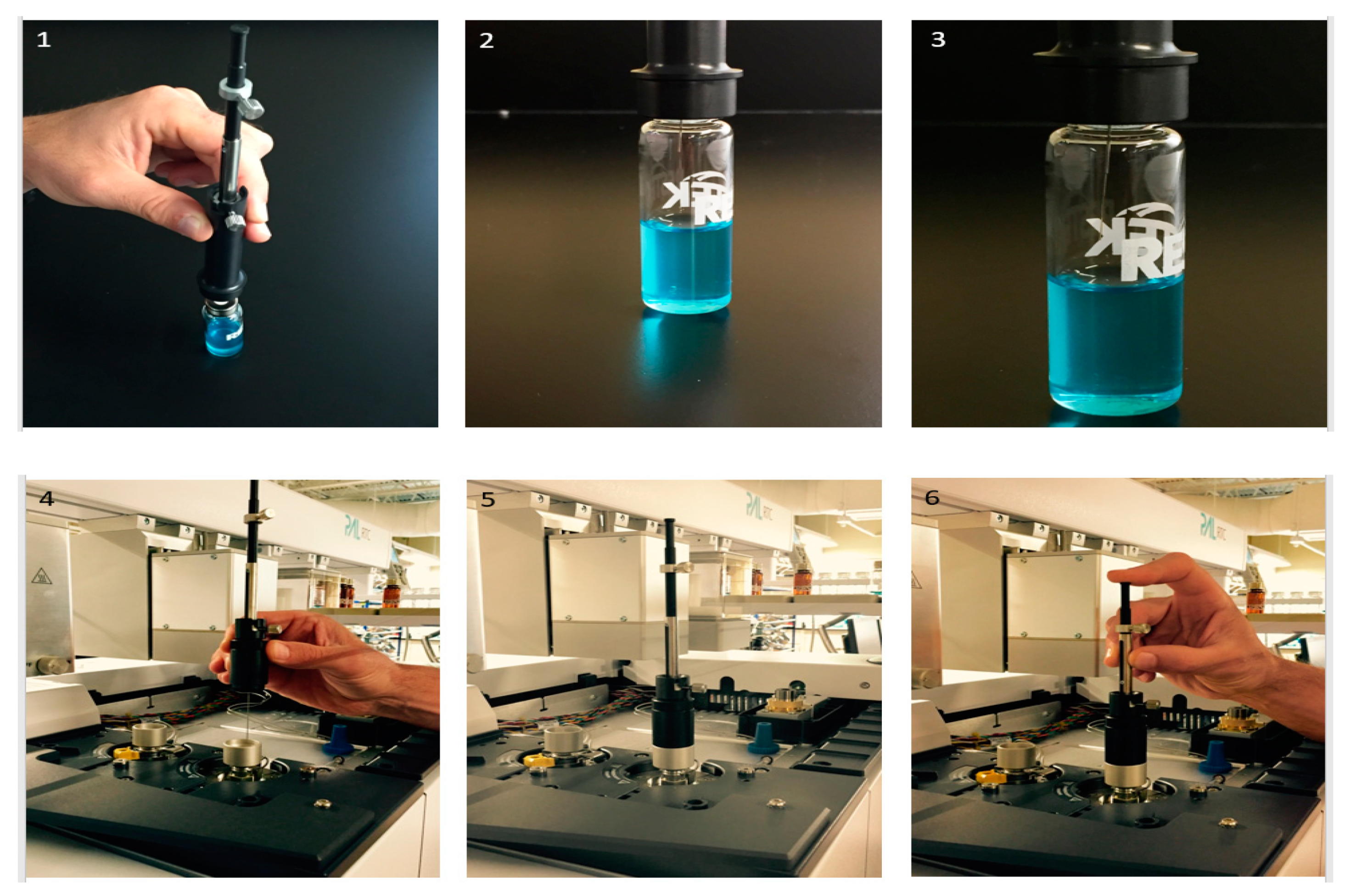
As portrayed in Figure 3, the SPME device workflow comprises several steps including the following: 1. piercing the septum of the vial with the outer needle; 2. exposing the extraction phase to the sample and collecting the analytes of interest for a fixed period of time; 3. withdrawing the extraction phase into the needle of the SPME device; 4. transporting the SPME device to the instrument station, and 5. transferring the SPME device into the injection port of the instrument, so the analytes are eluted from the extraction phase via thermal desorption.
SPME Arrows were designed to overcome short-comings associated with the workflow of traditional SPME fibers, such as limited mechanical robustness ([19]), poor inter-device reproducibility, and small extraction phase volumes ([9]). The body of an SPME fiber is made of stainless steel, and the extraction phase is typically coated on fused silica. Consequently, one of the most common complaints of traditional SPME fiber end-users is a lack of physical durability of the fused silica ([20]). Though this problem can be partially alleviated by coating the extraction phase on a metal core (e.g., nitinol) [20,21], the body of a traditional SPME fiber is also commonly reported as fragile (see Figure 4). Consequently, commercial vendors have begun offering pre-drilled GC inlet septa and thin-walled vial septa to help mitigate issues with the damage of the core during the extraction and desorption steps. It is not uncommon for a SPME device to become injured, even within the first use. Failure rates appear to be highest amongst new end users; however, experienced end users are not immune to these issues either. Due to the delicate nature of traditional SPME fiber devices, most of them cannot be repaired. Manual extractions and desorptions appear to increase traditional SPME fiber failure rates, so the adoption of robotic autosamplers (e.g., CTC PAL or more commonly referred to as “rail” systems) has helped alleviate this problem to some degree. However, regardless of manual or automated injection, traditional SPME fiber devices typically fail to reach their true potential lifetime due to some sort of physical damage. “Potential” lifetime is stated because the majority of these mechanical failures appear to take place well before the fiber phase has been exhausted (i.e., it has not reached the life time of the coating).
2.1. Physical Attributes
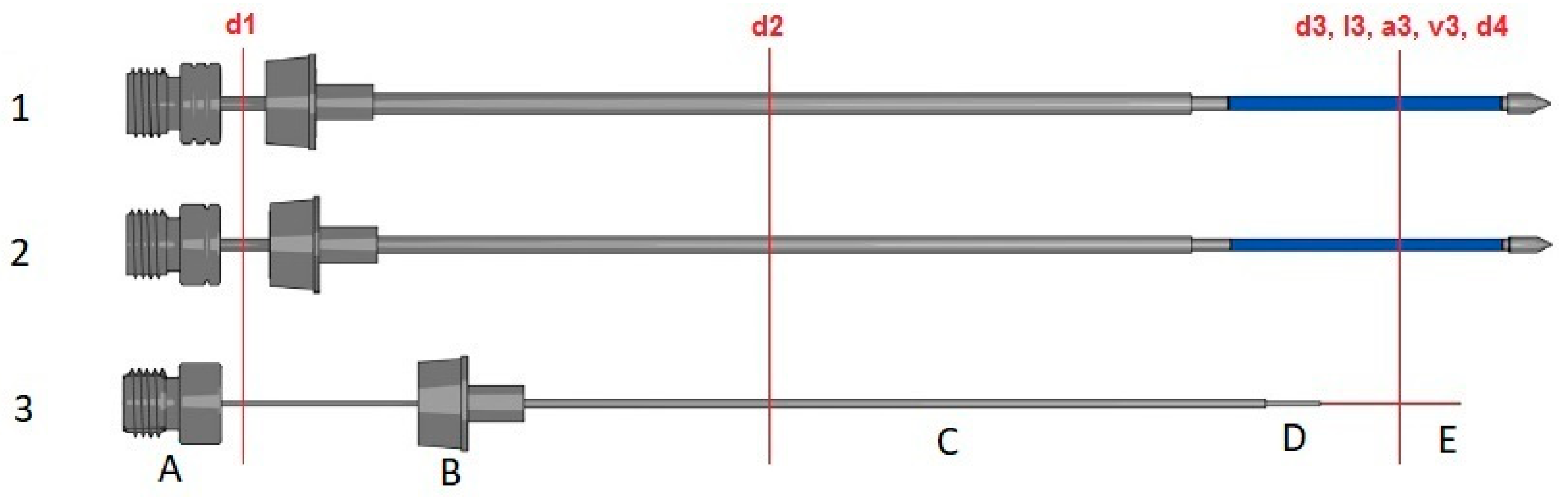
SPME Arrows only share the following physical attributes with traditional SPME fibers: 1. the dimensions and thread types on the color-coded hubs; 2. the dimensions of the needle ferrule; and 3. a stainless steel composition of the support tubing and septum piercing needle. Beyond the aforementioned, SPME Arrows diverge from traditional SPME fibers in an attempt to increase mechanical durability. Traditional SPME fibers have 23 or 24 gauge (i.e., 0.573 or 0.511 mm, respectively) external diameters on their septum piercing needle. The SPME Arrow was initially developed with 1.1 and 1.5 mm (i.e., ~17 and 15 gauge) external diameters on their septum piercing needle, which is approximately 2 and 3 times the diameter of traditional SPME fibers, respectively. Figure 1 provides a scale image for visual comparison of the physical attributes of SPME Arrows and a 23 gauge traditional SPME fiber. Furthermore, Table 1 breaks down the divergent physical attributes of the SPME Arrows and a 23 gauge traditional SPME fiber. Most notably, the support tubing (i.e., plunger), septum piercing needle, and phase support tubing of the 1.1 and 1.5 mm SPME Arrows have increased external diameters by 264–332%, 174–237%, and 583–449%, respectively over a 23 gauge traditional SPME fiber. These increased external diameters are largely responsible for the increased mechanical robustness of SPME Arrows. In particular, the increase in the support tubing appears to contribute the most improvement in durability, as this seems to be the most common failure point associated with traditional SPME fibers [13].
It is important to note that the SPME Arrows have “arrow” shaped tips from which they garner their name. These arrow tips have the same external diameter as the SPME Arrows’ septum piercing needle (i.e., 1.1 or 1.5 mm). The arrow tip helps increase the mechanical robustness of the SPME Arrow, as the force required to penetrate the vial and/or GC inlet septa is less on an SPME Arrow when compared to a traditional SPME fiber, despite the increase in diameter. It has been demonstrated that a 1.1 mm SPME Arrow only requires 799 g of force to penetrate and headspace vial septum, whereas, a 0.63 mm traditional SPME fiber requires 1188 g (~50% more than the SPME Arrow) of force to penetrate the same configuration [22]. Furthermore, the SPME Arrow tip has been demonstrated to cut slits in vial and GC inlet septa, as opposed to coring septa. Therefore, septa lifetime appears to be as good, if not better, than traditional SPME fibers [23]. Furthermore, when retracted (i.e., not extracting/desorbing), the Arrow tip serves the purpose of a protective cap, thereby minimizing the diffusion of compounds into the septum piercing needle and ultimately reaching the phase. This helps minimize background contamination in between analyses or losses of analytes while the fibers are store for analysis on the tray of the GC system [21].
More recently, the SPME Arrow has been advanced with a 0.804 mm support tubing housed inside a 1.5 mm septum piercing needle (denoted as 1.5* mm in Table 2). This design was released to overcome problems associated with using SPME Arrows for direct immersion (DI) extractions, such as phase swelling and the subsequently sloughing off and/or damaged when the SPME Arrow support tubing is retracted inside a 1.1 mm septum piercing needle. The wider 1.5 mm septum piercing needle provides enough clearance for the swollen phase on the 0.804 mm support tubing, thereby mitigating the sloughing issues.
2.2. Phase
SPME Arrows were also designed to overcome the small phase areas and volumes associated with traditional SPME fibers. As shown in the Table 1, a 23 gauge traditional SPME fiber with 100 µm of polydimethylsiloxane (PDMS) has a 9.40 mm² and 0.600 µL phase. The 1.1 mm Arrow with 100 µm of PDMS, which represents the most direct comparison to the aforementioned, has a 44.0 mm² and 3.80 µL phase; which is a 468% and 633% increase in area and volume, respectively. This increase in phase area and volume has resulted in an increase in sensitivity and/or capacity, which is addressed later on in the performance and applications sections of this manuscript.
As shown in Table 2, SPME Arrows have been developed with most of the traditional SPME fiber phase offerings. The carbowax-polyethylene glycol (PEG) and carbowax/templated resin (CW/TPR) phase are the only phases not currently available on the SPME Arrow platform. Though the SPME Arrow has most of the phases, it is important to note that not all phase thickness/configurations have been replicated in the SPME Arrow. For example, although the SPME arrow is available in PDMS, it is only available in 100 and 250 µm PDMS configurations. The 7 and 30 µm PDMS configurations found in the traditional SPME fiber offerings are not available in the SPME Arrow. Additional phase thickness deviations may be observed when looking at the other phases (e.g., 75/85 µm of carbon on the traditional SPME fiber compared to 120 µm of carbon on the SPME Arrow).
3. SPME Arrow Accommodations
End users may not purchase a SPME Arrow and begin using it as direct replacement for the traditional SPME fiber. There are several things an analyst will need to modify/replace in order to accommodate the SPME Arrows’ larger external diameters, including: 1. the injection tool (both manual and robotic); 2. the GC inlet; and 3. the GC inlet liner.
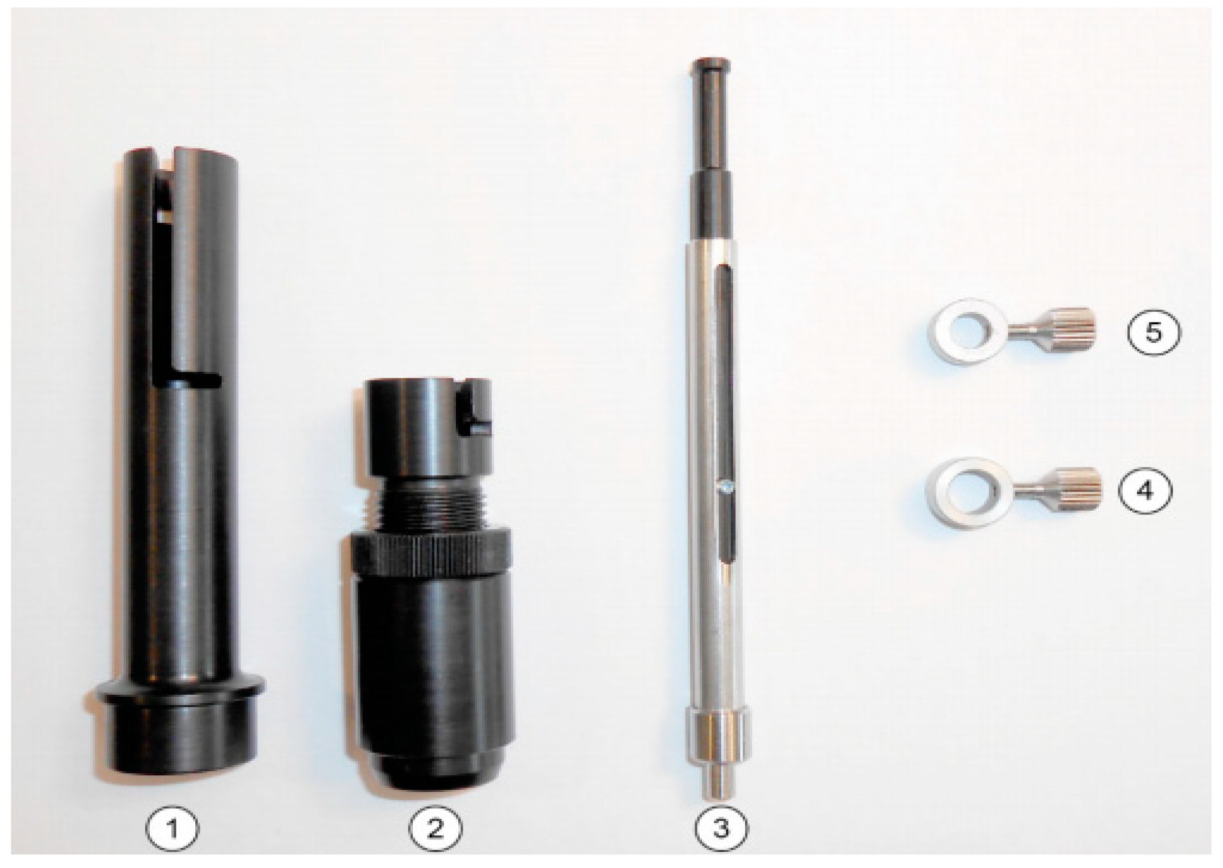
First, the manual tool may need modification or replacement (see Figure 2). For example, the traditional SPME fiber holder [19] does not accommodate SPME Arrows. Despite the SPME Arrows sharing a similar length and the same dimensions on the color-coded hubs, the Supelco manual holder tip diameter is too small. Some users have worked around this problem by drilling out the manual holder with a 3/64” metal compatible drill bit. Alternatively, other users have purchased the Restek PAL manual injection kit (Restek Corporation, Bellefonte, PA, USA), which accommodates both traditional SPME fibers and SPME Arrows without modification.
In terms of robotic platforms, CTC Analytics AG does not support the use of SPME Arrows on their second generation PAL systems (e.g., PAL/PAL-xt). Therefore, users with these systems will need to modify their existing SPME holder or acquire an appropriate SPME Arrow holder from Chromtech (Bad Camberg, Germany). Newer generations of robotic arms, such as PAL3 systems (e.g., RTC and RSI), fully support the SPME Arrow as long as users acquire the appropriate tool from CTC Analytics AG. In the case of rail systems commercialized by other vendors such as Agilent (Santa Clara, CA, USA), Gerstel (Linthicum Heights, MD, USA), Shimadzu (Kyoto, Kyoto Prefecture, Japan), and Thermo (Waltham, MA, USA), users need to contact the manufacturer to acquire the appropriate tool for their robotic system. Furthermore, the fiber conditioner found on second and third generation PAL systems does not accommodate the SPME Arrow diameters. Therefore, users need to directly condition the SPME devices in the GC inlet or acquire an SPME Arrow-specific conditioner module from the appropriate manufacturer.
In terms of the GC system, the inlets that are currently installed on the instruments made by major manufacturers (e.g., Agilent, Shimadzu, and Thermo) do not accommodate SPME Arrows. However, factory-modified GC inlets are commercially available for those manufacturers. Alternatively, users can modify their existing GC inlets by drilling out the excess of metal with a 3/64” metal drill bit. Likewise, traditional SPME fibers require the use of 0.75 mm inlet liners or greater. With the smallest outer needle diameter of 1.1 mm, SPME Arrows require the use of lager inlet liners. Several commercial vendors provide straight-walled inlet liners capable of accommodating SPME Arrows. For example, a 1.8 mm ID straight/inlet liner can accommodate both the 1.1 and 1.5 mm SPME Arrows. It is important to note, that in lieu of SPME Arrow-specific liners, end users may be able to use a standard 2.0 mm straight-walled liner and not suffer any significant performance losses [24].
4. SPME Arrow Performance
4.1. Benchmarking
In the previous sections, several advantages and disadvantages of SPME Arrows when compared to traditional SPME fibers were reviewed. However, those were mostly focused around the physical attributes associated with SPME Arrows. The current section focuses on how the aforementioned physical differences in phase area and volume translate into analytical performance. Several benchmarking applications have been chosen to compare SPME Arrows with traditional SPME fibers. It is important to note that the 1.1 mm SPME Arrow is the focus for the remainder of this section and for most of the manuscript, as this configuration is available in the most popular SPME phases (e.g., polydimethylsiloxane (PDMS) and divinylbenzene (DVB)), as shown in Table 2, whereas the 1.5 mm SPME Arrow is only available in a 250 µm polydimethylsiloxane (PDMS) configuration, which is anticipated to have less applications. It is important to point out that the 1.5* mm SPME Arrows commercially launched a few weeks prior to the writing of this manuscript, so there are little data to warrant a discussion beyond what was mentioned previously.
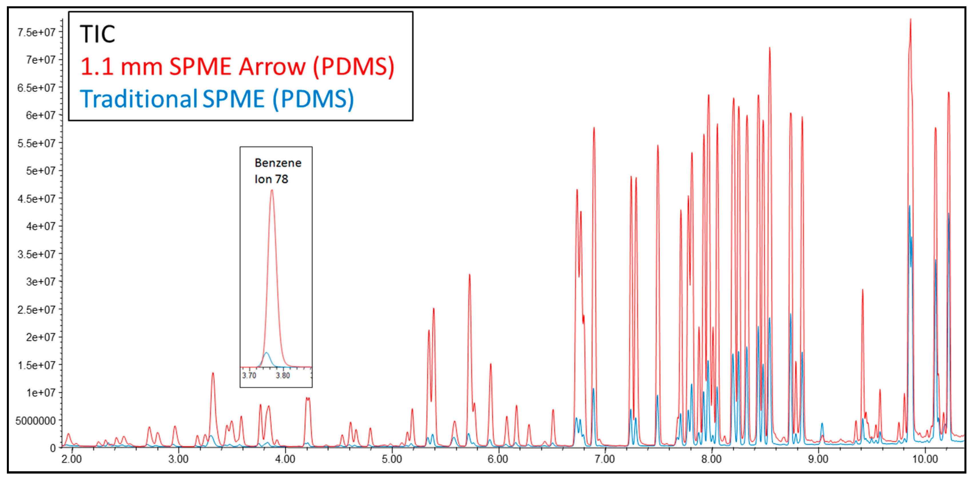
When comparing a 100 µm PDMS 1.1 mm SPME Arrow against a 100 µm PDMS 23 gauge SPME fiber after the headspace (HS) extraction of volatile organic compounds (VOCs) in drinking water [spiked at 2.5 ppb as per International Organization for Standardization (ISO) method 17943] via GC coupled to mass spectrometry, it was found that equilibration times, extraction times, and desorption temperatures were equivalent among the two devices. Figure 5 presents a chromatogram overlaying instrumental response of the SPME Arrow and SPME fiber for the 92 VOCs. The SPME Arrow’s response is higher than the traditional SPME fiber. On average, the SPME Arrow demonstrated a ~4× increase in response over traditional SPME fibers. This observation is consistent with the SPME Arrow’s larger phase volume, which correlates to a greater volume/mass of target analyte collected and thereby an increased analytical response. It is important to note that “on average” was stated in the previous point, because the increase in response for very volatile compounds like vinyl chloride was ~10× on the SPME Arrow vs. the traditional SPME fiber. However, a semi-volatile compound like naphthalene only saw ~2× increase in sensitivity on the SPME Arrow compared to the traditional SPME fiber. Such differences in extraction recoveries are correlated to different vapor pressures (i.e., Henry’s law constants) of said analytes and, expectedly, the availability in the headspace being the rate-limiting step. Likewise, it was observed that the SPME Arrow generated linear results (0.998 median R²) over a wide calibration range (0.0025–166 µg/L); with excellent precision (3.24% median RSD); and low sensitivity (30.0 ng/L median MDL) for all 92 ISO 17943 VOCs [25].
4.2. Method Development
Initial studies with SPME Arrows indicate that method development should follow the same logic and approach already demonstrated to be optimum for traditional SPME fibers [26]. For example, Herrington et al. evaluated SPME Arrows to see if there was a deviation in the extraction times required for SPME Arrows given the increase in phase volume. For this study, 100 µm PDMS 1.1 mm SPME Arrows and 100 µm PDMS 23 gauge traditional SPME fibers were evaluated for 92 HS VOCs, which had been spiked in drinking water at 2.5 ppb per method ISO 17943 [25]. Everything was equivalent (e.g., equilibration times and desorption temperatures), except for the extraction times. Extraction times of 15, 30, 60, 120, 240, 480, 960, and 1920 s were evaluated for each SPME (n = 3 for each SPME and each extraction time). Figure S1 shows the results from which the following two observations were made: 1. the SPME Arrow continued to demonstrate an increase in response (i.e., amount collected per unit of time) [9], which was again attributed to the increase in phase area over traditional SPME fibers; 2. both SPME types equilibrated at the same time (~120 s) for most of the 92 VOCs evaluated. This observation was consistent with the fact that both SPME types were 100 µm PDMS. Since the phase thickness was the same, it was deemed reasonable that the gas phase kinetics was the same; therefore, the equilibrium times were equivalent.
4.3. Troubleshooting
An extensive literature search only produced one reported issue associated with the SPME Arrow. Hartonen et al. reported chromatographic issues of double amine peaks when using SPME Arrows for extracting HS amines from water [27]. The root cause was determined to be an increased amount of water vapor extracted from the samples, due to the SPME Arrows’ increased surface area and volume. However, it is important to note that other work on HS volatiles from drinking water [27] did not report any chromatographic issues and/or water issues. However, this work was conducted on thermally conditioned fibers with the use of sodium chloride in samples and split mode during desorption: the latter two were important conclusions Hartonen et al. arrived at [27]. In addition, Helin et al. did not report any issues when using SPME Arrow for HS amines in water with an SPME Arrow [17]. Finally, Gionfriddo et al. demonstrated short-chain aliphatic amines are best analyzed with the use of derivatization [28].
Beyond the aforementioned works, it is important to note that other work on HS volatiles from drinking water investigated the use of the 1.5 mm SPME Arrow with 250 µm of PDMS over a 1.1 mm SPME Arrow with 100 µm of PDMS [25]. As shown in Figure S2, light gases like chloroethane had ~2.5 times the response on the 1.5 mm SPME Arrow compared to the 1.1 mm SPME Arrow. However, the chromatography began to tail and split. A split injection could overcome this poor chromatography; however, then any sensitivity gains would be lost. It is believed that this tailing was a product of the fact that the phase was so thick on the 1.5 mm SPME Arrow that these lighter gases deeply penetrated into the thick phase, which then caused tailing up desorption. This work, Hartonen et al.’s work, and work not shown here have tended to suggest that SPME Arrows perform best with the use of a small (e.g., 2:1 or 5:1) split during thermal desorption. However, more extensive work will have to investigate this in the future. For instance, forthcoming work should consider the use of programmed temperature vaporization (PTV)-type inlets for SPME Arrows [14].
5. SPME Arrow Applications
5.1. Environmental Analysis
SPME technology has applicability for environmental pollutants in air, water, soil and sediment, and it can be used in the field or in the laboratory for sample preparation [17,18]. An advantage of the SPME Arrow is the larger phase volume that allows for the collection of a higher amount of the target compounds, so reporting requirements for environmental data users can be met.
One drawback of the traditional SPME fibers for headspace analysis is that the amount of analyte enriched on the coating is not sufficient to meet the reporting limits established by environmental agencies. The SPME Arrow design allows for four-to-five times higher analytical responses of compounds than a traditional SPME fiber [29]. For instance, the work performed by Kremser et al. presented a comparison of several techniques for determining volatile organic compounds including: purge and trap (P and T), ITEX, sample loop, traditional SPME fiber, gas-tight syringe, and SPME Arrow. As can be seen in Table 3, the detection limits for the SPME Arrow are in the same range as P and T and ITEX techniques, which are representative of exhaustive extraction techniques. However, the automation of workflow for the SPME Arrow is not only simpler than for P and T but also compatible with direct immersion experiments, which are not doable by ITEX [14].
In another study, Kaziur and collaborators developed a method capable of detecting picogram-per-liter (0.05 and 0.6 ng L−1) levels of water taste and odor compounds (i.e., isopropyl-3-methoxypyrazine, 2-isobutyl-3-methoxypyrazine, geosmin, 2-methylisoborneol, 2,4,6-trichloroanisole, 2,4,6-bromoanisole, and beta-ionone) in water samples [18]. As a matter of fact, this fully automated workflow was more sensitive than existing methodologies (see Table 4).
Headspace work with SPME Arrow has been performed for the analysis of volatile amines in waste water. For example, after careful optimization of the extraction conditions, Helin et al. observed that the SPME Arrow produced lower detection limits, and higher recoveries of dimethyl amine (DMA) over the traditional SPME fiber (88% vs. 57%, respectively) [17].
The SPME Arrow has also been used for the analysis of semi-volatile compounds using immersion extraction. Typically, semi-volatile sample preparation for water samples has used liquid-liquid extraction (LLE) or solid phase extraction (SPE). Boyaci et al. did a comparison of SPME technology versus LLE and SPE, and it was found that the reduction of unwanted matrix interferences, the solventless extraction, the reusability, and the feasibility for high throughput sample analysis made SPME a more attractive technique [47]. Kremser et al. did extensive work with freely dissolved polycyclic aromatic hydrocarbons (PAHs) in water [48]. Extraction times and stirring rates were optimized to determine the method detection limits, and these were compared to previously published data for traditional SPME and SBSE (see Table 5). Direct immersion experiments using the SPME Arrow for freely dissolved semi-organic compounds showed detection limits five times lower than the traditional SPME fiber and similar results to stir bar sorptive extraction (SBSE). When comparing the SPME Arrow to SBSE, the authors highlighted the easiness of automation and utilization of shorter extraction times.
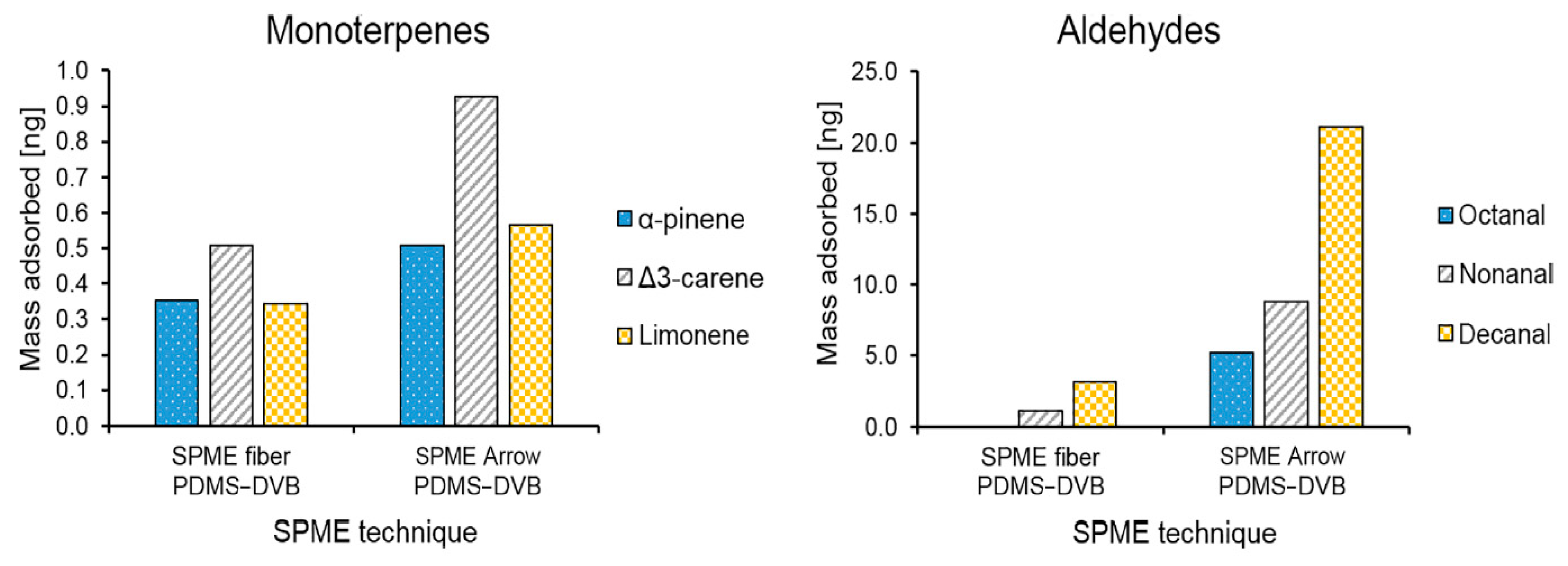
The SPME Arrow has also been used for environmental air sampling with the in-field analysis of biogenic volatile organic compounds (BVOCs). Monoterpenes (gamma-pinene and d3-carene) and aliphatic aldehydes (octanal and decanal) were selected by Barreira et al. to represent expected compounds to be found in field testing [49]. In-laboratory extraction efficiencies showed that the SPME Arrow had two-to-three times more area count than the traditional SPME fiber. Sampling was then performed in a boreal forest in Hyytiälä, Finland. Barreira et al. determined that the extraction efficiency of the SPME Arrow was two-times higher or greater, depending on compound, than the traditional SPME fiber, allowing for more sensitive testing (see Figure 6) [49].
5.2. Food Analysis
There are not as many publications on SPME Arrow in food analysis, as compared to traditional SPME fibers [8]. However, in the past couple of years, several applications have spanned the analysis of diverse matrices including fish and rice [52,53]. For instance, Song and coworkers compared carboxen/polydimethylsiloxane (CAR/PDMS) SPME sorbents in the fiber format to the arrow format for the HS extraction of volatiles present in salt-fermented sand lance fish sauce [53]. The researchers reported that alcohols, aldehydes and pyrazines, with the exception of 1-pentenol, were more effectively extracted using the SPME Arrow. Some compounds that are believed to be important to the flavor profile were only observed using the arrow device. Lan and coworkers described a modified zeolitic imidizolate framework (ZIF-8) as a solid phase microextraction support on the arrow construct [54]. The group compared the novel adsorbent to a commercially available carboxen/polydimethylsiloxane device for the sampling of volatile amines in wastewater and food samples (salmon and mushrooms). The researchers found that the commercial ZIF-8 material exhibited a small pore size (5.6 Å), which likely excluded the model amine compounds, resulting in a low extraction efficiency. The acidification of the material significantly increased the extraction of small volatile amines, presumably due to an increase in pore size. The modified ZIF-8 arrow design provided comparable extraction efficiencies for small, volatile amines from several matrices as compared to commercial carboxen/polydimethylsiloxane SPME Arrow devices. Yuan and coworkers investigated metal organic framework (MOF) sorbents applied to arrow SPME devices for the determination of PAH contaminants in fish samples [55]. The authors employed a zirconia based UiO-66-molybdenum disulfide composite and compared the recovery of PAH contaminants from seafood samples to both commercially-available arrow and fibers coated with PDMS/carboxen/DVB coatings. The combination of the MOF composite sorbent and the arrow format resulted in and increased number of PAH species detected. Lan et. al. described the analysis of low molecular weight aliphatic amines from various matrices using several different modifications of silica sorbent applied to the arrow format [56]. The scientists demonstrated that dimethylamine and trimethylamine could be effectively detected and quantitated in mushroom samples using these devices.
5.3. Terpenes in Cannabis
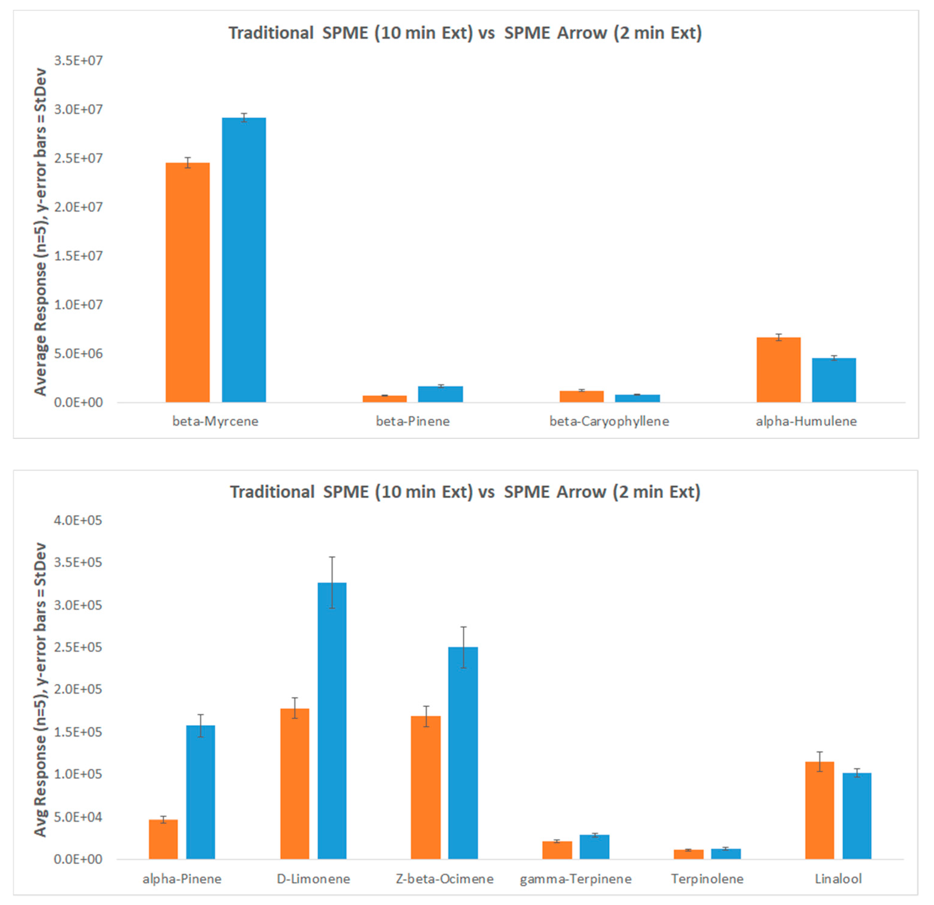
With the rapid growth of the cannabis market taking place, the need for analytical testing has become more critical. An area of interest in this market is being able to identify and place cannabis flowers into the correct chemical variety, otherwise known as chemovar. To properly classify cannabis chemovars, a comprehensive chemical profile examining compounds, such as terpenes and cannabinoids, is collected [57]. An analysis of terpenes is typically done via headspace—gas chromatography—mass spectrometry (HS-GC-MS). Herein, a 1.1 mm 120 µm divinylbenzene (DVB)/PDMS SPME Arrow was used to analyze the terpene content in Humulus lupulus (hops), and individual terpene responses were compared to that of a HS-syringe method typically used for this application and a 65 µm DVB/PDMS traditional SPME fiber. Hops were used in place of cannabis, as cannabis could not be legally obtained. Hops were ground using drying ice and then stored in the freezer until needed. Ten-to-fifteen milligrams of ground hops were added to a 20 mL crimp top HS vial.
Current methodologies recommend extraction times of 10 min for the traditional SPME fiber analysis of terpenes [58]. Improvements to this parameter alone have the potential to decrease instrument runtimes and increasing the number of samples that contract laboratories are able to test. As can be seen in Figure 7, average responses for a 10 min extraction time using a traditional SPME fiber are equivalent to that of a 2 min extraction time with the SPME Arrow. This can be done without sacrificing reproducibility, as both techniques showed % RSDs under ≤10%. Lighter terpenes (monoterpenes) gave better responses on the SPME Arrow, while the heavier terpenes (sesquiterpenes) were more comparable. However, the traditional fiber did provide slightly better responses for the sesquiterpenes.
6. Future Directions
The future of the SPME Arrow is promising, and among the multiple research and application avenues, this new geometry of SPME may grow rapidly. The following three areas are anticipated to expand: the development of new extraction phases [26,55,56], the Arrow’s direct interface with MS instrumentation [59], and the Arrow’s evolution towards smarter devices.
The first premise beyond the development of novel extraction phases is that the performance of these materials is better than those already commercially available by improving either robustness, extraction capabilities or device ruggedness [6,14,60]. For instance, the lack of inter-device reproducibility of one of the most popular extraction phases in SPME for GC analysis, the triple phase (DVB/Car/PDMS), has been its Achilles’ heel [61]. Therefore, the recently launched SPME Arrow with a blended triple phase is expected to make a greater impact in the food and fragrance industry [62]. The main issue with the “original” tri-phasic SPME fibers relies on the process used for its manufacturing given that, if the two extraction phases (DVB-PDMS and Car-PDMS) are not properly aligned, this can lead to differences in the amount of extraction phase per coating length, thus leading to significant differences in the amount of analyte extracted—particularly volatile compounds [61]. In tri-phasic SPME Arrows, the particles of DVB and Car are embedded on the PDMS, resolving the issue of multiphase irregularities and consequently leading to better inter-device reproducibility.
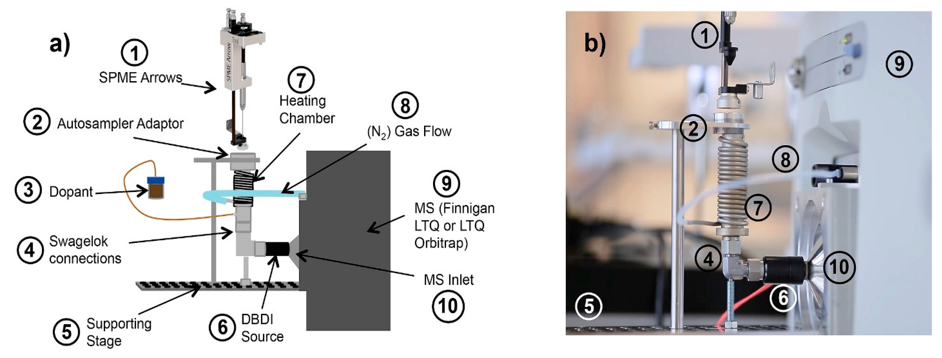
As recently reviewed by Gomez-Rios and Mirabelli [4], the direct interface of SPME devices to MS has been increasing in the last five years, and SPME Arrows have not been an exception. For instance, research from the Zenobi’s group demonstrated the applicability of an automated SPME Arrow workflow, hyphened with a dielectric barrier desorption ionization (DBDI) coupled to an LTQ Orbitrap, to analyze common organic contaminants in treated wastewater (see Figure 8) [59]. Limits of detection as low as 3 ng/L were attained with a total analysis time per sample of less than 10 min (see Figure S3). Given that the ionization mechanism by DBDI, as well as by other direct-to-MS technologies, primarily relies on proton transfer for analyte ionization, a good response is typically attained for polar compounds with high proton affinity, whereas low ionization efficiencies are observed for nonpolar compounds. To overcome this limitation, a dopant-assisted DBDI ionization approach has been proposed to more efficiently ionize PAHs extracted from water. Though this strategy allowed for an up-to-an-order-of-magnitude signal enhancement for the analytes monitored, others go in detriment, and, consequently, the use (or not) of dopant must be driven by the target analytes. Certainly, DBDI is only one of the possible means of interfacing SPME Arrows to MS instrumentation. Other options include but are not limited to direct analysis in real time (DART) [63,64,65], atmospheric pressure photon ionization (APPI) [66], and thermal desorption-electrospray ionization (TD-ESI) [67]. It is anticipated that SPME Arrow-like geometries [11] compatible with liquid desorption and complex biological matrices will be developed in the near future and potentially coupled to direct-to-MS technologies such nano-electrospray ionization (nano-ESI) [68], desorption electrospray ionization [69], and the microfluidic open interface (also known as Open Probe Sampling Interface, OPSI) [70,71,72], as means to enhance the speed of analysis and the sensitivity [73]. Areas where such devices can make a great impact include but are not limited to, food fraud [74], volatomics [64], and rapid screening in the clinical chemistry realm [75].
Finally, as part of the advances on analytical chemistry towards the Internet of Things and other intelligent platforms [76], the development of “smart” SPME devices has begun. The so-called “smart” SPME devices, currently commercialized by PAL, have been exclusively designed for their rail systems and comprise a chip that automatically informs instruments about the phase coated on the device and its usage history. Though the smartness of the device is strictly related to physical/mechanical aspects of the device, the development of Arrows with “smarter” coatings [6] (e.g., ionic liquids [77], MOF [78], and carbon nanotubes (CNT) [79]) and “smarter” geometries and substrates components [80] is expected.
Supplementary Materials
The following are available online at https://www.mdpi.com/2297-8739/7/1/12/s1, Figure S1: Extraction times for SPME Arrow and traditional SPME fiber for ISO 17943 HS-VOCs. Acquired on Agilent 7890B/5977B GC-MS, Figure S2: 1.5 mm SPME Arrow (red trace) versus 1.1 mm SPME Arrow (black trace) for analysis of chloroethane in water samples according to method ISO 17943 HS-VOCs. Acquired on Agilent 7890B/5977B GC-MS., Figure S3: SPME-Arrow and DBDI for determination of ppt of contaminants in waste water. Quantitation of four compounds of varying polarities and contaminant classes: (a) DEET, (b) Tamoxifen, (c) Pyrene, (d) Metolachlor. Figure reprinted with permission of Elsevier from Reference [59], 2020.
Author Contributions
C.M., G.S., D.S.B., J.S.H. and G.A.G.-R. wrote the review. J.S.H. and G.A.G.-R. planned and revised the document. All authors have read and agree to the published version of the manuscript.
Funding
This research received no external funding.
Conflicts of Interest
All the authors work at Restek Corporation and Restek Corporation commercializes SPME Arrows.
References
- Reyes-Garcés, N.; Gionfriddo, E.; Gómez-Ríos, G.A.; Alam, M.N.; Boyaci, E.; Bojko, B.; Singh, V.; Grandy, J.J.; Pawliszyn, J. Advances in solid phase microextraction and perspective on future directions. Anal. Chem. 2018, 90, 302–360. [Google Scholar] [CrossRef] [PubMed]
- Reyes-Garcés, N.; Gionfriddo, E. Recent developments and applications of solid phase microextraction as a sample preparation approach for mass-spectrometry-based metabolomics and lipidomics. TrAC Trends Anal. Chem. 2019, 113, 172–181. [Google Scholar] [CrossRef]
- Alam, M.N.; Nazdrajić, E.; Singh, V.; Tascon, M.; Pawliszyn, J. Effect of Transport Parameters and Device Geometry on Extraction Kinetics and Efficiency in Direct Immersion Solid-phase Microextraction. Anal. Chem. 2018. [Google Scholar] [CrossRef] [PubMed]
- Gómez-Ríos, G.A.; Mirabelli, M.F. Solid Phase Microextraction-Mass Spectrometry: Metanoia. TrAC Trends Anal. Chem. 2019, 112, 201–211. [Google Scholar] [CrossRef]
- Armenta, S.; Garrigues, S.; de la Guardia, M. The role of green extraction techniques in Green Analytical Chemistry. TrAC Trends Anal. Chem. 2015. [Google Scholar] [CrossRef]
- Gómez-Ríos, G.A.; Garcés, N.R.; Tascon, M. Smart Materials in Solid Phase Microextraction (SPME). In Handbook of Smart Materials in Analytical Chemistry; John Wiley & Sons, Ltd.: Chichester, UK, 2019; pp. 581–620. [Google Scholar] [CrossRef]
- Belardi, R.P.; Pawliszyn, J.B. The application of chemically modified fused silica fibers in the extraction of organics from water matrix samples and their rapid transfer to capillary columns. Water Pollut. Res. J. Can. 1989, 24, 179–191. [Google Scholar] [CrossRef]
- Godage, N.H.; Gionfriddo, E. A critical outlook on recent developments and applications of matrix compatible coatings for solid phase microextraction. TrAC Trends Anal. Chem. 2019, 111, 220–228. [Google Scholar] [CrossRef]
- Olcer, Y.A.; Tascon, M.; Eroglu, A.E.; Boyacı, E. Thin film microextraction: Towards faster and more sensitive microextraction. TrAC Trends Anal. Chem. 2019, 113, 93–101. [Google Scholar] [CrossRef]
- Gutiérrez-Serpa, A.; Schorn-García, D.; Jiménez-Moreno, F.; Jiménez-Abizanda, A.I.; Pino, V. Braid solid-phase microextraction of polycyclic aromatic hydrocarbons by using fibers coated with silver-based nanomaterials in combination with HPLC with fluorometric detection. Microchim. Acta 2019, 186, 311. [Google Scholar] [CrossRef]
- Poole, J.J.; Grandy, J.J.; Yu, M.; Boyaci, E.; Gómez-Ríos, G.A.; Reyes-Garcés, N.; Bojko, B.; Heide, H.V.; Pawliszyn, J. Deposition of a Sorbent into a Recession on a Solid Support to Provide a New, Mechanically Robust Solid-Phase Microextraction Device. Anal. Chem. 2017, 89, 8021–8026. [Google Scholar] [CrossRef]
- Truong, T.V.; Lee, E.D.; Black, B.D.; Truong, T.X.; Lee, M.L. Coiled wire filament sample introduction for gas chromatography–mass spectrometry. Int. J. Mass Spectrom. 2018, 427, 123–132. [Google Scholar] [CrossRef]
- Ziegler, M.; Schmarr, H.-G. Comparison of Solid-Phase Microextraction Using Classical Fibers Versus Mini-Arrows Applying Multiple Headspace Extraction and Various Agitation Techniques. Chromatographia 2019, 82, 635–640. [Google Scholar] [CrossRef]
- Dugheri, S.; Mucci, N.; Bonari, A.; Marrubini, G.; Cappelli, G.; Ubiali, D.; Campagna, M.; Montalti, M.; Arcangeli, G. Solid phase microextraction techniques used for gas chromatography: A review. Acta Chromatogr. 2019, 1–9. [Google Scholar] [CrossRef]
- Grandy, J.J.; Boyacı, E.; Pawliszyn, J. Development of a Carbon Mesh Supported Thin Film Microextraction Membrane As a Means to Lower the Detection Limits of Benchtop and Portable GC/MS Instrumentation. Anal. Chem. 2016, 88, 1760–1767. [Google Scholar] [CrossRef] [PubMed] [Green Version]
- Ruiz-Jimenez, J.; Zanca, N.; Lan, H.; Jussila, M.; Hartonen, K.; Riekkola, M.-L. Aerial drone as a carrier for miniaturized air sampling systems. J. Chromatogr. A 2019, 1597, 202–208. [Google Scholar] [CrossRef] [PubMed]
- Helin, A.; Rönkkö, T.; Parshintsev, J.; Hartonen, K.; Schilling, B.; Läubli, T.; Riekkola, M.-L. Solid phase microextraction Arrow for the sampling of volatile amines in wastewater and atmosphere. J. Chromatogr. A 2015, 1426, 56–63. [Google Scholar] [CrossRef] [PubMed]
- Kaziur, W.; Salemi, A.; Jochmann, M.A.; Schmidt, T.C. Automated determination of picogram-per-liter level of water taste and odor compounds using solid-phase microextraction arrow coupled with gas chromatography-mass spectrometry. Anal. Bioanal. Chem. 2019, 411, 2653–2662. [Google Scholar] [CrossRef]
- Risticevic, S.; Lord, H.; Górecki, T.; Arthur, C.L.; Pawliszyn, J. Protocol for solid-phase microextraction method development. Nat. Protoc. 2010, 5, 122–139. [Google Scholar] [CrossRef]
- Setkova, L.; Risticevic, S.; Linton, C.M.; Ouyang, G.; Bragg, L.M.; Pawliszyn, J. Solid-phase microextraction-gas chromatography-time-of-flight mass spectrometry utilized for the evaluation of the new-generation super elastic fiber assemblies. Anal. Chim. Acta 2007, 581, 221–231. [Google Scholar] [CrossRef]
- Gómez-Ríos, G.A.; Reyes-Garcés, N.; Pawliszyn, J. Evaluation of a multi-fiber exchange solid-phase microextraction system and its application to on-site sampling. J. Sep. Sci. 2015, 38. [Google Scholar] [CrossRef]
- Herrington, J. SPME Arrow Blog 7 ChromaBLOGraphy: Restek’s Chromatography Blog. Available online: https://blog.restek.com/?p=41704 (accessed on 31 July 2019).
- Herrington, J. SPME Arrow Blog 2 ChromaBLOGraphy: Restek’s Chromatography Blog. Available online: https://blog.restek.com/?p=37889 (accessed on 31 July 2019).
- Herrington, J.S. Solid-Phase Microextraction Liners for Headspace Volatile Organic Compounds. Column 2018, 14, 36–40. Available online: http://files.alfresco.mjh.group/alfresco_images/pharma//2019/03/07/aae3f1b3-af2d-4eb0-9d05-fdd7ed4bb372/LCTC121718%20North%20America.pdf (accessed on 31 July 2019).
- Herrington, J.S.; Myers, C.; Stidsen, G.; Kozel, S. Determination of Volatile Organic Compounds in Water (ISO 17943) with SPME Arrow. In Proceedings of the ExTech 2017, Santiago de Compostela, Spain, 27–30 June 2017. [Google Scholar]
- Cernosek, T.; Eckert, K.E.; Carter, D.O.; Perrault, A.P. Volatile Organic Compound Profiling from Postmortem Microbes using Gas Chromatography–Mass Spectrometry. J. Forensic Sci. 2020, 65, 134–143. [Google Scholar] [CrossRef] [PubMed] [Green Version]
- Hartonen, K.; Helin, A.; Parshintsev, J.; Riekkola, M.-L. Problems Caused by Moisture in Gas Chromatographic Analysis of Headspace SPME Samples of Short-Chain Amines. Chromatographia 2019, 82, 307–316. [Google Scholar] [CrossRef] [Green Version]
- Gionfriddo, E.; Passarini, A.; Pawliszyn, J. A facile and fully automated on-fiber derivatization protocol for direct analysis of short-chain aliphatic amines using a matrix compatible solid-phase microextraction coating. J. Chromatogr. A 2016, 1457, 22–28. [Google Scholar] [CrossRef] [PubMed] [Green Version]
- Kremser, A.; Jochmann, M.A.; Schmidt, T.C. Systematic comparison of static and dynamic headspace sampling techniques for gas chromatography. Anal. Bioanal. Chem. 2016, 408, 6567–6579. [Google Scholar] [CrossRef] [PubMed]
- Wardencki, W.; Curyło, J.; Namieśnik, J. Trends in Solventless Sample Preparation Techniques for Environmental Analysis. J. Biochem. Biophys. Methods 2007, 70, 275–288. [Google Scholar] [CrossRef]
- Namieśnik, J.; Zygmunt, B.; Jastrzȩbska, A. Application of Solid-Phase Microextraction for Determination of Organic Vapours in Gaseous Matrices. J. Chromatogr. A 2000, 885, 405–418. [Google Scholar] [CrossRef]
- Flórez Menéndez, J.C.; Fernández Sánchez, M.L.; Sánchez Uría, J.E.; Fernández Martínez, E.; Sanz-Medel, A. Static Headspace, Solid-Phase Microextraction and Headspace Solid-Phase Microextraction for BTEX Determination in Aqueous Samples by Gas Chromatography. Anal. Chim. Acta 2000, 415, 9–20. [Google Scholar] [CrossRef]
- Jochmann, M.A.; Yuan, X.; Schilling, B.; Schmidt, T.C. In-Tube Extraction for Enrichment of Volatile Organic Hydrocarbons from Aqueous Samples. J. Chromatogr. A 2008, 1179, 96–105. [Google Scholar] [CrossRef]
- Laaks, J.; Jochmann, M.A.; Schilling, B.; Schmidt, T.C. In-Tube Extraction of Volatile Organic Compounds from Aqueous Samples: An Economical Alternative to Purge and Trap Enrichment. Anal. Chem. 2010, 82, 7641–7648. [Google Scholar] [CrossRef]
- Jakubowska, N.; Polkowska, Ż.; Kujawski, W.; Konieczka, P.; Namieśnik, J. A Comparison of Three Solvent-Free Techniques Coupled with Gas Chromatography for Determining Trihalomethanes in Urine Samples. Anal. Bioanal. Chem. 2007, 388, 691–698. [Google Scholar] [CrossRef] [PubMed]
- Meyer-Monath, M.; Beaumont, J.; Morel, I.; Rouget, F.; Tack, K.; Lestremau, F. Analysis of BTEX and Chlorinated Solvents in Meconium by Headspace-Solid-Phase Microextraction Gas Chromatography Coupled with Mass Spectrometry. Anal. Bioanal. Chem. 2014, 406, 4481–4490. [Google Scholar] [CrossRef] [PubMed]
- Schulz, K.; Dreßler, J.; Sohnius, E.-M.; Lachenmeier, D.W. Determination of Volatile Constituents in Spirits Using Headspace Trap Technology. J. Chromatogr. A 2007, 1145, 204–209. [Google Scholar] [CrossRef] [PubMed]
- German Standard Procedure DIN 38 407-41. German Standard Procedure NA 119-01-03-02 AK. German Standard Method Procedure 32465:2008–11. Available online: http://www.wasserchemische-gesellschaft.de/dev/validierungsdokumente?download=32:f41-din-38407-41-2011-06&lang=de (accessed on 16 January 2020).
- Jeleń, H.H.; Wlazły, K.; Wąsowicz, E.; Kamiński, E. Solid-Phase Microextraction for the Analysis of Some Alcohols and Esters in Beer: Comparison with Static Headspace Method. J. Agric. Food Chem. 1998, 64, 1469–1473. [Google Scholar] [CrossRef]
- Sriseadka, T.; Wongpornchai, S.; Kitsawatpaiboon, P. Rapid Method for Quantitative Analysis of the Aroma Impact Compound, 2-Acetyl-1-Pyrroline, in Fragrant Rice Using Automated Headspace Gas Chromatography. J. Agric. Food Chem. 2006, 54, 8183–8189. [Google Scholar] [CrossRef] [PubMed]
- Keith, L.H.; Crummett, W.; Deegan, J.; Libby, R.A.; Taylor, J.K.; Wentler, G. Principles of Environmental Analysis. Anal. Chem. 1983, 55, 2210–2218. [Google Scholar] [CrossRef]
- Ma, J.; Lu, W.; Li, J.; Song, Z.; Liu, D.; Chen, L. Determination of Geosmin and 2-Methylisoborneol in Water by Headspace Liquid-Phase Microextraction Coupled with Gas Chromatography-Mass Spectrometry. Anal. Lett. 2011, 44, 1544–1557. [Google Scholar] [CrossRef]
- Yu, S.; Xiao, Q.; Zhu, B.; Zhong, X.; Xu, Y.; Su, G.; Chen, M. Gas Chromatography–Mass Spectrometry Determination of Earthy–Musty Odorous Compounds in Waters by Two Phase Hollow-Fiber Liquid-Phase Microextraction Using Polyvinylidene Fluoride Fibers. J. Chromatogr. A 2014, 1329, 45–51. [Google Scholar] [CrossRef]
- Nakamura, S.; Nakamura, N.; Ito, S. Determination of 2-Methylisoborneol and Geosmin in Water by Gas Chromatography-Mass Spectrometry Using Stir Bar Sorptive Extraction. J. Sep. Sci. 2001, 24, 674–677. [Google Scholar] [CrossRef]
- Bagheri, H.; Salemi, A. Headspace Solvent Microextraction as a Simple and Highly Sensitive Sample Pretreatment Technique for Ultra Trace Determination of Geosmin in Aquatic Media. J. Sep. Sci. 2006, 29, 57–65. [Google Scholar] [CrossRef]
- Wu, D.; Duirk, S.E. Quantitative Analysis of Earthy and Musty Odors in Drinking Water Sources Impacted by Wastewater and Algal Derived Contaminants. Chemosphere 2013, 91, 1495–1501. [Google Scholar] [CrossRef] [PubMed] [Green Version]
- Boyaci, E.; Rodriguez-Lafuente, A.; Gorynski, K.; Mirnaghi, F.; Souza-Silva, E.A.; Hein, D.; Pawliszyn, J. Sample Preparation with Solid Phase Microextraction and Exhaustive Extraction Approaches: Comparison for Challenging Cases. Anal Chim Acta 2015, 873, 14–30. [Google Scholar] [CrossRef] [PubMed]
- Kremser, A.; Jochmann, M.A.; Schmidt, T.C. PAL SPME Arrow—Evaluation of a Novel Solid-Phase Microextraction Device for Freely Dissolved PAHs in Water. Anal. Bioanal. Chem. 2016, 408, 943–952. [Google Scholar] [CrossRef] [PubMed] [Green Version]
- Barreira, L.M.F.; Parshintsev, J.; Kärkkäinen, N.; Hartonen, K.; Jussila, M.; Kajos, M.; Kulmala, M.; Riekkola, M.-L. Field Measurements of Biogenic Volatile Organic Compounds in the Atmosphere by Dynamic Solid-Phase Microextraction and Portable Gas Chromatography-Mass Spectrometry. Atmos. Environ. 2015, 115, 214–222. [Google Scholar] [CrossRef]
- Cheng, X.; Forsythe, J.; Peterkin, E. Some Factors Affecting SPME Analysis and PAHs in Philadelphia’s Urban Waterways. Water Res. 2013, 47, 2331–2340. [Google Scholar] [CrossRef]
- Pérez-Carrera, E.; León, V.M.L.; Parra, A.G.; González-Mazo, E. Simultaneous Determination of Pesticides, Polycyclic Aromatic Hydrocarbons and Polychlorinated Biphenyls in Seawater and Interstitial Marine Water Samples, Using Stir Bar Sorptive Extraction–Thermal Desorption–Gas Chromatography–Mass Spectrometry. J. Chromatogr. A 2007, 1170, 82–90. [Google Scholar] [CrossRef]
- Nam, T.G.; Lee, J.-Y.; Kim, B.-K.; Song, N.-E.; Jang, H.W. Analyzing Volatiles in Brown Rice Vinegar by Headspace Solid-Phase Microextraction (SPME)–Arrow: Optimizing the Extraction Conditions and Comparisons with Conventional SPME. Int. J. Food Prop. 2019, 22, 1195–1204. [Google Scholar] [CrossRef] [Green Version]
- Song, N.-E.; Lee, J.-Y.; Lee, Y.-Y.; Park, J.-D.; Jang, H.W. Comparison of Headspace–SPME and SPME-Arrow–GC–MS Methods for the Determination of Volatile Compounds in Korean Salt–Fermented Fish Sauce. Appl. Biol. Chem. 2019, 62, 16. [Google Scholar] [CrossRef] [Green Version]
- Lan, H.; Rönkkö, T.; Parshintsev, J.; Hartonen, K.; Gan, N.; Sakeye, M.; Sarfraz, J.; Riekkola, M.-L. Modified Zeolitic Imidazolate Framework-8 as Solid-Phase Microextraction Arrow Coating for Sampling of Amines in Wastewater and Food Samples Followed by Gas Chromatography-Mass Spectrometry. J. Chromatogr. A 2017, 1486, 76–85. [Google Scholar] [CrossRef] [Green Version]
- Yuan, Y.; Lin, X.; Li, T.; Pang, T.; Dong, Y.; Zhuo, R.; Wang, Q.; Cao, Y.; Gan, N. A Solid Phase Microextraction Arrow with Zirconium Metal–Organic Framework/Molybdenum Disulfide Coating Coupled with Gas Chromatography–Mass Spectrometer for the Determination of Polycyclic Aromatic Hydrocarbons in Fish Samples. J. Chromatogr. A 2019. [Google Scholar] [CrossRef]
- Lan, H.; Zhang, W.; Smått, J.-H.; Koivula, R.T.; Hartonen, K.; Riekkola, M.-L. Selective Extraction of Aliphatic Amines by Functionalized Mesoporous Silica-Coated Solid Phase Microextraction Arrow. Microchim. Acta 2019, 186, 412. [Google Scholar] [CrossRef] [PubMed] [Green Version]
- Russo, E.B. Taming THC: Potential Cannabis Synergy and Phytocannabinoid-Terpenoid Entourage Effects. Br. J. Pharmacol. 2011, 163, 1344–1364. [Google Scholar] [CrossRef] [PubMed]
- Halpenny, M.; Stenerson, K.K. Quantitative Determination of Terpenes in Cannabis Using Headspace Solid Phase Microextraction and GC/MS. Available online: https://www.gerstel.com/pdf/AppNote-189.pdf (accessed on 31 July 2019).
- Huba, A.K.; Mirabelli, M.F.; Zenobi, R. High-Throughput Screening of PAHs and Polar Trace Contaminants in Water Matrices by Direct Solid-Phase Microextraction Coupled to a Dielectric Barrier Discharge Ionization Source. Anal. Chim. Acta 2018, 1030, 125–132. [Google Scholar] [CrossRef] [Green Version]
- Gionfriddo, E.; Boyacı, E.; Pawliszyn, J. New Generation of Solid-Phase Microextraction Coatings for Complementary Separation Approaches: A Step toward Comprehensive Metabolomics and Multiresidue Analyses in Complex Matrices. Anal. Chem. 2017, 89, 4046–4054. [Google Scholar] [CrossRef] [Green Version]
- Gomez-Rios, G.A. High Throughput Analysis for On-Site Sampling. MSc. Thesis, University of Waterloo, Waterloo, ON, Canada, November 2012. [Google Scholar]
- Stilo, F.; Cordero, C.; Sgorbini, B.; Bicchi, C.; Liberto, E.; Stilo, F.; Cordero, C.; Sgorbini, B.; Bicchi, C.; Liberto, E. Highly Informative Fingerprinting of Extra-Virgin Olive Oil Volatiles: The Role of High Concentration-Capacity Sampling in Combination with Comprehensive Two-Dimensional Gas Chromatography. Separations 2019, 6, 34. [Google Scholar] [CrossRef] [Green Version]
- Bee, M.Y.; Jastrzembski, J.A.; Sacks, G.L. Parallel Headspace Extraction onto Etched Sorbent Sheets Prior to Ambient-Ionization Mass Spectrometry for Automated, Trace-Level Volatile Analyses. Anal. Chem. 2018, 90, 13806–13813. [Google Scholar] [CrossRef]
- Rafson, J.P.; Bee, M.Y.; Sacks, G.L. Spatially Resolved Headspace Extractions of Trace-Level Volatiles from Planar Surfaces for High-Throughput Quantitation and Mass Spectral Imaging. J. Agric. Food Chem. 2019, 67, 13840–13847. [Google Scholar] [CrossRef]
- Vasiljevic, T.; Gómez-Ríos, G.A.; Pawliszyn, J. Single-Use Poly(Etheretherketone) Solid-Phase Microextraction—Transmission Mode Devices for Rapid Screening and Quantitation of Drugs of Abuse in Oral Fluid and Urine via Direct Analysis in Real-Time Tandem Mass Spectrometry. Anal. Chem. 2018, 90, 952–960. [Google Scholar] [CrossRef]
- Mirabelli, M.F.; Zenobi, R. Solid-Phase Microextraction Coupled to Capillary Atmospheric Pressure Photoionization-Mass Spectrometry for Direct Analysis of Polar and Nonpolar Compounds. Anal. Chem. 2018, 90, 5015–5022. [Google Scholar] [CrossRef] [Green Version]
- Wang, C.H.; Su, H.; Chou, J.H.; Huang, M.Z.; Lin, H.J.; Shiea, J. Solid Phase Microextraction Combined with Thermal-Desorption Electrospray Ionization Mass Spectrometry for High-Throughput Pharmacokinetics Assays. Anal. Chim. Acta 2018, 1021, 60–68. [Google Scholar] [CrossRef]
- Hu, B.; Zheng, B.; Rickert, D.; Gómez-Ríos, G.A.; Bojko, B.; Pawliszyn, J.; Yao, Z.-P. Direct Coupling of Solid Phase Microextraction with Electrospray Ionization Mass Spectrometry: A Case Study for Detection of Ketamine in Urine. Anal. Chim. Acta 2019, 1075, 112–119. [Google Scholar] [CrossRef]
- Lendor, S.; Gómez-Ríos, G.A.; Boyacı, E.; Vander Heide, H.; Pawliszyn, J. Space-Resolved Tissue Analysis by Solid-Phase Microextraction Coupled to High-Resolution Mass Spectrometry via Desorption Electrospray Ionization. Anal. Chem. 2019, 91, 10141–10148. [Google Scholar] [CrossRef] [PubMed]
- Tascon, M.; Alam, M.N.; Gómez-Ríos, G.A.; Pawliszyn, J. Development of a Microfluidic Open Interface with Flow Isolated Desorption Volume for the Direct Coupling of SPME Devices to Mass Spectrometry. Anal. Chem. 2018, 90, 2631–2638. [Google Scholar] [CrossRef] [PubMed]
- Gómez-Ríos, G.A.; Liu, C.; Tascon, M.; Reyes-Garcés, N.; Arnold, D.W.; Covey, T.R.; Pawliszyn, J. Open Port Probe Sampling Interface for the Direct Coupling of Biocompatible Solid-Phase Microextraction to Atmospheric Pressure Ionization Mass Spectrometry. Anal. Chem. 2017, 89. [Google Scholar] [CrossRef] [PubMed]
- Looby, N.T.; Tascon, M.; Acquaro, V.R.; Reyes-Garcés, N.; Vasiljevic, T.; Gomez-Rios, G.A.; Wąsowicz, M.; Pawliszyn, J. Solid Phase Microextraction Coupled to Mass Spectrometry via a Microfluidic Open Interface for Rapid Therapeutic Drug Monitoring. Analyst 2019, 144, 3721–3728. [Google Scholar] [CrossRef] [PubMed]
- Gómez-Ríos, G.A.; Reyes-Garcés, N.; Bojko, B.; Pawliszyn, J. Biocompatible Solid-Phase Microextraction Nanoelectrospray Ionization: An Unexploited Tool in Bioanalysis. Anal. Chem. 2016, 88, 1259–1265. [Google Scholar] [CrossRef]
- Gómez-Ríos, G.A.; Vasiljevic, T.; Gionfriddo, E.; Yu, M.; Pawliszyn, J. Towards On-Site Analysis of Complex Matrices by Solid-Phase Microextraction-Transmission Mode Coupled to a Portable Mass Spectrometer via Direct Analysis in Real Time. Analyst 2017, 142, 2928–2935. [Google Scholar] [CrossRef]
- Feider, C.L.; Krieger, A.; DeHoog, R.J.; Eberlin, L.S. Ambient Ionization Mass Spectrometry: Recent Developments and Applications. Anal. Chem. 2019, 91, 4266–4290. [Google Scholar] [CrossRef]
- Prabhu, G.R.D.; Urban, P.L. The Dawn of Unmanned Analytical Laboratories. TrAC Trends Anal. Chem. 2017, 88, 41–52. [Google Scholar] [CrossRef]
- Trujillo-Rodriguez, M.J.; Pino, V.; Psillakis, E.; Anderson, J.L.; Ayala, J.H.; Yiantzi, E.; Afonso, A.M. Vacuum-Assisted Headspace-Solid Phase Microextraction for Determining Volatile Free Fatty Acids and Phenols. Investigations on the Effect of Pressure on Competitive Adsorption Phenomena in a Multicomponent System. Anal. Chim. Acta 2017, 962, 41–51. [Google Scholar] [CrossRef] [Green Version]
- Zhang, S.; Yang, Q.; Wang, W.; Wang, C.; Wang, Z. Covalent Bonding of Metal-Organic Framework-5/Graphene Oxide Hybrid Composite to Stainless Steel Fiber for Solid-Phase Microextraction of Triazole Fungicides from Fruit and Vegetable Samples. J. Agric. Food Chem. 2016, 64, 2792–2801. [Google Scholar] [CrossRef]
- Song, X.-Y.; Chen, J.; Shi, Y.-P. Different Configurations of Carbon Nanotubes Reinforced Solid-Phase Microextraction Techniques and Their Applications in the Environmental Analysis. TrAC Trends Anal. Chem. 2017, 86, 263–275. [Google Scholar] [CrossRef]
- Kalsoom, U.; Nesterenko, P.N.; Paull, B. Current and Future Impact of 3D Printing on the Separation Sciences. TrAC Trends Anal. Chem. 2018, 105, 492–502. [Google Scholar] [CrossRef]
Figure 1.
Solid phase microextraction (SPME) Arrows and a traditional SPME fiber; (1) 1.5 mm SPME Arrow; (2) 1.1 mm SPME Arrow; and (3) 23 gauge traditional SPME fiber. d1: Support tubing; d2: septum piercing needle; d3: phase diameter; d4: phase support tubing diameter; l3: phase length; a3: phase area; and v3: phase volume.

Figure 2.
Restek PAL traditional SPME fiber and SPME Arrow manual extraction and injection kit. (1) Extraction guide; (2) injection guide; (3) Arrow/fiber syringe; (4) large inner diameter (ID) locking screw; and (5) small ID locking screw.
Figure 2.
Restek PAL traditional SPME fiber and SPME Arrow manual extraction and injection kit. (1) Extraction guide; (2) injection guide; (3) Arrow/fiber syringe; (4) large inner diameter (ID) locking screw; and (5) small ID locking screw.

Figure 3.
SPME (Arrow and traditional fiber) workflow for gas chromatography (GC) analysis. Manual SPME extraction (1–3) and injection (4–6). (1) Load SPME in manual syringe and extraction holder and position over sample vial. (2) Penetrate sample vial septum. (3) Press down on manual holder plunger to expose SPME extraction phase and begin sampling. (4) Load SPME in manual injection holder and position over GC inlet. (5) Penetrate GC inlet septum. (6) Press down on manual holder plunger to expose SPME extraction phase and begin desorption.

Figure 4.
Mechanical failures common to traditional SPME fibers.

Figure 5.
SPME Arrow overlay (TIC) on traditional SPME fiber for ISO 71943 headspace- volatile organic compounds (HS-VOCs) in drinking water. Acquired on Agilent 7890B/5977B GC-MS.
Figure 5.
SPME Arrow overlay (TIC) on traditional SPME fiber for ISO 71943 headspace- volatile organic compounds (HS-VOCs) in drinking water. Acquired on Agilent 7890B/5977B GC-MS.

Figure 6.
Comparison between the average mass of identified monoterpenes (α-pinene, 13-carene, and limonene) and aldehydes (octanal, nonanal, and decanal) collected with different polydimethylsiloxane-divinylbenzene (PDMS-DVB) SPME devices (fiber and Arrow) from ambient air and measured by GC-MS. Figure reprinted with permission of Elsevier from Reference [48], 2019.

Figure 7.
Extraction of VOCs in cannabis-related applications. Comparison of the traditional SPME fiber vs. SPME Arrow. Acquired on Agilent 7890B/5977B GC-MS.
Figure 7.
Extraction of VOCs in cannabis-related applications. Comparison of the traditional SPME fiber vs. SPME Arrow. Acquired on Agilent 7890B/5977B GC-MS.

Figure 8.
Schematic drawing (a) and image (b) of the sample introduction system (with and without dopant), source, and dielectric barrier desorption ionization (DBDI) interface with the mass spectrometer. Figure reprinted with permission of Elsevier from Reference [59], 2020.
Figure 8.
Schematic drawing (a) and image (b) of the sample introduction system (with and without dopant), source, and dielectric barrier desorption ionization (DBDI) interface with the mass spectrometer. Figure reprinted with permission of Elsevier from Reference [59], 2020.


Table 1.
Dimension breakdown of 23 gauge traditional SPME fiber (100 µm polydimethylsiloxane (PDMS), 1.1 mm SPME Arrow (100 µm PDMS), and 1.5 mm SPME Arrow (250 µm PDMS).
Table 1.
Dimension breakdown of 23 gauge traditional SPME fiber (100 µm polydimethylsiloxane (PDMS), 1.1 mm SPME Arrow (100 µm PDMS), and 1.5 mm SPME Arrow (250 µm PDMS).
| Label | Description | Units | Traditional SPME | 1.1 mm SPME Arrow | 1.5 mm SPME Arrow | 1.1 mm% Increase | 1.5 mm% Increase |
|---|---|---|---|---|---|---|---|
| d1 | Support tubing | mm | 0.304 | 0.804 | 1.01 | 264 | 332 |
| d2 | Septum piercing needle | mm | 0.634 | 1.10 | 1.50 | 174 | 237 |
| d3 | Phase diameter | mm | 0.285 | 0.721 | 0.912 | 253 | 320 |
| d4 | Phase support tubing diameter | mm | 0.111 | 0.647 | 0.498 | 583 | 449 |
| l3 | Phase length | mm | 10.0 | 20.0 | 20.0 | 200 | 200 |
| a3 | Phase area | mm² | 9.40 | 44.0 | 62.8 | 468 | 668 |
| v3 | Phase volume | µL | 0.600 | 3.80 | 11.8 | 633 | 1967 |
Table 2.
Current SPME Arrow phase configurations.
| Analytes | Molecular Weight * | Stationary Phase | Thickness (µm) | Needle Diameter (mm) |
|---|---|---|---|---|
| Volatile | 60–275 | Polydimethylsiloxane (PDMS) | 100 | 1.1 and 1.5 * |
| Volatile (high capacity) | 60–275 | PDMS | 250 | 1.5 |
| Polar, semi-volatile | 80–300 | Polyacrylate | 100 | 1.1 |
| Very volatile | 30–225 | Carbon Wide Range (WR)/PDMS | 120 | 1.1 and 1.5 * |
| Aromatic, semi-volatile | 60–300 | Divinylbenzene (DVB)/PDMS | 120 | 1.1 and 1.5 * |
| Volatile and semi-volatile | 40–275 | DVB/Carbon WR/PDMS | 120 | 1.1 and 1.5 * |
* 0.804 mm support tubing housed inside of a 1.5 mm septum piercing needle to allow for phase swelling during direction immersion (DI) extractions.
Table 3.
Comparison of different headspace technologies towards the analysis of VOCs. Table reprinted with permission of Springer from Reference [29], 2019.
Table 3.
Comparison of different headspace technologies towards the analysis of VOCs. Table reprinted with permission of Springer from Reference [29], 2019.
| Method Name | Sources with Reported Detection Limits in ng L−1 | |||||||||||
|---|---|---|---|---|---|---|---|---|---|---|---|---|
| This Work | [30] | [31] | [32] | [33] | [34] | [35] | [36] | [37] | [38] | [39] | [40] | |
| Syringe | 25–143 | / | / | 1–2 × 10³ | / | / | 25–53 | / | 66–570 × 103 | / | / | / |
| Loop | 25–168 | / | / | / | / | / | / | / | / | / | 8–20 × 103 | 5–20 × 103 |
| SPME | 0.5–79.6 | 5.0–50 | 2.0–550 | 80–600 | / | / | / | 8.0–12 | / | / | 2–26 × 103 | / |
| PAL SPME Arrow | 0.7–4.9 | / | / | / | / | / | / | / | / | / | / | / |
| Trap | 0.6–9.6 | / | / | / | / | 1.0–10 | / | / | 7–149 × 103 | 0.5–91 | / | / |
| ITEX | 0.9–9.1 | / | / | / | 28–799 | 1.0–70 | / | / | / | / | / | / |
| Sample matrix | Lab Water | Spirit | Wastewater | Water | Lab water | Lab water | Urine | Meconium | Spirit | Water | Beer | Wet rice |
| MDL method | a | b | NS | b | a | a | NS | NS | c | d | e | b |
Table 4.
Comparison of the developed PAL SPME Arrow with similar methods for determination of taste and odor compounds in water samples. Table reprinted with permission of Springer from Reference [18], 2019.
Table 4.
Comparison of the developed PAL SPME Arrow with similar methods for determination of taste and odor compounds in water samples. Table reprinted with permission of Springer from Reference [18], 2019.
| Method Name | Analytes | Sample Volume (mL) | Calibration Range (ng L−1) | LOD (ng L−1) | RSD (%) | Ref. |
|---|---|---|---|---|---|---|
| Purge and trap | IPMP, IBMP, GSM, MIB, TCA | 20 | 10–200 | 0.2–2 | <8 | [42] |
| Solvent microextraction | GSM | 5 | 5–900 | 0.8 | <5 | [43] |
| SBSE | GSM, MIB, TCA | 60 | 0.1–100 | 0.02–0.16 | <3.7 | [44] |
| DLLME | GSM, MIB | 12 | 10–1000 | 2 and 9 | <11 | [45] |
| SPME | IPMP, GSM, MIB, TCA, BIN | 40 | 5–100 | 0.2–0.5 | <7 | [46] |
| PAL SPME Arrow | IPMP, IBMP, GSM, MIB, TCA, BIN, TBA | 10 | 1 (2.6)–1000 (2600) | 0.05–0.6 | <11 | - |
Table 5.
Comparison of SPME, SPME Arrow and stir bar sorbtive extraction (SBSE) for determination of polycyclic aromatic hydrocarbons (PAHs) in water via direct immersion. MDL and RSD results were obtained with PAL SPME Arrow (250 μm × 20 mm, 10.2 μL) for PAHs in water in comparison with literature data for classical SPME fibers and SBSE bars. Table reprinted from Reference [48] (open access).
| Compound | PAL SPME Arrow | SPME (Cheng et al.) [50] | SBSE (Carrera et al.) [51] | |||
|---|---|---|---|---|---|---|
| MDL (ng L−1) | RSD (%) (at 10 ng L−1) | LOD (SD × 3) | RSD (conc. at S/N = 3 × 3) | LOD (conc. at S/N = 3 × 3) | RSD (%) (at 50 ng L−1) | |
| Naphthalene | 0.3 | 5.7 | 2.7 | 9 | / | / |
| Acenaphthylene | 0.2 | 6 | 1.8 | 6 | 0.1 | / |
| Acenaphthene | 0.1 | 7.1 | 0.9 | 3 | / | / |
| Fluorene | 0.2 | 5.6 | 3 | 10 | 0.1 | 8.3 |
| Phenanthrene | 0.2 | 5.5 | 2.1 | 7 | 0.1 | 1.1 |
| Anthracene | 0.3 | 7.6 | 2.1 | 7 | 0.2 | 2.1 |
| Pyrene | 0.2 | 6.4 | 3.6 | 12 | 0.2 | / |
| Fluoranthene | 0.2 | 6.2 | 2.1 | 7 | 0.2 | / |
| 1,2-Benzanthracene | 0.1 | 6.2 | 2.1 | 7 | 0.2 | 6 |
| Chrysene | 0.1 | 11 | 1.5 | 5 | 0.2 | 10.6 |
| Benzo(b)fluoranthene | 0.2 | 10.5 | 2.7 | 9 | 0.1 | / |
| Benzo(k)fluoranthene | 0.2 | 8.6 | 1.8 | 6 | 0.1 | / |
| Benzo[a]pyrene | 0.3 | 7.2 | 3.6 | 12 | 0.1 | / |
| Indeno(1,2,3 cd)pyrene | 0.8 | 9.2 | 3.6 | 12 | 0.3 | / |
| Dibenz(ah)anthracene | 0.6 | 11.3 | / | / | 0.3 | / |
| Benzo(ghi)perylene | 0.8 | 11.9 | 1.8 | 6 | 0.3 | / |
| MDL values calculated with a 99% confidence interval/not determined | ||||||
© 2020 by the authors. Licensee MDPI, Basel, Switzerland. This article is an open access article distributed under the terms and conditions of the Creative Commons Attribution (CC BY) license (http://creativecommons.org/licenses/by/4.0/).
Share and Cite
MDPI and ACS Style
Herrington, J.S.; Gómez-Ríos, G.A.; Myers, C.; Stidsen, G.; Bell, D.S. Hunting Molecules in Complex Matrices with SPME Arrows: A Review. Separations 2020, 7, 12. https://doi.org/10.3390/separations7010012
AMA Style
Herrington JS, Gómez-Ríos GA, Myers C, Stidsen G, Bell DS. Hunting Molecules in Complex Matrices with SPME Arrows: A Review. Separations. 2020; 7(1):12. https://doi.org/10.3390/separations7010012
Chicago/Turabian StyleHerrington, Jason S., German A. Gómez-Ríos, Colton Myers, Gary Stidsen, and David S. Bell. 2020. "Hunting Molecules in Complex Matrices with SPME Arrows: A Review" Separations 7, no. 1: 12. https://doi.org/10.3390/separations7010012
Note that from the first issue of 2016, this journal uses article numbers instead of page numbers. See further details here.