
A Review on the Development of Gold and Silver Nanoparticles-Based Biosensor as a Detection Strategy of Emerging and Pathogenic RNA Virus


Abstract
:1. Introduction
2. The Optical and Electrochemical Properties of Metal Nanoparticles
2.1. Gold Nanoparticles
2.2. Silver Nanoparticles
2.3. The Gold-Silver Nanoparticles
3. The Choice of Ligands
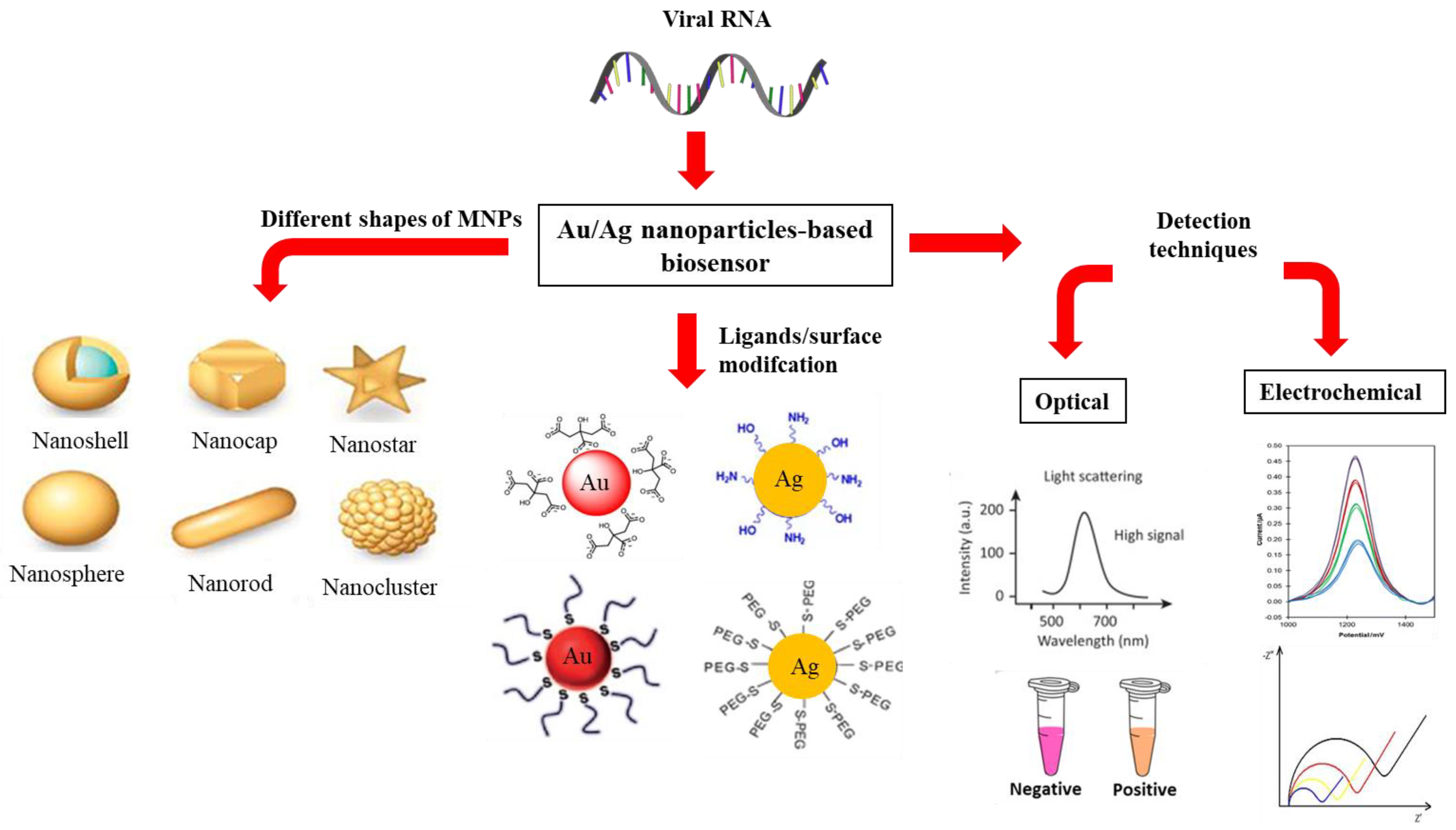
4. The Development of Optical and Electrochemical Metal Nanoparticles-Based Biosensors for the Detection of Viral RNA
4.1. Optical Nanobiosensors
4.1.1. Fluorescence-Based Assay
4.1.2. Localized Surface Plasmon Resonance
4.1.3. Colorimetric
4.1.4. Reflectance
4.2. Electrochemical Nanobiosensor
4.2.1. Voltammetric Detection
4.2.2. Impedimetric Detection
5. Conclusions
6. Recommendations
Author Contributions
Funding
Institutional Review Board Statement
Informed Consent Statement
Data Availability Statement
Conflicts of Interest
References
- Peiris, J.S.M.; Poon, L.L.M. Severe Acute Respiratory Syndrome (SARS). Encycl. Virol. 2008, 552–556. [Google Scholar] [CrossRef] [Green Version]
- Zhao, G.P. SARS molecular epidemiology: A Chinese fairy tale of controlling an emerging zoonotic disease in the genomics era. Philos. Trans. R. Soc. B Biol. Sci. 2007, 362, 1063–1081. [Google Scholar] [CrossRef] [PubMed] [Green Version]
- Vijay, R.; Perlman, S. Middle East Respiratory Syndrome and Severe Acute Respiratory Syndrome. Curr. Opin. Virol. 2016, 16, 70–76. [Google Scholar] [CrossRef] [PubMed]
- Hajjar, S.A.; Memish, Z.A.; McIntosh, K. Middle East Respiratory Syndrome Coronavirus (MERS-CoV): A perpetual challenge. Ann. Saudi Med. 2013, 33, 427–436. [Google Scholar] [CrossRef]
- Zhu, Z.; Lian, X.; Su, X.; Wu, W.; Marraro, G.A.; Zeng, Y. From SARS and MERS to COVID-19: A brief summary and comparison of severe acute respiratory infections caused by three highly pathogenic human coronaviruses. Respir. Res. 2020, 21, 1–14. [Google Scholar] [CrossRef]
- Rojas, M.; Monsalve, D.M.; Pacheco, Y.; Acosta-Ampudia, Y.; Ramírez-Santana, C.; Ansari, A.A.; Gershwin, M.E.; Anaya, J.M. Ebola virus disease: An emerging and re-emerging viral threat. J. Autoimmun. 2020, 106, 1–26. [Google Scholar] [CrossRef]
- World Health Organization (WHO), Ebola Virus Disease. 2021. Available online: https://www.who.int/news-room/fact-sheets/detail/ebola-virus-disease (accessed on 26 May 2021).
- Jacob, S.T.; Crozier, I.; Fischer, W.A.; Hewlett, A.; Kraft, C.S.; Vega, M.A.; Soka, M.J.; Wahl, V.; Griffiths, A.; Bollinger, L.; et al. Ebola virus disease. Nat. Rev. Dis. Primers 2020, 6, 1–31. [Google Scholar] [CrossRef] [PubMed] [Green Version]
- Gubler, D.J.; Vasilakis, N.; Musso, D. History and emergence of Zika virus. J. Infect. Dis. 2017, 216, 1–8. [Google Scholar] [CrossRef] [Green Version]
- Carod-Artal, F.J. Neurological complications of Zika virus infection. Expert Rev. Anti-Infect. Ther. 2018, 16, 399–410. [Google Scholar] [CrossRef]
- Póvoa, T.F.; Alves, A.M.; Oliveira, C.A.; Nuovo, G.J.; Chagas, V.L.; Paes, M.V. The pathology of severe dengue in multiple organs of human fatal cases: Histopathology, ultrastructure and virus replication. PLoS ONE 2014, 9, 1–16. [Google Scholar] [CrossRef] [Green Version]
- Zeng, Z.; Zhan, J.; Chen, L.; Chen, H.; Cheng, S. Global, regional, and national dengue burden from 1990 to 2017: A systematic analysis based on the global burden of disease study 2017. EClinicalMedicine 2021, 32, 1–7. [Google Scholar] [CrossRef] [PubMed]
- Altawalah, H.; Essa, S.; Ezzikouri, S.; Al-Nakib, W. Hepatitis B virus, hepatitis C virus and human immunodeficiency virus infections among people who inject drugs in Kuwait: A cross-sectional study. Sci. Rep. 2019, 9, 1–5. [Google Scholar] [CrossRef]
- Utsumi, T.; Lusida, M.I. Viral hepatitis and human immunodeficiency virus co-infections in Asia. World J. Virol. 2015, 4, 96–104. [Google Scholar] [CrossRef] [PubMed]
- Cevik, M.; Kuppalli, K.; Kindrachuk, J.; Peiris, M. Virology, transmission, and pathogenesis of SARS-CoV-2. BMJ 2020, 371, 1–6. [Google Scholar] [CrossRef]
- Zhang, X.S.; Vynnycky, E.; Charlett, A.; De Angelis, D.; Chen, Z.; Liu, W. Transmission dynamics and control measures of COVID-19 outbreak in China: A modelling study. Sci. Rep. 2021, 11, 1–12. [Google Scholar]
- Abebe, G.M. Emerging and Re-Emerging Viral Diseases: The case of coronavirus disease-19 (COVID-19). Int. J. Virol. AIDS 2020, 7, 67. [Google Scholar]
- Reta, D.H.; Tessema, T.S.; Ashenef, A.S.; Desta, A.F.; Labisso, W.L.; Gizaw, S.T.; Abay, S.M.; Melka, D.S.; Reta, F.A. Molecular and immunological diagnostic techniques of medical viruses. Int. J. Microbiol. 2020, 4, 1–19. [Google Scholar] [CrossRef]
- Tan, A.S.; Nerurkar, S.N.; Tan, W.C.C.; Goh, D.; Lai, C.P.T.; Poh Sheng, Y.J. The virological, immunological, and imaging approaches for COVID-19 diagnosis and research. SLAS Technol. 2020, 25, 1–23. [Google Scholar]
- Cui, X.; Shi, Y.; Zhao, L.; Gu, S.; Wei, C.; Yang, Y.; Wen, S.; Chen, H.; Ge, J. Application of real-time quantitative PCR to detect mink circovirus in naturally and experimentally infected minks. Front. Microbiol. 2018, 9, 1–8. [Google Scholar] [CrossRef]
- Wang, M.; Zhang, W.; Gao, Y.; Hu, B.; Ge, X.; Yang, X.; Zhang, Y.; Shi, Z. Longitudinal surveillance of SARS-like coronaviruses in bats by quantitative real-time PCR. Virol. Sin. 2016, 31, 78–80. [Google Scholar] [CrossRef] [PubMed] [Green Version]
- Choi, J.R. Development of point-of-care biosensors for COVID-19. Front. Chem. 2020, 8, 1–10. [Google Scholar] [CrossRef]
- Samson, R.; Navale, G.R.; Dharne, M.S. Biosensors: Frontiers in rapid detection of COVID-19. 3 Biotech 2020, 10, 1–9. [Google Scholar] [CrossRef] [PubMed]
- Wu, Q.; Zhang, Y.; Yang, Q.; Yuan, N.; Zhang, W. Review of electrochemical DNA biosensors for detecting food borne pathogens. Sensors 2019, 19, 4916. [Google Scholar] [CrossRef] [PubMed] [Green Version]
- Medhi, R.; Srinoi, P.; Ngo, N.; Tran, H.-V.; Lee, T.R. Nanoparticle-based strategies to combat COVID-19. ACS Appl. Nano Mater. 2020, 3, 8557–8580. [Google Scholar] [CrossRef]
- Khan, I.; Saeed, K.; Khan, I. Nanoparticles: Properties, applications and toxicities. Arab. J. Chem. 2019, 12, 908–931. [Google Scholar] [CrossRef]
- Malhotra, B.D.; Ali, M.A. Nanomaterials in biosensors: Fundamentals and applications. Nanomater. Biosens. 2018, 1–74. [Google Scholar] [CrossRef]
- Malekzad, H.; Zangabad, S.P.; Mirshekari, H.; Karimi, M.; Hamblin, M.R. Noble metal nanoparticles in biosensors: Recent studies and applications. Nanotechnol. Rev. 2017, 6, 301–329. [Google Scholar] [CrossRef]
- Loiseau, A.; Asila, V.; Boitel-Aullen, G.; Lam, M.; Salmain, M.; Boujday, S. Silver-based plasmonic nanoparticles for and their use in biosensing. Biosensors 2019, 9, 78. [Google Scholar] [CrossRef] [PubMed] [Green Version]
- Draz, M.S.; Shafiee, H. Applications of gold nanoparticles in virus detection. Theranostics 2018, 8, 1985–2017. [Google Scholar] [CrossRef]
- Tzeng, Y.; Lin, B.Y. Silver SERS adenine sensors with a very low detection limit. Biosensors 2020, 10, 53. [Google Scholar] [CrossRef] [PubMed]
- Xu, S.; Huang, X.; Chen, Y.; Liu, Y.; Zhao, W.; Sun, Z.; Zhu, Y.; Liu, X.; Wong, C.-P. Silver nanoparticle-enzyme composite films for hydrogen peroxide detection. ACS Appl. Nano Mater. 2019, 2, 5910–5921. [Google Scholar] [CrossRef]
- Yang, M.; Hood, Z.D.; Yang, X.; Chi, M.; Xia, Y. Facile synthesis of Ag@Au core-sheath nanowires with greatly improved stability against oxidation. Chem. Commun. 2017, 53, 1965–1968. [Google Scholar] [CrossRef] [PubMed]
- Parang, Z.; Keshavarz, A.; Farahi, S.; Elahi, S.M.; Ghoranneviss, M.; Parhoodeh, S. Fluorescence emission spectra of silver and silver/cobalt nanoparticles. Sci. Iran. 2012, 19, 943–947. [Google Scholar] [CrossRef] [Green Version]
- Amendola, V.; Pilot, R.; Frasconi, M.; Maragò, O.M.; Iatì, M.A. Surface plasmon resonance in gold nanoparticles: A review. J. Phys. Condens. Matter. 2017, 29, 1–49. [Google Scholar] [CrossRef] [PubMed]
- Chen, Y.; Munechika, K.; Ginger, D.S. Dependence of fluorescence intensity on the spectral overlap between fluorophores and plasmon resonant single silver nanoparticles. Nano Lett. 2007, 7, 690–696. [Google Scholar] [CrossRef] [PubMed]
- Matveeva, E.G.; Shtoyko, T.; Gryczynski, I.; Akopova, I.; Gryczynski, Z. Fluorescence quenching/enhancement surface assays: Signal manipulation using silver-coated gold nanoparticles. Chem. Phys. Lett. 2008, 454, 85–90. [Google Scholar] [CrossRef] [Green Version]
- Obliosca, J.M.; Liu, C.; Batson, R.A.; Babin, M.C.; Werner, J.H.; Yeh, H.C. DNA/RNA detection using DNA-templated few-atom silver nanoclusters. Biosensors 2013, 3, 185–200. [Google Scholar] [CrossRef] [Green Version]
- Cao, Q.; Teng, Y.; Yang, X.; Wang, J.; Wang, E. A label-free fluorescent molecular beacon based on DNA-Ag nanoclusters for the construction of versatile biosensors. Biosens. Bioelectron. 2015, 74, 318–321. [Google Scholar] [CrossRef] [PubMed]
- Malecka, K.; Kaur, B.; Cristaldi, D.A.; Chay, C.S.; Mames, I.; Radecka, H.; Radecki, J.; Stulz, E. Silver or gold? A comparison of nanoparticle modified electrochemical genosensors based on cobalt porphyrin-DNA. Bioelectrochemistry 2021, 138, 107723. [Google Scholar] [CrossRef]
- Kaur, B.; Malecka, K.; Cristaldi, D.A.; Chay, C.S.; Mames, I.; Radecka, H.; Radecki, J.; Stulz, E. Approaching single DNA molecule detection with an ultrasensitive electrochemical genosensor based on gold nanoparticles and cobalt-porphyrin DNA conjugates. Chem. Commun. 2018, 54, 11108–11111. [Google Scholar] [CrossRef] [Green Version]
- Rauwel, P.; Küünal, S.; Ferdov, S.; Rauwel, E. A review on the green synthesis of silver nanoparticles and their morphologies studied via TEM. Adv. Mater. Sci. Eng. 2015, 2015, 1–10. [Google Scholar] [CrossRef] [Green Version]
- Ogarev, V.A.; Rudoi, V.M.; Dement’eva, O.V. Gold nanoparticles: Synthesis, optical properties, and application. Inorg. Mater. Appl. Res. 2018, 9, 134–140. [Google Scholar] [CrossRef]
- Pinheiro, T.; Ferrão, J.; Marques, A.C.; Oliveira, M.J.; Batra, N.M.; Costa, P.M.F.J.; Macedo, M.P.; Águas, H.; Martins, R.; Fortunato, E. Paper-based in-situ gold nanoparticle synthesis for colorimetric, non-enzymatic glucose level determination. Nanomaterials 2020, 10, 2027. [Google Scholar] [CrossRef]
- Yang, Y.; Li, C.; Yin, L.; Liu, M.; Wang, Z.; Shu, Y.; Li, G. Enhanced charge transfer by gold nanoparticle at DNA modified electrode and its application to label-free DNA detection. ACS Appl. Mater. Interfaces 2014, 6, 7579–7584. [Google Scholar] [CrossRef]
- Carnerero, J.M.; Jimenez-Ruiz, A.; Castillo, P.M.; Prado-Gotor, R. Covalent and non-covalent DNA-gold-nanoparticle interactions: New avenues of research. Chemphyschem 2017, 18, 17–33. [Google Scholar] [CrossRef]
- Rasheed, P.A.; Sandhyarani, N. Femtomolar level detection of BRCA1 gene using a gold nanoparticle labeled sandwich type DNA sensor. Colloids Surf. B Biointerfaces 2014, 117, 7–13. [Google Scholar] [CrossRef]
- Hartati, Y.W.; Suryani, A.A.; Agustina, M.; Gaffar, S.; Anggraeni, A. A gold nanoparticle-DNA bioconjugate-based electrochemical biosensor for detection of Sus scrofa mtDNA in raw and processed meat. Food Anal. Methods 2019, 12, 2591–2600. [Google Scholar] [CrossRef] [Green Version]
- Zouari, M.; Campuzano, S.; Pingarrón, J.M.; Raouafi, N. Femtomolar direct voltammetric determination of circulating miRNAs in sera of cancer patients using an enzymeless biosensor. Anal. Chim. Acta 2020, 1104, 188–198. [Google Scholar] [CrossRef]
- Tao, Y.; Yin, D.; Jin, M.; Fang, J.; Dai, T.; Li, Y.; Li, Y.; Pu, Q.; Xie, G. Double-loop hairpin probe and doxorubicin-loaded gold nanoparticles for the ultrasensitive electrochemical sensing of microRNA. Biosens. Bioelectron. 2017, 96, 99–105. [Google Scholar] [CrossRef] [PubMed]
- Liang, H.; Wei, H.; Pan, D.; Xu, H. Chemically synthesized noble metal nanostructures for plasmonics. Nanotechnol. Rev. 2015, 4, 289–302. [Google Scholar] [CrossRef]
- Li, M.; Cushing, S.K.; Wu, N. Plasmon-enhanced optical sensors: A review. Analys 2015, 140, 386–406. [Google Scholar] [CrossRef] [PubMed] [Green Version]
- Liu, B.; Yan, H.; Chen, S.; Guan, Y.; Wu, G.; Jin, R.; Li, L. Stable and controllable synthesis of silver nanowires for transparent conducting film. Nanoscale Res. Lett. 2017, 12, 1–6. [Google Scholar] [CrossRef] [PubMed] [Green Version]
- Elsner, C.; Prager, A.; Sobottka, A.; Lotnyk, A.; Abel, B. Coated triangular Ag nanoprisms as optical sensors: Control of stability and spectral response with a thermo-responsive polymer. Anal. Methods 2017, 9, 4663–4672. [Google Scholar] [CrossRef]
- Roy, S.; Muhammed Ajmal, C.; Baik, S.; Kim, J. Silver nanoflowers for single-particle SERS with 10 pM sensitivity. Nanotechnology 2017, 28, 1–8. [Google Scholar] [CrossRef]
- Chang, Q.; Shi, X.; Liu, X.; Tong, J.; Liu, D.; Wang, Z. Broadband plasmonic silver nanoflowers for high-performance random lasing covering visible region. Nanophotonics 2017, 6, 1151–1160. [Google Scholar] [CrossRef]
- Ali, W.; Shabani, V.; Linke, M.; Sayin, S.; Gebert, B.; Altinpinar, S.; Hildebrandt, M.; Gutmann, J.S.; Mayer-Gall, T. Electrical conductivity of silver nanoparticle doped carbon nanofibres measured by CS-AFM. RSC Adv. 2019, 9, 4553–4562. [Google Scholar] [CrossRef] [Green Version]
- Santos, V.M.; Ribeiro, R.S.A.; Bosco, A.J.T.; Alhadeff, E.M.; Bojorge, N.I. Characterization and evaluation of silver-nanoparticle-incorporated in composite graphite aiming at their application in biosensors. Braz. J. 2017, 34, 647–657. [Google Scholar] [CrossRef] [Green Version]
- Tran, H.V.; Nguyen, T.V.; Nguyen, L.T.N.; Hoang, H.S.; Huynh, C.D. Silver nanoparticles as a bifunctional probe for label-free and reagentless colorimetric hydrogen peroxide chemosensor and cholesterol biosensor. J. Sci. Adv. Mater. Dev. 2020, 5, 385–391. [Google Scholar] [CrossRef]
- Godfrey, I.J.; Dent, A.J.; Parkin, I.P.; Maenosono, S.; Sankar, G. Structure of gold-silver nanoparticles. J. Phys. Chem. C 2017, 121, 1957–1963. [Google Scholar] [CrossRef]
- Mott, D.; Lee, J.; Thuy, N.T.B.; Aoki, Y.; Singh, P.; Maenosono, S. A study on the plasmonic properties of silver core gold shell nanoparticles: Optical assessment of the particle structure. Jpn. J. Appl. Phys. 2011, 50, 065004. [Google Scholar] [CrossRef]
- Zhao, K.; Ge, L.; Wong, T.I.; Zhou, X.; Lisak, G. Gold-silver nanoparticles modified electrochemical sensor array for simultaneous determination of chromium(III) and chromium(VI) in wastewater samples. Chemosphere 2021, 281, 130880. [Google Scholar] [CrossRef]
- Arvinte, A.; Crudu, I.-A.; Doroftei, F.; Timpu, D.; Pinteala, M. Electrochemical codeposition of silver-gold nanoparticles on CNT-based electrode and their performance in electrocatalysis of dopamine. J. Electroanal. Chem. 2018, 829, 184–193. [Google Scholar] [CrossRef]
- Heuer-Jungemann, A.; Feliu, N.; Bakaimi, I.; Hamsjaly, M.; Alkilany, A.; Chakraborty, I.; Masood, A.; Casula, M.F.; Kostopoulou, A.; Oh, E.; et al. The role of ligands in the chemical synthesis and applications of inorganic nanoparticles. Chem. Rev. 2019, 119, 4819–4880. [Google Scholar] [CrossRef] [PubMed] [Green Version]
- Guerrini, L.; Alvarez-Puebla, R.A.; Pazos-Perez, N. Surface modifications of nanoparticles for stability in biological fluids. Materials 2018, 11, 1154. [Google Scholar] [CrossRef] [Green Version]
- Li, B.; Lane, L.A. Probing the biological obstacles of nanomedicine with gold nanoparticles. Wiley Interdiscip. Rev. Nanomed. Nanobiotechnol. 2019, 11, 1–27. [Google Scholar] [CrossRef] [PubMed]
- Song, J.; Tan, Y.N.; Jańczewski, D.; Hempenius, M.A.; Xu, J.W.; Tan, H.R.; Vancso, G.J. Poly (ferrocenylsilane) electrolytes as a gold nanoparticle foundry: “two-in-one” redox synthesis and electrosteric stabilization, and sensing applications. Nanoscale 2017, 9, 19255–19262. [Google Scholar] [CrossRef] [PubMed] [Green Version]
- Moaseri, E.; Bollinger, J.A.; Changalvaie, B.; Johnson, L.; Schroer, J.; Johnston, K.P.; Truskett, T.M. Reversible self-assembly of glutathione-coated gold nanoparticle clusters via pH-tunable interactions. Langmuir 2017, 33, 12244–12253. [Google Scholar] [CrossRef] [PubMed]
- Mazloomi-Rezvani, M.; Salami-Kalajahi, M.; Roghani-Mamaqani, H.; Pirayesh, A. Effect of surface modification with various thiol compounds on colloidal stability of gold nanoparticles. Appl. Organometal. Chem. 2018, 32, 1–11. [Google Scholar] [CrossRef]
- Lee, S.H.; Jun, B.H. Silver nanoparticles: Synthesis and application for nanomedicine. Int. J. Mol. Sci. 2019, 20, 865. [Google Scholar] [CrossRef] [Green Version]
- Imran, M.; Ehrhardt, C.J.; Bertino, M.F.; Shah, M.R.; Yadavalli, V.K. Chitosan stabilized silver nanoparticles for the electrochemical detection of lipopolysaccharide: A facile biosensing approach for gram-negative bacteria. Micromachines 2020, 11, 413. [Google Scholar] [CrossRef] [Green Version]
- Kyrychenko, A.; Pasko, D.A.; Kalugin, O.N. Poly (vinyl alcohol) as a water protecting agent for silver nanoparticles: The role of polymer size and structure. Phys. Chem. Chem. Phys. 2017, 19, 8742–8756. [Google Scholar] [CrossRef] [PubMed]
- Swamidoss, V.F.; Bangaru, M.; Nalathambi, G.; Sangeetha, D.; Selvam, A.K. Silver-incorporated poly vinylidene fluoride nanofibers for bacterial filtration. Aerosol. Sci. Technol. 2019, 53, 196–206. [Google Scholar] [CrossRef]
- Pinzaru, I.; Coricovac, D.; Dehelean, C.; Moacă, E.A.; Mioc, M.; Baderca, F.; Sizemore, I.; Brittle, S.; Marti, D.; Calina, C.D.; et al. Stable PEG-coated silver nanoparticles—A comprehensive toxicological profile. Food Chem. Toxicol. 2018, 111, 546–556. [Google Scholar] [CrossRef] [PubMed]
- Mohamad Kasim, A.S.; Ariff, A.B.; Mohamad, R.; Wong, F. Interrelations of synthesis method, polyethylene glycol coating, physico-chemical characteristics, and antimicrobial activity of silver nanoparticles. Nanomaterials 2020, 10, 2475. [Google Scholar] [CrossRef]
- Vizzini, P.; Braidot, M.; Vidic, J.; Manzano, M. Electrochemical and optical biosensors for the detection of Campylobacter and Listeria: An update look. Micromachines 2019, 10, 500. [Google Scholar] [CrossRef] [Green Version]
- Peltomaa, R.; Glahn-Martínez, B.; Benito-Peña, E.; Moreno-Bondi, M.C. Optical biosensors for label-free detection of small molecules. Sensors 2018, 18, 4126. [Google Scholar] [CrossRef] [PubMed] [Green Version]
- Damborský, P.; Švitel, J.; Katrlík, J. Optical biosensors. Essays Biochem. 2016, 60, 91–100. [Google Scholar] [PubMed] [Green Version]
- Camarca, A.; Varriale, A.; Capo, A.; Pennacchio, A.; Calabrese, A.; Giannattasio, C.; Murillo, A.C.; D’Auria, S.; Staiano, M. Emergent biosensing technologies based on fluorescence spectroscopy and surface plasmon resonance. Sensors 2021, 21, 906. [Google Scholar] [CrossRef]
- Mozhgani, S.H.; Kermani, H.A.; Norouzi, M.; Arabi, M.; Soltani, S. Nanotechnology based strategies for HIV-1 and HTLV-1 retroviruses gene detection. Heliyon 2020, 6, e04048. [Google Scholar] [CrossRef] [PubMed]
- Abu-Salah, K.M.; Zourob, M.M.; Mouffouk, F.; Alrokayan, S.A.; Alaamery, M.A.; Ansari, A.A. DNA-based nanobiosensors as an emerging platform for detection of disease. Sensors 2015, 15, 14539–14568. [Google Scholar] [CrossRef] [Green Version]
- Tagit, O.; Hildebrandt, N. Fluorescence sensing of circulating diagnostic biomarkers using molecular probes and nanoparticles. ACS Sens. 2017, 2, 31–45. [Google Scholar] [CrossRef] [PubMed]
- Bayda, S.; Adeel, M.; Tuccinardi, T.; Cordani, M.; Rizzolio, F. The history of nanoscience and nanotechnology: From chemical-physical applications to nanomedicine. Molecules 2019, 25, 112. [Google Scholar] [CrossRef] [Green Version]
- Wen, Q.-L.; Peng, J.; Liu, A.-Y.; Wang, J.; Hu, Y.-L.; Ling, J.; Cao, Q.-E. DNA bioassays based on the fluorescence ‘turn off’ of silver nanocluster beacon. Luminescence 2020, 35, 702–708. [Google Scholar] [CrossRef] [PubMed]
- Jia, Z.; Tu, K.; Xu, Q.; Gao, W.; Liu, C.; Fang, B.; Zhang, M. A novel disease-associated nucleic acid sensing platform based on split DNA-scaffolded sliver nanocluster. Anal. Chim. Acta 2021, 1175, 338734. [Google Scholar] [CrossRef]
- Latorre, A.; Lorca, R.; Somoza, A. Fluorescent DNA stabilized silver nanoclusters as biosensors. J. Chem. 2013, 2013, 1–7. [Google Scholar] [CrossRef]
- Zarei-Ghobadi, M.; Mozhgani, S.H.; Dashtestani, F.; Yadegari, A.; Hakimian, F.; Norouzi, M.; Ghourchian, H. A genosensor for detection of HTLV-I based on photoluminescence quenching of fluorescent carbon dots in presence of iron magnetic nanoparticle-capped Au. Sci. Rep. 2018, 8, 1–8. [Google Scholar] [CrossRef] [PubMed]
- Pudza, M.Y.; Abidin, Z.Z. Synthesis and applications of organic-based fluorescent carbon dots: Technical review. In Novel Nanomaterials; IntechOpen: London, UK, 2021; ISBN 978-1-83881-026-9. [Google Scholar]
- Zhou, Z.; Xiao, R.; Cheng, S.; Wang, S.; Shi, L.; Wang, C.; Qi, K.; Wang, S. A universal SERS-label immunoassay for pathogen bacteria detection based on Fe3O4@Au-aptamer separation and antibody-protein A orientation recognition. Anal. Chim. Acta 2021, 1160, 338421. [Google Scholar] [CrossRef] [PubMed]
- Unser, S.; Bruzas, I.; He, J.; Sagle, L. Localized surface plasmon resonance biosensing: Current challenges and approaches. Sensors 2015, 15, 15684–15716. [Google Scholar] [CrossRef]
- Bukovinszky, K.; Szalóki, M.; Csarnovics, I.; Bonyár, A.; Petrik, P.; Kalas, B.; Daróczi, L.; Kéki, S.; Kökényesi, S.; Hegedűs, C. Optimization of plasmonic gold nanoparticle concentration in green LED light active dental photopolymer. Polymers 2021, 13, 275. [Google Scholar] [CrossRef]
- Qiu, G.; Gai, Z.; Tao, Y.; Schmitt, J.; Kullak-Ublick, G.A.; Wang, J. Dual-functional plasmonic photothermal biosensors for highly accurate severe acute respiratory syndrome coronavirus 2 detection. ACS Nano 2020, 14, 5268–5277. [Google Scholar] [CrossRef] [Green Version]
- Zhang, Y.; An, W.; Zhao, C.; Dong, Q. Radiation induced plasmonic nanobubbles: Fundamentals, applications and prospects. AIMS Energy 2021, 9, 676–713. [Google Scholar] [CrossRef]
- Qiu, G.; Gai, Z.; Saleh, L.; Tang, J.; Gui, T.; Kullak-Ublick, G.A.; Wang, J. Thermoplasmonic-Assisted Cyclic Cleavage Amplification for Self-Validating Plasmonic Detection of SARS-CoV-2. ACS Nano 2021, 15, 7536–7754. [Google Scholar] [CrossRef] [PubMed]
- Aldewachi, H.; Chalati, T.; Woodroofe, M.N.; Bricklebank, N.; Sharrack, B.; Gardiner, P. Gold nanoparticle-based colorimetric biosensors. Nanoscale 2017, 10, 18–33. [Google Scholar] [CrossRef] [PubMed] [Green Version]
- Jazayeri, M.H.; Aghaie, T.; Avan, A.; Vatankhah, A.; Ghaffari, S.M.R. Colorimetric detection based on gold nano particles (GNPs): An easy, fast, inexpensive, low-cost and short time method in detection of analytes (protein, DNA, and ion). Sens. Biosensing Res. 2018, 20, 1–8. [Google Scholar] [CrossRef]
- Kim, H.; Park, M.; Hwang, J.; Kim, J.H.; Chung, D.R.; Lee, K.S.; Kang, M. Development of label-free colorimetric assay for MERS-CoV using gold nanoparticles. ACS sensors 2019, 4, 1306–1312. [Google Scholar] [CrossRef]
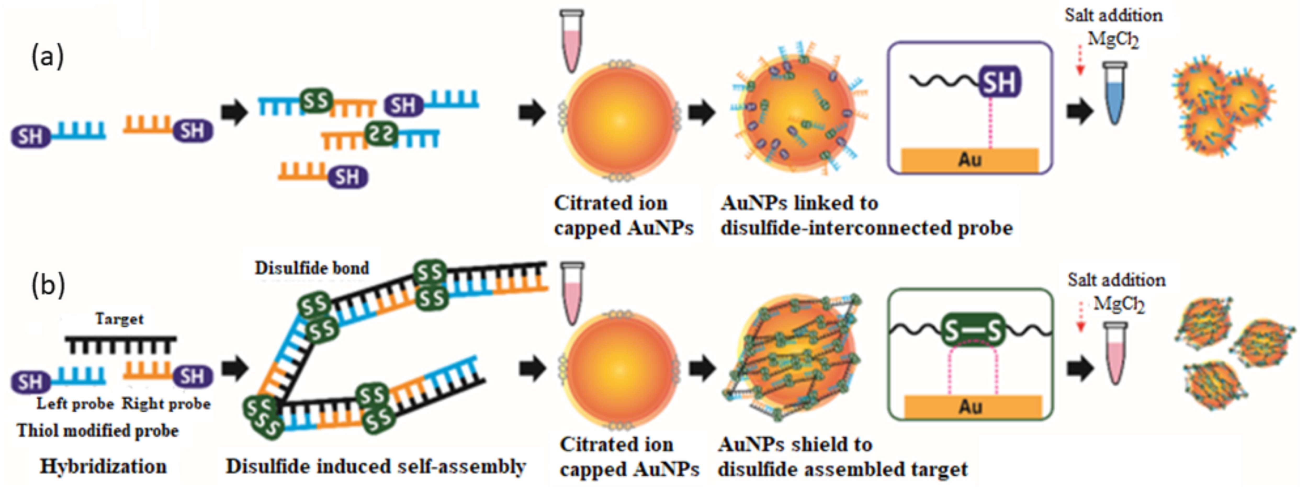
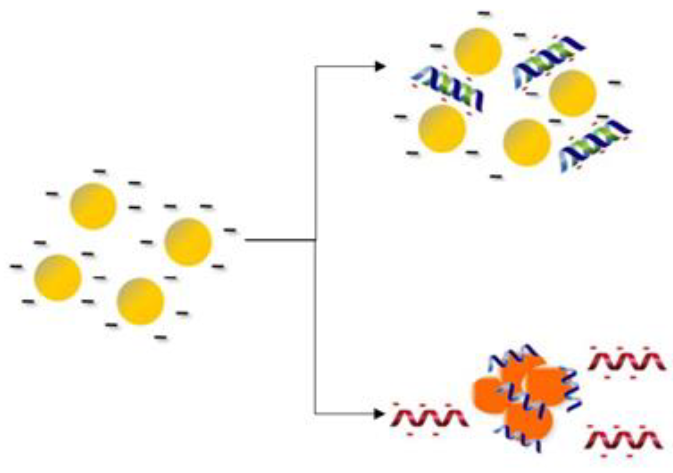
- Shokri, E.; Hosseini, M.; Davari, M.D.; Ganjali, M.R.; Peppelenbosch, M.P.; Rezaee, F. Disulfide-induced self-assembled targets: A novel strategy for the label free colorimetric detection of DNAs/RNAs via unmodified gold nanoparticles. Sci. Rep. 2017, 7, 1–11. [Google Scholar]
- Teengam, P.; Siangproh, W.; Tuantranont, A.; Vilaivan, T.; Chailapakul, O.; Henry, C.S. Multiplex paper-based colorimetric DNA sensor using pyrrolidinyl peptide nucleic acid-induced AgNPs aggregation for detecting MERS-CoV, MTB, and HPV oligonucleotides. Anal. Chem. 2017, 89, 5428–5435. [Google Scholar] [CrossRef] [PubMed] [Green Version]
- Charoenpakdee, C.; Vilaivan, T. Quenching of fluorescently labeled pyrrolidinyl peptide nucleic acid by oligodeoxyguanosine and its application in DNA sensing. Org. Biomol. Chem. 2020, 18, 5951–5962. [Google Scholar] [CrossRef]
- Singh, A.T.; Lantigua, D.; Meka, A.; Taing, S.; Pandher, M.; Camci-Unal, G. Paper-based sensors: Emerging themes and applications. Sensors 2018, 18, 2838. [Google Scholar] [CrossRef] [Green Version]
- Kuswandi, B.; Ensafi, A.A. Perspective—Paper-Based Biosensors: Trending Topic in Clinical Diagnostics Developments and Commercialization. J. Electrochem. Soc. 2019, 167, 037509. [Google Scholar] [CrossRef]
- Gauglitz, G. Analytical evaluation of sensor measurements. Anal. Bioanal Chem. 2018, 410, 5–13. [Google Scholar] [CrossRef] [Green Version]
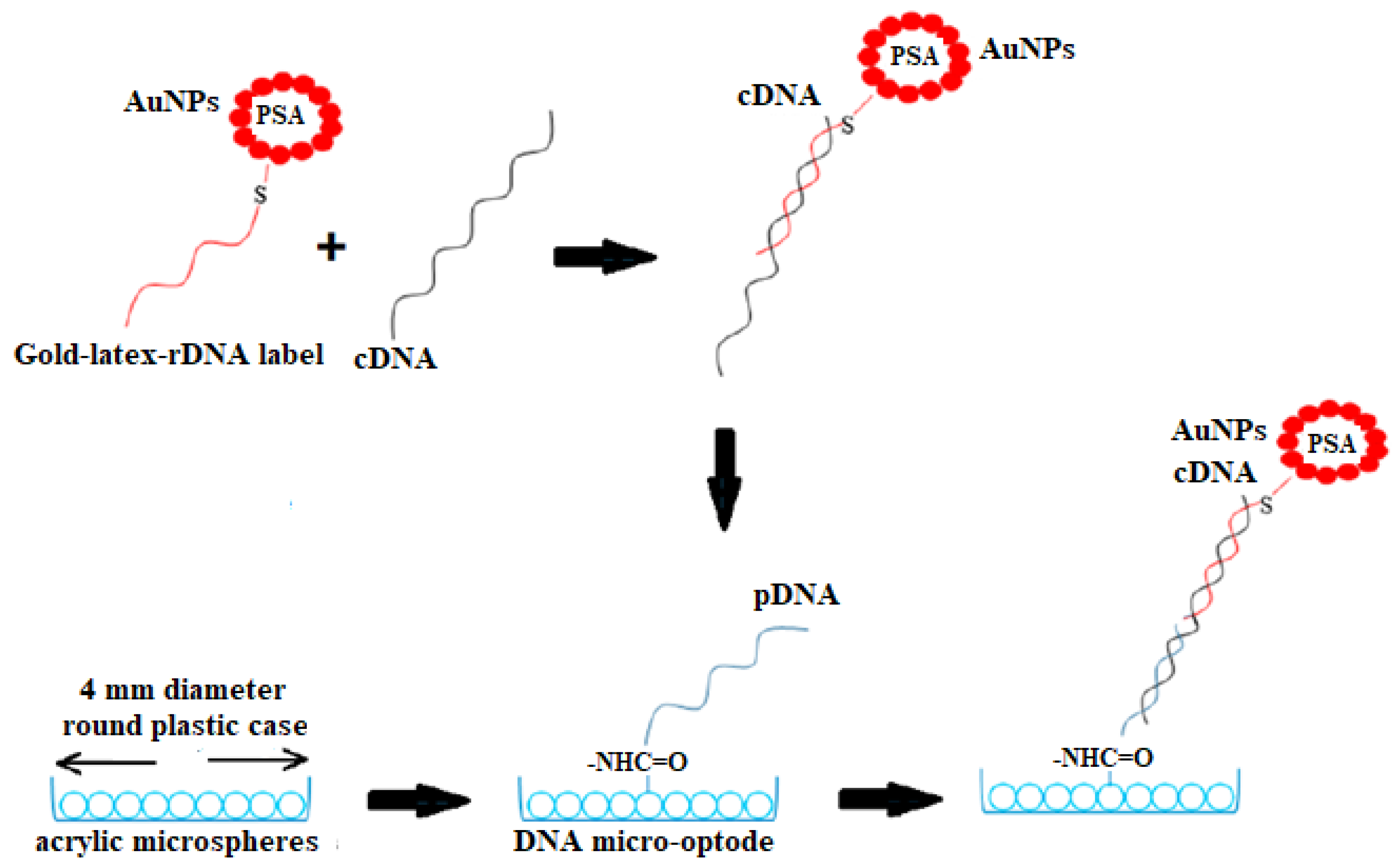
- Jeningsih; Tan, L.L.; Ulianas, A.; Heng, L.Y.; Mazlan, N.F.; Jamaluddin, N.D.; Mohd. Yusof, N.Y.; Khalid, B.; Ta, G.C. Sandwich-type DNA micro-optode based on gold–latex spheres label for reflectance dengue virus detection. Sensors 2020, 20, 1820. [Google Scholar] [CrossRef] [Green Version]
- Bayan, S.; Chakraborty, P. Secondary ion mass spectrometry and photoluminescence study on microstructural characteristics of chemically synthesized ZnO nanowalls. Appl. Surf. Sci. 2014, 303, 233–240. [Google Scholar] [CrossRef]
- Santhanam, M.; Algov, I.; Alfonta, L. DNA/RNA electrochemical biosensing devices a future replacement of PCR methods for a fast epidemic containment. Sensors 2020, 20, 4648. [Google Scholar] [CrossRef] [PubMed]
- Aryand, M.; Shabani, A.; Ardaki, M.S. A new electrochemical sensing platform based on binary composite of graphene oxide-chitosan for sensitive rutin determination. Food Anal. Methods 2017, 10, 2332–2345. [Google Scholar] [CrossRef]
- Jiang, Y.; Wu, J. Recent development in chitosan nanocomposites for surface-based biosensor applications. Electrophoresis 2019, 40, 2084–2097. [Google Scholar] [CrossRef]
- Mahato, K.; Nagpal, S.; Shah, M.A.; Srivastava, A.; Maurya, P.K.; Roy, S.; Jaiswal, A.; Singh, R.; Chandra, P. Gold nanoparticle surface engineering strategies and their applications in biomedicine and diagnostics. Biotech 2019, 9, 1–19. [Google Scholar] [CrossRef]
- Abdul Rashid, J.I.; Yusof, N.A. The strategies of DNA immobilization and hybridization detection mechanism in the construction of electrochemical DNA sensor: A review. Sens Biosensing Res. 2017, 16, 19–31. [Google Scholar] [CrossRef]
- Dziabowska, K.; Czaczyk, E.; Nidzworski, D. Application of electrochemical methods in biosensing technologies. In Biosensing Technologies for the Detection of Pathogens—A Prospective Way for Rapid Analysis; IntechOpen: London, UK, 2017; ISBN 978-953-51-3916-4. [Google Scholar]
- Li, H.; Liu, X.; Li, L.; Mu, X.; Genov, R.; Mason, A.J. CMOS electrochemical instrumentation for biosensor microsystems: A review. Sensors 2017, 17, 74. [Google Scholar] [CrossRef]
- Zhang, Z.; Li, Q.; Du, X.; Liu, M. Application of electrochemical biosensors in tumor cell detection. Thorac. Cancer 2020, 11, 840–850. [Google Scholar] [CrossRef]
- Zamfir, L.G.; Puiu, M.; Bala, M.C. Advances in electrochemical impedance spectroscopy detection of endocrine disruptors. Sensors 2020, 2, 6443. [Google Scholar] [CrossRef]
- Gil, R.; Amorim, C.G.; Araújo, A.N.; Montenegro, M.C.B.S.M. Process analysis|Electroanalytical techniques. In Encyclopedia of Analytical Science, 3rd ed.; Worsfold, P., Poole, C., Townshend, A., Miró, M., Eds.; Academic Press: Cambridge, MA, USA, 2019; pp. 384–388. [Google Scholar]
- Hussain, G.; Silvester, D.S. Comparison of voltammetric techniques for ammonia sensing in ionic liquids. Electroanalysis 2018, 30, 75–83. [Google Scholar] [CrossRef]
- Scott, K. Electrochemical principles and characterization of bioelectrochemical systems. In Microbial Electrochemical and Fuel Cells; Scott, K., Yu, E.H., Eds.; Woodhead Publishing: Cambridge, UK, 2016; pp. 29–66. [Google Scholar]
- Simoes, F.R.; Xavier, M.G. Electrochemical Sensors. In Micro and Nano Technologies, Nanoscience and its Applications; Da Róz, A.L., Ferreira, M., de Lima Leite, F., Oliveira, O.N., Eds.; William Andrew Publishing: Norwich, NY, USA, 2017; pp. 155–178. [Google Scholar]
- Cajigas, S.; Alzate, D.; Orozco, J. Gold nanoparticle/DNA-based nanobioconjugate for electrochemical detection of Zika virus. Mikrochim. Acta 2020, 187, 1–10. [Google Scholar] [CrossRef]
- Alafeef, M.; Dighe, K.; Moitra, P.; Pan, D. Rapid, ultrasensitive, and quantitative detection of SARS-CoV-2 using antisense oligonucleotides directed electrochemical biosensor chip. ACS Nano 2020, 14, 17028–17045. [Google Scholar] [CrossRef]
- Leva-Bueno, J.; Peyman, S.A.; Millner, P.A. A review on impedimetric immunosensors for pathogen and biomarker detection. Med. Microbiol. Immunol. 2020, 209, 343–362. [Google Scholar] [CrossRef] [PubMed] [Green Version]
- Karash, S.; Wang, R.; Kelso, L.; Lu, H.; Huang, T.J.; Li, Y. Rapid detection of avian influenza virus H5N1 in chicken tracheal samples using an impedance aptasensor with gold nanoparticles for signal amplification. J. Virol. Methods 2016, 236, 147–156. [Google Scholar] [CrossRef] [PubMed] [Green Version]
- Teengam, P.; Siangproh, W.; Tuantranont, A.; Vilaivan, T.; Chailapakul, O.; Henry, C.S. Electrochemical impedance-based DNA sensor using pyrrolidinyl peptide nucleic acids for tuberculosis detection. Anal. Chim. Acta 2018, 1044, 102–109. [Google Scholar] [CrossRef] [PubMed]
- Ilkhani, H.; Farhad, S. A novel electrochemical DNA biosensor for Ebola virus detection. Anal. Biochem. 2018, 557, 151–155. [Google Scholar] [CrossRef]
- Yang, X.; Gao, Z. Enzyme-catalysed deposition of ultrathin silver shells on gold nanorods: A universal and highly efficient signal amplification strategy for translating immunoassay into a litmus-type test. Chem. Commun. 2015, 51, 6928–6931. [Google Scholar] [CrossRef] [Green Version]
- Kemp, N.T. A tutorial on electrochemical impedance spectroscopy and nanogap electrodes for biosensing applications. IEEE Sens. J. 2021. [CrossRef]
- Bhalla, N.; Jolly, P.; Formisano, N.; Estrela, P. Introduction to biosensors. Essays Biochem. 2016, 60, 1–8. [Google Scholar] [PubMed] [Green Version]
- Islam, T.; Hasan, M.M.; Awal, A.; Nurunnabi, M.; Ahammad, A. Metal nanoparticles for electrochemical sensing: Progress and challenges in the clinical transition of point-of-care testing. Molecules 2020, 25, 5787. [Google Scholar] [CrossRef] [PubMed]
- Martines-Arano, H.; García-Pérez, B.E.; Vidales-Hurtado, M.A.; Trejo-Valdez, M.; Hernández-Gómez, L.H.; Torres-Torres, C. Chaotic signatures exhibited by plasmonic effects in Au nanoparticles with cells. Sensors 2019, 19, 4728. [Google Scholar] [CrossRef] [PubMed] [Green Version]
- Ekrami, E.; Pouresmaieli, M.; Barati, F.; Asghari, S.; Ziarani, F.R.; Shariati, P.; Mamoudifard, M. Potential diagnostic systems for coronavirus detection: A critical review. Biol. Proced. Online 2020, 22, 1–19. [Google Scholar] [CrossRef] [PubMed]


| Nanoparticles | Design of the Probe | The Approaches for Analyte Detection | Target Genes | Technique | LOD |
|---|---|---|---|---|---|
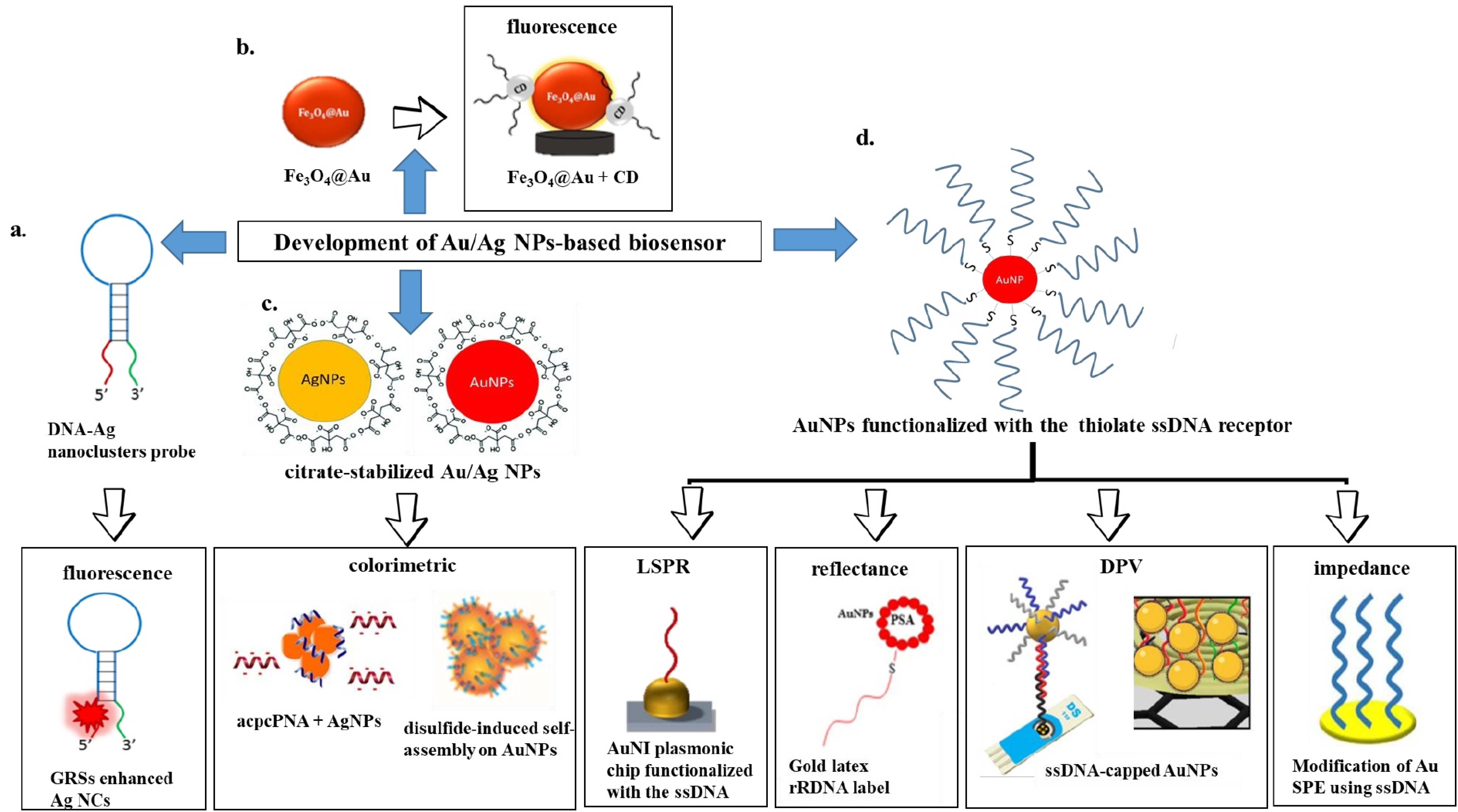
| AgNCs | The probe is tagged with AgNCs and GRSs two terminals with a loop whose sequence was complementary to cDNA (HIV, HBV and HTLV-I) | The detection of analyte was based on the fluorescence quenching of AgNCs in the presence of cDNA, which open the hairpin-shape probe and keeps AgNCs away from GRSs. This results in the decrease of fluorescence intensity. | HIV gene, HBV gene and HTLV-I gene | Fluorescence | HIV = 4.4 nM HBV = 6.8 nM HTLV-I = 8.5 nM [39] |
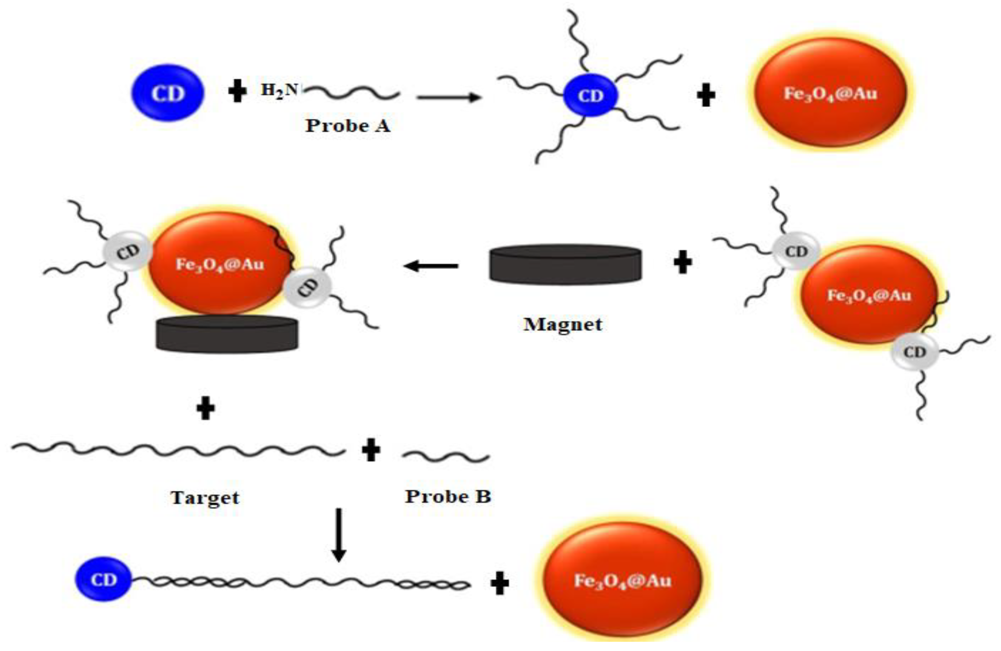
| CDs and Fe3O4@Au | Two probes were designed; CDs/DNA (probe A) and probe (B) to complete the DNA hybridization | The detection of analyte was based on the fluorescence quenching of CDs in the proximity of Fe3O4@Au. In the presence of cDNA, the fluorescence emission of CDs was recovered, since the CDs/DNA hybridized with the cDNA and formed double-strand DNA, which cannot adsorb on the Fe3O4@Au surface | HTLV-I | Fluorescence | 10 nM [87] |
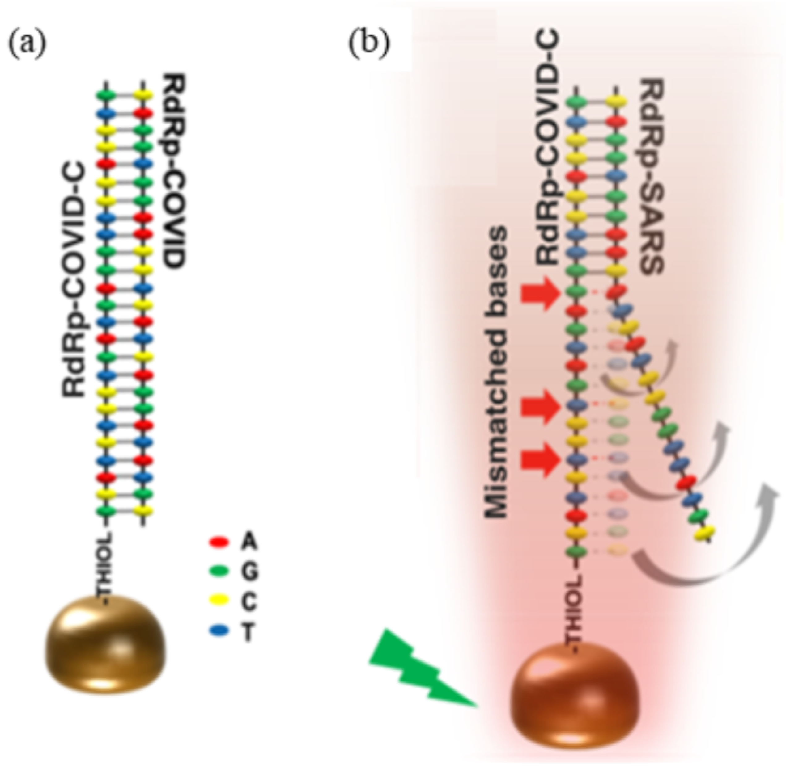
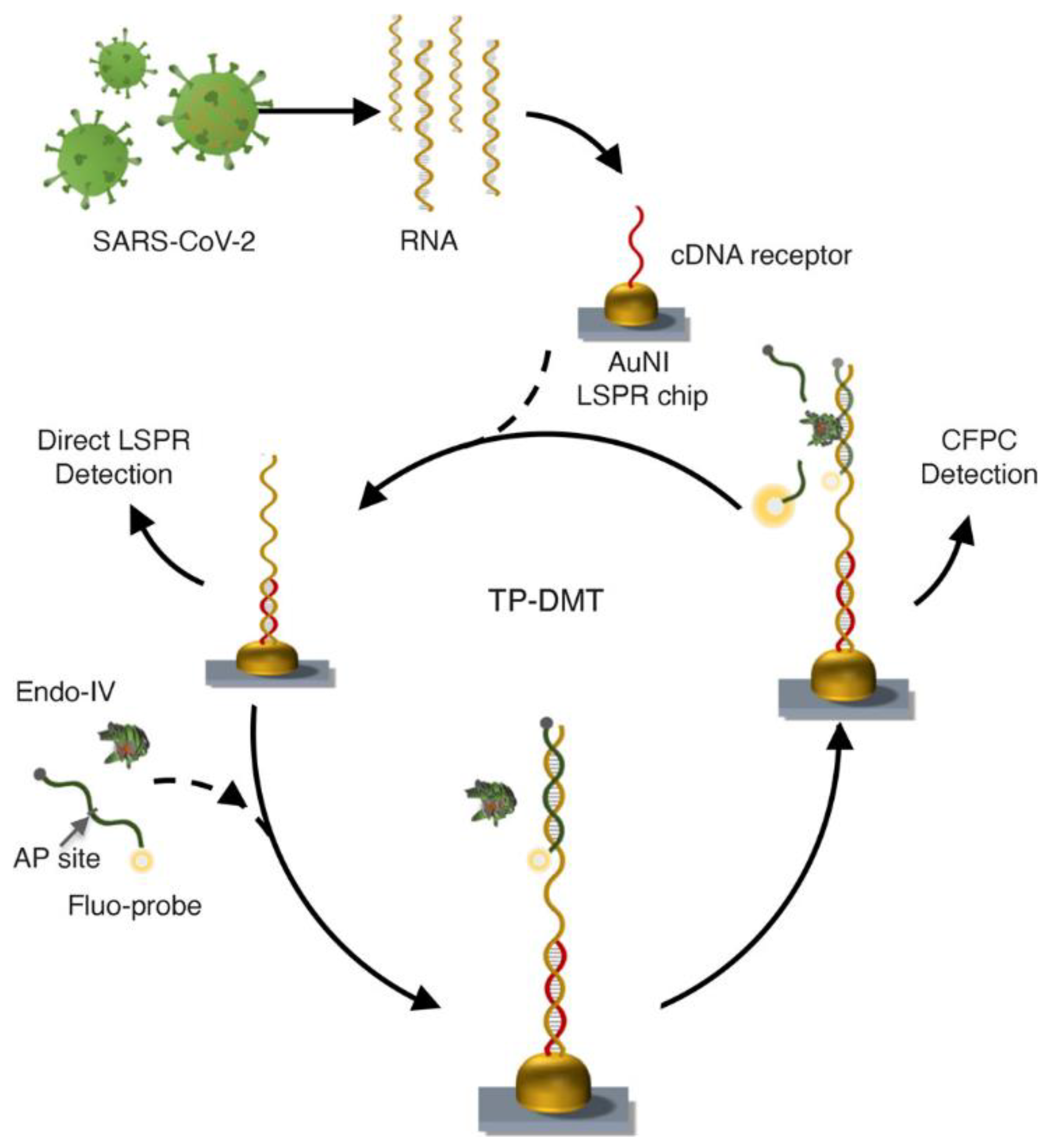
| The two-dimensional AuNIs | The AuNI surface was functionalized with thiol-cDNA | A dual-functional plasmonic biosensor combining the PPT effect and LSPR sensing transduction was used to detect the analyte. | SARS-CoV-2 | LSPR | 0.22 ± 0.08 pM [92] |
| The LSPR transduction signal based on the hybridization between the target sequences and the functionalized thiol-DNA receptors was involved in the direct sensing system. In CFPC-based detection, the mixture of API-site-modified fluorescent probe and endo IV were used. The cleaved strand of the probes stimulates the LSPR response. | SARS-CoV-2 | LSPR | Direct sensing system = 0.1 ± 0.04 pM CFPC-based detection = 0.275 ± 0.051 fM [94] | ||
| AuNPs | Two thiol modified probes at the 5′ site (right) and 3′ site (left) | The formation of a disulfide-induced interconnection between the probes in the absence of target caused AuNPs aggregation (color changed from red to blue) and the formation of disulfide-induced long self-assembled complex prevented AuNPs aggregation in the presence of target (no color change). | MERS-CoV | Colorimetric test | 1 pmol μL−1 [97] |
| AgNPs | The acpcPNA was designed as a probe. | The color change of AgNPs (yellow to red) was observed due to the electrostatic interaction between acpcPNA with AgNPs (aggregated). In the presence of cDNA, the interaction of acpcPNA and DNA led to the depletion of acpcPNA-AgNPs interaction, which changed the color from red to yellow again (non-aggregated). | MERS-CoV MTB HPV | Colorimetric test | MERS-CoV: 1.53 nM MTB: 1.27 nM HPV: 1.03 nM [99] |
| AuNPs-PSA (latex) | AuNPs-PSA latex particles were attached to the thiolated reporter probe (rDNA) by Au–thiol. | Sandwich hybridization strategy of DNAs was performed on poly(nBA-NAS) microspheres. Microsphere poly(nBA-NAS) conjugated to the aminated pDNA via a peptide covalent bond hybridized to the cDNA-rDNA-AuNP-PSA complex, which attenuated the reflectance signal. | DENV serotype 2 | Reflectance spectrophotometer | 1 × 10⁻²⁹ M [104] |
| SPAuE or a SPCE/Au | ssDNA monolayer consisting of thiolated signal and spacer probes linked to AuNPs | In the presence of the target RNA, it is sandwiched in between the capture probe and the signal probe from the nanobioconjugate. Then, the Ru3+ complex is coupled by electrostatic interaction, serving as a reporter of the electrochemical measured by DPV. If the target is absent, the resultant DPV signal is much smaller due to no assembly of the genosensor. | Zika virus | DPV | SPAuE: 0.2 fM SPCE/Au: 33 fM [119] |
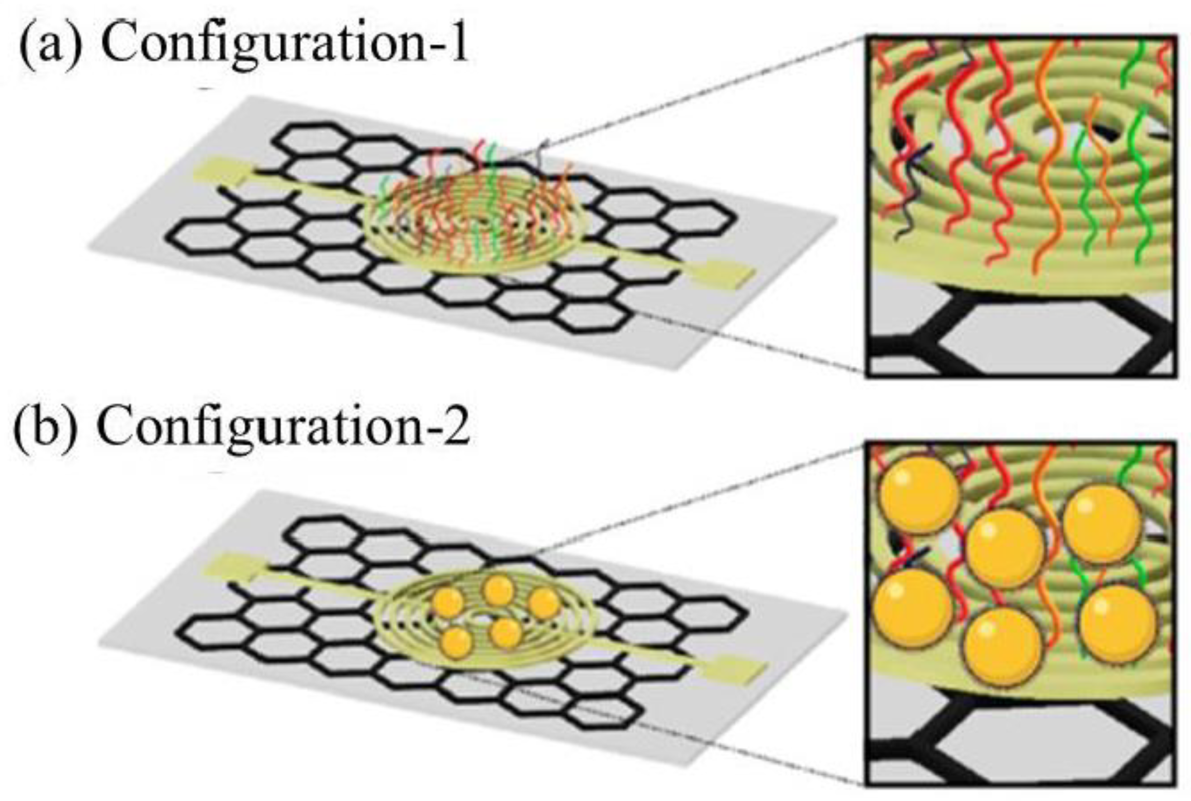
| AuNPs | Two antisense probes, P1 and P3, were modified with thiol at the 5′ end and another two thiol-modified antisense probes at the 3′ end (P2 and P4) | In the presence of SARS-CoV-2 RNA, the specific RNA−DNA hybridization led to the change in charge and electron mobility on the graphene surface, which brought the change in sensor output voltage. | SARS-CoV-2 | DPV | 6.9 copies μL−1 [120] |
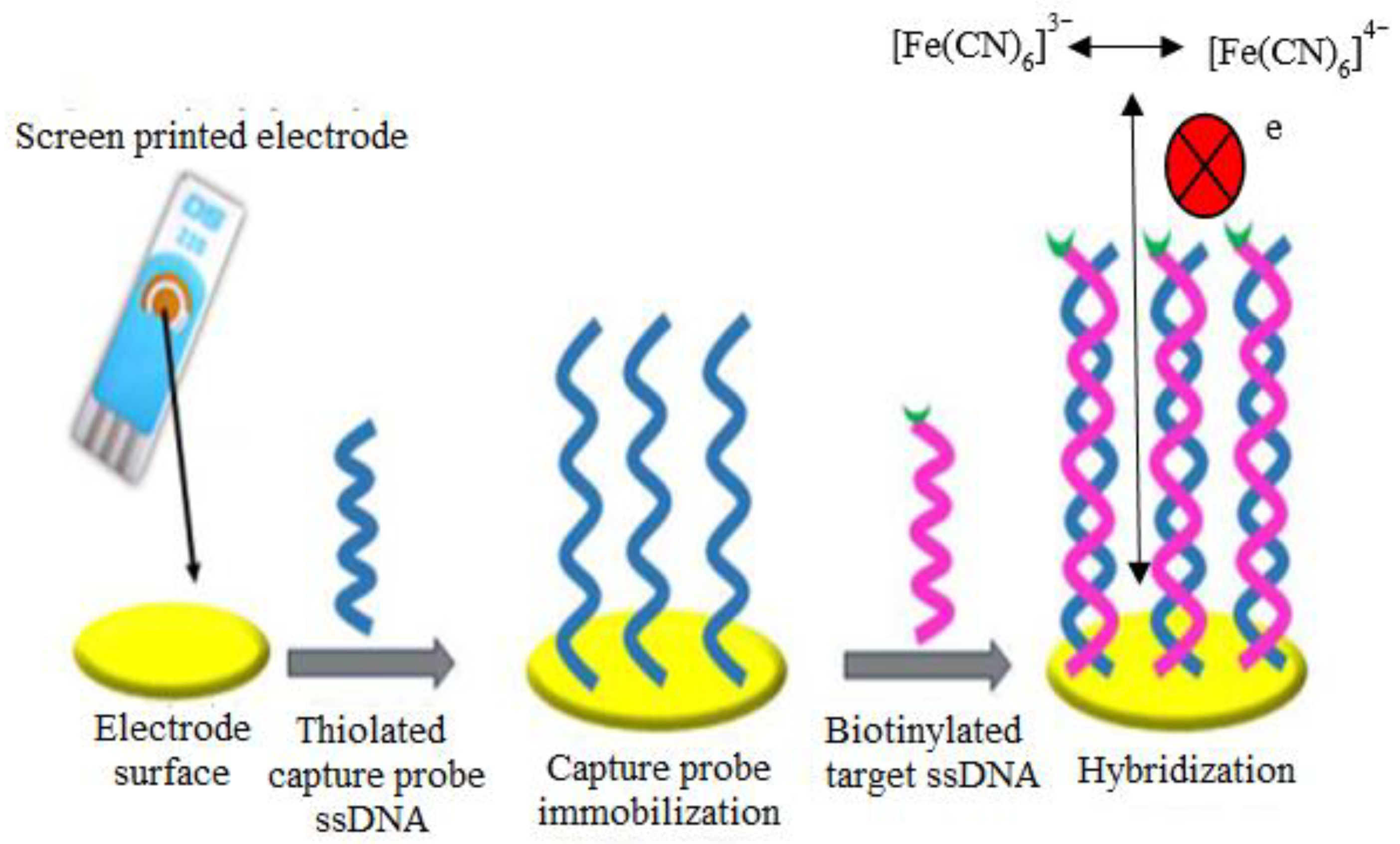
| AuNPs modified SPE | A thiolated DNA capture probe sequence was immobilized on the SPE surface | The redox probe [Fe(CN)6]3−/4− was used to measure the Rct values after the DNA hybridization. The interaction of [Fe(CN)6]3−/4− with dsDNA formed an electrostatic repulsion due to the presence of negatively charged phosphate backbone of DNA molecule. As a result, the electron transfer could be blocked, which led to the increase of impedance value (Rct values). | Ebola virus | EIS | 4.7 nM [124] |
Publisher’s Note: MDPI stays neutral with regard to jurisdictional claims in published maps and institutional affiliations. |
© 2021 by the authors. Licensee MDPI, Basel, Switzerland. This article is an open access article distributed under the terms and conditions of the Creative Commons Attribution (CC BY) license (https://creativecommons.org/licenses/by/4.0/).
Share and Cite
Ibrahim, N.; Jamaluddin, N.D.; Tan, L.L.; Mohd Yusof, N.Y. A Review on the Development of Gold and Silver Nanoparticles-Based Biosensor as a Detection Strategy of Emerging and Pathogenic RNA Virus. Sensors 2021, 21, 5114. https://doi.org/10.3390/s21155114
Ibrahim N, Jamaluddin ND, Tan LL, Mohd Yusof NY. A Review on the Development of Gold and Silver Nanoparticles-Based Biosensor as a Detection Strategy of Emerging and Pathogenic RNA Virus. Sensors. 2021; 21(15):5114. https://doi.org/10.3390/s21155114
Chicago/Turabian StyleIbrahim, Nadiah, Nur Diyana Jamaluddin, Ling Ling Tan, and Nurul Yuziana Mohd Yusof. 2021. "A Review on the Development of Gold and Silver Nanoparticles-Based Biosensor as a Detection Strategy of Emerging and Pathogenic RNA Virus" Sensors 21, no. 15: 5114. https://doi.org/10.3390/s21155114