Review

The Role of Oxidative Stress and the Importance of miRNAs as Potential Biomarkers in the Development of Age-Related Macular Degeneration

1
Department of Clinical Chemistry and Biochemistry, Medical University of Lodz, 90-136 Lodz, Poland
2
Department of Gerontology, Geriatrics and Social Work, Jesuit University Ignatianum, 31-501 Krakow, Poland
3
Department of Ophthalmology, SPKSO Ophthalmic Hospital, Medical University of Warsaw, 00-576 Warsaw, Poland
4
Laser Eye Microsurgery Center, Clinic of Jerzy Szaflik, 00-215 Warsaw, Poland
*
Authors to whom correspondence should be addressed.
Processes 2021, 9(8), 1328; https://doi.org/10.3390/pr9081328
Submission received: 3 May 2021 / Revised: 22 June 2021 / Accepted: 8 July 2021 / Published: 30 July 2021
(This article belongs to the Special Issue Advances of Redox Status in Disease)

Abstract

:
Age-related macular degeneration (AMD) is the primary cause of blindness in developed countries. With the progressive aging of the population, AMD is a significant ophthalmological problem in the population over 50 years of age. The etiology of AMD is known to be based on various biochemical, immunological and molecular pathways and to be influenced by a range of genetic and environmental elements. This review provides an overview of the pathophysiological role of oxidative stress and free radicals in the retina with a special focus on the DNA repair efficiency and enzymatic antioxidant defense. It also presents a correlation between miRNA profile and AMD, and indicates their involvement in inflammation, angiogenesis, increased oxidation of cellular components, enzymatic antioxidant capacity and DNA repair efficiency, which play particularly important roles in AMD pathogenesis. Gene silencing by miRNAs can induce changes in antioxidant enzymes, leading to a complex interplay between redox imbalance by free radicals and miRNAs in modulating cellular redox homeostasis.

1. Introduction

Age-related macular degeneration (AMD) is a progressive, neurodegenerative disease that affects the macula of the retina. The incidence of AMD was estimated to be approximately 170 million people worldwide in 2016, but this number is predicted to increase to 288,000,000 in 2040, thus becoming the third leading cause of vision loss worldwide [1]. AMD is a slow and progressive disease that attacks retinal cells in the macula, the region of the eye responsible for central vision. The macula, which is at the center of retina, is particularly prone to age-related degenerative changes. The retinal pigment epithelium RPE, adjacent to Bruch’s membrane, has many important functions, including light absorption, phagocytosis of photoreceptor outer segments, heat exchange, vitamin A metabolism, retinal outer blood barrier, and maintenance of choriocapillaris.
AMD is a disease affecting four components of the visual organ: photoreceptors, retinal pigment epithelium (RPE), Bruch’s membrane, and choroidal capillaries. This process results in the destabilization of four key physiological functions: homeostasis/stress response, extracellular remodeling, complement-related inflammation, and phagocytosis. Disruption of these functions leads to distortion of Bruch’s membrane, the RPE, and the subretinal space. The pigmentary abnormalities in the RPE, drusen and Bruch’s membrane, and the molecular changes associated with this disease, are believed to be caused by inflammation, angiogenesis, and increased oxidation of cellular components, which play a central role in the pathogenesis and progression of AMD [2].
Although the etiology of AMD is still not well understood, the disease is divided into early and late stages. The early stage is characterized by the accumulation of lipofuscin and drusen deposits between Bruch’s membrane and the RPE, the specialized cells that form the outer blood–retinal barrier. Detection of vision deterioration in early stage of AMD is important for both prevention and treatment.
Late-stage AMD is divided into dry and wet forms, which account for about 85% and 15% of cases, respectively. Dry AMD (geographic atrophy (GA) or exudation-free form) is associated with slowly progressive RPE and choriocapillaris dysfunction, photoreceptor loss, and retinal degeneration. In addition, dry AMD is characterized by the formation of pale or yellow deposits called druses in the space between the RPE and Bruch’s membrane. The drusen are composed of various components, including lipid and amyloid-β deposits [3]. It has been documented that the total number of drusen or the measured area and volume of drusen can be risk factors for progression to dry or wet AMD [4].
Wet AMD, i.e., the neovascular (nAMD) or exudative form, is characterized by subretinal neovascularization that leads to fibrovascular scarring and subsequent loss of central vision. The hallmark of wet AMD is abnormal growth of blood vessels under the macula. In addition, there is pigment epithelial detachment, the presence of intraretinal and subretinal fluid, macular hemorrhage, hard exudates or subretinal fibrovascular scarring. In addition, retinal neovascularization can also take place following the rupture of Bruch’s membrane, which damages the macula and results in blurred or macular vision [2,5].
Poor antioxidant capacity, and the resulting overproduction of free radicals, is thought to be a major factor affecting RPE in the pathophysiology of AMD. This theory is underpinned by both environmental and genetic factors. The macula lives in an environment of high oxidative stress. Normal antioxidant capacity is influenced by the activity of enzymatic and non-enzymatic defense mechanisms, as well as the efficiency of DNA repair. DNA repair efficiency depends on the expression of genes encoding DNA repair proteins, which is mainly regulated by the interaction of regulatory proteins and specific sequences regulatory proteins and specific sequences in DNA repair genes. Changes in miRNA expression profile occur during organ development, aging, or cell death. The expression of miRNAs is also altered in the pathophysiology of complex diseases such as inflammation or neurodegenerative diseases including AMD. Furthermore, in addition to its role in regulating gene expression, miRNAs released into body fluids have emerged as potential blood serum biomarkers. Some researchers have reported that mRNA transcription factors are sensitive to redox status [6,7].

2. AMD—Risk Factors

AMD is believed to arise in response to a combination of environmental, demographic, and genetic risk factors. The most strongly associated environmental factors are age, family history, smoking and previous cataract surgery. Other risk factors include female gender, iris color, white race, high body mass index, history of cardiovascular disease, hypertension, and lifetime exposure to sunlight [8,9,10,11,12]. In addition, dietary factors, eating an unbalanced diet, and high fat intake may also be associated with an increased risk of AMD.
The known genetic risk factors associated with AMD susceptibility include the presence of single nucleotide polymorphisms in Complement factor H identified CFH on chromosome 1q32; LOC387715/ARMS2 on 10q26, associated with susceptibility to age-related maculopathy; HTRA1 on 10q26, a factor that plays a role in extracellular matrix homeostasis and cell growth or survival. In addition, BF and complement component 2 (C2), an activator of the classical complement pathway, are paralogous genes located just 500 bp apart on human chromosome 6p21. These two genes, along with genes encoding complement components 4A (C4A) and 4B (C4B), reside in the C2/BF class III region of the major tissue compatibility complex (MHC) on chromosome 6p21 as major AMD susceptibility genes [13]. In addition, studies of genetic variants in the components of the complement system suggest that immune system disorders and inflammation may influence the development of AMD [14,15,16,17].
The most important risk factor for AMD is age over 50 years. The retina and RPE show structural changes during aging. Moreover, with age, extracellular deposits (drusen) associated with degenerative pathology develop in the retina, inflammatory mediators indicative of local inflammation typically associated with subretinal deposits increase [18,19,20]. It has been found that in Europe, the prevalence of early AMD increases from 3.5% in people aged 55–59 years to 17.6% in people aged 85 years and older, with the prevalence increasing from 0.1% to 9.8%, respectively, for late AMD [21].
It is believed that aging of Bruch’s membrane and RPE, i.e., accumulation of waste material, leads to a decrease in metabolic function, and adds to the potential for new blood vessel growth. Furthermore, dysfunction of retinal pigment epithelial cells with age leads to the accumulation of lipofuscin, a biomarker of aging. It is a yellowish pigment made of free radical-damaged proteins and fat that accumulates gradually mainly in aging post-mitotic cells. This pigment accumulates in RPE cells after birth as byproducts from phagocytosis of photochemically oxidized photoreceptor outer segments. Lipofuscin is composed of visual cycle proteins, membrane proteins, and phospholipids, retinoids formed by incomplete enzymatic degradation in phagolysosomes. Oxidation of N-retinylidene-N-retinyl ethanolamine (A2E), a photocytotoxic component of age pigment, can lead to the formation of toxic free radicals that can damage the structure of RPE cell biomolecules [22,23].
When the eye is exposed to sunlight, in the presence of oxygen, plasma membrane phospholipids i.a. docosahexaenoic acid (DHA) combines with phosphatidylethanolamine PE (DHA-PA) and phosphatidylcholine PC (DHA-PC) and then forms carboxyethylpyrrole-protein adducts (CEPs) when combined with protein. CEP adduct levels are higher in the drusen, Bruch’s membrane, choroid, and retinal pigment epithelium (RPE). It has also been suggested that CEPs may initiate neovascularization in the wet form of the disease [24].
Smoking is an epidemiologically modifiable risk factor for AMD and is associated with a 2–4-fold increased risk of any type of AMD. Smoking and inhaled toxins may also account for the increased AMD risk associated with high levels of reactive oxygen species (ROS) and reactive nitrogen species (RNS), which are known to cause, among other things, oxidative damage to cellular components, RPE cell inflammation, and vascular modifications in choroidal vessels. While oxidative modifications of phospholipids and lipoproteins are generated, they demonstrate pro-inflammatory and atherosclerotic effects by activating macrophages and stimulating foam cell formation. In addition, components of cigarette smoke can cause insulin resistance and dyslipidemia, promote thrombosis and vasculitis, induce abnormal changes in vascular development and angiogenesis, and impair homeostatic and regenerative endothelial functions [9,25].
However, oxidative stress can arise in the eye in many forms due to a variety of causes other than smoking. The retina is the most oxygen-consuming tissue in the body with consumption levels ~50% higher than the brain or kidney. Therefore, it is susceptible to overproduction of free radicals due to its high oxygen consumption, polyunsaturated fatty acid content, and exposure to visible light. Free radicals damage the fats necessary for the proper functioning of photoreceptor outer segments and lead to progressive degradation of the RPE.
In addition, many factors can alter the structure of DNA, including ionizing radiation, ROS and various harmful chemicals. Each type of DNA damage is source-specific. Oxidatively damaged molecules, such as carboxyethylpyrrole, malondialdehyde, malondialdehyde-acetaldehyde, oxidized phosphocholine, carboxymethyllysine, 4-hydroxynonenal, advanced glycation end products, accumulate in the macula and are a source of chronic oxidative stress. AMD is characterized by mitochondrial dysfunction, elevated levels of insoluble protein–fat complex, oxidative stress, redox imbalance of DNA repair systems, and dysregulation of microRNA profile [26,27,28,29,30].

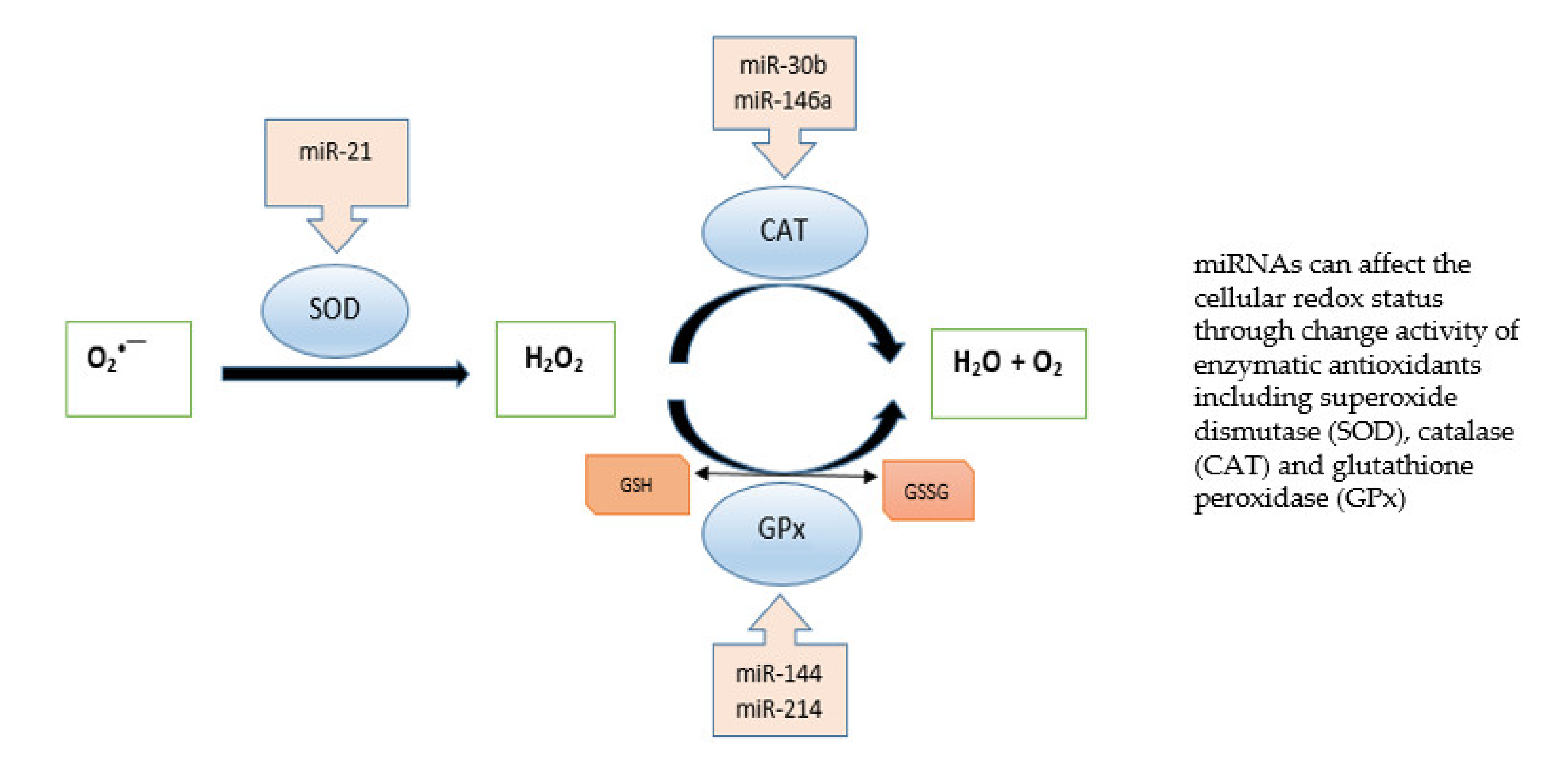
3. AMD—Enzymatic Antioxidant Capacity

Although the macula lives in an environment of high oxidative stress caused by the presence of many sources of reactive oxygen species (ROS), it contains many enzymatic and non-enzymatic antioxidants in the photoreceptor and RPE cells. Macular pigment is a natural barrier that protects the central part of the retina from oxidative damage. It is formed by two dihydrocarotenoids: lutein and zeaxanthin. In addition, the antioxidant defense includes various enzymes including superoxide dismutase (SOD), catalase (CAT) and glutathione peroxidase (GPx), and non-enzymatic antioxidants such as glutathione, albumin, bilirubin, uric acid, vitamin A, C, E, lipoic acid, carotenoids, plant polyphenols, zinc and selenium, among others.
Antioxidant enzymes reduce lipid hydroperoxide and hydrogen peroxide (H2O2) levels, thus playing an important role in preventing lipid peroxidation and maintaining cell membrane structure and function.
The copper-zinc superoxide dismutase (CuZnSOD) plays a key role in the removal of superoxide anion and protects cells from oxidative injury caused by free radicals. Located in the cytosol and mitochondria, SOD catalytically converts the superoxide anion (O2˙) to O2 and H2O2 in the presence of metal ion cofactors such as copper (Cu), zinc (Zn) or manganese (Mn). It catalyzes the one-electron dismutation of the superoxide anion. The human SOD1 gene, located on chromosome 21q22.11, is highly polymorphic. Few studies have examined the relationships between SOD1 polymorphisms with AMD. A study conducted in a Polish AMD population [31] reported that a polymorphism in the SOD1 gene protected against AMD. Many works implicate SOD2 polymorphisms as potential determinants of AMD susceptibility. SOD2, a mitochondrial enzyme, has manganese (Mn) in its reactive center and active site, which functions as a metal cofactor. The enzyme scavenges toxic superoxide anions formed as a byproduct of oxidative phosphorylation, which enables the removal of mitochondrial ROS and provides protection against cell death. The SOD2 gene (6q25) encodes MnSOD and has several single nucleotide polymorphisms (SNPs), among which is rs4880 (C/T) (also designated as C47T, Ala16Val, Ala-9Val or A16V). The T allele of this SNP (rs4880-T) has been linked to changes in MnSOD activity due to modification of the mitochondrial target sequence, which may play a role in neurodegenerative processes including primary open angle glaucoma, AMD [32].
Catalase (CAT) is present in the peroxisome, where it converts hydrogen peroxide to water and molecular oxygen. The CAT gene is located on chromosome 11p13 and contains 13 exons. Glutathione peroxidase (GSHPx) is found both in the cytoplasm and extracellularly in almost every human tissue, where it converts H2O2 into water. GSHPx has strong activity against both H2O2 and fatty acid hydroperoxides. It is encoded by the GPX1 gene, located on chromosome 3p21, which contains two exons. The results from previous studies investigating the association between genes encoding antioxidant enzymes as risk factors for the development of eye diseases have gained mixed results [33,34,35,36,37].
Moreover, recent studies indicate that miRNAs play an essential role in ROS generation by targeting genes associated with antioxidant response. Gene silencing by miRNAs can induce changes in antioxidant enzymes, leading to a complex interplay between redox imbalance by free radicals and miRNAs in modulating cellular redox homeostasis. The transcription, biogenesis and translocation of miRNAs, as well as their function, are connected with oxidative stress, and miRNAs can regulate the expression of redox markers such as cellular antioxidant status. Figure 1 shows miRNAs that affect enzymatic antioxidants. Expression analyses indicate that Cu/Zn SOD is downregulated by miRNAs in human bronchial cells [38], and the suppression of miR-146a was found to effectively restore catalase expression [39]. miR-30b downregulated CAT expression in human retinal pigment epithelial cell line [40]. Furthermore, dysregulation of miR-214 affected glutathione peroxidase activity: miR-214 overexpression increased GPx activity and reduced oxidative stress in an in vitro diabetic nephropathy model [41]. In contrast, miR-144 suppressed GPx expression in sickle cell disease [42].

4. AMD—DNA Damage and Repair

An important role in macular function is played by an imbalance between pro- and antioxidant reactions (e.g., during aging), which is associated with oxidative regulation and gene modification. Elevated levels of free radicals can destroy cellular macromolecules, and induce lipid peroxidation and the modification of DNA, RNA, proteins and carbohydrates. Not all ROS damage DNA directly. For instance, superoxide anions and hydrogen peroxide can initiate DNA damage by interacting with transition metal ion chelates. Oxidative stress is thought to play an essential role in the development of AMD. One source of oxidative stress in the macula may be iron ions, which are capable of promoting the generation of hydroxyl radicals through the Fenton reaction. The hydroxyl radical is a harmful species which forms a range of products when it attacks bases in DNA. Major oxidative DNA damage products include 8-hydroxyadenine, 8-oxoguanine (8-OH-G), thymine glycol, and ring lesions: 4,6-diamino-5-formamidopyrimidine or 2,6-diamino-4-hydroxy-5-formamidopyrimidine. Other deamination products, such as uracil, 5-hydroxyuracil (5-OH-Ura), 5-hydroxymethylouracil (5-HMU), hypoxanthine, and xanthine, represent a subset of all ROS-induced base damage. The results of clinical studies have shown that patients with AMD have elevated levels of oxidative DNA damage, and DNA repair efficiency is reduced in these patients [43,44].
Oxidative stress induces different types of DNA damage such as base modifications, DNA breaks and alkali-prone sites. ROS are a known cause of DNA damage, from single base oxidation to single bases and DSBs, indicating that high levels of ROS have inappropriate effects on genome integrity. Base excision repair (BER) is an important pathway involved in the repair of oxidative damage in DNA. A specialized DNA glycosylase recognizes and removes the modified base. This creates a-base or a-purine/pyrimidine (AP) sites in double-stranded DNA. The system also repairs single-stranded DNA breaks that may be caused by free radical reactions. The OGG1 gene located at 3p26.2 on chromosome 3 encodes 8-oxoguanine DNA glycosylase (OGG1), the major human glycosylase that cleaves the glycosyl bond between an oxidase base and a sugar. OGG1 removes oxidized guanine (8-oxoG) causing G:C to T:A transversion [45]. The most common polymorphic variant of OGG1 is the Ser326Cys variant (rs1052133). Homozygous carriers of the OGG1 Ser326Cys variant present a lower ability to remove 8-oxoG and to complete the repair fusion step. In addition, under oxidative conditions, this variant exhibits lower catalytic activity and a higher propensity for dimerization [46]. Recent studies have reported a vital association between the rs1052133 and rs125701 genetic polymorphisms of the OGG1 DNA repair gene and the development of AMD [44,47,48]. Previous literature demonstrates the pathogenic role of oxidative stress in ocular diseases such as glaucoma. The most common type of glaucoma is primary open-angle glaucoma (POAG), which accounts for more than 50% of diagnosed glaucoma cases in developed countries. Szaflik et al. [49] report an association between the OGG1 Ser326Cys polymorphism and POAG progression.
Another important DNA glycosylase is MUTYH (MYH), whose key role is to prevent oxidative DNA damage. The MUTYH gene located on the short arm of chromosome 1p34.1 encodes an A/G-specific adenine DNA glycosylase (MUTYH) that removes adenine paired with 8-oxoG. In humans, two isoforms of MUTYH differing in localization (mitochondrial for type 1 protein and nuclear for type 2 protein) have been documented. MUTYH interacts with proteins belonging to several DNA repair pathways, such as APE1, which induces its glycosylase activity and promotes its turnover, and MMR and PCNA proteins, which ensure its coupling to DNA replication [50]. Mutation of this gene may be bound up with progression of AMD and primary open-angle glaucoma [47,49]. A common polymorphism of the MUTYH gene is also rs10527342 caused by the insertion of an Alu element in the 15th intron of this gene, resulting in increasing levels of genomic 8-oxoG. Individuals homozygous for this variant have been shown to demonstrate low expression of mitochondrial MUTYH type 1 protein and reduced mitochondrial homeostasis, which perhaps may account for the associated age-related diseases [51].
It has been found that the appearance of uracil in DNA is associated with oxidative stress. The presence of uracil in DNA leads to genomic instability; as such, precise mechanisms are needed to remove uracil from DNA, thus preventing its incorporation and maintaining DNA integrity. Under normal conditions, such lesions are repaired by base excision repair, which is initiated by uracil-DNA glycosylases (UDG). In humans, four uracil glycosylases remove uracil from DNA: UNG, SMUG1, MBD4 and TDG.
The potential role of the rs2337395 UNG polymorphism and its connection with SMUG1 rs3087404 SNP has been suggested in the pathogenesis of eye disease resulting in progressive and irreversible loss of central vision (AMD) [52]. Mehdizadeh et al. [53] report that in an Iranian population, heterozygous individuals who carry the AG genotype of -31A>G rs3087404 polymorphism of SMUG1 gene represent a lower risk of AMD.
AP-endonuclease (APE1) is a key enzyme for the next step in the repair pathway by base excision. This step is called short-patch BER, and is predominant in mammalian cells; in contrast, long-patch BER is characterized by the incorporation of a longer fragment into DNA. The APE1 gene, which encodes AP endonuclease in the human body, is located on chromosome 14q11.2. APE1 interacts with DNA polymerase by only one nucleotide embedded in the DNA backbone. Studies have confirmed that the -141 G/G genotype of the rs1760944 APE1 polymorphism appears to play a protective role against cataracts, while the T allele appears to support cataract development [54].
Another key enzyme for BER is the XRCC1 protein encoded by the X-ray repair cross-complementing group 1 (XRCC1) gene. XRCC1 is located on chromosome 19, at position 19q13.2. XRCC1 interacts with DNA polymerase DNAligase III, AP endonuclease and poly(ADP-ribose) polymerase (PARP, ADPRT). XRCC1 attaches to single-stranded DNA breaks upon removal of 5′ deoxyribose phosphate. Previous studies have shown that the inheritance of Arg194Trp, as well as Arg399Glu genotypes of the XRCC1 gene, is associated with the development of neurodegenerative disease of the lung [55] and brain [56], and the growth of head and neck cancers [57]. In addition, in one study, among several studied BER-related factors, only Polβ (DNA polymerase beta) and XRCC1 showed a correlation between expression level and age, and that restoration of XRCC1 or Polβ in old cells failed to restore BER efficiency to a similar repair capacity as observed in young cells [58].
PARP polymerase is encoded by the ADPRT gene located on chromosome 1q42.1. The main role of PARP is to bind to DNA strand breaks and recruit the XRCC1-Lig3 complex. Changes in its function may be associated with the Val726Ala polymorphism of the ADPRT gene.
Wang et al. report reduced gene expression of key DNA repair enzymes in the retinal pigment epithelium cells and choroid of old rodents: OGG1, MUTYH, TDG (DNA thymine glycosylase), PARP1 and NTH1 (they remove modified bases in the first step of the DNA repair process) and therefore may have a relatively lower DNA repair capacity [59,60]. In addition, Zhang et al. note the expression of mRNA genes and proteins of the key BER enzymes OGG1, APE1, and Polgamma (DNA polymerase gamma) in rats decreased with aging, resulting in reduced mtDNA repair capacity and accumulation of mtDNA damage [61]. Other data indicate that patients with colorectal cancer demonstrate changes in the expression of the BER genes APE1, XRCC1, PARP1, OGG1, and APE1 [62]. Interestingly, Haque at al. [40] found that H2O2 increased the mRNA expression of human catalase in a human retinal pigment epithelial cell line (ARPE-19), this being an important component of cellular antioxidant defense that detoxifies hydrogen peroxide radicals.
Regarding the practical applicability of these studies for prognosis and rapid diagnosis, it is important to note that in organisms displaying polymorphisms of genes involved in ROS scavenging, increased levels of oxidative DNA damage are detected not only in tissues and organs, but also in lymphocytes isolated from the peripheral blood of AMD patients.
miRNAs are engaged in the regulation of mitochondrial signaling pathways such as the apoptotic pathway. Several reports have suggested that miRNAs are involved in the redox regulation of DNA damage and DNA repair pathways [63]. Tinaburri et al. [64] report that overexpression of miR-200a downregulates the BER enzymes OGG1, APE1, LIG3 and XRCC1 and the expression of UNG1, FEN1 (flap endonuclease) as well as LIG1; it can also decrease the efficiency of oxidative DNA repair in aging skin. In contrast, overexpression of miR-4673 reduced OGG1 accumulation and induced oxidative stress and ROS generation in a human lung cancer cell line [65].

5. AMD—miRNA Expression and Diagnostic Markers

Patients with this disease usually become dependent on others, and their quality of life is significantly reduced. AMD is also a significant ophthalmological problem in the population under 50 years of age. As in all chronic diseases, early diagnosis and treatment play important roles in the outcome of AMD.
Due to very poor prognosis, lack of causal treatment options and high costs of symptomatic therapy, it is very important to identify new methods of preventing the development of the disease. The discovery of numerous small non-coding RNAs, including microRNAs (miRNAs), has shown that human biology is even more complex than previously thought.
Alterations in the expression profile of miRNAs have been successively demonstrated in many diseases, including AMD. Differences in miRNA gene expression can be accounted for by the localization of genes in disease-related regions, changes in miRNA processing mechanisms, and epigenetic mechanisms.
MicroRNAs (miRNAs) are RNA molecules belonging to a family of small non-coding RNAs 20–25 nucleotides long, located within introns and exons of protein-coding genes or in intergenic regions. MiRNAs are involved in gene silencing by repressing protein synthesis through imperfect binding to the 3’-UTR (untranslated region) of the target mRNA, leading to mRNA degradation. MicroRNAs play a key role in the regulation of gene expression. Moreover, they are involved in many important biological processes such as cell proliferation, differentiation, angiogenesis, growth control, organogenesis, and apoptosis [66].
Studies in recent years have shown dysregulation of microRNAs in AMD. The discovery of new specific microRNAs as biomarkers for AMD facilitates early detection of the disease and constant checking of its progression. Understanding how miRNAs coordinate disease progression may allow for a better understanding of the disease itself and the initiation of new therapeutic approaches.
Researchers have detected hundreds of small RNAs in a spectrum of body fluids, such as whole blood [67], blood plasma [68], blood serum [69], peripheral blood nuclear cells [70], urine, vitreous [71], saliva, cerebrospinal fluid, retinal tissues [72], and various others [73,74]. Studies have shown that in addition to being detectable in blood cells, miRNAs circulate in serum-secreted exosomes [75,76], microtubules, apoptotic bodies [77], associated with the AGO2 complex [78] and in lipoproteins [79].
miRNAs play key roles in regulating pathological processes involved in AMD development, including angiogenesis, the imbalance of complement activation, inflammation and oxidative stress. In addition to vascular endothelial growth factor (VEGF), other growth factors disrupt angiogenesis and vascular balance and may also serve as significant markers in AMD: platelet-derived growth factor (PDGF), fibroblast growth factors (FGFs), placental growth factor (PlGF), hepatocyte growth factor (HGF), fibroblast growth factor-2 (FGF-2), pigment epithelium-derived growth factor (PEDF), and angiopoietins (ANGPTs).
Previous studies have shown a link between oxidative stress and inflammation. It has been indicated that oxidative stress induces inflammation during the pathological process of AMD [80]. Pathological oxidative damage contributes to protein, lipid and DNA damage and mitochondrial dysfunction, and generates toxic free radicals and high concentrations of harmful compounds such as AGEs and MDA; it also induces pro-inflammatory responses, and promotes the recruitment of macrophages, which release pro-inflammatory and pro-angiogenic mediators [81]. Many pro-inflammatory cytokines and chemokines, including IL-1, IL-6, IL-8, TNF, INF-γ, MCP-1, have been shown to accelerate AMD progression [82].
Some of the circulating miRNAs are involved in the regulation of angiogenesis, neovascularization and inflammatory process, which play a role in the pathophysiology of AMD. Recent reviews indicate that dysregulation of miRNAs is associated with the development of retinal disease: miR-9, miR21, miR-23a, miR-27a, miR-126, miR-144, miR-146, miR-150, miR-155 and Let-7. Alterations in these miRNAs are most commonly demonstrated in biological materials from AMD patients (Table 1).
miR-9 is highly expressed in the adult brain and targets several genes that play key roles in neurogenesis. It has been documented that miR-9 can combine neurogenesis with angiogenesis and is potentially associated with the generation of new vasculature [83]. In the eye, this miRNA plays a key role in maintaining RPE cell function. Depending on cell type and developmental stage, miR-9 has been found to be regulated by retinoic acid, pro-inflammatory cytokines and ROS, all of which are found in high concentrations in the diseased retina [72]. Bhattacharjee et al. [84] report that miRNA-9 was upregulated in human retinal tissues.
miR-21 has a very vital function in the regulation of angiogenesis, tumor growth and metastasis. It demonstrates very high expression in endothelial cells and is believed to be involved in endothelial cell differentiation, migration and angiogenesis [85]. Samples from patients with wet AMD showed downregulation of miR-21 and involvement in regulation of blood vessel growth, as indicated by its elevated expression in retinal endothelial cells [68,86].
miR-23a plays an important role in the regulation of development, differentiation, cell growth and apoptosis. Moreover, miR-23a is a key factor in regulating oligodendroglial development and myelin formation [87]. miR-23a was identified as overexpressed in peripheral blood nuclear cells of both dry and wet AMD. Additionally, miR-23a was upregulated in the serum of patients with wet AMD [70,88].
The miRNA-27 family consists of miR-27a and miR-27b, which are transcribed from different chromosomes, and display different nucleotides at the 3’ end. This miRNA has been linked to inflammation, angiogenesis, the oxidative stress response and age-related macular degeneration. Ren et al. [67,89] demonstrate that samples from patients with wet AMD showed an overexpression of miR-27a-3p compared to dry AMD patients, and that whole blood miR-27a could act as a potential diagnostic biomarker of AMD.
miR-126, an endothelial cell-restricted miRNA, was found to regulate developmental angiogenesis. miR-126 is located within intron 7 of epidermal growth factor-like domain 7 (EGFL7). Expression analysis revealed abundant levels of miR-126 in highly vascularized tissues. Previous studies have identified miR-126 as the only miRNA known to be specifically expressed in endothelial cell lines, hematopoietic progenitor cells, and bone marrow-derived endothelial cell lines, which play important roles in blood vessel formation and angiogenesis [89,90]. Recently, Ulańczyk et al. [68] reported that miR-126 expression is increased and promotes angiogenesis in AMD. In addition, a small scale study comprising 11 AMD patients and 11 healthy control subjects, found a higher expression of miR-126 in the blood serum of AMD patients compared to controls [88]. In contrast, however, lower expression of miR-126 has been recorded in blood serum samples from 76 wet AMD and 70 controls [91]. These discrepancies may result from differences in sample size, patient inclusion criteria, the specific arm of the miRNA studied, or the strategies for miRNA quantification.
miR-144 is the only miRNA that is consistently elevated in the brains of elderly humans; it is believed to play a role in connecting the dysregulated antioxidant defense system with the imbalanced redox state in a number of age-related diseases. Jadeja et al. [92] note high expression levels of miR-144-3p and miR-144-5p in an animal model. Nuclear factor erythroid 2-related factor 2 (NRF2), a potential target of miR-144, is believed to be a primary regulator of the antioxidant defense, and to play a significant role in glutathione (GSH) metabolism. The overexpression of the miRNA protects against oxidative stress-induced outer retinal degeneration.
miR-146 is composed of miRNA-146a and miRNA-146b-5p. It has been shown that miRNA-146a and miRNA-146b-5p are involved in inflammation in tissues. miR-146a has been shown to play an important role in modulating the innate immune response. miR-146a is induced in response to lipopolysaccharide (LPS) and pro-inflammatory mediators, and miR-146a induction is controlled by nuclear factor kappa-light-chain-enhancer of activated B cells (NF-κB). MiR-146a can directly inhibit pro-inflammatory cytokine production by acting as a negative feedback effector of the inflammatory signaling pathway initiated by NF-κB [93].
Samples from AMD patients showed upregulation of miRNA-146a and its involvement in complement factor H induced inflammation. Induction of miRNA-146a has been reported in the PBNCs of patients with dry AMD [70], in the vitreous humor of patients with wet AMD [71] and in patients with diabetic retinas [94], or in whole blood samples of individuals with wet AMD [89]. Moreover, circulating miRNA-146a is upregulated in AMD retinal tissues collected postmortem [72].
miR-150 has been associated with high expression in immune cells and has a function in the maturation, proliferation and differentiation of myeloid and lymphoid cells. Many of the miR-150 target transcripts identified to date are pro-apoptotic and differentiation proteins such as early growth response 2 (EGR2) and c-myb. It is believed that miR-150 plays an essential function in angiogenesis because it is enriched to a greater degree in retinal endothelial cells than in any other nuclear layers of the retina [95]. Furthermore, Lin et al. [96] propose a novel role for miR-150 in macrophages in regulating cholesterol metabolism and lipid trafficking genes involved in AMD. The authors propose that disorders in plasma membrane lipids secondary to the observed miR-150 upregulation in aging macrophages modulate the inflammatory balance of aging macrophages and thereby predispose them toward a pathogenic, pro-angiogenic phenotype. Studies suggest an important role for miR-150 as an intrinsic inhibitor of pathological ocular angiogenesis.
Another miRNA with a possible role in inflammation and ocular angiogenesis in eye diseases is miR-155. This miRNA is upregulated and disturbs the normal retinal vessel growth in mice [97], is upregulated in human patients with AMD and plays an important role in the regulation of several important pathways of immune responses. Research has shown that inflammatory process increased miRNA-155 expression in human RPE cells by activation of the JAK/STAT signaling pathway [98]. Pogue and Lukiw [72] report that miR-155 demonstrates increased expression in AMD tissues compared to controls.
Let-7 (lethal-7) was one of the first miRNAs to be discovered. Among humans, the let-7 family consists of nine mature let-7 miRNAs encoded by 12 different genomic loci, some of which are clustered together. Two major biological roles have been elucidated for let-7 miRNAs: an essential regulator of terminal differentiation and a primary tumor suppressor [99]. Szemraj et al. found let-7 to be expressed at significantly higher levels in wet AMD, and that let-7 expression was positively correlated with VEGF and VEGFR2 expression, at both the mRNA and protein levels [100]. Let-7 family members are known to be pro-angiogenic. These findings strongly indicate that increased Let-7 expression in serum may reflect neovascularization in AMD patients.
microRNAs play key roles in regulating the transcription and translation of genetic material. miRNAs, in contrast to RNA, are very stable, due to the connection with the Ago protein, and are not degraded by RNAse. The high stability of circulating miRNA is mainly due to the encapsulation in lipid vesicles or the formation of complexes with different kinds of proteins that protect them against denaturation. The identification of the miRNA profile in a disease entity and correlation of this profile with the corresponding genes allows for the identification of new, previously unknown pathophysiological processes [101].
miRNAs have also been shown to be a promising target for potential therapeutic intervention. The use of a synthetic, stable antagonist of miRNAs (antagomir) may allow the key gene for the disease development to be regulated [102].
As of today, there is no therapy that can stop or reverse changes caused by neurodegenerative diseases such as AMD, Alzheimer’s or Parkinson’s disease. It is therefore particularly important to identify a biomarker that would counteract the effects of the disease before it progresses to a chronic form. At the moment, no validated biomarker for the early detection of AMD is available. miRNA profiling may disclose potential diagnostic biomarkers of early-stage AMD and exudative AMD. Most of the available research comprises preliminary and experimental studies, in small groups of patients, which do not meet the criteria of validation analyses. In addition, their function can vary according to the stage of the disease, making it difficult to be used as a precise target. As such, the role of miRNAs as biomarkers is not yet clear. The size of the study group, prospective and epidemiological studies and validation are needed to ensure the correct interpretation of miRNA test results.
Previous studies have confirmed the role of microRNAs in the development of neurodegenerative diseases, linking them to chronic inflammation in the elderly. The mechanisms of aging that lead to age-related diseases have been shown to help identify new therapeutic approaches [103]. For example, treatments targeting Alzheimer’s disease may be applicable to AMD [72,84].
Research on miRNAs as biomarkers is still in an early stage, and at present, the findings are predominantly not reproducible. miRNAs can be detected using various sensitive methods, including in situ hybridization [104], real-time PCR [100], Northern blot analysis [105], miRNA microarray [71,84] and next-generation sequencing (NGS) [106].
To be able to apply miRNAs in in vitro diagnostics, it is necessary to discover specific miRNAs that can be used as biomarkers and to introduce a new methodology based on simple and inexpensive methods with standardized procedures before and after analysis.
Moreover, one of the more interesting observations was made by in vivo studies that documented an amelioration of AMD-associated damage with manipulation of miRNAs in AMD mice. Although most of the tested miRNAs were not homologous to those that were dysregulated in the AMD profiling studies, miR-155 and miR-184 were found to overlap [107]. This further demonstrates the potential of miRNAs as a target for treating AMD.
According to the epigenetic hypothesis, a number of environmental stimuli can, through miRNAs, influence the phenotype of an organism. Discovering the phenomena of miRNAs regulation enables understanding of AMD pathomechanism. The characteristic profile of changes in miRNA expression levels may be helpful in early diagnosis. However, the role of miRNAs as biomarkers is not yet clear. Most of the studies presented above are preliminary and experimental studies, in small groups of patients, which did not meet criteria for validation analyses. Undoubtedly, in the coming years many subsequent studies devoted to this issue will be carried out. Whether the altered miRNA expression profile is due to the course or the cause of the disease remains under investigation. Nonetheless, many studies clearly show the usefulness of the analysis of circulating miRNA expression as diagnostic, prognostic, predictive markers and potential therapeutic targets.
However, it should be remembered that single molecule expression may be increased in both types of AMD or other ophthalmic diseases, which significantly limits the diagnostic utility. Therefore, more and more authors emphasize the importance of the greater sensitivity and diagnostic specificity of the selected miRNAs panel analyzed. The advantage of the analyzed selected circulating miRNAs panel of (miR-15b, miR-17, miR21, miR-26b, miR145) over single molecule analysis was, for example, confirmed in colorectal cancer [108]. Perhaps in the near future, the analysis of miRNAs will be an important stage of diagnosis and the introduction to treatment consistent with the endotype of a given disease entity.

6. Conclusions

In chronic diseases, such as AMD, early diagnosis and treatment play an important role in the outcome. AMD is a highly complex disease that is influenced by many factors, such as aging, genetic predisposition, environmental elements, oxidative stress, and inflammatory factors. Despite the fact that AMD is not a classic inflammatory disease, inflammatory cells play a significant role in its pathogenesis and progression [81]. In addition, several risk factors have been presented that are associated with oxidative stress, such as age, smoking, alcohol consumption, diet and obesity [109,110,111]. Elevated levels of oxidative DNA damage connected with polymorphisms of genes involved in ROS inactivation, inefficient enzymatic antioxidant system, ineffective BER repair system, and changes in gene expression may be responsible for degenerative changes in the macula [43,112]. The correlation between miRNA profile and AMD progression indicates their involvement in molecular pathways known to play an important role in AMD pathogenesis, such as inflammation in RPE cells and vascular changes in choroidal vessels, oxidative defense system, and DNA repair efficiency.
The profiling of miRNAs in body fluids offers promise in the diagnosis, monitoring and prognosis of AMD. One of the most important applications of miRNA profiling is the development of clinically useful molecular diagnostic tests; miRNAs are present at relatively stable levels in human serum and plasma, and miRNA level has been reported to be much more stable than mRNA in a variety of sample types—including plasma/serum and urine, and is measurable at much greater sensitivity than proteins. The development of therapeutic strategies based on the analysis of miRNA panels that target inflammation, ROS and repair of oxidative damage in DNA, has the potential to significantly impact the treatment of AMD [107].

Author Contributions

Conceptualization, M.M. and I.M.; data curation, J.M., E.K. and B.S.; writing—original draft preparation, M.M.; writing—review and editing, M.M. and I.M.; supervision, I.M., J.P.S. and J.S.; ophthalmological review J.P.S. and J.S. All authors have read and agreed to the published version of the manuscript.

Funding

This work was supported by Medical University of Lodz (503/5-108-05/503-51-001-19-00).

Institutional Review Board Statement

Not applicable.

Informed Consent Statement

Not applicable.

Data Availability Statement

Data is contained within the article.

Acknowledgments

The authors are grateful for financial support to the Medical University of Lodz (503/5-108-05/503-51-001-19-00).

Conflicts of Interest

The authors declare no conflict of interest.

References

  1. Xiayan, X.; Jing, W.; Xiaoning, Y.; Yelei, T.; Xiajing, T.; Xingchao, S. Regional differences in the global burden of age-related macular degeneration. BMC Public Health 2020, 20, 410–418. [Google Scholar] [CrossRef]
  2. Shaw, P.X.; Stiles, T.; Douglas, C.; Ho, D.; Fan, W.; Du, H.; Xiao, X. Oxidative stress, innate immunity, and age-related macular degeneration. AIMS Mol. Sci. 2016, 3, 196–221. [Google Scholar] [CrossRef]
  3. Fleckenstein, M.; Mitchell, P.; Freund, K.B.; Sadda, S.V.; Holz, F.G.; Brittain, C.; Henry, E.C.; Ferrara, D. The Progression of Geographic Atrophy Secondary to Age-Related Macular degeneration. Ophthalmology 2018, 125, 369–390. [Google Scholar] [CrossRef] [PubMed]
  4. Heesterbeek, T.J.; Lores-Motta, L.; Hoyng, C.B.; Lechanteur, Y.T.E.; den Hollander, A.I. Risk factors for progression of age-related macular degeneration. Ophthalmic. Physiol. Opt. 2020, 40, 140–170. [Google Scholar] [CrossRef] [PubMed] [Green Version]
  5. Treister, A.D.; Nesper, P.L.; Fayed, A.E.; Gill, M.K.; Mirza, G.; Fawzi, A.A. Prevalence of Subclinical CNV and Choriocapillaris Nonperfusion in Fellow Eyes of Unilateral Exudative AMD on OCT Angiography. Transl. Vis. Sci. Technol. 2018, 7, 19–28. [Google Scholar] [CrossRef] [PubMed] [Green Version]
  6. Johnston, H.; Dickinson, P.; Ivens, A.; Buck, A.H.; Levine, R.D.; Remacle, F.; Campbell, C.J. Intracellular redox potential is correlated with miRNA expression in MCF7 cells under hypoxic conditions. Proc. Natl. Acad. Sci. USA 2019, 116, 19753–19759. [Google Scholar] [CrossRef] [PubMed] [Green Version]
  7. Konovalova, J.; Gerasymchuk, D.; Parkkinen, I.; Chmielarz, P.; Domanskyi, A. Interplay between MicroRNAs and oxidative stress in neurodegenerative diseases. Int. J. Mol. Sci. 2019, 20, 6065. [Google Scholar] [CrossRef] [Green Version]
  8. Algvere, P.V.; Kvanta, A.; Seregard, S. Drusen maculopathy: A risk factor for visual deterioration. Acta Ophthalmol. 2016, 94, 427–433. [Google Scholar] [CrossRef]
  9. Woodell, A.; Rohrer, B. A mechanistic review of cigarette smoke and agerelated macular degeneration. Adv. Exp. Med. Biol. 2014, 801, 301–307. [Google Scholar]
  10. Pennington, K.L.; DeAngelis, M.M. Epidemiology of age-related macular degeneration (AMD): Associations with cardiovascular disease phenotypes and lipid factors. Eye Vision 2016, 3, 34–54. [Google Scholar] [CrossRef] [Green Version]
  11. Zetterberg, M. Age-related eye disease and gender. Maturitas 2016, 83, 19–26. [Google Scholar] [CrossRef]
  12. Zhang, Q.Y.; Tie, L.J.; Wu, S.S.; Lv, P.L.; Huang, H.W.; Wang, W.Q.; Wang, H.; Ma, L. Overweight, Obesity, and Risk of Age-Related Macular Degeneration. Investig. Opthalmol. Vis. Sci. 2016, 57, 1276–1283. [Google Scholar] [CrossRef] [PubMed] [Green Version]
  13. Xiaohua, L.; Shikun, H.; Mingwei, Z. An Updated Review of the Epigenetic Mechanism Underlying the Pathogenesis of Age-related Macular Degeneration. Aging Dis. 2020, 11, 1219–1234. [Google Scholar]
  14. Geerlings, M.J.; de Jong, E.K.; den Hollander, A.I. The complement system in age-related macular degeneration: A review of rare genetic variants and implications for personalized treatment. Mol. Immunol. 2017, 84, 65–76. [Google Scholar] [CrossRef]
  15. Hong, N.; Shen, Y.; Yu, C.Y.; Wang, S.Q.; Tong, J.P. Association of the polymorphism Y402H in the CFH gene with response to anti-VEGF treatment in age-related macular degeneration: A systematic review and meta-analysis. Acta Ophthalmol. 2016, 94, 334–345. [Google Scholar] [CrossRef] [PubMed] [Green Version]
  16. Clark, S.J.; Bishop, P.N. Role of factor H and related proteins in regulating complement activation in the macula, and relevance to age-related macular degeneration. J. Clin. Med. 2014, 4, 18–31. [Google Scholar] [CrossRef] [Green Version]
  17. Qian-Qian, Y.; Yong, Y.; Jing, Z.; Xin, B.; Tian-Hua, X.; Chao, S.; Jia, C. Nonsynonymous single nucleotide polymorphisms in the complement component 3 gene are associated with risk of age-related macular degeneration: A meta-analysis. Gene 2015, 561, 249–255. [Google Scholar] [CrossRef]
  18. Lim, L.S.; Mitchell, P.; Seddon, J.M.; Holz, F.G.; Wong, T.Y. Age-related macular degeneration. Lancet 2012, 379, 1728–1738. [Google Scholar] [CrossRef]
  19. Hallak, J.A.; de Sisternes, L.; Osborne, A.; Yaspan, B.; Rubin, D.L.; Leng, T. Imaging, Genetic, and Demographic Factors Associated With Conversion to Neovascular Age-Related Macular Degeneration: Secondary Analysis of a Randomized Clinical Trial. JAMA Ophthalmol. 2019, 137, 738–744. [Google Scholar] [CrossRef] [PubMed]
  20. Wong, W.L.; Su, X.; Li, X.; Cheung, C.M.G.; Klein, R.; Cheng, C.Y.; Wong, T.Y. Global prevalence of age-related macular degeneration and disease burden projection for 2020 and 2040: A systematic review and meta-analysis. Lancet Glob. Health 2014, 2, e106–e116. [Google Scholar] [CrossRef] [Green Version]
  21. Colijn, J.M.; Buitendijk, G.H.; Prokofyeva, E.; Alves, D.; Cachulo, M.L.; Khawaja, A.P.; Cougnard-Gregoire, A.; Merle, B.M.; Korb, C.; Erke, M.G.; et al. Prevalence of age-related macular degeneration in Europe: The past and the future. Ophthalmology 2017, 124, 1753–1763. [Google Scholar] [CrossRef] [PubMed] [Green Version]
  22. Usui, H.; Nishiwaki, A.; Landiev, L.; Kacza, J.; Eichler, W.; Wako, R.; Kato, A.; Takase, N.; Kuwayama, S.; Ohashi, K.; et al. In vitro drusen model – three-dimensional spheroid culture of retinal pigment epithelial cells. J. Cell Sci. Spec. Issue Reconst. Cell Biol. 2019, 132, jcs215798–jcs215805. [Google Scholar] [CrossRef] [Green Version]
  23. Upadhyay, M.; Milliner, C.; Bell, B.A.; Bonilha, V.L. Oxidative stress in the retina and retinal pigment epithelium (RPE): Role of aging, and DJ-1. Redox Biol. 2020, 37, 101623–101635. [Google Scholar] [CrossRef] [PubMed]
  24. Renganathan, K.; Gu, J.; Rayborn, M.E.; Crabb, J.S.; Salomon, R.G.; Collier, R.J.; Kapin, M.A.; Romano, C.; Hollyfield, J.G.; Crabb, J.W.; et al. CEP Biomarkers as Potential Tools for Monitoring Therapeutics. PLoS ONE 2013, 8, e76325. [Google Scholar] [CrossRef] [PubMed]
  25. Cooke, J.P. New Insights Into Tobacco-Induced Vascular Disease: Clinical Ramifications. Methodist Debakey Cardiovasc. J. 2015, 11, 156–159. [Google Scholar] [CrossRef] [PubMed] [Green Version]
  26. Hou, L.; Yang, G.; Tang, S.; Alcazar, C.; Joshi, P.; Strassberg, Z.; Kim, M.; Kawamura, M.; Woo, Y.J.; Shrager, J.; et al. Small Molecule Derived From Carboxyethylpyrrole Protein Adducts Promotes Angiogenesis in a Mouse Model of Peripheral Arterial Disease. J. Am. Heart Assoc. 2018, 7, e009234–e009245. [Google Scholar] [CrossRef] [Green Version]
  27. Leńska-Mieciek, M.; Korczak-Kowalska, G.; Bocian, K.; Fiszer, U. Pentosidine, advanced glycation end product, in acute ischaemic stroke patients with and without atrial rhythm disturbances. Neurol. Neurochir. Pol. 2020, 54, 323–328. [Google Scholar] [CrossRef]
  28. Joo, Y.S.; Areum, S.; Hyunsoo, K.; Youngsoo, K.; Hyeong, G.Y. Carboxymethyl-lysine-modified Plasma Proteins in Age-related Macular Degeneration. Biotechnol. Bioproc. E 2017, 22, 52–59. [Google Scholar] [CrossRef]
  29. Handa, J.T.; Cano, M.; Wang, L.; Datta, S.; Liu, T. Lipids, Oxidized Lipids, Oxidation-specific Epitopes, and Age-related Macular Degeneration. Biochim. Biophys. Acta. 2017, 1862, 430–440. [Google Scholar] [CrossRef] [Green Version]
  30. Je-Hyun, B.; Daehan, L.; Park, K.H.; Chae, J.B.; Jang, H.; Lee, J.; Chung, H. Quantitative proteomic analysis of aqueous humor from patients with drusen and reticular pseudodrusen in age-related macular degeneration. BMC Ophthalmol. 2018, 18, 289–303. [Google Scholar] [CrossRef] [Green Version]
  31. Mrowicka, M.; Mrowicki, J.; Szaflik, J.P.; Szaflik, M.; Ulinska, M.; Szaflik, J.; Majsterek, I. Analysis of antioxidative factors related to AMD risk development in the polish patients. Acta Ophthalmol. 2017, 95, 530–536. [Google Scholar] [CrossRef]
  32. Yeye, C.; Hezheng, Z. Role of two common SNPs of superoxide dismutase 2 gene in the development of primary open angle glaucoma. Biomed. Res. 2017, 28, 7503–7508. [Google Scholar]
  33. Abokyi, S.; To, C.H.; Lam, T.T.; Tse, D.Y. Central Role of Oxidative Stress in Age-Related Macular Degeneration: Evidence from a Review of the Molecular Mechanisms and Animal Models. Hindawi Oxidative Med. Cell. Longev. 2020, 2020, 19. [Google Scholar] [CrossRef] [Green Version]
  34. Saghar, Y. Association between genetic polymorphism of catalase (CAT) C-262T, Cu/Zn superoxide dismutase (SOD1) A251G and risk of age-related macular degeneration. Mol. Bioch. Diagn. J. 2014, 1, 77–88. [Google Scholar]
  35. Kucukgergin, C.; Gokpinar, M.; Sanli, O.; Tefik, T.; Oktar, T.; Seckin, S. Association between genetic variants in glutathione peroxidase 1 (GPx1) gene, GPx activity and the risk of prostate cancer. Minerva Urol. Nefrol. 2011, 63, 183–190. [Google Scholar] [PubMed]
  36. Hansen, R.D.; Krath, B.N.; Frederiksen, K.; Tjønneland, A.; Overvad, K.; Roswall, N.; Loft, S.; Dragsted, L.O.; Vogel, U.; Raaschou-Nielsen, O. GPX1 Pro(198)Leu polymorphism, erythrocyte GPX activity, interaction with alcohol consumption and smoking, and risk of colorectal cancer. Mutat. Res. 2009, 664, 13–19. [Google Scholar] [CrossRef]
  37. Hu, J.; Zhou, G.W.; Wang, N.; Wang, Y.J. GPX1 Pro198Leu polymorphism and breast cancer risk: A meta-analysis. Breast Cancer Res.Treat. 2010, 124, 425–431. [Google Scholar] [CrossRef]
  38. Zhang, X.; Ng, W.L.; Wang, P.; Tian, L.L.; Werner, E.; Wang, H.; Doetsch, P.; Wang, Y. MicroRNA-21 modulates the levels of reactive oxygen species by targeting SOD3 and TNFα. Canc. Res. 2012, 72, 4707–4713. [Google Scholar] [CrossRef] [PubMed] [Green Version]
  39. Wang, Q.; Chen, W.; Bai, L.; Chen, W.; Padilla, M.T.; Lin, A.S.; Shi, S.; Wang, X.; Lin, Y. Receptor-interacting protein 1 increases chemoresistance by maintaining inhibitor of apoptosis protein levels and reducing reactive oxygen species through a microRNA-146a-mediated catalase pathway. J. Biol. Chem. 2014, 289, 5654–5663. [Google Scholar] [CrossRef] [Green Version]
  40. Haque, R.; Chun, E.; Howell, J.C.; Sengupta, T.; Chen, D.; Kim, H. MicroRNA-30b-Mediated Regulation of Catalase Expression in Human ARPE-19 Cells. PLoS ONE 2012, 7, e42542. [Google Scholar] [CrossRef] [PubMed]
  41. Yang, S.; Fei, X.; Lu, Y.; Xu, B.; Ma, Y.; Wan, H. miRNA-214 suppresses oxidative stress in diabetic nephropathy via the ROS/Akt/mTOR signaling pathway and uncoupling protein 2. Exp. Ther. Med. 2019, 7, 3530–3538. [Google Scholar] [CrossRef] [Green Version]
  42. Sangokoya, C.; Telen, M.; Chi, J. microRNA miR-144 modulates oxidative stress tolerance and associates with anemia severity in sickle cell disease. Blood 2010, 116, 4338–4348. [Google Scholar] [CrossRef] [PubMed] [Green Version]
  43. Szaflik, J.P.; Janik-Papis, K.; Synowiec, E.; Ksiazek, D.; Zaras, M.; Wozniak, K.; Szaflik, J.; Blasiak, J. DNA damage and repair in age-related macular degeneration. Mutat. Res. 2009, 669, 169–176. [Google Scholar] [CrossRef]
  44. Wozniak, K.; Szaflik, J.P.; Zaras, M.; Sklodowska, A.; Janik-Papis, K.; Poplawski, T.R.; Blasiak, J.; Szaflik, J. DNA damage/repair and polymorphism of the hOGG1 gene in lymphocytes of AMD patients. J. Biomed. Biotechnol. 2009, 2009, 827562. [Google Scholar] [CrossRef] [Green Version]
  45. Wang, R.; Hao, W.; Pan, L.; Boldogh, I.; Ba, X. The roles of base excision repair enzyme OGG1 in gene expression. Cell. Mol. Life Sci. 2018, 75, 3741–3750. [Google Scholar] [CrossRef] [Green Version]
  46. Simonelli, V.; Camerini, S.; Mazzei, F.; van Loon, B.; Allione, A.; D’Errico, M.; Barone, F.; Minoprio, A.; Ricceri, F.; Guarrera, S.; et al. Genotype-phenotype analysis of S326C OGG1 polymorphism: A risk factor for oxidative pathologies. Free Radic. Biol. Med. 2013, 63, 401–409. [Google Scholar] [CrossRef]
  47. Synowiec, E.; Blasiak, J.; Zaras, M.; Szaflik, J.; Szaflik, J.P. Association between polymorphisms of the DNA base excision repair genes MUTYH and hOGG1 and age-related macular degeneration. Exp. Eye Res. 2012, 98, 58–66. [Google Scholar] [CrossRef]
  48. Shengqun, J.; Nan, H.; Jing, Z.; Junfang, Z.; Ruifang, G.; Jianyan, H.; Huaijin, G. Polymorphisms of the WRN gene and DNA damage of peripheral lymphocytes in age-related cataract in a Han Chinese population. Age 2013, 35, 2435–2444. [Google Scholar] [CrossRef] [Green Version]
  49. Szaflik, J.P.; Cuchra, M.; Przybylowska-Sygut, K.; Dziki, L.; Kurowska, A.K.; Gacek, M.; Drzewoski, J.; Szaflik, J.; Majsterek, I. Association of the 399Arg/Gln XRCC1, the 194 Arg/Trp XRCC1, the 326Ser/Cys OGG1, and the 324Gln/His MUTYH gene polymorphisms with clinical parameters and the risk for development of primary open-angle glaucoma. Mutat. Res. 2013, 753, 12–22. [Google Scholar] [CrossRef] [PubMed]
  50. Boal, A.K.; Genereux, J.C.; Sontz, P.A.; Gralnick, J.A.; Newman, D.K.; Barton, J.K. Redox signaling between DNA repair proteins for efficient lesion detection. Proc. Natl. Acad. Sci. USA 2009, 106, 15237–15242. [Google Scholar] [CrossRef] [Green Version]
  51. Sun, C.; Chen, H.; Guo, W.; Zhang, K.; Qi, Q.; Gu, X.; Zhu, D.; Wang, Y. A common mutation of the MYH gene is associated with increased DNA oxidation and age-related diseases. Free Radic. Biol. Med. 2010, 48, 430–436. [Google Scholar] [CrossRef]
  52. Synowiec, E.; Wysokinski, D.; Zaras, M.; Kolodziejska, U.; Stoczynska-Fidelus, E.; Janik, K.; Szaflik, J.; Blasiak, J.; Szaflik, J.P. Association between polymorphism of the DNA repair SMUG1 and UNG genes and age-related macular degeneration. Retina 2014, 34, 38–47. [Google Scholar] [CrossRef]
  53. Mehdizadeh, F.; Bonyadi, M.; Bonyadi, M.H.; Javadzadeh, A. The Association of SMUG1 Gene Polymorphism with Age-related Macular Degeneration in Northwestern Iran. JAMSAT 2016, 2, 257–261. [Google Scholar] [CrossRef] [Green Version]
  54. Wang, C.; Lai, Q.; Zhang, S.; Hu, J. Senile cataract and genetic polymorphisms of APE1, XRCC1 and OGG1. Int. J. Clin. Exp. Pathol. 2015, 8, 16036–16045. [Google Scholar]
  55. Peng, Y.; Li, Z.; Zhang, S.; Xiong, Y.; Cun, Y.; Qian, C.; Li, M.; Ren, T.; Xia, L.; Cheng, Y.; et al. Association of DNA base excision repair genes (OGG1, APE1 and XRCC1) polymorphisms with outcome to platinum-based chemotherapy in advanced nonsmall-cell lung cancer patients. Int. J. Cancer. 2014, 35, 2687–2696. [Google Scholar] [CrossRef] [PubMed]
  56. Berger, F.; Vaslin, L.; Belin, L.; Asselain, B.; Forlani, S.; Humbert, S.; Durr, A.; Hall, J. The impact of single-nucleotide polymorphisms (SNPs) in OGG1 and XPC on the age at onset of Huntington disease. Mutat. Res. 2013, 755, 115–119. [Google Scholar] [CrossRef] [PubMed]
  57. Kowalski, M.; Przybylowska, K.; Rusin, P.; Olszewski, J.; Morawiec-Sztandera, A.; Bielecka-Kowalska, A.; Pietruszewska, W.; Mlynarski, W.; Szemaraj, J.; Majsterek, I. Genetic polymorphisms in DNA base excision repair gene XRCC1 and the risk of squamous cell carcinoma of the head and neck. J. Exp. Clin. Cancer Res. 2009, 28, 37. [Google Scholar] [CrossRef] [PubMed] [Green Version]
  58. Xu, Z.; Zhang, L.; Zhang, W.; Meng, D.; Zhang, H.; Jiang, Y.; Xu, X.; van Meter, M.; Seluanov, A.; Gorbunova, V.; et al. SIRT6 rescues the age related decline in base excision repair in a PARP1-dependent manner. Cell Cycle 2015, 14, 269–276. [Google Scholar] [CrossRef]
  59. Wang, A.L.; Lukas, T.J.; Yuan, M.; Neufeld, A.H. Increased mitochondrial DNA damage and down-regulation of DNA repair enzymes in aged rodent retinal pigment epithelium and choroid. Mol. Vis. 2008, 14, 644–651. [Google Scholar]
  60. Wang, A.L.; Lukas, T.J.; Yuan, M.; Neufeld, A.H. Age-related increase in mitochondrial DNA damage and loss of DNA repair capacity in the neural retina. Neurobiol. Aging 2010, 31, 2002–2010. [Google Scholar] [CrossRef]
  61. Zhang, Y.; Zhang, L.; Zhang, L.; Bai, J.; Ge, H.; Liu, P. Expression changes in DNA repair enzymes and mitochondrial DNA damage in aging rat lens. Mol. Vis. 2010, 16, 1754–1763. [Google Scholar]
  62. Santos, J.C.; Funck, A.; Silva-Fernandes, I.J.; Rabenhorst, S.H.; Martinez, C.A.; Ribeiro, M.L. Effect of APE1 T2197G (Asp148Glu) polymorphism on APE1, XRCC1, PARP1 and OGG1 expression in patients with colorectal cancer. Int. J. Mol. Sci. 2014, 29, 17333–17343. [Google Scholar] [CrossRef] [Green Version]
  63. Wang, Y.; Taniguchi, T. MicroRNAs and DNA damage response: Implications for cancer therapy. Cell Cycle. 2013, 12, 32–42. [Google Scholar] [CrossRef] [PubMed] [Green Version]
  64. Tinaburri, L.; D’Errico, M.; Sileno, S.; Maurelli, R.; Degan, P.; Magenta, A.; Dellambra, E. miR-200a Modulates the Expression of the DNA Repair Protein OGG1 Playing a Role in Aging of Primary Human Keratinocytes. Oxid. Med. Cell. Longev. 2018, 2018, 9147326. [Google Scholar] [CrossRef] [PubMed] [Green Version]
  65. Huang, H.L.; Shi, Y.P.; He, H.J.; Wang, Y.H.; Chen, T.; Yang, L.W.; Yang, T.; Chen, J.; Cao, J.; Yao, W.M.; et al. miR-4673 modulates paclitaxel-induced oxidative stress and loss of mitochondrial membrane potential by targeting 8-oxoguanine-DNA glycosylase-1. Cell. Physiol. Biochem. 2017, 42, 889–900. [Google Scholar] [CrossRef] [Green Version]
  66. Catalanotto, C.; Cogoni, C.; Zardo, G. MicroRNA in Control of Gene Expression: An Overview of Nuclear Functions. Int. J. Mol. Sci. 2016, 17, 1712. [Google Scholar] [CrossRef] [PubMed] [Green Version]
  67. Ren, C.; Liu, Q.; Wei, Q.; Cai, W.; He, M.; Du, Y.; Xu, D.; Wu, Y.; Yu, J. Circulating miRNAs as potential biomarkers of age-related macular degeneration. Cell. Physiol. Biochem. 2017, 41, 1413–1423. [Google Scholar] [CrossRef] [PubMed]
  68. Ulańczyk, Z.; Sobuś, A.; Łuczkowska, K.; Grabowicz, A.; Mozolewska-Piotrowska, K.; Safranow, K.; Kawa, M.P.; Pałucha, A.; Krawczyk, M.; Sikora, P.; et al. Associations of microRNAs, angiogenesis-regulating factors and CFH Y402H polymorphism–an attempt to search for systemic biomarkers in age-related macular degeneration. Int. J. Mol. Sci. 2019, 20, 5750. [Google Scholar] [CrossRef] [PubMed] [Green Version]
  69. Szemraj, M.; Oszajca, K.; Szemraj, J.; Jurowski, P. MicroRNA expression analysis in serum of patients with congenital hemochromatosis and age-related macular degeneration (AMD). Med. Sci. Monit. 2017, 23, 4050–4060. [Google Scholar] [CrossRef] [Green Version]
  70. Litwińska, Z.; Sobuś, A.; Łuczkowska, K.; Grabowicz, A.; Mozolewska-Piotrowska, K.; Safranow, K.; Kawa, M.P.; Machaliński, B.; Machalińska, A. The interplay between systemic inflammatory factors and microRNAs in age-related macular degeneration. Front. Aging Neurosci. 2019, 11, 286–298. [Google Scholar] [CrossRef] [PubMed]
  71. Ménard, C.; Rezende, F.A.; Miloudi, K.; Wilson, A.; Tétreault, N.; Hardy, P.; SanGiovanni, J.P.; de Guire, V.; Sapieha, P. MicroRNA signatures in vitreous humour and plasma of patients with exudative AMD. Oncotarget 2016, 7, 19171–19184. [Google Scholar] [CrossRef] [Green Version]
  72. Pogue, A.I.; Lukiw, W.J. Up-regulated pro-inflammatory microRNAs (miRNAs) in Alzheimer’s disease and age-related macular degeneration (AMD). Cell. Mol. Neurobiol. 2018, 38, 1021–1031. [Google Scholar] [CrossRef] [PubMed]
  73. Weber, J.A.; Baxter, D.H.; Zhang, S.; Huang, D.Y.; Huang, K.H.; Lee, M.J.; Galas, D.J.; Wang, K. The microRNA spectrum in 12 body fluids. Clin. Chem. 2010, 56, 1733–1741. [Google Scholar] [CrossRef] [PubMed]
  74. Ragusa, M.; Caltabiano, R.; Russo, A.; Puzzo, L.; Avitabile, T.; Longo, A.; Toro, M.D.; di Pietro, C.; Purrello, M.; Reibaldi, M. MicroRNAs in vitreus humor from patients with ocular diseases. Mol. Vis. 2013, 19, 430–440. [Google Scholar] [PubMed]
  75. Elbay, A.; Ercan, Ç.; Akbaş, F.; Bulut, H.; Ozdemir, H. Three new circulating microRNAs may be associated with wet age-related macular degeneration. Scand. J. Clin. Lab. Investig. 2019, 79, 388–394. [Google Scholar] [CrossRef] [PubMed]
  76. Valadi, H.; Ekstrom, K.; Bossios, A.; Sjostrand, M.; Lee, J.J.; Lotvall, J.O. Exosome-mediated transfer of mRNAs and microRNAs is a novel mechanism of genetic exchange between cells. Nat. Cell Biol. 2007, 9, 654–659. [Google Scholar] [CrossRef] [Green Version]
  77. Zhang, D.; Lee, H.; Wang, X.; Groot, M.; Sharma, L.; dela Cruz, C.S.; Jin, Y. A potential role of microvesicle-containing miR-223/142 in lung inflammation. Thorax 2019, 74, 865–874. [Google Scholar] [CrossRef]
  78. Panshina, D.D.; Kondratov, K.A. The Efficiency of Immunoprecipitation of microRNA/AGO2 Complexes from Human Blood Plasma is Protocol Dependent. Mol. Biol. 2020, 54, 213–219. [Google Scholar] [CrossRef]
  79. Axmann, M.; Meier, S.M.; Karner, A.; Strobl, W.; Stangl, H.; Plochberger, B. Serum and Lipoprotein Particle miRNA Profile in Uremia Patients. Genes 2018, 9, 533. [Google Scholar] [CrossRef] [Green Version]
  80. Datta, S.; Cano, M.; Ebrahimi, K.; Wang, L.; Handa, J.T. The Impact of Oxidative Stress and Inflammation on RPE Degeneration in Non-neovascular AMD. Prog. Retin. Eye Res. 2017, 60, 201–218. [Google Scholar] [CrossRef]
  81. Wei, T.; Jingling, Z.; Shigeo, Y.; Bing, J.; Yedi, Z. The Role of Inflammation in Age-Related Macular Degeneration. Int. J. Biol. Sci. 2020, 16, 2989–3001. [Google Scholar] [CrossRef]
  82. Knickelbein, J.E.; Chan, C.C.; Sen, H.N.; Ferris, F.L.; Nussenblatt, R.B. Inflammatory mechanisms of age-related macular degeneration. Int. Ophthalmol. Clin. 2015, 55, 63–78. [Google Scholar] [CrossRef] [Green Version]
  83. Madelaine, R.; Sloan, S.A.; Huber, N.; Notwell, J.H.; Leung, L.C.; Skariah, G.; Halluin, C.; Pasca, S.P.; Bejerano, G.; Krasnow, M.A.; et al. MicroRNA-9 couples brain neurogenesis and angiogenesis. Cell Rep. 2017, 20, 1533–1542. [Google Scholar] [CrossRef] [PubMed] [Green Version]
  84. Bhattacharjee, S.; Zhao, Y.; Dua, P.; Rogaev, E.I.; Lukiw, W.J. microRNA34a-mediated down-regulation of the microglial-enriched triggering receptor and phagocytosis-sensor TREM2 in age-related macular degeneration. PLoS ONE 2016, 11, e0150211. [Google Scholar] [CrossRef]
  85. Krzywińska, O.; Bracha, M.; Jeanniere, C.; Recchia, E.; Kędziora Kornatowska, K.; Kozakiewicz, M. Meta-Analysis of the Potential Role of miRNA-21 in Cardiovascular System Function Monitoring. BioMed Res. Int. 2020, 2020, 4525410. [Google Scholar] [CrossRef] [PubMed] [Green Version]
  86. Ertekin, S.; Yildirim, O.; Dinc, E.; Ayaz, L.; Fidanci, S.B.; Tamer, L. Evaluation of circulating miRNAs in wet age-related macular degeneration. Mol. Vis. 2014, 20, 1057–1066. [Google Scholar]
  87. Lin, S.T.; Fu, Y.H. miR-23 regulation of lamin B1 is crucial for oligodendrocyte development and myelination. Dis. Model Mech. 2009, 2, 178–188. [Google Scholar] [CrossRef] [PubMed] [Green Version]
  88. Romano, G.L.; Platania, C.B.M.; Drago, F.; Salomone, S.; Ragusa, M.; Barbagallo, C.; di Pietro, C.; Purrello, M.; Reibaldi, M.; Avitabile, T.; et al. Retinal and circulating miRNAs in age-related macular degeneration: An in vivo animal and human study. Front. Pharmacol. 2017, 8, 168–177. [Google Scholar] [CrossRef] [Green Version]
  89. Harris, T.A.; Yamakuchi, M.; Ferlito, M.; Mendell, J.T.; Lowenstein, C.J. MicroRNA-126 regulates endothelial expression of vascular cell adhesion molecule 1. Proc. Natl. Acad. Sci. USA 2008, 105, 1516–1521. [Google Scholar] [CrossRef] [Green Version]
  90. Fish, J.E.; Santoro, M.M.; Morton, S.U.; Yu, S.; Yeh, R.F.; Wythe, J.D.; Ivey, K.N.; Bruneau, B.G.; Stainier, D.Y.; Srivastava, D. miR-126 regulates angiogenic signaling and vascular integrity. Dev. Cell 2008, 15, 272–284. [Google Scholar] [CrossRef] [Green Version]
  91. Blasiak, J.; Watala, C.; Tuuminen, R.; Kivinen, N.; Koskela, A.; Uusitalo-Järvinen, H.; Tuulonen, A.; Winiarczyk, M.; Mackiewicz, J.; Zmorzyński, S.; et al. Expression of VEGFA-regulating miRNAs and mortality in wet AMD. J. Cell Mol. Med. 2019, 23, 8464–8471. [Google Scholar] [CrossRef] [Green Version]
  92. Jadeja, R.N.; Jones, M.A.; Abdelrahman, A.A.; Powell, F.L.; Thounaojam, M.C.; Gutsaeva, D.; Bartoli, M.; Martin, P.M. Inhibiting microRNA-144 potentiates Nrf2-dependent antioxidant signalingin RPE and protects against oxidative stress-induced outer retinaldegeneration. Redox Biol. 2019, 28, 101336. [Google Scholar] [CrossRef] [PubMed]
  93. Rusca, N.; Monticelli, S. MiR-146a in Immunity and Disease. Mol. Biol. Int. 2011, 2011, 437301. [Google Scholar] [CrossRef] [PubMed] [Green Version]
  94. Wang, Q.; Bozack, S.N.; Yan, Y.; Boulton, M.E.; Grant, M.B.; Busik, J.V. Regulation of retinal inflammation by rhythmic expression of MiR-146a in diabetic retina. Investig. Ophthalmol. Vis. Sci. 2014, 55, 3986–3994. [Google Scholar] [CrossRef] [Green Version]
  95. Liu, C.-H.; Sun, Y.; Li, J.; Gong, Y.; Tian, K.T.; Evans, L.P.; Morss, P.C.; Fredrick, T.W.; Saba, N.J.; Chen, J. Endothelial microRNA-150 is an intrinsic suppressor of pathologic ocular neovascularization. Proc. Natl. Acad. Sci. USA 2015, 112, 12163–12168. [Google Scholar] [CrossRef] [Green Version]
  96. Lin, J.B.; Moolani, H.V.; Sene, A.; Sidhu, R.; Kell, K.; Lin, J.B.; Dong, Z.; Ban, N.; Ory, D.S.; Apte, R.S. Macrophage microRNA-150 promotes pathological angiogenesis as seen in age-related macular degeneration. JCI Insight 2018, 3, e120157. [Google Scholar] [CrossRef] [PubMed]
  97. Yan, L.; Lee, S.; Lazzaro, D.R.; Aranda, J.; Grant, M.B.; Chaqour, B. Single and Compound Knock-outs of MicroRNA (miRNA)-155 and Its Angiogenic Gene Target CCN1 in Mice Alter Vascular and Neovascular Growth in the Retina via Resident Microglia. J. Biol. Chem. 2015, 290, 23264–23281. [Google Scholar] [CrossRef] [PubMed] [Green Version]
  98. Lukiw, W.J.; Surjyadipta, B.; Dua, P.; Alexandrov, P.N. Original Article Common micro RNAs (miRNAs) target complement factor H (CFH) regulation in Alzheimer’s disease (AD) and in age-related macular degeneration (AMD). Int. J. Biochem. Mol. Biol. 2012, 3, 105–116. [Google Scholar]
  99. Lee, H.; Han, S.; Kwon, C.S.; Lee, D. Biogenesis and regulation of the let-7 miRNAs and their functional implications. Protein Cell 2016, 7, 100–113. [Google Scholar] [CrossRef] [PubMed] [Green Version]
  100. Szemraj, M.; Bielecka-Kowalska, A.; Oszajca, K.; Krajewska, M.; Gos, R.; Jurowski, P.; Kowalski, M.; Szemraj, J. Serum MicroRNAs as Potential Biomarkers of AMD. Med. Sci. Monit. 2015, 21, 2734–2742. [Google Scholar] [CrossRef] [Green Version]
  101. Turchonovich, A.; Weiz, L.; Langheinz, A.; Burwinkel, B. Characterization of extracellular circulating microRNA. Nucleid Acids Res. 2011, 39, 7223–7233. [Google Scholar] [CrossRef]
  102. Mattes, J.; Yang, M.; Foster, P.S. Regulation of microRNA by antagomirs: A new class of pharmacological antagonists for the specific regulation of gene function? Am. J. Respir. Cell Mol. Biol. 2007, 36, 8–12. [Google Scholar] [CrossRef]
  103. Sharma, S.; Khadimallah, I.; Corya, A.W.; Ali, Y.O.; Rao, X.; Liu, Y.; Lu, H.C. Presymptomatic change in microRNAs modulates Tau pathology. Sci. Rep. 2018, 8, 9251. [Google Scholar] [CrossRef] [Green Version]
  104. Rui, S.; Seungil, R.; Wei, Y. In situ hybridization detection of microRNAs. Methods Mol Biol. 2010, 629, 287–294. [Google Scholar] [CrossRef] [Green Version]
  105. Jiawei, Y.; Mingcheng, X.; Xueke, T.; Sheng, C.; Su, Z. Research advances in the detection of miRNA. J Pharm. Anal. 2019, 9, 217–226. [Google Scholar]
  106. Sato, F.; Tsuchiya, S.; Terasawa, K.; Tsujimoto, G. Intra-platform repeatability and inter-platform comparability of microRNA microarray technology. PLoS ONE 2009, 4, e5540. [Google Scholar] [CrossRef] [PubMed] [Green Version]
  107. Berber, P.; Grassmann, F.; Kiel, C.; Weber, B.H. An Eye on Age-Related Macular Degeneration: The Role of MicroRNAs in Disease Pathology. Mol. Diagn. Ther. 2017, 21, 31–43. [Google Scholar] [CrossRef] [PubMed] [Green Version]
  108. Pan, C.; Yan, X.; Li, H.; Huang, L.; Yin, M.; Yang, Y.; Gao, R.; Hong, L.; Ma, Y.; Shi, C.; et al. Systematic literature review and clinical validation of circulating microRNAs as diagnostic biomarkers for colorectal cancer. Oncotarget 2017, 8, 68317–68328. [Google Scholar] [CrossRef] [PubMed] [Green Version]
  109. Paradowska, D.; Kucharska, E. Nutrition assessment of patients with AMD. World Sci. News 2020, 139, 221–232. [Google Scholar]
  110. Jonasson, F.; Fisher, D.E.; Eiriksdottir, G.; Sigurdsson, S.; Klein, R.; Launer, L.J.; Harris, T.; Gudnason, V.; Cotch, M.F. Five-year incidence, progression and risk factors for age-related macular degeneration: The Age, Gene/Environment Susceptibility Study. Ophthalmology 2014, 121, 1766–1772. [Google Scholar] [CrossRef] [PubMed] [Green Version]
  111. Pugazhendhi, A.; Hubbell, M.; Jairam, P.; Ambati, B. Neovascular Macular Degeneration: A Review of Etiology, Risk Factors, and Recent Advances in Research and Therapy. Int. J. Mol. Sci. 2021, 22, 1170. [Google Scholar] [CrossRef] [PubMed]
  112. Domènech, E.B.; Marfany, G. The Relevance of Oxidative Stress in the Pathogenesis and Therapy of Retinal Dystrophies. Antioxidants 2020, 9, 347. [Google Scholar] [CrossRef] [PubMed] [Green Version]
Figure 1. Enzymatic antioxidant capacity and age-related macular degeneration (AMD).
Processes 09 01328 g001
Table 1. miRNAs associated with age-related macular degeneration (AMD).
miRNAsSampleComparisonEffectsReference
miR-9Human retinal tissueDry AMD vs. HCUpregulatedBhattacharjee et al. 2016
miR-21Blood plasmaAMD vs. HCDownregulatedUlanczyk et al. 2019
miR-23aPBNCsWet AMD vs. HCUpregulatedLitwińska et al. 2019
Blood serumWet AMD vs. HCUpregulatedRomano et al. 2017
miR-27aBloodWet vs. dry AMDUpregulatedRen et al. 2017
miR-126PBNCsDry AMD vs. HCUpregulatedLitwińska et al. 2019
miR-144BloodWet vs. dry AMDUpregulatedRen et al. 2017
miR-146aPBNCsDry AMD vs. HCUpregulatedLitwińska et al. 2019
Blood plasmaWet AMD vs. HCUpregulatedMenard et al. 2016
Blood serumWet AMD vs. HCUpregulatedRomano et al. 2017
Retinal tissuesAMD vs. HCUpregulatedPogue and Lukiw 2018
miR-150PBNCsAMD vs. HCUpregulatedLin et al. 2018
miR-155Human retinal tissueDry AMD vs. HCUpregulatedBhattacharjee et al. 2016
Let-7Blood serumWet AMD vs. HCUpregulatedSzemraj et al. 2015
Publisher’s Note: MDPI stays neutral with regard to jurisdictional claims in published maps and institutional affiliations.

