
Skin-Beautifying Effects of Magnolol and Honokiol Glycosides


1
Department of Applied Biological Chemistry, Faculty of Agriculture, Kindai University, 3327-204 Nakamachi, Nara 631-8505, Japan
2
Graduate School of Agriculture, Kindai University, 3327-204 Nakamachi, Nara 631-8505, Japan
3
Department of Biotechnology and Chemistry, Faculty of Engineering, Kindai University, 1 Takaya, umenobe, Higashihiroshima 739-2116, Japan
4
Department of Nutritional Sciences for Well-being, Faculty of Health Sciences for Welfare, Kansai University of Welfare Sciences, Asahigaoka 3-11-1, Kashiwara City, Osaka 582-0026, Japan
*
Author to whom correspondence should be addressed.
Processes 2022, 10(7), 1241; https://doi.org/10.3390/pr10071241
Submission received: 7 June 2022
/
Revised: 17 June 2022
/
Accepted: 18 June 2022
/
Published: 22 June 2022
(This article belongs to the Special Issue Plants as Functional Food Ingredients and Food Preservative)
Abstract
:Glycosides have been synthesized using the starting materials magnolol (1) and honokiol (4), isolated from the Japanese white-bark magnolia, and their anti-aging effects on the skin (skin-beautifying effects) have been examined. The advanced glycation end-product (AGE) inhibitory activity test (anti-glycation test) and glycation induction model test, using human-derived dermal fibroblasts, TIG-110 cells, have been conducted to evaluate the anti-aging effects. The synthesized glycoside compounds, 5,5′-di(prop-2-en-1-yl)[1,1′-biphenyl]-2-hydroxy-2′-glucopyranoside (3a), 5,5′-di(prop-2-en-1-yl)[1,1′-biphenyl]-2,2′-diglucopyranoside (3b), 3′,5-di(prop-2-en-1-yl)[1,1′-biphenyl]-4′-hydroxy-2-glucopyranoside (6a) and 3′,5-di(prop-2-en-1-yl)[1,1′-biphenyl]-2,4′-diglucopyranoside (6b), have shown significant anti-glycation activities of less than 0.10 mM in IC50. The glycation induction model test with the fibroblasts, TIG-110 cells, demonstrates that the aforementioned glycosides significantly inhibit the decrease in cell viability. These newly synthesized glycoside compounds are expected to be used as cosmetic ingredients, health foods, and pharmaceutical ingredients, which have inhibitory effects against AGE formation.
1. Introduction
As public health awareness has grown rapidly in recent years, the cosmetic and health food market sectors have increased rapidly. In addition, an increased nature orientation requires the use of natural materials instead of synthetic materials, and the exploration of novel organic compounds has become an important research project.
In Japan, the aging of society continues to increase due to decreased overall population and increased average life expectancy, and the aging rate has reached 28%. In this situation, the challenge is how to live a long life healthily. For the purposes of anti-aging, we are studying glycation reactions, which are intrinsically related to aging.
Glycation reactions are catalyzed easily in the body of the elderly. This reaction is also known as the Maillard reaction, which is named after Louis Camille Maillard [1], who discovered that a yellow-brown color develops when amino acids are heated with reducing sugars.
At first, glycation was considered important in the field of food chemistry as a browning reaction; however, its necessity has been argued in recent years, with regard to novel biological reactions [2]. The most common example is hemoglobin A1c (HbA1c) [3,4]. Advanced glycation end-products (AGEs) are generated as the final products of the glycation reaction, specifically at the N-terminal valine of the β chain. These reactions are also observed in many other proteins, including collagen and albumin.
AGEs are the final products of the glycation reaction, which are glycated proteins that cause damage to lysine, arginine, and tryptophan residues [5]. These glycated proteins cause browning, fluorescence emission, crosslink formation, and the cleavage of peptide bonds. Crosslinkers derived from reduced sugars of the glycation reaction cause crosslinking within and between proteins in the body, resulting in vascular sclerosis and impaired joint mobility [6,7].
The modified amino acid residues also alter the nature of the protein. For example, it may lead to a decline in the isoelectric point or a change in the three-dimensional structure of proteins. Since these phenomena are correlated with aging, the glycation reaction is attracting attention as a factor in aging. For example, collagen fibers are proteins essential for the strength and resilience of bone, skin, and blood vessels. It is thought that with aging, AGEs accumulate in proteins such as collagen, which have a very slow turnover rate, and it has been confirmed that large amounts of fluorescent substances accumulate in the dura mater of the elderly and patients with diabetes [2]. When collagen is glycated, a crosslink is formed by AGEs, which decreases tissue strength and the resilience of the skin and blood vessels. The degeneration of collagen caused by AGE-induced crosslinking also contributes to atherosclerosis and skin aging [8,9].
Glycation-induced insolubilization, hardening, and increased resistance to the proteases of proteins are thought to contribute to aging and the development of lifestyle related diseases. These may be attributed to the active oxygen species generated during AGE formation and glycation reaction. Thus, it is believed that inhibiting the glycation reaction may be one of the ways to prevent lifestyle-related diseases and aging [10].
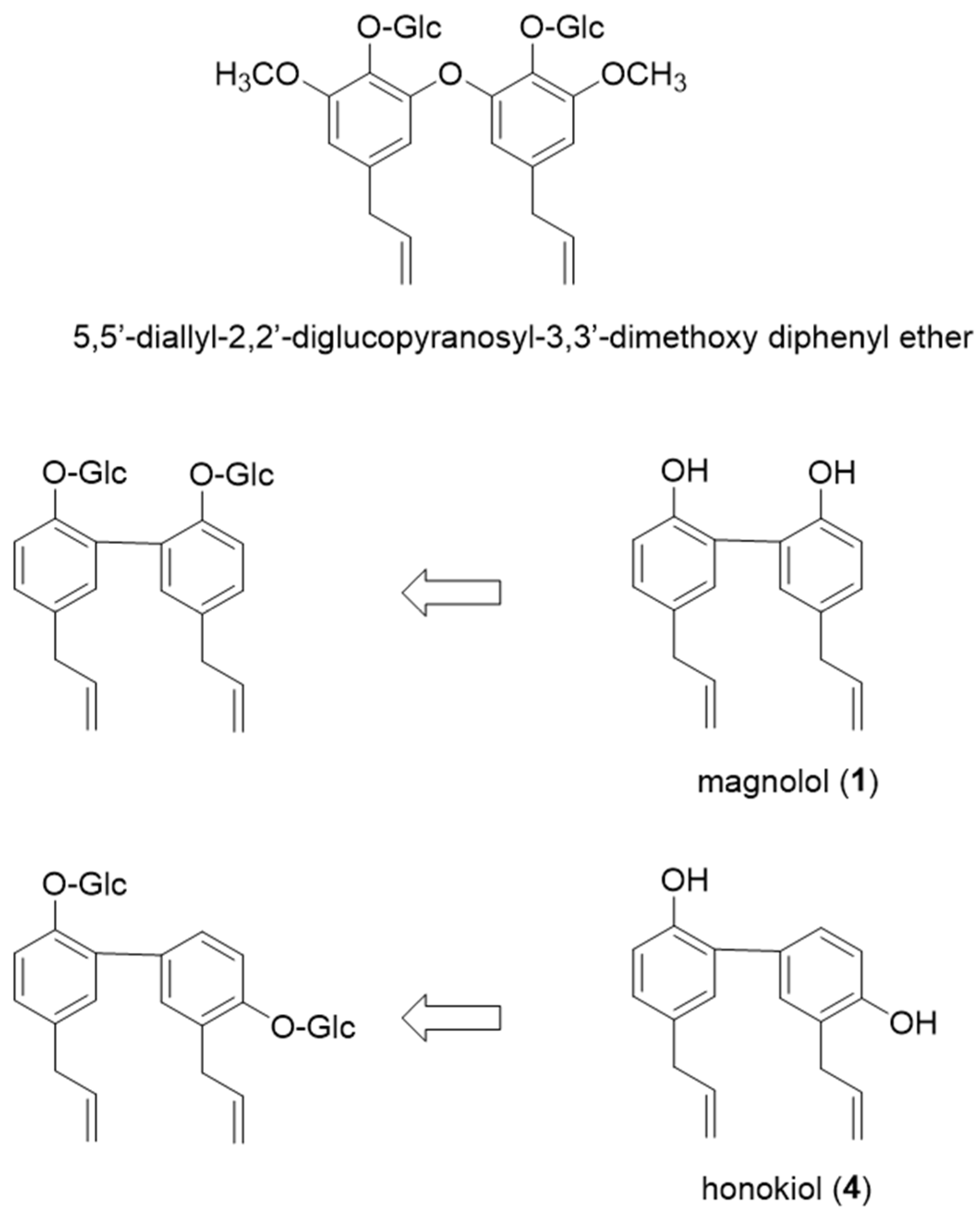
In our previous report, we established an evaluation method for the glycation induction model test using human-derived dermal fibroblasts, TIG-110 cells, and demonstrated that it is effective in evaluating antiaging effects on the skin [11]. Using this evaluation method, we also found that the novel compound, 5,5’-diallyl-2,2’-diglucopyranosyl-3,3’-dimethoxy diphenyl ether, which was isolated from fennel seeds, shows significant antiglycation activity. We are currently working on its effective use in cosmetics and health foods. The glycosides of magnolol (1) and honokiol (4), which are very similar to 5,5’-diallyl-2,2’-diglucopyranosyl-3,3’-dimethoxy diphenyl ether in terms of chemical structure, were newly synthesized (Figure 1) and examined for their anti-aging effects on the skin in this study.
Magnolol (1) accumulates in the bark of the Japanese white-bark magnolia (Magnolia obovata). Japanese white-bark magnolia is a species in the genus Magnolia, and family Magnoliaceae. It is also known as Ho or Hogashiwa in Japan. “Ho” means “wrap”, which is a name derived from the economic importance of its broad leaves for food packaging purposes. For medicinal purposes, the bark is used in the herbal medicine Koboku, which is dispensed in traditional medicines for gastrointestinal disorders, laxatives, anti-tussives, and expectorants [12]. Magnolol (1) and honokiol (4) have shown muscle relaxant, neuroprotective, antioxidative, anti-atherosclerosis, anti-inflammatory and anti-microbial effects [13,14,15,16].
We have previously reported on the features of magnolol (1), honokiol (4), and their glycosides, revealing their antioxidant potency, cytotoxicity, and inhibitory effects on histamine release and tyrosinase activity [17,18], under consideration of their application to cosmetics and food additives [19]. Based on these findings, this study focused on anti-glycation and examined the anti-aging effects of synthesized magnolol and honokiol glycosides on the skin (skin-beautifying effects) by conducting the glycation induction model test using human-derived dermal fibroblasts, TIG-110 cells [11].
2. Materials and Methods
2.1. General Experimental Procedures
For silica gel column chromatography, glass columns (Φ2.5 cm × 30 cm) were used, and silica gel (C-300 Wako gel, Fujifilm Wako Pure Chemical Co., Tokyo, Japan) was used as a packing material. The eluting solvent was prepared with a mixture of 1-hexane and ethyl acetate, or chloroform and methanol in appropriate proportions. Using a JEOL JMS-700 M Station mass spectrometer (FAB/MS) and an LCMS-2020 mass spectrometer (Shimadzu instrument, Kyoto, Japan), the mass counts of the synthesis compounds were measured. Then, nuclear magnetic resonance (NMR) spectra were measured using a BRUKER AVANCETM III Nanobay nuclear magnetic resonance spectrometer (400 MHz) with CDCl3 or CD3OD as a solvent.
2.2. Plant Material
Magnolol (1) and honokiol (4), which were isolated from commercially provided bark powders of the magnoliae (Magnolia obovata), by the previously reported method [11], were used as starting materials.
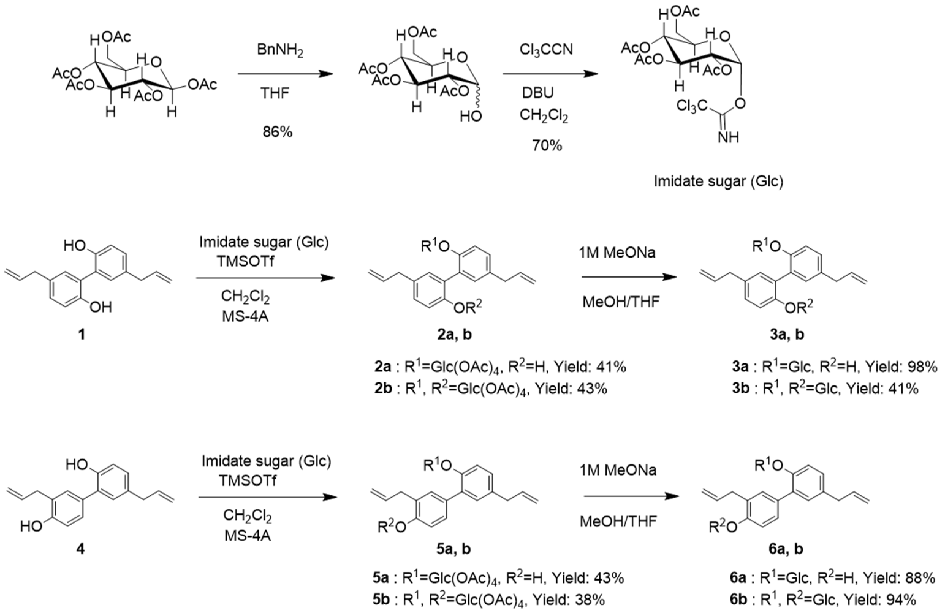
2.3. Preparation of Imidate Sugar (Glc) [2,3,4,6-Tetra-O-acetyl-β-D-glucopyaranosyl-2,2,2-trichloroacet imidate]
Penta acetyl-β-D-glucopyranoside [Glc(OAc)5] (10 mmol) was dissolved in tetrahydrofuran (40 mL), and benzylamine 11 mmol was added. The mixture was stirred for 24 h at room temperature and concentrated to a yellow syrup under vacuum to obtain tetra acetyl-β-D-glucopyranoside (Glc(OAc)4; yield—86%). The mixture of Glc(OAc)4 (10 mmol) was dissolved in dichloromethane (20 mL), and the solution was cooled to 0 °C. Trichloroacetonitrile 15 mmol was added, followed by 1,8-Diazabicyclo [5.4.0]undec-7-ene (DBU) 0.25 mmol. The reaction was warmed to room temperature and stirring was continued for 12 h. Concentration gave a dark brown syrup that was purified by silica gel, to obtain imidate Sugar (Glc) (yield—70%).
2.4. Synthesis of Magnolol and Honokiol Glycoside
A solution of magnolol (1) or honokiol (4) (0.56 mmol) in dichloromethane (15 mL) was stirred for 15 min with molecular sieves 4A (MS 4A 1/8, 20 grains) at −78 °C in an ice CO2/acetone bath. TMS-OTf 0.06 mmol was added under argon. Then, a solution of imidate sugar (Glc) (0.84 mmol) in dichloromethane (5 mL) was added dropwise over 10 min. The reaction was allowed to warm at room temperature over 4 h. The reaction was quenched by addition of triethylamine (2.24 mmol). The solvent was evaporated under reduced pressure and the resulting residue was purified by silica gel, to obtain magnolol or honokiol acetyl glycoside (yield—38–47%; 2a, 2b, 5a, and 5b).
To a room temperature solution of magnolol or honokiol acetyl glycoside (2a, 2b, 5a, and 5b) in methanol, tetrahydrofuran (1:1, 15 mL) was added with 1 M sodium methoxide (1.5 mL), and was then stirred for 30 min. To the reaction was added DOWEX 50WX8-100 ion-exchange resin (Aldrich Chemical Co., Inc., Saint Louis, MO, USA) and was adjusted to pH 7.0. Then, DOWEX 50WX8-100 was filtered. The solvent was evaporated under reduced pressure and the resulting residue was purified by silica gel, to obtain magnolol or honokiol glycoside (yield—88–98%; 3a, 3b, 6a, and 6b).
2.5. Synthesized Compounds
5,5′-Di(prop-2-en-1-yl)[1,1′-biphenyl]-2-hydroxy-2′-glucopyranoside (3a)
White crystal, m.p. 77–78 °C
FAB-MS: m/z 427 [M-H]−
1H-NMR (CD3OD, δppm): 3.36 (4H, br. d, J = 6.5 Hz), 3.37~3.47 (3H, m), 3.66 (1H, dd, J = 6, 12 Hz), 3.87 (1H, d, J = 2, 12 Hz), 4.97 (1H, d, J = 8 Hz), 5.00 (1H, m), 5.02 (1H, m), 5.04 (1H, m), 5.01 (1H, m), 5.96 (2H, m), 6.82 (1H, d, J = 8 Hz), 6.95 (1H, d, J = 2 Hz), 7.00 6.95 (1H, dd, J = 2, 8 Hz), 7.01 6.95 (1H, d, J = 2 Hz), 7.13 6.95 (1H, dd, J = 2, 8 Hz), 7.16 6.95 (1H, d, J = 8 Hz).
13C-NMR (CD3OD, δppm): 40.4 (×2), 62.5, 71.2, 74.8, 77.8, 78.2, 102.4, 115.6, 115.8, 115.9, 117.6, 128.1, 129.7, 129.8 (×2), 132.7, 132.8, 133.1 (×2), 135.2 (×2), 139.1, 139.4.
5,5′-Di(prop-2-en-1-yl)[1,1′-biphenyl]-2,2′-diglucopyranoside (3b)
White crystal, m.p. 107–108 °C
FAB-MS: m/z 589 [M-H]−
1H-NMR (CD3OD, δppm): 3.33 (4H, br. d, J = 6 Hz), 3.34~3.40 (6H, m), 3.65 (2H, dd, J = 5, 12 Hz), 3.83 (2H, J = 2, 12 Hz), 5.01 (1H, m), 5.04 (2H, m), 5.09 (1H, m), 5.97 (2H, m), 7.04 (2H, d, J = 2 Hz), 7.11 (2H, dd, J = 2, 8.5 Hz), 7.16 (2H, d, J = 8.5 Hz).
13C-NMR (CD3OD, δppm): 41.4 (×2), 62.5 (×2), 71.3 (×2), 74.8 (×2), 78.0 (×4), 101.9 (×2), 115.9 (×2), 117.1 (×2), 129.7 (×2), 130.3 (×2), 132.8 (×2), 135.2 (×2), 139.1 (×2), 154.0 (×2).
3′,5-Di(prop-2-en-1-yl)[1,1′-biphenyl]-4′-hydroxy-2-glucopyranoside (6a)
White crystal, m.p. 78–79 °C
FAB-MS: m/z 427 [M-H]−
1H-NMR (CD3OD, δppm): 3.34 (4H, br. d, J = 6.5 Hz), 3.37~3.52 (3H, m), 3.67 (1H, dd, J = 6, 12 Hz), 3.86 (1H, dd, J = 2, 12 Hz), 4.98 (1H, m), 5.01 (H, m), 5.03 (1H, m), 5.08 (1H, m), 5.99 (1H, m), 6.78 (1H, d, J = 8 Hz), 7.04 (1H, dd, J = 2, 8 Hz), 7.05 (1H, d, J = 2 Hz), 7.15 (1H, d, J = 8.5 Hz), 7.26 (1H, dd, J = 2, 8.5 Hz), 7.29 (1H, d, J = 2 Hz).
13C-NMR (CD3OD, δppm): 35.3, 40.4, 62.5, 71.3, 75.0, 78.1, 78.2, 102.0, 115.4, 115.5, 115.8, 116.6, 127.3, 128.9, 129.6, 131.0, 131.7, 132.5, 1322.9, 135.3, 138.55, 139.2, 153.7, 155.3.
3′,5-Di(prop-2-en-1-yl)[1,1′-biphenyl]-2,4′-diglucopyranoside (6b)
White crystal, m.p. 132–133 °C
FAB-MS: m/z 589 [M-H]−
1H-NMR (CD3OD, δppm): 3.35 (4H, br.d, J = 6.5Hz), 3.37~3.50(6H, m), 3.68(2H, br.d, J = 11.5 Hz), 3.80 (6H, m), 383~3.88(4H, m), 4.99 (1H, m), 5.02 (2H, m), 5.07(1H, m), 5.89 (1H, dt, J = 6.5, 17 Hz), 5.92 (1H, dt, J = 6.5, 17 Hz), 6.38 (2H, d, J = 2 Hz), 6.52 (2H, d, J = 2 Hz)
13C-NMR (CD3OD, δppm): 35.3, 40.4, 62.5, 62.6, 71.3, 71.4, 75.0, 75.1, 78.1, 78.2, 78.3 (×2), 102.0, 102.7, 115.7, 115.8, 115.9, 116.7, 129.3, 129.8, 130.4, 131.8, 132.3, 132.4, 133.9, 135.3, 138.7, 139.1, 153.8, 155.7.
2.6. In Vitro Inhibition Test of AGEs Generation
The mixture of the sample (20 μL), which was adjusted to each concentration, 0.1 mol/L phosphate buffer solution (PBS) (pH7.4) (500 mL), distilled water (180 μL), 40 mg/mL of Bovine serum albumin (BSA, Sigma Chemical Co., Ltd., Missouri, USA) (200 mL), and 2 mmol/L of glucose aqueous solution (100 μL) was stirred. Two samples of the same concentrations were prepared, to identify a difference of incubation. In addition, as a blank (controlled trial), methanol, instead of a sample, was used. Each sample was incubated for 30 h at 60 °C (A) and 25 °C (B). After incubation, trichloroacetic acid (100 μL) was added to each mixture and stirred. Then, each mixture was centrifuged at 4 °C, 15,000 rpm for 4 min. Each precipitate (AGEs) was dissolved with 1 mL of 0.25 N sodium hydroxide water solution-PBS and poured by 200 μL into a white microplate. The AGE-derived fluorescence was measured using a microplate reader TECAN F200 (Tecan Group Ltd., Männedorf, Switzerland), at an excitation wavelength of 360 nm and fluorescent wavelength of 440 nm. Percentage inhibition of AGEs generation was calculated as:
AGEs inhibition rate (%) = {(blank A − blank B) − (sample A − sample B)/(blank A − blank B)} × 100
2.7. Cell Culture
TIG-110 cells (JCRB-05423) are normal diploid fibroblasts isolated from the skin of a 33-year-old Japanese woman. TIG-110 cells were cultured in T-25 flasks using DMEM containing fetal bovine serum and antibiotics (antibiotic–antimicrobial agent mixture solution (100× concentration), Nakalai Tesque, INC., Kyoto, Japn) as cell culture medium. After 2–3 days of culturing in an incubator (37 °C, 5% CO2), the cells grew to 80% confluency in the flasks. The 80% confluent cells were washed with PBS(-) solution, and then, Trypsin solution (TrypLETM Express, ThermoFisher, MA, USA) was added, and it was left to stand in a CO2 5% incubator at 37 °C for 5–8 min. Then, the cells were detached by gently tapping the flasks, checked under a microscope, and collected by centrifugation. After the cell count was measured, the number of cells was adjusted to the specified number, and then plates were seeded or passaged. TIG-110 cells were used for experiments up to 15 passages.
2.8. Cell Viability
Synthesized compounds were co-cultured with TIG-110 cells for 48 h to examine the cytotoxicity of the various spice seed extracts and isolates. Twenty-four hours before the start of the test, TIG-110 cells were seeded into 96-well plates. To ensure a uniform seeding concentration, DMEM was used to adjust the cell count to 5.0 × 10⁴ cells/100 μL. After 24 h, sample-DMEM cultures containing a diluted thawed compound and a control were prepared and added to the seeded wells in 100 µL increments. Sample-DMEM was diluted and dissolved for each sample to achieve a final concentration of 25 µg/mL (0.4% DMSO concentration). The control was prepared from DMEM mixed with 0.4% DMSO. After 48 h, cell viability was checked by the MTT method. Cell viability was calculated as follows:
Cell viability (%) = (absorbance of Sample − absorbance DMEM/absorbance of control) × 100
2.9. Determination of Glyoxal Concentration
The GO concentration was examined to establish a glycation induction model test method. To obtain a final concentration of 5 mM of GO, 40% glyoxal (Fujifilm Wako Pure Chemical Co., Tokyo, Japan) was diluted and dissolved in DMEM. This 5 mM GO-DMEM solution was further diluted to prepare four concentrations (5 mM, 2.5 mM, 1.25 mM, and 0.625 mM) of GO-DMEM. Cells were seeded in the same manner as indicated previously herein. The prepared GO-DMEM was added to the seeded wells in 100 µL increments. The control was DMEM mixed with 0.4% DMSO. After 48 h, cell viability was examined by the MTT method. Cell viability was calculated as follows:
Cell viability (%) = (absorbance of GO − absorbance DMEM/absorbance of control) × 100
2.10. Assay of AGE Formation Inhibitory Effects in Glyoxal System
Based on the preliminary test results, GO-DMEM was set to 1.25 mM, and the culture time was set to 48 h. Using these conditions, the samples that did not show cytotoxicity were co-cultured. Cell seeding and preparation of GO-DMEM were performed using the same method described previously herein. Samples were also diluted and dissolved in GO-DMEM. The control was 0.4% DMSO mixed with DMEM. After 48 h, cell viability was assessed by the MTT method. Cell viability was calculated as follows:
Cell viability (%) = (absorbance of GO − absorbance DMEM, absorbance of sample-GO − absorbance DMEM/absorbance of control) × 100
2.11. Statistical Processing
Statistical processing was performed using SAS University Edition (SAS Institute, Cary, NC, USA) with data expressed as the mean ± S.D. A risk rate of less than 5% (* p < 0.05, ** p < 0.01) was considered a significant difference.
3. Results
3.1. Synthesis of Glycosides
Glycoside synthesis of magnolol (1) was carried out using Imidate Sugar (Glc) [22]. Namely, magnolol (1) was exposed to TMS-OTf and imidate sugar (Glc) in dichloromethane in the presence of argon. The resulting acetylated glycosides 2a and 2b were obtained with yields of 41 and 43%, respectively. Then, 2a and 2b were deacetylated with 1.0 M sodium methoxide in a methanol-THF (1:1) solution. Then, the resulting glycosides 3a and 3b were obtained with yields of 98 and 97%, respectively (Scheme 1).
A similar operation was performed on honokiol (4), and the resulting acetylated glycosides 5a and 5b were obtained with yields of 47 and 38%, respectively. Then, the resulting 5a and 5b were treated in the same manner as in the case of 3a and 3b synthesis, and the glucose glycosides 6a and 6b were obtained with yields of 88 and 94%, respectively (Scheme 1).
The structures of the following synthesized compounds were determined by MS, NMR and HMBC spectra: 5,5′-di(prop-2-en-1-yl)[1,1′-biphenyl]-2-hydroxy-2′-glucopyranoside (3a), 5,5′-di(prop-2-en-1-yl)[1,1′-biphenyl]-2,2′-diglucopyranoside (3b), 3′,5-di(prop-2-en-1-yl)[1,1′-biphenyl]-4′-hydroxy-2-glucopyranoside (6a) and 3′,5-di(prop-2-en-1-yl)[1,1′-biphenyl]-2,4′-diglucopyranoside (6b).
3.2. AGEs’ Inhibitory Activity
The starting materials, 1 and 4, and resulting glycosides, 3a, 3b, 6a and 6b, were tested for their inhibitory effect on AGE production (Table 1). The results showed that these substances had higher inhibitory activity than the positive control aminoguanidine. Specifically, significant inhibitions were observed in 3a, 3b, 6a, and 6b, with IC50 values below 0.10 mmol/L.
3.3. Cytotoxicity of Synthesized Compounds
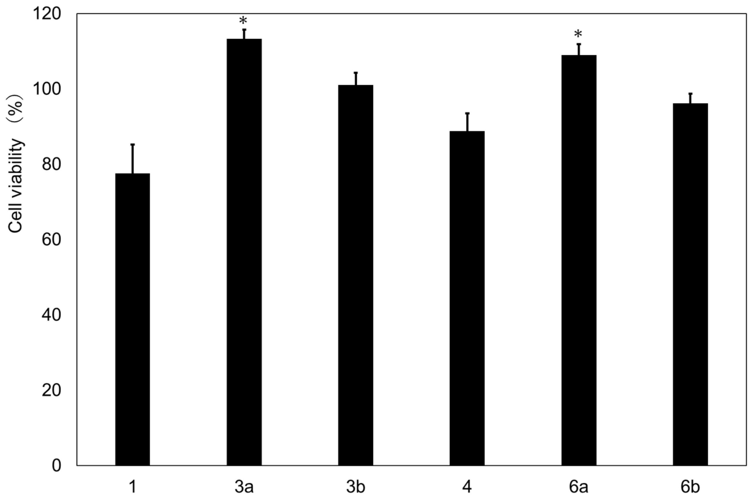
Fibroblasts produce the most major components of the epidermis; therefore, the effects of the synthesized compounds on fibroblasts, TIG-110 cells, were examined (Figure 2). Glycosides 3a and 3b synthesized from magnolol (1) and 6a and 6b synthesized from honokiol (4) showed no cytotoxicity at a concentration of 25 μg/mL. However, the starting material magnolol (1) and honokiol (4) showed some cytotoxicity. These results indicate that the synthesized glycoside compounds are expected to be applied to cosmetics and food additives with skin-beautifying effects.
3.4. Effect of Synthesized Compounds on Cell Viability of TIG-110 Cells Exposed to Glyoxal
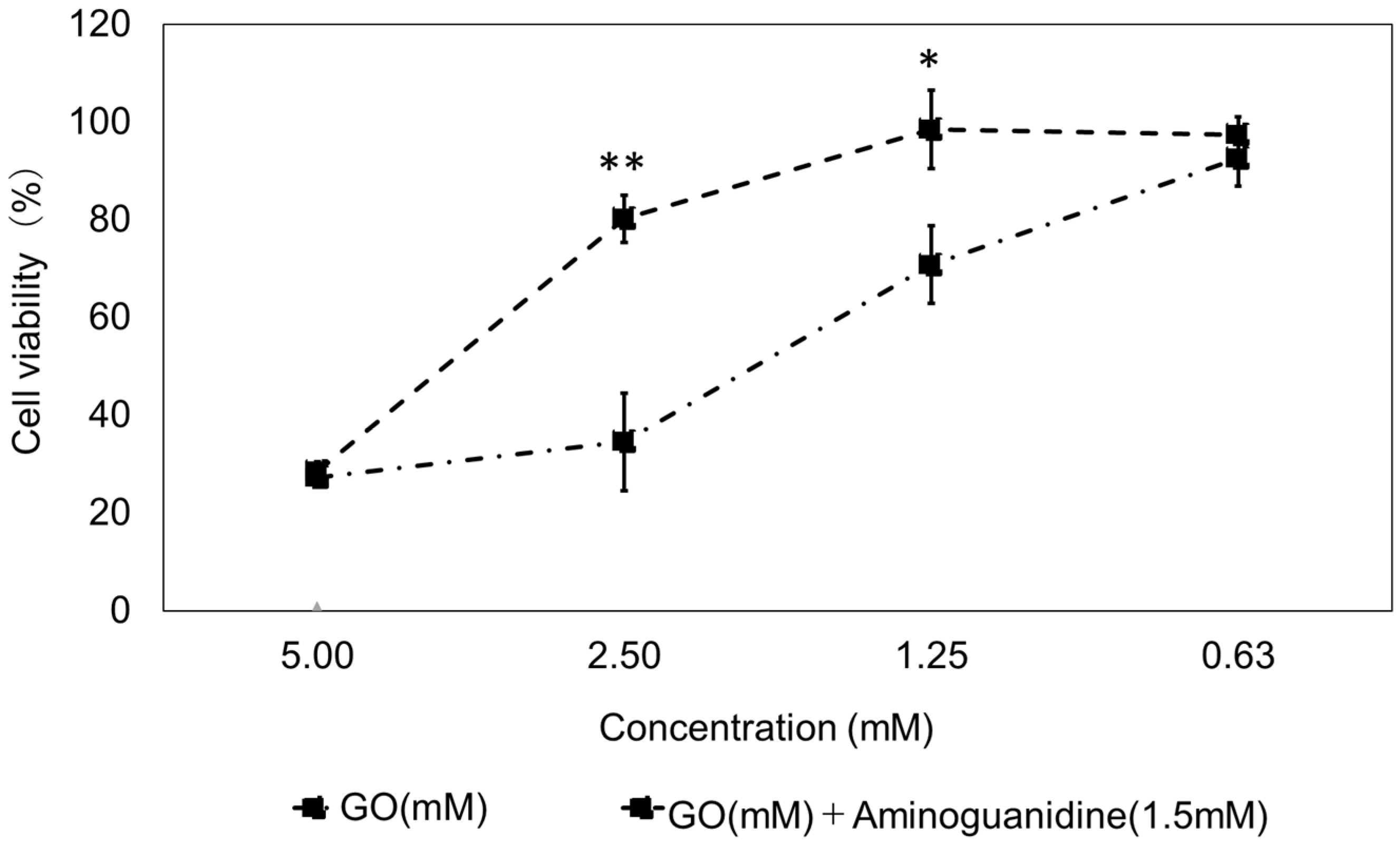
In order to conduct a glycation suppression test in cells, the concentration of glyoxal (GO), which is a glycation inducer, was examined. Aminoguanidine, which has anti-glycation activity, was used as a positive control. Cell viability was compared in a GO mixed medium and aminoguanidine–GO mixed medium. In each medium, GO concentrations were adjusted to 0.625, 1.25, 2.5 and 5.0 mM, and aminoguanidine concentrations were adjusted to 0 and 1.5 mM. In the previous report, the optimum concentration of GO was 1.25 mM, as shown in Figure 3 (GO concentration of 2.5 mM: 34.5 ± 10.0; GO concentration of 2.5 mM + aminoguanidine: 80.2 ± 4.9; GO concentration of 1.25 mM: 70.8 ± 8.0; GO concentration of 1.25 mM + aminoguanidine: 98.5 ± 8.0).
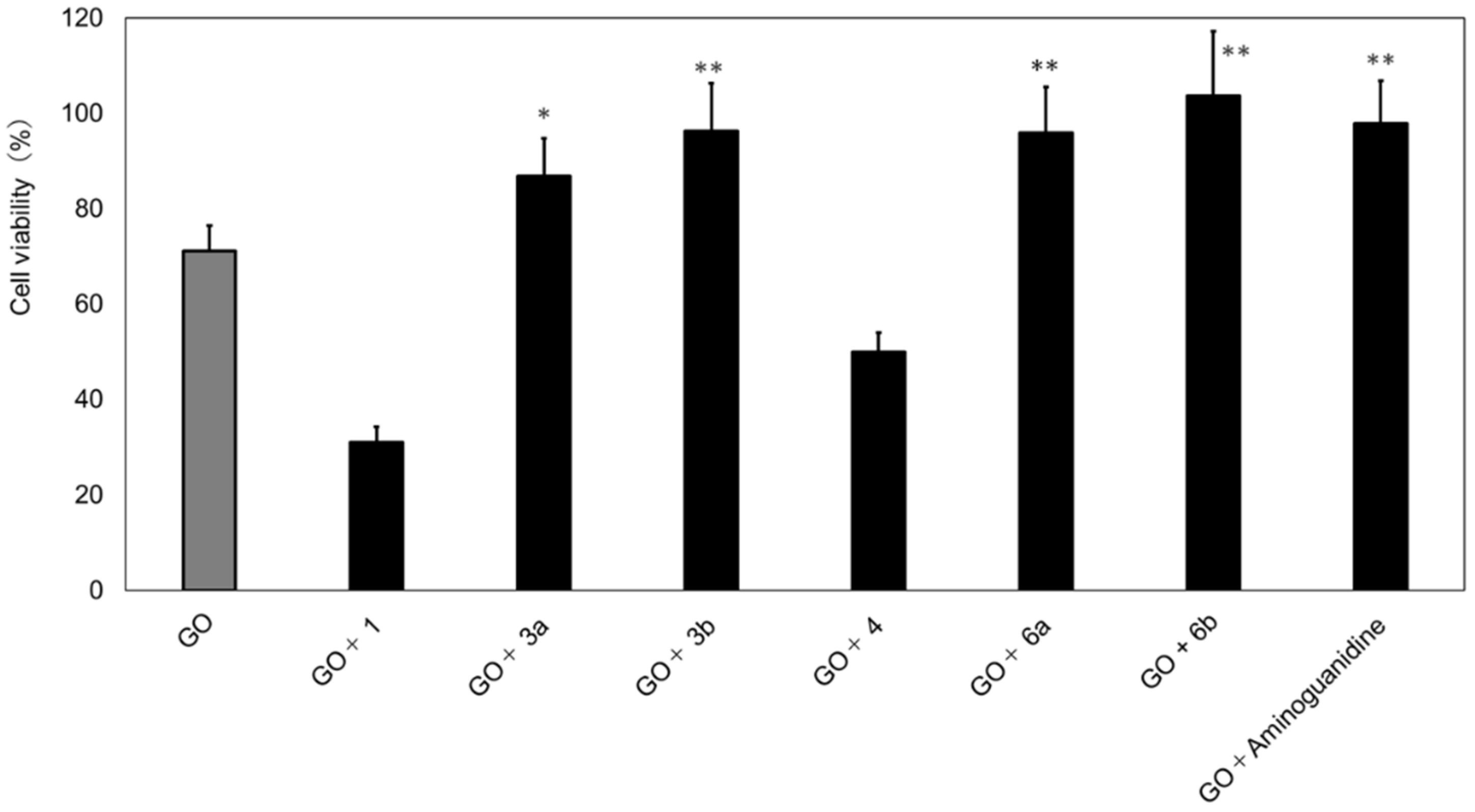
In the glycation induction model test [11], the starting materials and synthesized glycosides were evaluated. As shown in Figure 4, the glycoside compounds 3a, 3b, 6a, and 6b significantly inhibited the decrease in cell viability (GO concentration of 1.25 mm: 71.1 ± 4.8; GO concentration of 1.25 mm + 3a: 86.8 ± 7.9; GO concentration of 1.25 mm + 3b: 96.3 ± 1.0; GO concentration of 1.25 mm + 6a: 95.9 ± 9.6; GO concentration of 1.25 mm + 6b: 103.7 ± 13.5).
4. Discussion
The glycation reaction is a nonenzymatic binding of sugars and proteins in the body. Various AGEs have been attracting attention as factors contributing to the development of diabetic complications and aging. In this study, the effects of glycation on the skin were examined. In humans, collagen comprises approximately 30% of the total protein in the body, and aside from the skin, it is also widely distributed in bones and blood vessels. Supple skin has collagen in which chains of amino acids assemble into a triple-helix structure. When glycation-induced AGEs are formed in collagen, the protein function is reduced, resulting in decreased skin suppleness and resilience. Previous studies have confirmed that AGEs are expressed in human skin tissues, and the expression levels have been reported to increase age dependently [9]. In addition to the skin, the presence of AGEs in the collagen of the vascular wall has been revealed, which is likely to lead to aging-related diseases such as atherosclerosis [22]. Therefore, these diseases can be thought of as the result of AGEs, indicating that the control of the AGEs will lead the suppression of the development of pathologic aging [23] and that it can be an effective approach for prevention of aging.
In order to search for substances that prevent skin aging due to glycation, the anti-glycation effects of four glycosides were evaluated by the AGE inhibitory activity test and the glycation induction model test using human-derived dermal fibroblasts, TIG-110 cells.
The glycosides were synthesized with the starting materials magnolol (1) and honokiol (4), isolated from the Japanese white-bark magnolia, and evaluated by the AGE inhibitory activity test. The results showed that the synthesized glycosides have a high anti-glycation activity. In addition, the glycation induction model test in which GO (a glycation inducer) was added into a cell-culture medium showed that glycosides 3a, 3b, 6a, and 6b significantly inhibited the decrease in cell viability.
The IC50 value in the AGE inhibitory activity test for the compound 5.5′-diallyl-2,2′-diglucopyranosyl-3,3′-diphenyl ether, which was isolated from the fennel seeds in our previous study [11], was 0.08 mM. Comparablly, the IC50 values were observed with the newly synthesized glycosides, 3a and 6b. In the glycation induction model test, glycosides 3b, 6a, and 6b inhibited the decrease in cell viability at the level of 0.01 of significancy, while the inhibitory effect of glycosides 3a was significant at the 0.05 level.
Aminoguanidine, the positive control, is a known inhibitor of carbonyl compounds. Since the amino groups of aminoguanidine capture GO, the reactivity of GO is greatly reduced when treated with aminoguanidine, resulting in the inhibition of the production of carboxymethyl lysine (CML) [24]. The glycoside compounds 3a, 3b, 6a, and 6b, which were synthesized in this study, inhibit the production of CML. These compounds have the same structural characteristics as the 5,5′-diallyl-2,2′-diglucopyranosyl-3,3′-dimethoxy diphenyl ether, which has a double bond at the terminal position. It is highly likely that this terminal double bond captures the aldehyde group of GO, resulting in the inhibition of CML production.
5. Conclusions
Four glycosides were synthesized using the starting materials magnolol (1) and honokiol (4), isolated from the Japanese white-bark magnolia, and their anti-aging effects on the skin (skin-beautifying effects) have been examined. The AGE inhibitory activity test (anti-glycation test) and glycation induction model test using human-derived dermal fibroblasts, TIG-110 cells, were conducted to evaluate the anti-aging effects. The synthesized glycoside compounds, 5,5′-di(prop-2-en-1-yl)[1,1′-biphenyl]-2-hydroxy-2′-glucopyranoside (3a), 5,5′-di(prop-2-en-1-yl)[1,1′-biphenyl]-2,2′-diglucopyranoside (3b), 3′,5-di(prop-2-en-1-yl)[1,1′-biphenyl]-4′-hydroxy-2-glucopyranoside (6a) and 3′,5-di(prop-2-en-1-yl)[1,1′-biphenyl]-2,4′-diglucopyranoside (6b), showed remarkable anti-glycation activities. The glycation induction model test with the fibroblasts, TIG-110 cells, demonstrates that the aforementioned glycosides significantly inhibit the decrease in cell viability. These newly synthesized glycoside compounds are expected to be used as cosmetic ingredients, health foods, and pharmaceutical ingredients, which have inhibitory effects against AGE formation.
Author Contributions
A.S. and R.T. conceived and designed the research. A.T. carried out all experiments. A.S. carried out the synthesis of functional ingredients. M.N. performed the simulations. A.S. was responsible for writing—review and editing. All authors have read and agreed to the published version of the manuscript.
Funding
A part of this research was funded by the 21st Century Joint Research Enhancement Grant from Kindai University, grant number KD201705 and KD2003.
Data Availability Statement
The data presented in this study are available on request from the corresponding author. The data are not publicly available due to privacy.
Acknowledgments
We would also like to extend our gratitude to Kazuho Yokono and Maya Ishii, who were graduate school students when they were part of the research team, for their participation in the project.
Conflicts of Interest
The authors declare no conflict of interest.
References
- Maillard, L.C. Action des amines sur les sucres: Formation des mélanoïdines par voie méthodique, C.R. Acad. Sci. 1912, 2, 154–166. [Google Scholar]
- Taniguchi, N. TanpakuTouka Han-nou no Seitai ni Okeru Igi (Significance of Protein Glycation in Living Organisms); Shigeta, Y., Taniguchi, N., Eds.; Tanpaku no Touka (Protein Glycation); Igaku Shoin Ltd.: Bunkyō, Tokyo, Japan, 1997; pp. 2–8, 9–15. [Google Scholar]
- Rahber, S. An abnormal hemoglobin in red cells of diabetics. Clin. Chim. Acta 1968, 22, 296–298. [Google Scholar] [CrossRef]
- Holmquist, W.R.; Schoeder, W.A. A new N-terminal blocking group involving a Schiff base in hemoglobin A1c. Biochemistry 1966, 5, 2489–2503. [Google Scholar] [CrossRef] [PubMed]
- Hayase, F. Recent Development of 3-Deoxyosone Related Maillard Reaction Products. Food Sic. Technol. Res. 2000, 6, 79–86. [Google Scholar] [CrossRef]
- Singh, V.P.; Bali, A.; Singh, N.; Jaggi, A.S. Advanced Glycation End Products and Diabetic Complications. Korean J. Physiol. Pharmacol. 2014, 18, 1–14. [Google Scholar] [CrossRef] [PubMed] [Green Version]
- Vistoli, G.; Maddis, D.D.; Cipak, A.; Zarkovic, N.; Carini, M.; Aldini, G. Advanced glycoxidation and lipoxidation end products (AGEs and ALEs): An overview of their mechanisms of formation. Free. Radic. Res. 2013, 47, 3–27. [Google Scholar] [CrossRef] [Green Version]
- Sato, T.; Wu, X.; Shimogaito, N.; Takino, J.; Yamagishi, S.; Takeuchi, M. Effects of high-AGE beverage on RAGE and VEGF expressions in the liver and kidneys. Eur. J. Nutr. 2009, 48, 6–11. [Google Scholar] [CrossRef]
- Jeanmaire, C.; Danoux, L.; Pauly, G. Glycation during human dermal intrinsic and actinic aging: An in vivo and in vitro model study. Br. J. Dermatol. 2001, 145, 10–18. [Google Scholar] [CrossRef] [PubMed]
- Monnier, V.M.; Kohn, R.R.; Cerami, A. Accelerated age-related browning of human collagen in diabetes mellitus. Proc. Nat. Acad. Sci. USA 1984, 81, 583–587. [Google Scholar] [CrossRef] [Green Version]
- Sawabe, A.; Yamashita, A.; Fujimatsu, M.; Takeda, R. Development of Evaluation Methods for Anti-glycation Activity and their Functional Ingredients Contained in Coriander and Fennel Seeds. Processes 2022, 10, 982. [Google Scholar] [CrossRef]
- Mitsuhashi, H. Pharmacognosy; Nankodo Co., Ltd.: Tokyo, Japan, 1983; pp. 100–101, 186–190. [Google Scholar]
- Lee, Y.-J.; Lee, Y.M.; Lee, C.-K.; Jung, J.K.; Han, S.B.; Hong, J.T. Therapeutic applications of compounds in the Magnolia family. Pharmacol. Ther. 2011, 130, 157–176. [Google Scholar] [CrossRef] [PubMed]
- Woodbury, A.; Yu, S.P.; Wei, L.; GarcõÂa, P. Neuro-modulating effects of honokiol: A review. Front. Neurol 2013, 4, 130. [Google Scholar] [CrossRef] [PubMed] [Green Version]
- Shen, J.-L.; Man, K.-M.; Huang, P.-H.; Chen, W.-C.; Chen, D.-C.; Cheng, Y.-W.; Liu, P.-L.; Chou, M.-C.; Chen, Y.-H. Honokiol and magnolol as multifunctional antioxidative molecules for dermatologic disorders. Molecules 2010, 15, 6452–6465. [Google Scholar] [CrossRef] [PubMed]
- Maioli1, M.; Basoli1, V.; Carta, P.; Fabbri, D.; Dettori, M.A.; Cruciani, S.; Serra, P.A.; Delogu, G. Synthesis of magnolol and honokiol derivatives and their effect against hepatocarcinoma cells. PLoS ONE 2018, 13, e0192178. [Google Scholar] [CrossRef] [PubMed] [Green Version]
- Nomura, M.; Ishii, M.; Motoya, T.; Inoue, T. Isolation of polyphenols from general of herbal medicines and their physiological activities. Res. Rep. Fac. Eng. Kinkdai Univ. 2021, 55, 15–26. [Google Scholar]
- Nomura, M.; Murakami, Y.; Ishi, M.; Yokono, K.; Tanimoto, S.; Murai, Y.; Sawabe, A. Synthesis of Glycosides of Magnolol and Honokiol and their physiological activities. Res. Rep. Fac. Eng. Kinkdai Univ. 2021, 55, 27–34. [Google Scholar]
- Nomura, M.; Sawabe, A. Glycoside synthesis and functionality as the cosmetics material using the chlorogenic acid metabolites derived from a plant. In Synthesis and Application of a Functional Glycoside—Mainly on Glycosyltransferase; Hamda, H., Ed.; CMC Publishing Co., Ltd.: Tokyo, Japan, 2013; pp. 185–194. [Google Scholar]
- Sawabe, A.; Ohnishi, N.; Yoshioka, S.; Kusudo, K.; Kanno, K.; Watanabe, Y. Functional Ingredients and Food Preservative in Immature Persimmon “Tekka-Kaki”. Processes 2021, 9, 1989. [Google Scholar] [CrossRef]
- Das, R.; Mukhopadhyay, B. Chemical O-Glycosylations: An Overview. ChemistryOpen 2016, 5, 401–433. [Google Scholar] [CrossRef] [PubMed] [Green Version]
- Semba, R.D.; Nicklett, E.J.; Ferrucci, L. Does accumulation of advanced glycation end products contribute to the aging phenotype? J. Gerontol. Ser. A Biol. Sci. MED. Sci. 2010, 65, 963–975. [Google Scholar] [CrossRef] [PubMed] [Green Version]
- Saito, M. (Ed.) Anti-Aging Medicine, 3rd ed.; Medical View, Co., Ltd.: Tokyo, Japan, 2015; pp. 7–151. (In Japanese) [Google Scholar]
- Brownlee, M.; Vlassara, H.; Kooney, A.; Ulrich, P.; Cerami, A. Aminoguanidine presents diabets-induced arterial wall protein crosslinking. Science 1986, 232, 1629–1632. [Google Scholar] [CrossRef]
Figure 1.
Structures of 5,5′-diallyl-2,2′-diglucopyranosyl-3,3′-dimethoxy diphenyl ether, magnolol (1) and honokiol (4).
Figure 1.
Structures of 5,5′-diallyl-2,2′-diglucopyranosyl-3,3′-dimethoxy diphenyl ether, magnolol (1) and honokiol (4).

Scheme 1.
Synthesis of glycosides of magnolol (1) and honokiol (4).

Figure 2.
Cytotoxicity of compounds. n = 3; sample concentration: 25 µg/mL; compound (1: magnolol; 3a: 5,5′-di(prop-2-en-1-yl)[1,1′-biphenyl]-2-hydroxy-2′-glucopyranoside; 3b: 5,5′-di(prop-2-en-1-yl)[1,1′-biphenyl]-2,2′-diglucopyranoside; 4: honokiol; 6a: 3′,5-di(prop-2-en-1-yl)[1,1′-biphenyl]-4′-hydroxy-2-glucopyranoside; 6b: 3′,5-di(prop-2-en-1-yl)[1,1′-biphenyl]-2,4′-diglucopyranoside). The bars represent the mean ± SD, * p < 0.05 versus control; Student’s t-test.

Figure 3.
Cell viability of cells exposed to glyoxal (GO) at 48 h of culture. Four glyoxal (GO) concentrations of 5 mM, 2.5 mM, 1.25 mM, and 0.625 mM were studied, and their corresponding effects on cell viability are shown. n = 3; GO: Glyoxal (mM); aminoguanidine: positive control (1.5 mM). The bars represent the mean ± SD; * p < 0.05; ** p < 0.01 versus GO; Student’s t-test.

Figure 4.
Glycation suppression with selected purified compounds in the presence of glyoxal (GO) in TIG-110 cells. n = 3; GO: Glyoxal (1.25 mM), aminoguanidine: positive control (1.5 mM); compounds (1: magnolol; 3a: 5,5′-di(prop-2-en-1-yl)[1,1′-biphenyl]-2-hydroxy-2′-glucopyranoside; 3b: 5,5′-di(prop-2-en-1-yl)[1,1′-biphenyl]-2,2′-diglucopyranoside; 4: honokiol; 6a: 3′,5-di(prop-2-en-1-yl)[1,1′-biphenyl]-4′-hydroxy-2-glucopyranoside; 6b: 3′,5-di(prop-2-en-1-yl)[1,1′-biphenyl]-2,4′-diglucopyranoside)); sample concentration: 25 µg/mL (0.4% DMSO). The bars represent the mean ± SD; * p < 0.05; ** p < 0.01 versus GO; Student’s t-test.


Table 1.
IC50 values of the AGEs inhibitory activity test of the compounds.
| Compound | AGEs Inhibitory Activity IC50 Values |
|---|---|
| Aminoguanidine (positive control) | 0.42 |
| 1 | 0.21 |
| 3a | 0.09 |
| 3b | 0.04 |
| 4 | 0.17 |
| 6a | 0.06 |
| 6b | 0.07 |
Unit: mM.
Publisher’s Note: MDPI stays neutral with regard to jurisdictional claims in published maps and institutional affiliations. |
© 2022 by the authors. Licensee MDPI, Basel, Switzerland. This article is an open access article distributed under the terms and conditions of the Creative Commons Attribution (CC BY) license (https://creativecommons.org/licenses/by/4.0/).
Share and Cite
MDPI and ACS Style
Sawabe, A.; Tanaka, A.; Nomura, M.; Takeda, R. Skin-Beautifying Effects of Magnolol and Honokiol Glycosides. Processes 2022, 10, 1241. https://doi.org/10.3390/pr10071241
AMA Style
Sawabe A, Tanaka A, Nomura M, Takeda R. Skin-Beautifying Effects of Magnolol and Honokiol Glycosides. Processes. 2022; 10(7):1241. https://doi.org/10.3390/pr10071241
Chicago/Turabian StyleSawabe, Akiyoshi, Ayato Tanaka, Masato Nomura, and Ryuji Takeda. 2022. "Skin-Beautifying Effects of Magnolol and Honokiol Glycosides" Processes 10, no. 7: 1241. https://doi.org/10.3390/pr10071241
Note that from the first issue of 2016, this journal uses article numbers instead of page numbers. See further details here.