Article

Gallic Acid Enhances the Anti-Cancer Effect of Temozolomide in Human Glioma Cell Line via Inhibition of Akt and p38-MAPK Pathway

1
Department of Neurosurgery, Chang Gung Memorial Hospital, Chiayi 61363, Taiwan
2
College of Medicine, Chang Gung University, Taoyuan 33302, Taiwan
3
Department of Medical Research, Chang Gung Memorial Hospital, Chiayi 61363, Taiwan
4
Division of General Surgery, Department of Surgery, Chang Gung Memorial Hospital, Chiayi 61363, Taiwan
5
Graduate Institute of Clinical Medical Sciences, College of Medicine, Chang Gung University, Taoyuan 33302, Taiwan
6
Institute of Medicine, Chung Shan Medical University, Taichung 40201, Taiwan
7
Department of Microbiology, Immunology and Biopharmaceuticals, College of Life Sciences, National Chiayi University, Chiayi 60004, Taiwan
*
Author to whom correspondence should be addressed.
Processes 2022, 10(3), 448; https://doi.org/10.3390/pr10030448
Submission received: 26 November 2021 / Revised: 18 February 2022 / Accepted: 21 February 2022 / Published: 23 February 2022
(This article belongs to the Section Pharmaceutical Processes)

Abstract

:
(1) Background: Temozolomide (TMZ), an oral alkylating agent, is used to treat malignant gliomas and other difficult-to-treat tumors. TMZ can enter the cerebrospinal fluid p.o. (per os) and does not need hepatic metabolism for activation of its use as a standard chemotherapeutic regimen after surgical resection of malignant glioma of the brain. However, the prognosis remains poor for most patients, and the survival rate is still unsatisfactory. Gallic acid (Ga) is a secondary metabolite existent in numerous plants. Ga shows various bioactivities, including antioxidant, anti-inflammatory, anticancer and antimicrobial effects. In this study, the latent enhanced anti-cancer efficacy of Ga in TMZ-treated U87MG cells (a human glioma line) was evaluated. (2) Methods: The U87MG cell line was cultured for 24 h. The cells were incubated with Ga alone, TMZ alone, or their combination for various time points. Cell viability and the drug combination index were evaluated by an XTT-based analysis and isobologram analysis, respectively. DNA destruction and intracellular reactive oxygen species (ROS) generation were analyzed by flow cytometer. The expression of various proteins was assessed via Western blotting. (3) Results: Compared with the action of TMZ alone or Ga alone, TMZ/Ga combination augmented the inhibition of cellular viability and apoptotic level in the U87MG glioma cell line. This enhanced anti-cancer effect correlated with the decreased expression of Bcl-2 and p-Akt, and corresponded with the activation of the p38 mitogen-activated protein kinase (MAPK) pathway. In addition, Ga suppressed the TMZ-promoted ROS generation. (4) Conclusions: Ga can augment the anti-cancer effect of TMZ via the repression of Bcl-2 expression and Akt activation and the enhancement of the p38 MAPK pathway. Our results offer a novel probable approach for the medical treatment of malignant glioma.

1. Introduction

Temozolomide (TMZ) is an oral alkylating drug with a 3-methyl derivative of mitozolomide. This compound can readily spread across the blood–brain barrier. TMZ is a well-tolerated chemotherapeutic drug in the therapy of glioblastoma multiforme [1]. Although numerous studies have demonstrated that TMZ is a chemotherapeutic agent for gliomas, gliomas displaying TMZ resistance are frequently observed [2]. Several reports have showed that the anti-glioma action of TMZ can be enhanced by combining it with other treatment agents [3]. Guanosine promotes cellular toxicity through adenosine receptors and provokes apoptosis in A172 glioma cells by TMZ treatment [4]. The combination of sonodynamic therapy with TMZ reduces the migration of C6 glioma and stimulates the intrinsic mitochondrial apoptotic route via the inhibition of NHE-1 expression [5]. The treatment with honokiol notably increases TMZ-provoked apoptosis and G1 phase arrest in U87MG glioma cells [6]. The treatment of glioma cells with fluoxetine synergistically enhances TMZ to trigger apoptosis via the endoplasmic reticulum stress [7]. Our earlier report showed that valproic acid augmented the TMZ-induced apoptosis via the redox-regulated pathway [8]. Other studies demonstrated that the suppression of intracellular ROS and nuclear factor-κB mechanism resulted in an anti-migration effect on the propyl gallate-enhanced anti-cancer effect of TMZ in malignant glioma [9].
Gallic acid (Ga) contains a polyhydroxylphenolic structure and is broadly dispensed in various plants, fruits and foods. Ga can penetrate into the blood–brain barrier and extend to a rat’s brain, that is, into the middle cerebral artery occlusion in animal models [10]. Ga provokes the suppression of invasiveness through the inhibition of ADAM17, Ras/MAPK and the PI3K/Akt signaling pathways [11]. Ga significantly caused cytotoxicity, increased ROS generation, and provoked the intrinsic mitochondrial apoptosis in DBTRG-05MG cells [12]. Ga may be a potential compound for the treatment of brain glioma. Studies displayed that a synergistic effect can be induced by combining Ga with TMZ via regulating the apoptosis of U87MG glioma cells. In this study, we evaluated whether Ga can enhance the anti-cancer forces of TMZ in malignant glioma and clarify the probable molecular mechanisms.

2. Materials and Methods

2.1. Cell Line, Reagents, and Chemicals

The U87MG cell line (a human primary glioblastoma cell line) was purchased from the Bioresource Collection and Research Center (Food Industry Research and Development Institute, Hsinchu, Taiwan). Fetal bovine serum (FBS) and GibcoTM Eagle’s minimum essential medium were bought from Thermo Fisher Scientific (Waltham, MA, USA). Primary antibodies against Bcl-2, Bax, p53, β-actin, p-Akt, p-c-Jun N-terminal kinase (JNK), JNK, p-c-Jun N-terminal kinase (ERK), ERK, p-p38 and p38 were acquired from Santa Cruz Biotechnology, Inc. (Dallas, TX, USA). Protein concentration analysis was performed using the Bradford protein assay kit (Bio-Rad Laboratories, Inc. Irvine, CA, USA). Dimethyl sulfoxide (DMSO, Sigma-Aldrich Corp., Saint Louis, MO, USA), Ga, TMZ, 2′,7′-dichlorodihydrofluorescein diacetate (Sigma-Aldrich Corp., Saint Louis, MO, USA), propidium iodide (PI, Sigma-Aldrich Corp., Saint Louis, MO, USA), trypan blue solution and other experimental compounds were supplied by Sigma-Aldrich Corp. (Saint Louis, MO, USA).

2.2. Drug Treatment and Cell Culture

The U87MG cell line was incubated in Dulbecco’s Modified Eagle Medium (DMEM) containing 100 µg/mL streptomycin, 100 units/mL penicillin G, 2 mM L-glutamine and 10% FBS and retained in a 95% air/5% CO2 incubator maintained at 37 °C. TMZ or Ga was dissolved in DMSO for stock solutions. All incubated doses were diluted with cultured media. The dose of DMSO did not exceed 0.05%.

2.3. Cellular Viability Assay

A total of 1 × 10⁴ cells/well was seeded in a 96-well cultured-plate prior to treatment with various compounds. After the treatment with various compounds, cells were added with XTT (50 μL/well) for 2 h. The absorbance at 450 nm of all wells was assessed by deducting a background control medium as a blank group. The non-specific absorbance at 690 nm was detected and deducted from the absorbance at 450 nm with an EnSpire® plate analyzer (Billerica, MA, USA).

2.4. Intracellular ROS Analysis

The level of generation of intracellular ROS was evaluated using a DCFDA staining and flow cytometer. After the incubation of the compounds, the cells were added with DCFDA (20 μM) for 30 min at 37 °C and washed with 1 × phosphate-buffered saline (PBS) twice to eliminate DCFDA. After washing, single-cell suspensions were obtained by trypsinization. The dichlorofluorescein (DCF) fluorescence was detected by a FACSCantoTM II flow cytometer (BD, San Jose, CA, USA). A total of 1 × 10⁴ cells were accumulated and evaluated per trial situation by utilizing the mean fluorescence intensity.

2.5. Western Blot Analysis

Cells were scraped and lysed in a lysis buffer. The lysis buffer is PRO-PREP protein extraction solution. It is a commercial solution and purchased from iNtRon Biotechnology (Cat No. 17081.1, iNtRon Biotechnology, Gyeonggi, Korea). The lysates were centrifuged at 12,000× g at 4 °C for 10 min. The supernatants were acquired. Exactly 50 μg of proteins was subjected to a 12% sodium dodecyl sulfate-polyacrylamide gel electrophoresis and then transferred to polyvinylidene fluoride membranes. The membranes were blocked with 5% skimmed milk, soaked overnight with several primary antibodies and cleaned with 1 × PBS with Tween® 20 (PBST) solution (Sigma-Aldrich Corp., Saint Louis, MO, USA). After cleaning, the appropriate secondary antibodies conjugated to horseradish peroxidase were added to the polyvinylidene fluoride membrane, incubated for 1 h and then cleaned with 1 × PBST solution. The dilution for the first antibodies was 5% bovine serum albumin. The dilution for the secondary antibodies was PBST solution. The antigen-antibody complexes were evaluated by AmershamTM ECLTM prime Western blotting detection reagent (GE Healthcare Life Sciences; Uppsala, Sweden). Autoradiographic signs were revealed by X-ray film (Roche, Mannheim, Germany). The sign intensity was quantified by GeneTools analysis software (SYNGEN, Cambridge, UK).

2.6. Statistical Analysis

Data were evaluated by Student’s t-tests and are shown as the mean ± standard deviation (SD) from at least three independent trials. A p value less than 0.05 was considered statistically significant. Isobologram analysis of the Ga combined with TMZ in the U87MG cell line was determined by CalcuSyn software, and a combination index (CI) assessment was performed.

3. Results

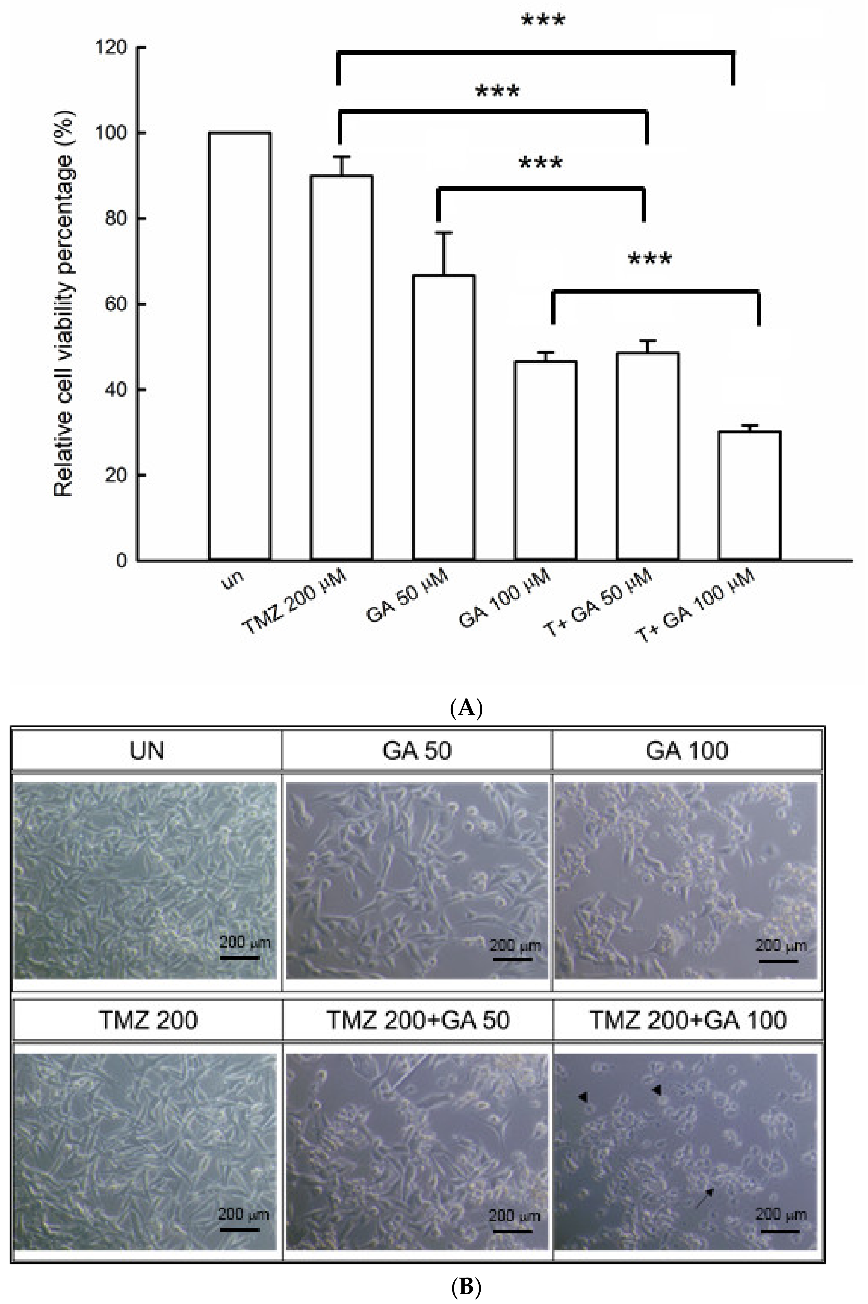
3.1. Ga Enhanced the Cell Viability Inhibition during the TMZ Treatment of U87MG Glioma Cells

As shown in Figure 1A, the cell viability of U87MG cells decreased to 89%, 68% and 48% when treated with TMZ (200 μM) and Ga (50 and 100 μM) after 48 h. The suppression of cell viability showed dose dependence when cells were incubated with Ga alone. The cell viability was retained at values of TMZ (200 μM) treatment larger than 85%. Co-incubation of Ga and TMZ induced the significant suppression of cell viability compared with single-compound incubation. The co-incubation of Ga (100 μM) and TMZ (200 μM) evidently suppressed the cell viability to 30%. Figure 1B shows the morphological findings of U87MG cells that were untreated or treated with TMZ (200 μM), Ga (50 and 100 μM) and Ga (50 and 100 μM) plus TMZ (200 μM) for 48 h. The untreated cells and TMZ (200 μM) treatment showed undamaged cellular morphology. Ga (100 μM) decreased total cell numbers. Ga (100 μM) plus TMZ (200 μM) induced a numerous of damaged cells, apoptotic bodies, and membrane blebbing development. The concentrations of 200 μM for TMZ and 50 and 100 μM for Ga were selected in advanced studies.

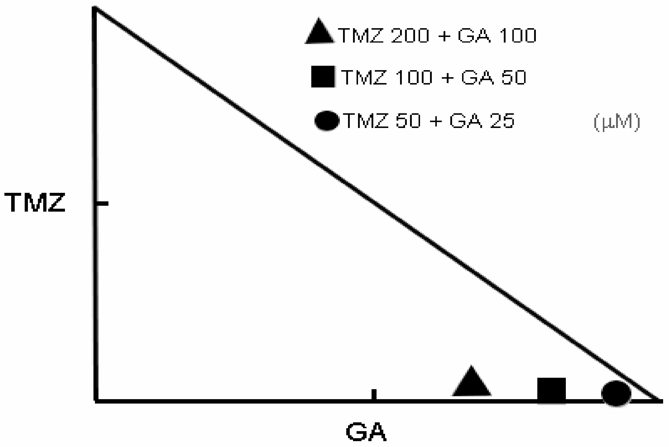
3.2. Synergism of Ga and TMZ Combination in Cell Viability Inhibition

The isobologram analysis was used to determine the synergism of Ga/TMZ combination on cell viability inhibition. As shown in Figure 2, 50, 100 and 200 μM TMZ were combined with 25, 50 and 100 μM Ga, respectively, resulting in the synergistic effect on cell viability inhibition in U87MG glioma cells.

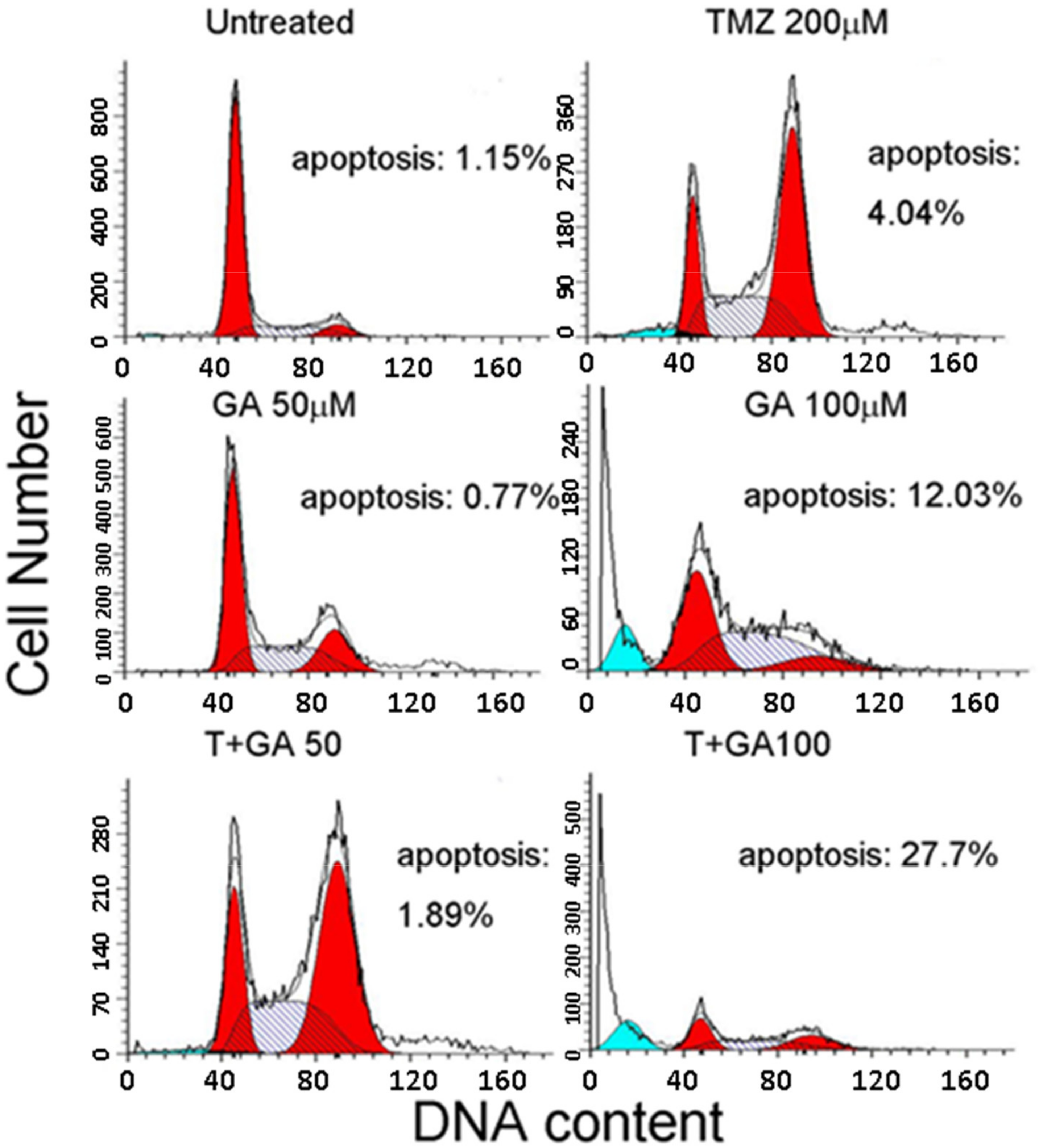
3.3. Ga Enhanced the Apoptosis during TMZ Treatment of U87MG Glioma Cells

We further detected whether Ga could increase the apoptosis-correlated DNA impairment caused by TMZ. The SubG1 phase percentages were measured in the Ga treatment, TMZ treatment and the Ga and TMZ co-treatment. The results showed extremely low percentages of SubG1 phase (<5.0%) in untreated and TMZ-treated cells at 48 h (Figure 3). The SubG1 phase percentages were 0.77% and 12.03% in the incubation with 50 and 100 μM of Ga alone, respectively. However, we detected a great augmentation in the SubG1 percentage up to 27.7% when the U87MG glioma cell line was incubated with Ga (100 μM)/TMZ drug combination for 48 h.

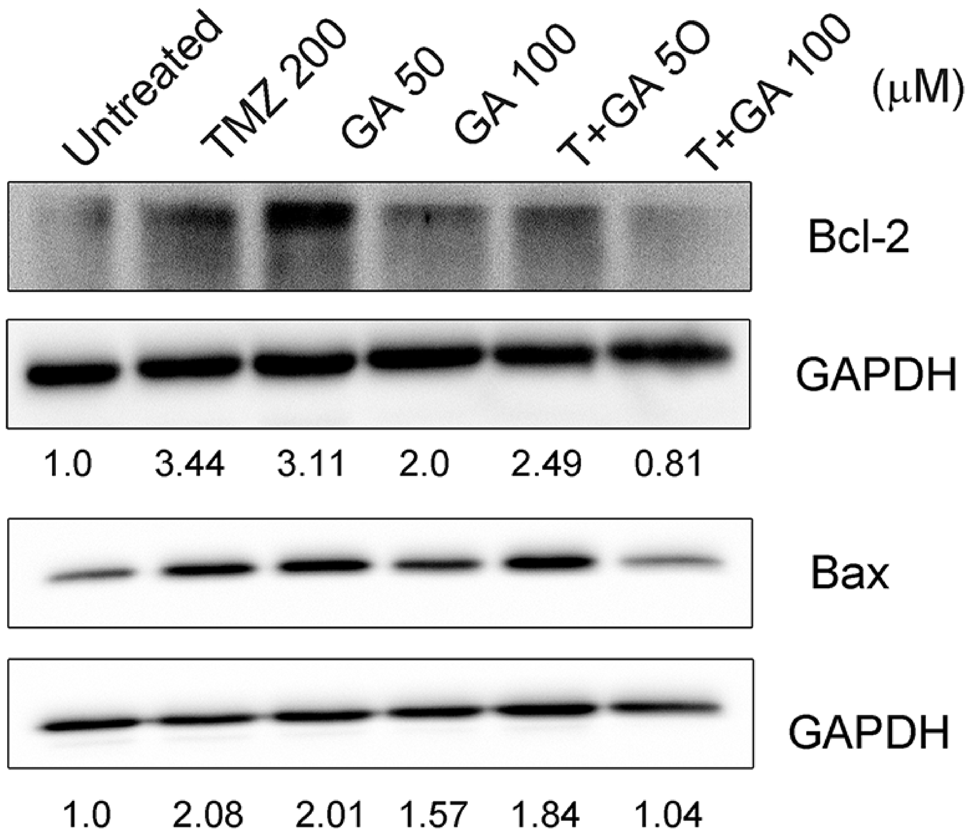
3.4. Ga Enhanced the Bcl-2 Inhibition of TMZ in U87MG Glioma Cell Line

Bcl-2 and Bax are two proteins important for the regulation of apoptosis. The Western blotting method was used to verify whether Ga has a latent role in these regulated proteins in Ga treatment or Ga plus TMZ incubation. As presented in Figure 4, the expression of Bcl-2 significantly decreased by approximately 0.81-fold in Ga (100 μM)/TMZ combination compared cells treated with Ga (100 μM) alone (2.0-fold) and TMZ alone (3.44-fold). The expressions of Bax showed no marked increase in Ga/TMZ combination. These results suggest that the inhibition of Bcl-2 is the major event during apoptosis in Ga and TMZ co-treatment.

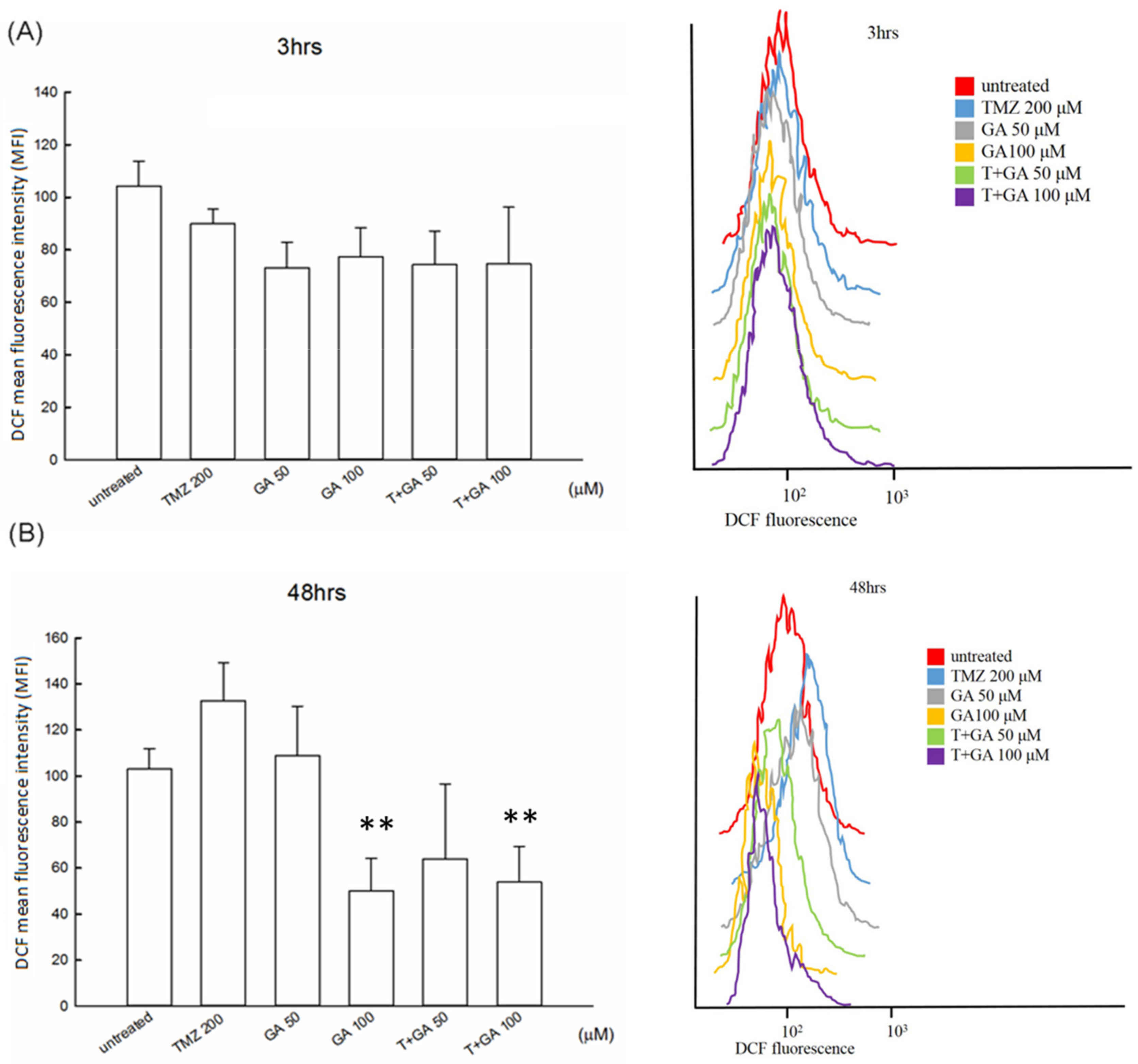
3.5. ROS Level in the Ga/TMZ Treatment

To assess the probable participation of ROS in Ga and TMZ co-incubation, we identified ROS generation in the drug-treated cells. No significant differences were observed in all treated cells at 3 h treatment (Figure 5). TMZ alone treatment resulted in a slight increase in the ROS generation at 48 h. The MFI of DCF was 138.3 ± 14.8 under the TMZ alone treatment. The ROS expression decreased by 53.3 ± 11.6 of DCF under the treatment with Ga (100 μM) alone, by 68.0 ± 24.9 of DCF under the Ga (50 μM)/TMZ combination and by 54.0 ± 15.4 of DCF under the Ga (100 μM)/TMZ combination. These results demonstrated that Ga presented an antioxidant effect in the combination treatment.

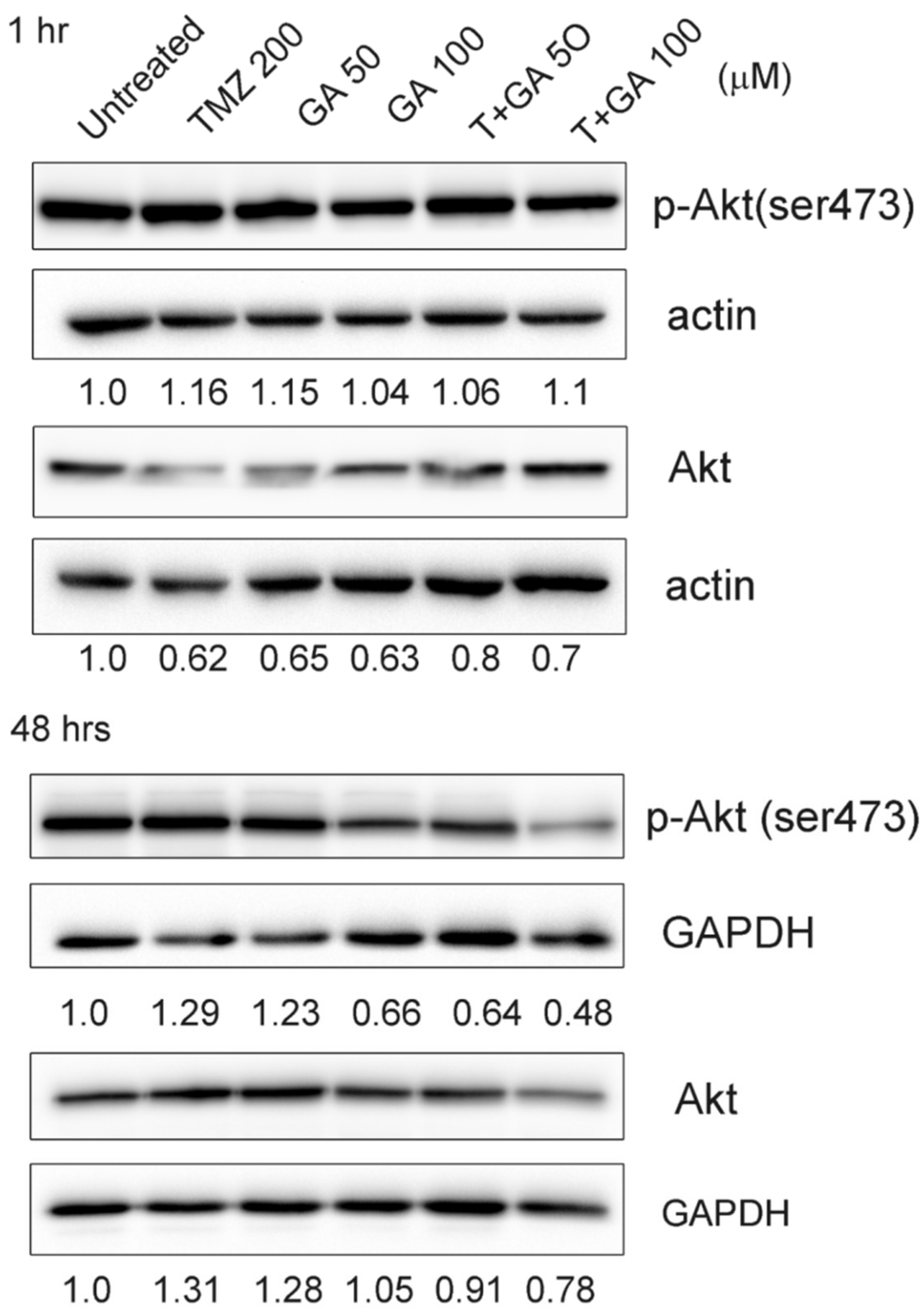
3.6. Ga Increased the Repression of Akt Phosphorylation on TMZ Incubation in U87MG Glioma Cells

The Akt pathway is a signal transduction pathway that regulates cell survival and apoptosis in response to extracellular signals. We analyzed the Akt signaling effect of TMZ and Ga on the U87MG cell line. The p-Akt expression was evaluated by Western blot assay. As shown in Figure 6, no remarkable increase was observed in all treated groups compared with the untreated group at 1 h. TMZ alone and Ga (50 μM) alone increased the p-Akt expression by 1.29- and 1.23-fold, respectively, at 48 h treatment. Ga (100 μM) and Ga (50 μM)/TMZ combination decreased the p-Akt expression by 0.66- and 0.64-fold, respectively. The p-Akt expression on Ga (100 μM)/TMZ combination was effectively suppressed by 0.48-fold.

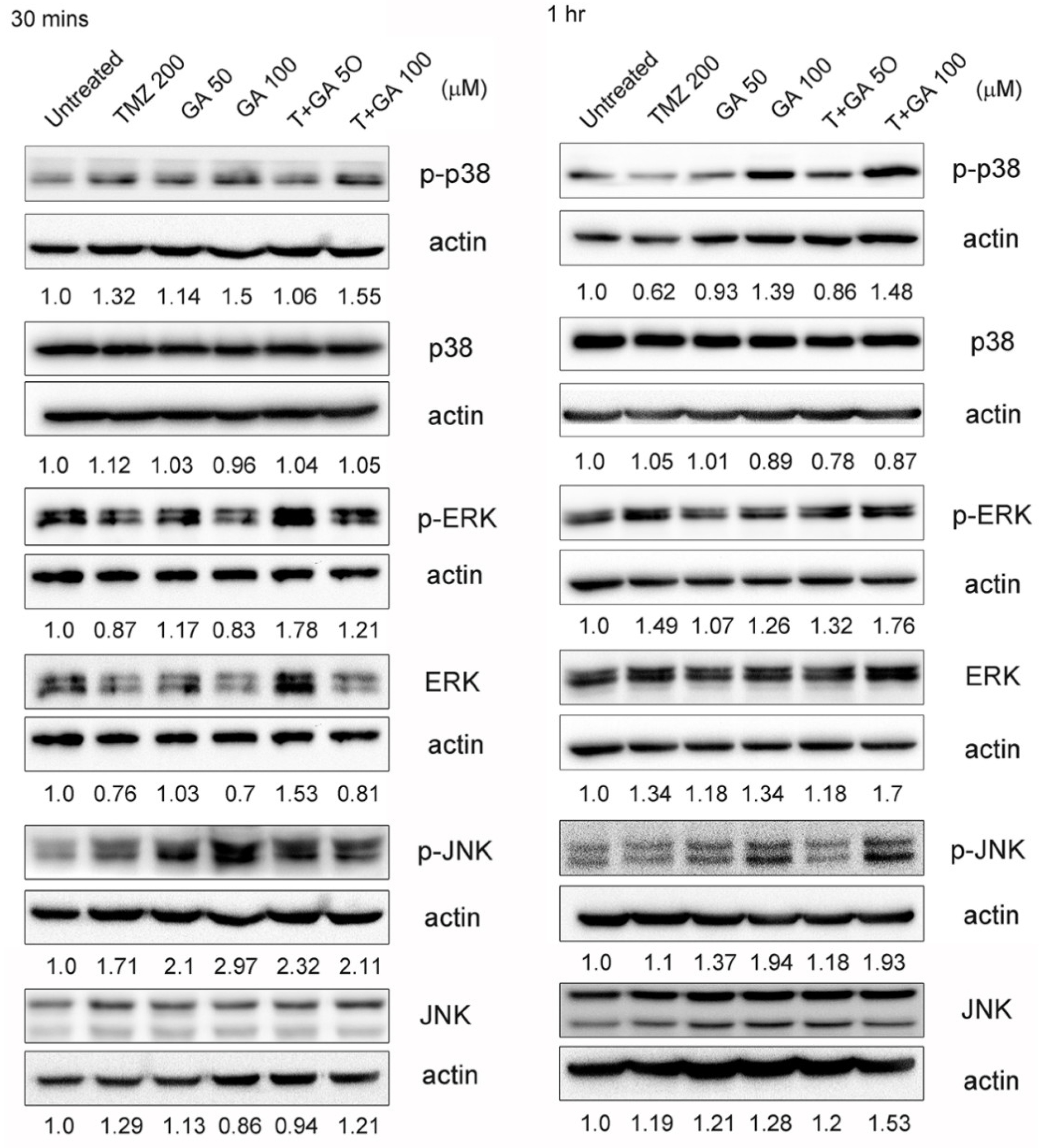
3.7. MAPK Expression in the Ga/TMZ Treatment

The MAPK pathway plays a major role in the sensitivity and is a potential target for anticancer drugs. We then detected the effect of TMZ and Ga on the MAPK pathway in the U87MG cel line. As shown in Figure 7, the expressions of p-38 increased by 1.55- and 1.48-fold in Ga (100 μM)/TMZ combination at 30 min and 1 h treatment, respectively. These increases were 1.32- and 0.62-fold in TMZ alone and 1.5- and 1.39-fold in Ga (100 μM) alone. The expressions of p-ERK increased by 1.21- and 1.76-fold in Ga (100 μM)/TMZ combination at 30 min and 1 h treatment, respectively, and the increases were 0.87- and 1.49-fold in TMZ alone and 0.83- and 1.26-fold in Ga (100 μM) alone. These results suggest that MAPK signaling may be involved in the anti-cancer effect in Ga/TMZ combination treatment.

4. Discussion

Ga, a trihydroxybenzoic acid, has an anti-cancer effect against a variety of cancer cells. At great concentrations, Ga induces a diminution in mitochondrial antioxidant activity, inhibits cell proliferation and decreases the power to repair the destruction of glioma T98G and U251 cells [13,14]. Ga decreased the proliferation and induced apoptosis through the activation of p-38 signaling and the decrease in ERK1/2 and JNK signaling in osteosarcoma cells [15]. Moghtaderi et al. demonstrated that Ca/curcumin combination significantly induced the apoptosis effect through the inhibition of Bcl-2 expression and augmentation of Bax expression, poly(ADP-ribose) polymerase cleavage and caspase3 activation in the human breast MDA-MB-231 cancer cell line [16]. These results indicate that Ga in combination with clinical chemotherapeutic drugs may be a probable applicant for a chemopreventive compound for cancer. Previous studies have demonstrated the plasma concentration of Ga after oral acidum gallicum tablets (10% of Ga) and black tea brew (93% of Ga) in healthy humans [17]. A single oral dose of acidum gallicum tablets or tea (each including 0.3 mM Ga) was administrated to 10 healthy humans; Ga from both the tablets and tea expressed mean maximum concentrations of 1.83 ± 0.16 and 2.09 ± 0.22 μM in plasma, respectively. These results show that Ga is as available from drinking tea as it is from consuming tablets of Ga.
Oxidative stress is a regulating mechanism of apoptosis during anti-cancer drug treatment in numerous cancer cells. Wang et al. indicated that Ga augments the anti-cancer effects of cisplatin via augmentation of intracellular ROS generation. They conclude the synergistic effect of Ga and cisplatin through the ROS-related mitochondrial apoptotic route in the human small-cell lung-cancer H446 cell line [18]. However, our results show that Ga/TMZ combination did not augment the intracellular ROS generation in U87MG glioma cells at 3 and 48 h treatments (Figure 5). Ga alone and Ga/TMZ combination decreased the ROS level at 48 h of treatment. In agreement with our results, vitamin C, curcumin, quercetin, metformin and other compounds decrease the ROS level in cellular apoptotic progression, and several of these compounds trigger apoptosis in tumor cells [19,20]. We speculate that Ga enhanced the anti-cancer effect of TMZ treatment on U87MG glioma cells in a ROS generation-independent manner.
Bcl-2 is an anti-apoptotic protein. It avoids the interruption of the membrane potential of mitochondria and cytochrome c release and represses apoptosis. Bax counteracts the effect of Bcl-2, which induces cytochrome c release to cytosol. When cytochrome c flows to the cytosol, it results in the establishment of apoptosome and the following caspase activation [21]. Our results revealed that the treatment with TMZ alone increased the Bcl-2 expression, suggesting the drug-resistant effect of TMZ on U87MG glioma cells. The level of Bcl-2 was also augmented in Ga alone and Ga (50 μM)/TMZ combination, indicating that the combination of low-concentration Ga with TMZ did not abolish the drug-resistant effect. Ga (100 μM)/TMZ co-incubation evidently inhibited the Bcl-2 expression, which may be an important event to explain the synergistic effect on its anti-cancer properties. Our results are similar to those of another study, which showed that Ga augmented the anticancer forces of cisplatin in the suppression of cancer cell multiplication, and the generation of apoptosis subsequently suppressed the Bcl-2 level in non-small-cell lung cancer A549 cells [22].
Our results demonstrated that the treatment with TMZ alone did not inhibit p-Akt at 1 and 48 h. p-Akt increased in TMZ-alone treatment, suggesting that U87MG glioma cells used the Akt pathway against the TMZ. In addition, p-Akt inhibition is involved in the anti-cancer effect of Ga [23,24]. Ga suppresses the activation of EGFR/Src-mediated ERK and Akt, triggers the decreased amounts of p65/c-Jun-mediated DNA circling and suppresses matrix metalloproteinase-9 generation in EGF-incubated MCF-7 cells [25]. Combination therapy with Ga may augment the antileukemic efficiency of typical chemotherapeutic compounds in acute myeloid leukemia [23]. Ga suppresses angiogenesis, cell viability, proliferation and invasion accompanied by the suppression of p-Akt in the human U87 glioma cell line [11]. We also observed a significant inhibition of the level of p-Akt by Ga/TMZ combination after 48 h (Figure 6). These mechanisms can explain the observed effects of p-Akt inhibition on the Ga/TMZ combination in U87MG glioma cells.
MAPK signaling plays a critical factor in various anti-cancer effects. MAPK signaling is regulated during Ga treatment. Ga induces intrinsic and extrinsic-mediated apoptosis, which involves the activation of p38-MAPK. Ga can suppress EGFR-related survival signals and proliferation and provoke p38-related apoptosis in malignant mesothelioma cells [26]. Ga also suppressed the cellular proliferation and triggered the apoptosis of osteosarcoma cells, accompanied by the induction of p38 activation [10]. In our study, Ga/TMZ combination resulted in the upregulation of p38 in U87MG glioma cells. We believe that p38-MAPK activation may be involved in the Ga/TMZ combination-induced apoptosis in U87MG glioma cells. In conclusion, we discovered for the first time that Ga has innovative enhanced regulatory characteristics in the anti-cancer effect of TMZ treatment by regulating Bcl-2 inhibition and p38-MAPK activation to induce apoptosis. In addition, the intracellular ROS generation did not involve an anti-cancer effect in Ga/TMZ combination treatment. Therefore, these findings provide a novel vision into Ga-enhanced TMZ anti-cancer activity, which is a critical procedure in devising and developing new therapeutic methods to treat glioma.

5. Conclusions

Our results offer scientific suggestions, such as the following: (1) Ga augments the anti-cancer effect of TMZ via the inhibition of Bcl-2 expression and the Akt pathway; (2) the anti-cancer effect of the Ga/TMZ combination occurs in a ROS-generation independent manner.

Author Contributions

Conceptualization, J.-T.Y. and C.-H.C. (Ching-Hsein Chen); methodology, Y.-C.C., C.-H.C. (Chun-Han Chen), J.-Y.P. and C.-H.C. (Ching-Hsein Chen); software, Y.-C.C., C.-H.C. (Chun-Han Chen), J.-Y.P., C.-Y.C. and C.-H.C. (Ching-Hsein Chen); validation, K.-T.C., J.-T.Y. and C.-H.C. (Ching-Hsein Chen); formal analysis, I.-N.L. and C.-H.C. (Ching-Hsein Chen); investigation, M.-H.L.; resources, C.-Y.C.; data curation, C.-H.C. (Ching-Hsein Chen); writing—original draft preparation, C.-H.C. (Ching-Hsein Chen); writing—review and editing, J.-T.Y. and C.-H.C. (Ching-Hsein Chen); visualization, F.-J.L.; supervision, C.-H.C. (Ching-Hsein Chen); project administration, C.-H.C. (Ching-Hsein Chen); funding acquisition, J.-T.Y. All authors have read and agreed to the published version of the manuscript.

Funding

This research was funded by [Chang Gung Medical Research Council, Taiwan, ROC.] grant number [CMRPG6F0441] and [CMRPG6F0442].

Institutional Review Board Statement

Not applicable.

Informed Consent Statement

Not applicable.

Data Availability Statement

The datasets used in the current study are available from the corresponding author on reasonable request.

Acknowledgments

We like to acknowledge the BD FACSCantoTM II flow cytometer service provided by the Expensive Advanced Instrument Core Laboratory, Department of Medical Research and Development, Chang Gung Memorial Hospital at Chiayi.

Conflicts of Interest

The authors declare no conflict of interest.

References

  1. Lombardi, F.; Augello, F.R.; Artone, S.; Gugu, M.K.; Cifone, M.G.; Cique, B.; Palumbo, P. Up-regulation of cyclooxygenase-2 (COX-2) expression by temozolomide (TMZ) in human glioblastoma (GBM) cell lines. Int. J. Mol. Sci. 2022, 23, 1545. [Google Scholar] [CrossRef] [PubMed]
  2. Wei, Y.; Lu, C.; Zhou, P.; Zhao, L.; Lyu, X.; Yin, J.; Shi, Z.; You, Y. EIF4A3-induced circular RNA ASAP1 promotes tumorigenesis and temozolomide resistance of glioblastoma via NRAS/MEK1/ERK1-2 signaling. Neuro-Oncology 2021, 23, 611–624. [Google Scholar] [CrossRef] [PubMed]
  3. Chen, L.; Pastorino, F.; Berry, P.; Bonner, J.; Kirk, C.; Wood, K.M.; Thomas, H.D.; Zhao, Y.; Daga, A.; Veal, G.J.; et al. Preclinical evaluation of the first intravenous small molecule MDM2 antagonist alone and in combination with temozolomide in neuroblastoma. Int. J. Cancer 2019, 144, 3146–3159. [Google Scholar] [CrossRef] [PubMed] [Green Version]
  4. Oliveira, K.A.; Dal-Cim, T.A.; Lopes, F.G.; Nedel, C.B.; Tasca, C.I. Guanosine promotes cytotoxicity via adenosine receptors and induces apoptosis in temozolomide-treated A172 glioma cells. Purinergic Signal. 2017, 13, 305–318. [Google Scholar] [CrossRef] [PubMed] [Green Version]
  5. Chen, L.; Cong, D.; Li, Y.; Wang, D.; Li, Q.; Hu, S. Combination of sonodynamic with temozolomide inhibits C6 glioma migration and promotes mitochondrial pathway apoptosis via suppressing NHE-1 expression. Ultrason. Sonochemistry 2017, 39, 654–661. [Google Scholar] [CrossRef]
  6. Chio, C.-C.; Chen, K.-Y.; Chang, C.-K.; Chuang, J.-Y.; Liu, C.-C.; Liu, S.-H.; Chen, R.-M. Improved effects of honokiol on temozolomide-induced autophagy and apoptosis of drug-sensitive and-tolerant glioma cells. BMC Cancer 2018, 18, 379. [Google Scholar] [CrossRef] [Green Version]
  7. Ma, J.; Yang, Y.-R.; Chen, W.; Chen, M.-H.; Wang, H.; Wang, X.-D.; Sun, L.-L.; Wang, F.-Z.; Wang, D.-C. Fluoxetine synergizes with temozolomide to induce the CHOP-dependent endoplasmic reticulum stress-related apoptosis pathway in glioma cells. Oncol. Rep. 2016, 36, 676–684. [Google Scholar] [CrossRef] [Green Version]
  8. Chen, C.-H.; Chang, Y.-J.; Ku, M.S.; Chung, K.-T.; Yang, J.-T. Enhancement of temozolomide-induced apoptosis by valproic acid in human glioma cell lines through redox regulation. J. Mol. Med. 2011, 89, 303–315. [Google Scholar] [CrossRef]
  9. Yang, J.-T.; Lee, I.; Lu, F.-J.; Chung, C.-Y.; Lee, M.-H.; Cheng, Y.-C.; Chen, K.-T.; Chen, C.-H. Propyl gallate exerts an antimigration effect on temozolomide-treated malignant glioma cells through inhibition of ROS and the NF-κB pathway. J. Immunol. Res. 2017, 2017, 9489383. [Google Scholar] [CrossRef] [Green Version]
  10. Kumar, P.P.; Madhuri, D.; Reddy, L.S.S.; Reddy, Y.D.; Somasekhar, G.; Sirisha, N.V.L.; Nagaraju, K.; Shouib, M.S.; Rizwaan, A.S. A new cerebral ischemic injury model in rats, preventive effect of gallic acid and in silico approaches. Saudi J. Biol. Sci. 2021, 28, 5204–5213. [Google Scholar] [CrossRef]
  11. Lu, Y.; Jiang, F.; Jiang, H.; Wu, K.; Zheng, X.; Cai, Y.; Katakowski, M.; Chopp, M.; To, S.-S.T. Gallic acid suppresses cell viability, proliferation, invasion and angiogenesis in human glioma cells. Eur. J. Pharmacol. 2010, 641, 102–107. [Google Scholar] [CrossRef] [PubMed] [Green Version]
  12. Hsu, S.-S.; Chou, C.-T.; Liao, W.-C.; Shieh, P.; Kuo, D.-H.; Kuo, C.-C.; Jan, C.-R.; Liang, W.-Z. The effect of gallic acid on cytotoxicity, Ca2+ homeostasis and ROS production in DBTRG-05MG human glioblastoma cells and CTX TNA2 rat astrocytes. Chem.-Biol. Interact. 2016, 252, 61–73. [Google Scholar] [CrossRef] [PubMed]
  13. Paolini, A.; Curti, V.; Pasi, F.; Mazzini, G.; Nano, R.; Capelli, E. Gallic acid exerts a protective or an anti-proliferative effect on glioma T98G cells via dose-dependent epigenetic regulation mediated by miRNAs. Int. J. Oncol. 2015, 46, 1491–1497. [Google Scholar] [CrossRef] [PubMed] [Green Version]
  14. Jing, Z.; Li, M.; Wang, H.; Yang, Z.; Zhou, S.; Ma, J.; Meng, E.; Zhang, H.; Liang, W.; Hu, W.; et al. Gallic acid-gold nanoparticles enhance radiation-induced cell death of human glioma U251 cells. IUBMB Life 2021, 73, 398–407. [Google Scholar] [CrossRef] [PubMed]
  15. Liang, C.-Z.; Zhang, X.; Li, H.; Tao, Y.-Q.; Tao, L.-J.; Yang, Z.-R.; Zhou, X.-P.; Shi, Z.-L.; Tao, H.-M. Gallic acid induces the apoptosis of human osteosarcoma cells in vitro and in vivo via the regulation of mitogen-activated protein kinase pathways. Cancer Biother. Radiopharm. 2012, 27, 701–710. [Google Scholar] [CrossRef] [PubMed] [Green Version]
  16. Moghtaderi, H.; Sepehri, H.; Delphi, L.; Attari, F. Gallic acid and curcumin induce cytotoxicity and apoptosis in human breast cancer cell MDA-MB-231. Bioimpacts 2018, 8, 185–194. [Google Scholar] [CrossRef] [Green Version]
  17. Shahrzad, S.; Aoyagi, K.; Winter, A.; Koyama, A.; Bitsch, I. Pharmacokinetics of Gallic Acid and Its Relative Bioavailability from Tea in Healthy Humans. J. Nutrition 2001, 131, 1207–1210. [Google Scholar] [CrossRef]
  18. Wang, R.; Ma, L.; Weng, D.; Yao, J.; Liu, X.; Jin, F. Gallic acid induces apoptosis and enhances the anticancer effects of cisplatin in human small cell lung cancer H446 cell line via the ROS-dependent mitochondrial apoptotic pathway. Oncol. Rep. 2016, 35, 3075–3083. [Google Scholar] [CrossRef] [Green Version]
  19. Jeong, C.-H.; Joo, S.-H. Downregulation of reactive oxygen species in apoptosis. J. Cancer Prev. 2016, 21, 13–20. [Google Scholar] [CrossRef] [Green Version]
  20. George, S.; Abrahamse, H. Redox potential of antioxidants in cancer progression and prevention. Antioxidants 2020, 9, 1156. [Google Scholar] [CrossRef]
  21. Dehkordi, M.H.; Tashakor, A.; O’Connell, E.; Fearnhead, H.O. Apoptosome-dependent myotube formation involves activation of caspase-3 in differentiating myoblasts. Cell Death Dis. 2020, 11, 308. [Google Scholar] [CrossRef] [PubMed]
  22. Zhang, T.; Ma, L.; Wu, P.; Li, W.; Li, T.; Gu, R.; Dan, X.; Li, Z.; Fan, X.; Xiao, Z. Gallic acid has anticancer activity and enhances the anticancer effects of cisplatin in non-small cell lung cancer A549 cells via the JAK/STAT3 signaling pathway. Oncol. Rep. 2019, 41, 1779–1788. [Google Scholar] [CrossRef] [PubMed] [Green Version]
  23. Gu, R.; Zhang, M.; Meng, H.; Xu, D.; Xie, Y. Gallic acid targets acute myeloid leukemia via Akt/mTOR-dependent mitochondrial respiration inhibition. Biomed. Pharmacother. 2018, 105, 491–497. [Google Scholar] [CrossRef]
  24. Zeng, M.; Su, Y.; Li, K.; Jin, D.; Li, Q.; Li, Y.; Zhou, B. Gallic acid inhibits bladder cancer T24 cell progression through mitochondrial dysfunction and PI3K/Akt/NF-κB signaling suppression. Front. Pharmacol. 2020, 11, 1222. [Google Scholar]
  25. Chen, Y.-J.; Lin, K.-N.; Jhang, L.-M.; Huang, C.-H.; Lee, Y.-C.; Chang, L.-S. Gallic acid abolishes the EGFR/Src/Akt/Erk-mediated expression of matrix metalloproteinase-9 in MCF-7 breast cancer cells. Chem.-Biol. Interact. 2016, 252, 131–140. [Google Scholar] [CrossRef]
  26. Demiroglu-Zergeroglu, A.; Candemir, G.; Turhanlar, E.; Sagir, F.; Ayvali, N. EGFR-dependent signalling reduced and p38 dependent apoptosis required by Gallic acid in Malignant Mesothelioma cells. Biomed. Pharmacother. 2016, 84, 2000–2007. [Google Scholar] [CrossRef] [PubMed]
Figure 1. Assessment of (A) cell viability and (B) morphological findings on TMZ, Ga or Ga, and TMZ incubation. U87MG cells were plated in 96-well plates (1 ×10⁴/well) for 24 h and incubated with Ga (50 or 100 μM), TMZ (200 μM) or TMZ/Ga co-treatment for 48 h. The cell viability was detected using an XTT assay. (A) The data are shown as mean ± SD (n = 5–8) of separate experiments. Significant difference is p < 0.001 (***). (B) The cellular morphology was examined by light microscope (scale bars are 200 μm, magnification 200×). The long arrow indicates membrane blebbing. The arrow heads show apoptotic bodies.
Processes 10 00448 g001
Figure 2. Isobologram analysis of Ga and TMZ co-incubation in U87MG cell line. CI data were computed by CalcuSyn software. Points under the backslash line imply synergestic effect. The trials were conducted at least 3 times. An illustrative trial is shown.
Processes 10 00448 g002
Figure 3. DNA damage analysis of the treatment with TMZ alone, Ga alone, or Ga/TMZ combination in U87MG glioma cell line. U87MG cell line was incubated with several concentrations of TMZ (200 μM), Ga (50 and 100 μM) or Ga/TMZ co-treatment for 48 h. The percentages of Sub G1 phase indicated that the apoptosis was evaluated by PI staining and flow cytometer. The red peak on the left represents the area of G1 phase. The red peak on the right represents the area of G/M phase. The backslash area in the middle represents the area of S phase. The light blue area in the left represents the area of apoptosis.
Processes 10 00448 g003
Figure 4. Expressions of Bcl-2 and Bax with TMZ, Ga or Ga/TMZ treatment. U87MG cells were cultured in 6 cm cultured dishes for 24 h and then incubated with 200 μM TMZ, 50 or 100 μM Ga, or co-incubated with both for 48 h. After incubation, whole proteins were extracted. The expressions of Bcl-2 and Bax were detected by Western blotting. Experiments were performed at least 3 times; an illustrative experiment was revealed. Numbers show the densitometric data of various incubated cellular groups normalized to their related untreated group.
Processes 10 00448 g004
Figure 5. Detection of intracellular ROS in TMZ, Ga or Ga and TMZ co-incubation. A total of 1 × 10⁶ U87MG cells were cultured in 6 cm cultured dishes for 24 h, incubated with 200 μM TMZ, 50 or 100 μM Ga or co-incubated with both for 3 or 48 h. After incubation, all cells were incubated with DCFDA for intracellular ROS detection and determined by a flow cytometer. (A) Data show the MFI inside the cells. The data are shown as mean ± SD (n = 5–8) of individual experiments. Significant difference is p < 0.01 (**) compared with untreated group. (B) The overlay plots of DCF flow cytometry data.
Processes 10 00448 g005
Figure 6. Expressions of p-Akt and Akt with TMZ, Ga or Ga/TMZ treatment. U87MG cells were cultured in 6 cm cultured dishes for 24 h, incubated with 200 μM TMZ, 50 or 100 μM Ga, or co-incubated with both for 1 and 48 h. After incubation, whole proteins were extracted. The expressions of Akt and p-Akt (Ser 473) were detected by Western blotting. These experiments were conducted at least 3 times; an illustrative experiment is shown. Numbers show the densitometric data of various incubated cellular groups normalized to their related untreated group.
Processes 10 00448 g006
Figure 7. Expressions of MAPKs with TMZ, Ga or Ga/TMZ incubation. U87MG cells were cultured in 6 cm cultured dishes for 24 h, incubated with 200 μM TMZ, 50 or 100 μM Ga, or co-incubated with both for 30 min and 1 h. After incubation, total proteins were extracted to assess various protein expressions. After incubation, total proteins were extracted. The expression of MAPKs was detected by Western blotting. These experiments were conducted at least 3 times; an illustrative experiment is shown. The numbers show the densitometric data of various incubated cellular groups normalized to their related untreated group.
Processes 10 00448 g007
Publisher’s Note: MDPI stays neutral with regard to jurisdictional claims in published maps and institutional affiliations.

