
Centrifugal Force-Spinning to Obtain Multifunctional Fibers of PLA Reinforced with Functionalized Silver Nanoparticles

 ,
,  , , , , and
Abstract
:1. Introduction
2. Materials and Methods
2.1. Materials
2.2. Synthesis of Based Chitosan Silver Nanoparticles
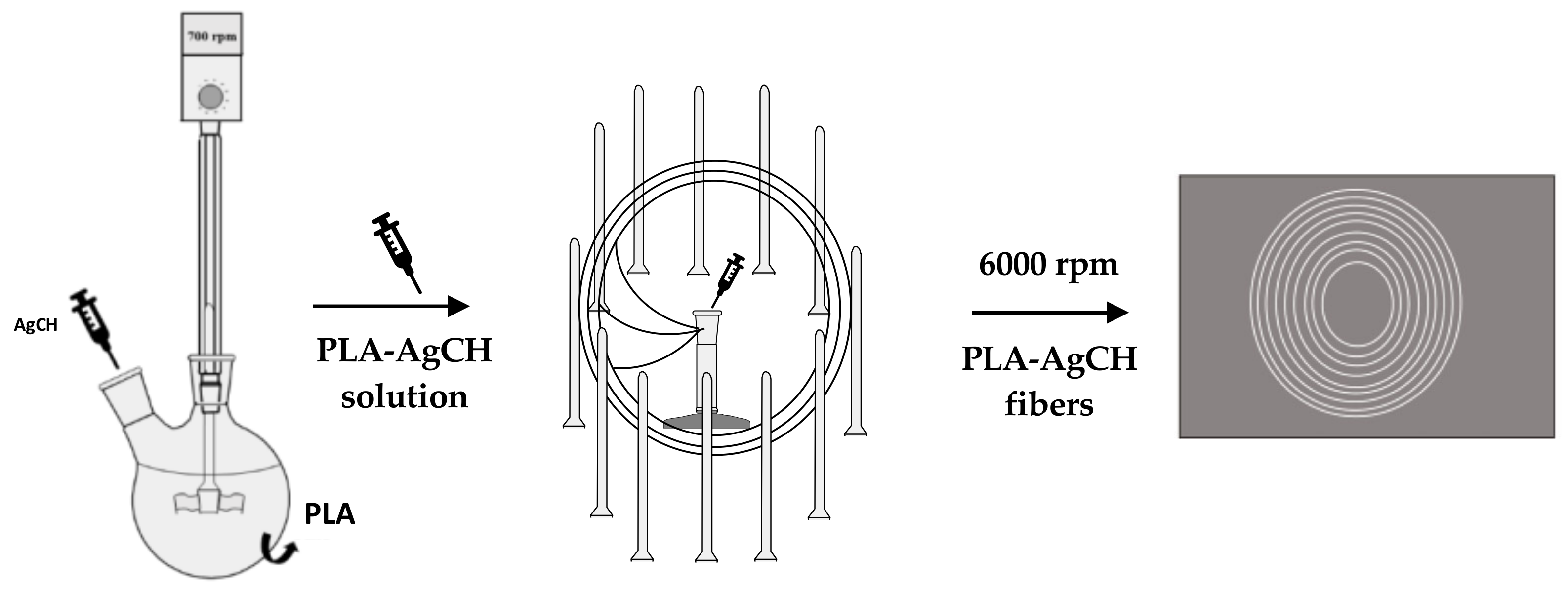
2.3. Preparation of Poly(Lactic Acid) Forced-Fibers Containing AgCH-NPs
2.4. Characterization Techniques
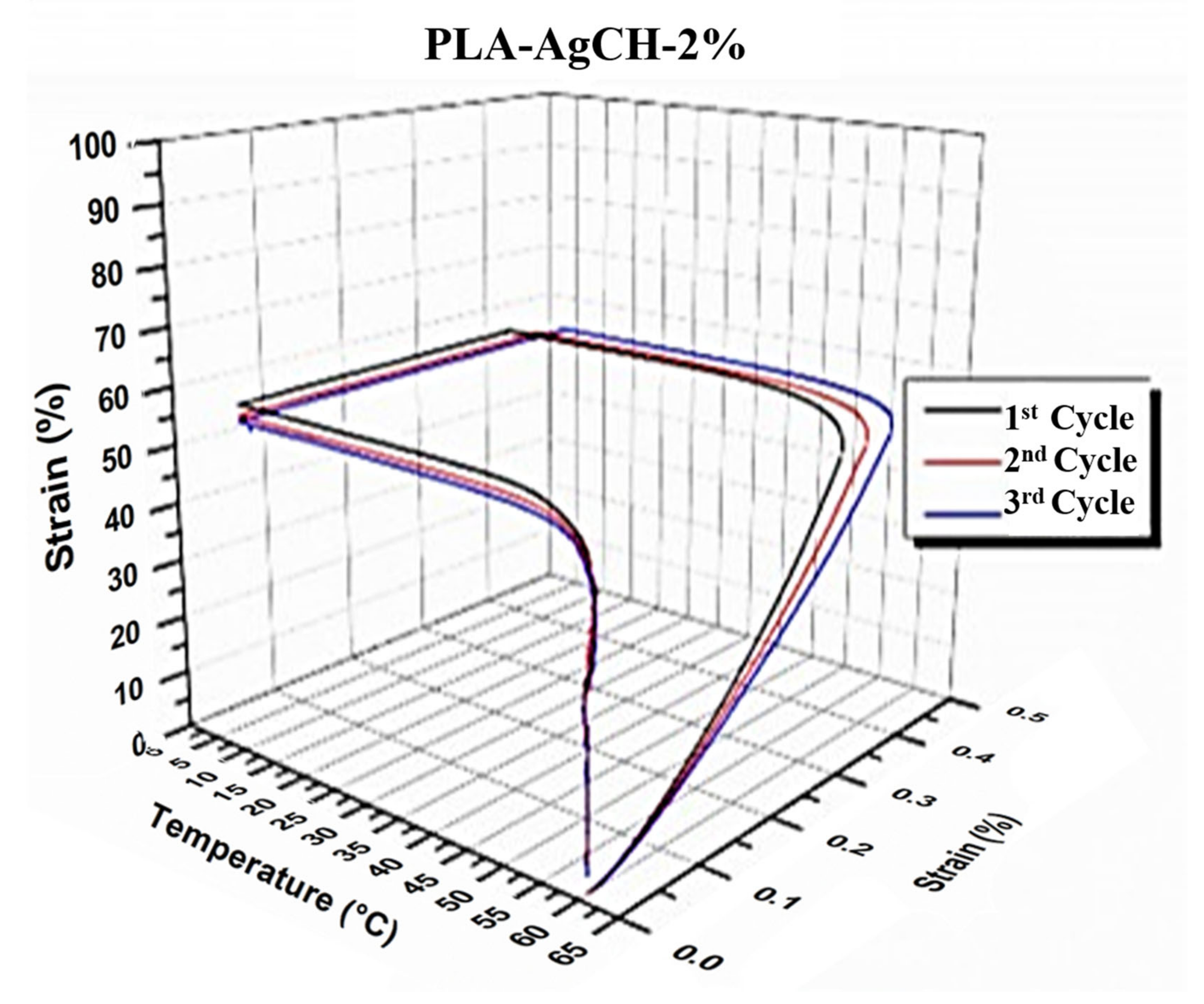
- the sample was equilibrated at the chosen switching temperature (Tsw) for 5 min, in this case, the Tg of PLA matrix, 60 °C;
- ramp stress of 0.2 MPa min−1 was applied until the sample reached 50% of deformation;
- the sample was subsequently cooled at a fixing temperature (Tfix) of 10 °C under constant stress in order to fix the temporary shape;
- after releasing the stress at 0.50 MPa min−1, the sample was heated at 3 °C min−1 up to Tsw and maintained for 30 min in order to recover the initial permanent shape.
3. Results and Discussion
4. Conclusions
Author Contributions
Funding
Institutional Review Board Statement
Data Availability Statement
Conflicts of Interest
References
- Sessini, V.; Raquez, J.M.; Lo Re, G.; Mincheva, R.; Kenny, J.M.; Dubois, P.; Peponi, L. Multiresponsive Shape Memory Blends and Nanocomposites Based on Starch. ACS Appl. Mater. Interfaces 2016, 8, 19197–19201. [Google Scholar] [CrossRef] [PubMed]
- Karger-Kocsis, J.; Kéki, S. Review of Progress in Shape Memory Epoxies and Their Composites. Polymers 2017, 10, 34. [Google Scholar] [CrossRef] [PubMed] [Green Version]
- Chow, W.S.; Mohd Ishak, Z.A. Smart Polymer Nanocomposites: A Review. Express Polym. Lett. 2020, 14, 416–435. [Google Scholar] [CrossRef]
- Peponi, L.; Navarro-Baena, I.; Kenny, J.M. Shape Memory Polymers: Properties, Synthesis and Applications; Woodhead Publishing: Sawston, UK, 2014; ISBN 9780857096951. [Google Scholar]
- Peponi, L.; Puglia, D.; Torre, L.; Valentini, L.; Kenny, J.M. Processing of Nanostructured Polymers and Advanced Polymeric Based Nanocomposites. Mater. Sci. Eng. R Rep. 2014, 85, 1–46. [Google Scholar] [CrossRef] [Green Version]
- Zhang, Z.; Lu, S.; Cai, R.; Tan, W. Sensors. Nano Today 2021, 38, 101202. [Google Scholar] [CrossRef]
- Qiu, X.; Hu, S. “Smart” Materials Based on Cellulose: A Review of the Preparations, Properties, and Applications. Materials 2013, 6, 738–781. [Google Scholar] [CrossRef] [Green Version]
- Mehrpouya, M.; Vahabi, H.; Janbaz, S.; Darafsheh, A.; Mazur, T.R.; Ramakrishna, S. 4D Printing of Shape Memory Polylactic Acid (PLA). Polymer 2021, 230, 124080. [Google Scholar] [CrossRef]
- Nathal, M.V.; Stefko, G.L. Smart Materials and Active Structures. J. Aerosp. Eng. 2013, 26, 491–499. [Google Scholar] [CrossRef]
- Sessini, V.; López Galisteo, A.J.; Leonés, A.; Ureña, A.; Peponi, L. Sandwich-Type Composites Based on Smart Ionomeric Polymer and Electrospun Microfibers. Front. Mater. 2019, 6, 301. [Google Scholar] [CrossRef] [Green Version]
- Merlettini, A.; Gigli, M.; Ramella, M.; Gualandi, C.; Soccio, M.; Boccafoschi, F.; Munari, A.; Lotti, N.; Focarete, M.L. Thermal Annealing to Modulate the Shape Memory Behavior of a Biobased and Biocompatible Triblock Copolymer Scaffold in the Human Body Temperature Range. Biomacromolecules 2017, 18, 2490–2508. [Google Scholar] [CrossRef]
- Arrieta, M.P.; Sessini, V.; Peponi, L. Biodegradable Poly(Ester-Urethane) Incorporated with Catechin with Shape Memory and Antioxidant Activity for Food Packaging. Eur. Polym. J. 2017, 94, 111–124. [Google Scholar] [CrossRef]
- Leonés, A.; Sonseca, A.; López, D.; Fiori, S.; Peponi, L. Shape Memory Effect on Electrospun PLA-Based Fibers Tailoring Their Thermal Response. Eur. Polym. J. 2019, 117, 217–226. [Google Scholar] [CrossRef]
- Leonés, A.; Lieblich, M.; Benavente, R.; Gonzalez, J.L.; Peponi, L. Potential Applications of Magnesium-Based Polymeric Nanocomposites Obtained by Electrospinning Technique. Nanomaterials 2020, 10, 1524. [Google Scholar] [CrossRef]
- Mujica-Garcia, A.; Navarro-Baena, I.; Kenny, J.M.; Peponi, L. Influence of the Processing Parameters on the Electrospinning of Biopolymeric Fibers. J. Renew. Mater. 2014, 2, 23–34. [Google Scholar] [CrossRef]
- El-hadi, A.; Al-Jabri, F. Influence of Electrospinning Parameters on Fiber Diameter and Mechanical Properties of Poly(3-Hydroxybutyrate) (PHB) and Polyanilines (PANI) Blends. Polymers 2016, 8, 97. [Google Scholar] [CrossRef] [Green Version]
- Manea, L.R.; Bertea, A.-P.; Nechita, E.; Popescu, C.V. Mathematical Modeling of the Relation between Electrospun Nanofibers Characteristics and the Process Parameters. In Electrospinning Method Used to Create Functional Nanocomposites Films; IntechOpen: London, UK, 2018; pp. 91–111. [Google Scholar] [CrossRef] [Green Version]
- Rodríguez-Sánchez, I.J.; Fuenmayor, C.A.; Clavijo-Grimaldo, D.; Zuluaga-Domínguez, C.M. Electrospinning of Ultra-Thin Membranes with Incorporation of Antimicrobial Agents for Applications in Active Packaging: A Review. Int. J. Polym. Mater. Polym. Biomater. 2020, 70, 1–24. [Google Scholar] [CrossRef]
- Basar, A.O.; Castro, S.; Torres-Giner, S.; Lagaron, J.M.; Turkoglu Sasmazel, H. Novel Poly(ε-Caprolactone)/Gelatin Wound Dressings Prepared by Emulsion Electrospinning with Controlled Release Capacity of Ketoprofen Anti-Inflammatory Drug. Mater. Sci. Eng. C 2017, 81, 459–468. [Google Scholar] [CrossRef]
- Rana, S.; Cho, J.W. Core-Sheath Polyurethane-Carbon Nanotube Nanofibers Prepared by Electrospinning. Fibers Polym. 2011, 12, 721–726. [Google Scholar] [CrossRef]
- Arrieta, M.P.; Gil, A.L.; Yusef, M.; Kenny, J.M.; Peponi, L. Electrospinning of PCL-Based Blends: Processing Optimization for Their Scalable Production. Materials 2020, 13, 3853. [Google Scholar] [CrossRef] [PubMed]
- Merlettini, A.; Pandini, S.; Agnelli, S.; Gualandi, C.; Paderni, K.; Messori, M.; Toselli, M.; Focarete, M.L. Facile Fabrication of Shape Memory Poly(ϵ-Caprolactone) Non-Woven Mat by Combining Electrospinning and Sol-Gel Reaction. RSC Adv. 2016, 6, 43964–43974. [Google Scholar] [CrossRef] [Green Version]
- Dziemidowicz, K.; Sang, Q.; Wu, J.; Zhang, Z.; Zhou, F.; Lagaron, J.M.; Mo, X.; Parker, G.J.M.; Yu, D.-G.; Zhu, L.-M.; et al. Electrospinning for Healthcare: Recent Advancements. J. Mater. Chem. B 2021, 9, 939–951. [Google Scholar] [CrossRef] [PubMed]
- Zhang, F.; Xia, Y.; Wang, L.; Liu, L.; Liu, Y.; Leng, J. Conductive Shape Memory Microfiber Membranes with Core-Shell Structures and Electroactive Performance. ACS Appl. Mater. Interfaces 2018, 10, 35526–35532. [Google Scholar] [CrossRef] [PubMed]
- Li, M.; Qiu, W.; Wang, Q.; Li, N.; Liu, L.; Wang, X.; Yu, J.; Li, X.; Li, F.; Wu, D. Nitric Oxide-Releasing Tryptophan-Based Poly(Ester Urea)s Electrospun Composite Nanofiber Mats with Antibacterial and Antibiofilm Activities for Infected Wound Healing. ACS Appl. Mater. Interfaces 2022, 14, 15911–15926. [Google Scholar] [CrossRef]
- Qi, Y.; Wang, C.; Wang, Q.; Zhou, F.; Li, T.; Wang, B.; Su, W.; Shang, D.; Wu, S. A Simple, Quick, and Cost-Effective Strategy to Fabricate Polycaprolactone/Silk Fibroin Nanofiber Yarns for Biotextile-Based Tissue Scaffold Application. Eur. Polym. J. 2023, 186, 111863. [Google Scholar] [CrossRef]
- Lu, Y.; Li, Y.; Zhang, S.; Xu, G.; Fu, K.; Lee, H.; Zhang, X. Parameter Study and Characterization for Polyacrylonitrile Nanofibers Fabricated via Centrifugal Spinning Process. Eur. Polym. J. 2013, 49, 3834–3845. [Google Scholar] [CrossRef]
- Zhang, X.; Lu, Y. Centrifugal Spinning: An Alternative Approach to Fabricate Nanofibers at High Speed and Low Cost Centrifugal Spinning: An Alternative Approach to Fabricate Nanofibers at High Speed and Low Cost. Polym. Rev. 2014, 3724, 677–701. [Google Scholar] [CrossRef]
- Krifa, M.; Hammami, M.A.; Wu, H. Occurrence and Morphology of Bead-on-String Structures in Centrifugal Forcespun PA6 Fibers. J. Text. Inst. 2015, 106, 284–294. [Google Scholar] [CrossRef]
- Zhang, Q.; Bao, N.; Wang, X.; Hu, X.; Miao, X.; Chaker, M.; Ma, D. Advanced Fabrication of Chemically Bonded Graphene/TiO2 Continuous Fibers with Enhanced Broadband Photocatalytic Properties and Involved Mechanisms Exploration. Sci. Rep. 2016, 6, 38066. [Google Scholar] [CrossRef] [Green Version]
- Zannini Luz, H.; Loureiro dos Santos, L.A. Centrifugal Spinning for Biomedical Use: A Review. Crit. Rev. Solid State Mater. Sci. 2022, 1–16. [Google Scholar] [CrossRef]
- Sarkar, K.; Gomez, C.; Zambrano, S.; Ramirez, M.; de Hoyos, E.; Vasquez, H.K.L. Electrospinning to Forcespinning. Mater. Today 2010, 13, 42–44. [Google Scholar] [CrossRef]
- Raquez, J.M.; Habibi, Y.; Murariu, M.; Dubois, P. Polylactide (PLA)-Based Nanocomposites. Prog. Polym. Sci. 2013, 38, 1504–1542. [Google Scholar] [CrossRef]
- Hamley, I.W.; Parras, P.; Castelletto, V.; Castillo, R.V.; Müller, A.J.; Pollet, E.; Dubois, P.; Martin, C.M. Melt Structure and Its Transformation by Sequential Crystallization of the Two Blocks within Poly(L-Lactide)-Block-Poly(ε-Caprolactone) Double Crystalline Diblock Copolymers. Macromol. Chem. Phys. 2006, 207, 941–953. [Google Scholar] [CrossRef]
- Navarro-Baena, I.; Kenny, J.M.; Peponi, L. Thermally-Activated Shape Memory Behaviour of Bionanocomposites Reinforced with Cellulose Nanocrystals. Cellulose 2014, 21, 4231–4246. [Google Scholar] [CrossRef] [Green Version]
- Sonseca, A.; Madani, S.; Muñoz-Bonilla, A.; Fernández-García, M.; Peponi, L.; Leonés, A.; Rodríguez, G.; Echeverría, C.; López, D. Biodegradable and Antimicrobial Pla–Ola Blends Containing Chitosan-Mediated Silver Nanoparticles with Shape Memory Properties for Potential Medical Applications. Nanomaterials 2020, 10, 1065. [Google Scholar] [CrossRef]
- Rizki, I.N.; Klaypradit, W. Patmawati Utilization of Marine Organisms for the Green Synthesis of Silver and Gold Nanoparticles and Their Applications: A Review. Sustain. Chem. Pharm. 2023, 31, 100888. [Google Scholar] [CrossRef]
- Beyth, N.; Farah, S.; Domb, A.J.; Weiss, E.I. Antibacterial Dental Resin Composites. React. Funct. Polym. 2014, 75, 81–88. [Google Scholar] [CrossRef]
- Vimala, K.; Mohan, Y.M.; Sivudu, K.S.; Varaprasad, K.; Ravindra, S.; Reddy, N.N.; Padma, Y.; Sreedhar, B.; MohanaRaju, K. Fabrication of Porous Chitosan Films Impregnated with Silver Nanoparticles: A Facile Approach for Superior Antibacterial Application. Colloids Surf. B Biointerfaces 2010, 76, 248–258. [Google Scholar] [CrossRef]
- Huang, H.; Yang, X. Synthesis of Chitosan-Stabilized Gold Nanoparticles in the Absence/Presence of Tripolyphosphate. Biomacromolecules 2004, 5, 2340–2346. [Google Scholar] [CrossRef]
- Antunes, J.C.; Domingues, J.M.; Miranda, C.S.; Silva, A.F.G.; Homem, N.C.; Amorim, M.T.P.; Felgueiras, H.P. Bioactivity of Chitosan-Based Particles Loaded with Plant-Derived Extracts for Biomedical Applications: Emphasis on Antimicrobial Fiber-Based Systems. Mar. Drugs 2021, 19, 359. [Google Scholar] [CrossRef] [PubMed]
- Chandrasekaran, M.; Kim, K.D.; Chun, S.C. Antibacterial Activity of Chitosan Nanoparticles: A Review. Processes 2020, 8, 1–21. [Google Scholar] [CrossRef]
- Peponi, L.; Navarro-Baena, I.; Báez, J.E.; Kenny, J.M.; Marcos-Fernández, A. Effect of the Molecular Weight on the Crystallinity of PCL-b-PLLA Di-Block Copolymers. Polymer 2012, 53, 4561–4568. [Google Scholar] [CrossRef] [Green Version]
- Peponi, L.; Navarro-Baena, I.; Sonseca, A.; Gimenez, E.; Marcos-Fernandez, A.; Kenny, J.M. Synthesis and Characterization of PCL-PLLA Polyurethane with Shape Memory Behavior. Eur. Polym. J. 2013, 49, 893–903. [Google Scholar] [CrossRef] [Green Version]
- ASTM E2149-13a; Standard Test Method for Determining the Antimicrobial Activity of Antimicrobial Agents under Dynamic Contact Conditions. American Society for Testing and Materials: West Conshohocken, PA, USA, 2013.
- Arrieta, M.P.; Perdiguero, M.; Fiori, S.; Kenny, J.M.; Peponi, L. Biodegradable Electrospun PLA-PHB Fibers Plasticized with Oligomeric Lactic Acid. Polym. Degrad. Stab. 2020, 179, 109226. [Google Scholar] [CrossRef]
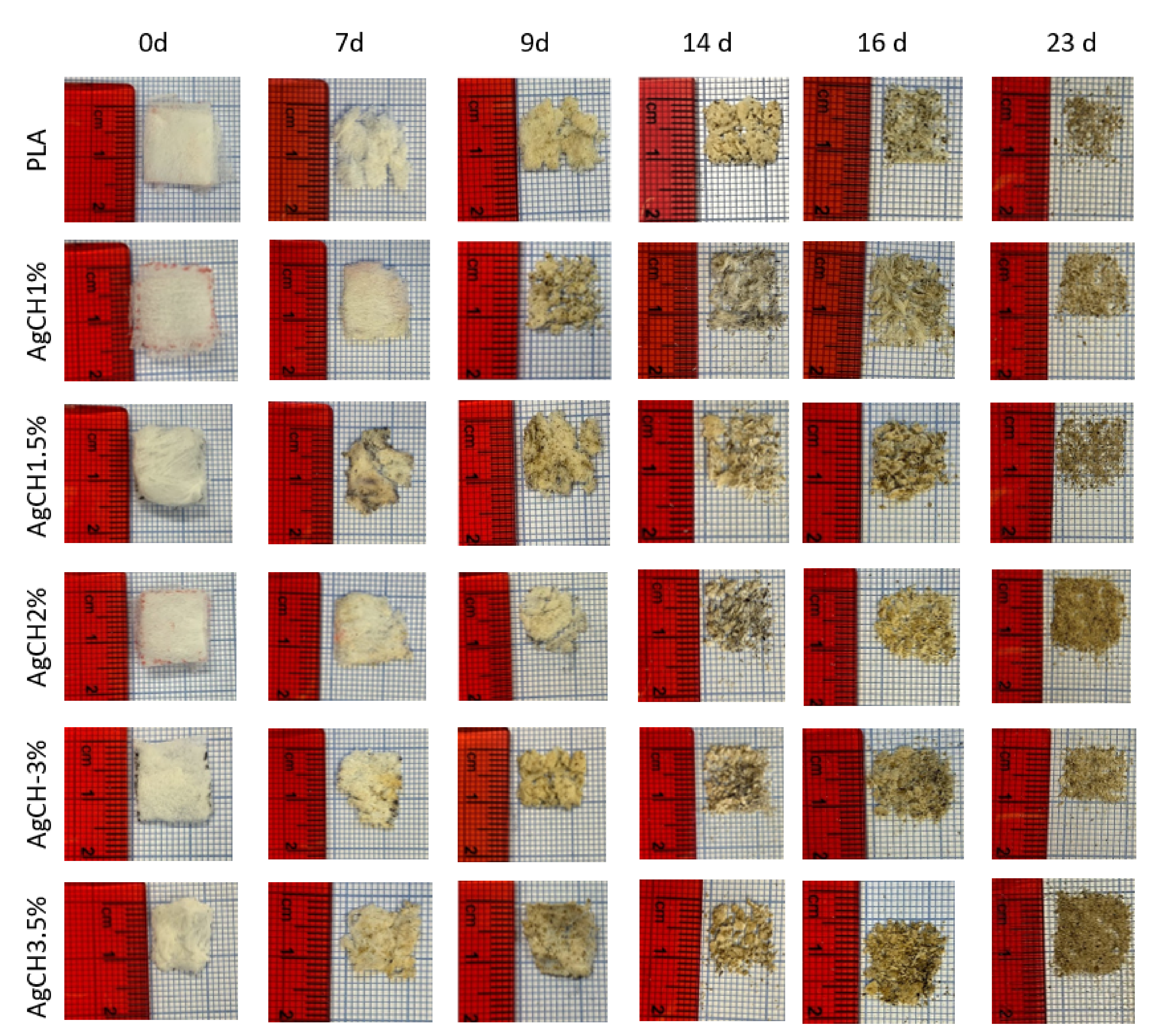
- UNE-EN ISO-20200; Determination of the Degree of Disintegration of Plastic Materials under Simulated Composting Conditions in a Laboratory-Scale Test. AENOR: Madrid, Spain, 2015.
- Sonseca, A.; Madani, S.; Rodríguez, G.; Hevilla, V.; Echeverría, C.; Fernández-García, M.; Muñoz-Bonilla, A.; Charef, N.; López, D. Multifunctional PLA Blends Containing Chitosan Mediated Silver Nanoparticles: Thermal, Mechanical, Antibacterial, and Degradation Properties. Nanomaterials 2020, 10, 22. [Google Scholar] [CrossRef] [Green Version]
- Jia, X.; Ma, X.; Wei, D.; Dong, J.; Qian, W. Direct Formation of Silver Nanoparticles in Cuttlebone-Derived Organic Matrix for Catalytic Applications. Colloids Surfaces A Physicochem. Eng. Asp. 2008, 330, 234–240. [Google Scholar] [CrossRef]
- Pawar, O.; Deshpande, N.; Dagade, S.; Waghmode, S.; Nigam Joshi, P. Green Synthesis of Silver Nanoparticles from Purple Acid Phosphatase Apoenzyme Isolated from a New Source Limonia Acidissima. J. Exp. Nanosci. 2016, 11, 28–37. [Google Scholar] [CrossRef]
- Wan, Y.; Wu, H.; Yu, A.; Wen, D. Biodegradable Polylactide/Chitosan Blend Membranes. Biomacromolecules 2006, 7, 1362–1372. [Google Scholar] [CrossRef]
- Senthilkumar, P.; Yaswant, G.; Kavitha, S.; Chandramohan, E.; Kowsalya, G.; Vijay, R.; Sudhagar, B.; Kumar, D.S.R.S. Preparation and Characterization of Hybrid Chitosan-Silver Nanoparticles (Chi-Ag NPs); A Potential Antibacterial Agent. Int. J. Biol. Macromol. 2019, 141, 290–297. [Google Scholar] [CrossRef]
- Dhanapal, J.; Ravindrran, M.; Baskar, S. Toxic Effects of Aflatoxin B1 on Embryonic Development of Zebrafish (Danio Rerio): Potential Activity of Piceatannol Encapsulated Chitosan/Poly (Lactic Acid) Nanoparticles. Anticancer. Agents Med. Chem. 2015, 15, 248–257. [Google Scholar] [CrossRef]
- Kawai, T.; Rahman, N.; Matsuba, G.; Nishida, K.; Kanaya, T.; Nakano, M.; Okamoto, H.; Kawada, J.; Usuki, A.; Honma, N.; et al. Crystallization and Melting Behavior of Poly (L-Lactic Acid). Macromolecules 2007, 40, 9463–9469. [Google Scholar] [CrossRef]
- Rudnik, E.; Briassoulis, D. Degradation Behaviour of Poly(Lactic Acid) Films and Fibres in Soil under Mediterranean Field Conditions and Laboratory Simulations Testing. Ind. Crops Prod. 2011, 33, 648–658. [Google Scholar] [CrossRef]


| Samples | PLA (wt%) | AgCH-NPs (wt%) |
|---|---|---|
| PLA | 100 | 0 |
| PLA-AgCH-1% | 99 | 1 |
| PLA-AgCH-1.5% | 98.5 | 1.5 |
| PLA-AgCH-2% | 98 | 2 |
| PLA-AgCH-3% | 97 | 3 |
| PLA-AgCH-3.5% | 96.5 | 3.5 |
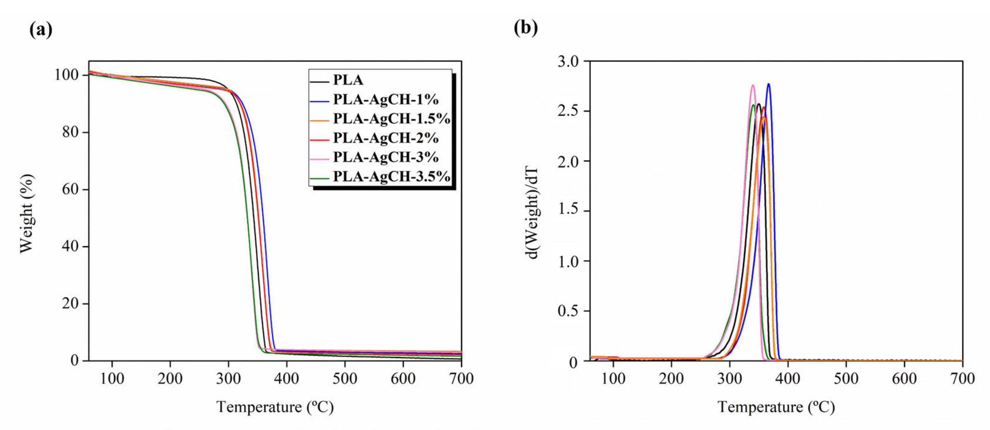
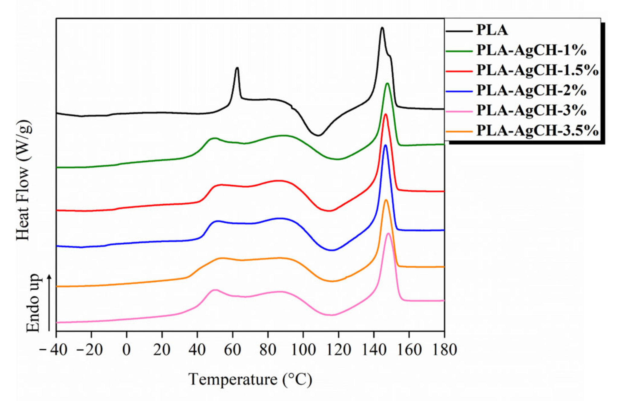
| Samples | Tmax | T5 | T700 | Tg (°C) | Tm (°C) | Xc (%) |
|---|---|---|---|---|---|---|
| PLA | 350 | 300 | 0.73 | 60 | 145 | 0.29 |
| PLA AgCH1% | 341 | 246 | 1.67 | 41 | 148 | 4.20 |
| PLA AgCH1.5% | 357 | 288 | 2.22 | 46 | 147 | 6.31 |
| PLA AgCH2% | 367 | 298 | 2.56 | 45 | 146 | 7.61 |
| PLA AgCH3% | 340 | 262 | 3.23 | 44 | 148 | 8.05 |
| PLA AgCH3.5% | 363 | 297 | 3.67 | 38 | 147 | 9.96 |
| Sample | E (MPa) | σ (MPa) | ε Break (%) |
|---|---|---|---|
| PLA | 209.07 ± 46.71 a | 9.51 ± 1.79 a | 15.79 ± 8.89 a,b |
| PLA-AgCH 1% | 76.73 ± 18.14 b | 3.32 ± 0.89 b | 11.31 ± 1.69 a,b |
| PLA-AgCH 1.5% | 43.98 ± 15.89 b | 1.34 ± 0.47 b | 5.92 ± 1.74 a |
| PLA-AgCH 2% | 35.32 ± 6.79 b | 1.67 ± 0.74 b | 10.04 ± 3.55 a |
| F ratio | 14.02 | 14.39 | 3.69 |
| p-Value | 0.0000 * | 0.0000 * | 0.0150 * |
Disclaimer/Publisher’s Note: The statements, opinions and data contained in all publications are solely those of the individual author(s) and contributor(s) and not of MDPI and/or the editor(s). MDPI and/or the editor(s) disclaim responsibility for any injury to people or property resulting from any ideas, methods, instructions or products referred to in the content. |
© 2023 by the authors. Licensee MDPI, Basel, Switzerland. This article is an open access article distributed under the terms and conditions of the Creative Commons Attribution (CC BY) license (https://creativecommons.org/licenses/by/4.0/).
Share and Cite
Martín-Alonso, M.D.; Salaris, V.; Leonés, A.; Hevilla, V.; Muñoz-Bonilla, A.; Echeverría, C.; Fernández-García, M.; Peponi, L.; López, D. Centrifugal Force-Spinning to Obtain Multifunctional Fibers of PLA Reinforced with Functionalized Silver Nanoparticles. Polymers 2023, 15, 1240. https://doi.org/10.3390/polym15051240
Martín-Alonso MD, Salaris V, Leonés A, Hevilla V, Muñoz-Bonilla A, Echeverría C, Fernández-García M, Peponi L, López D. Centrifugal Force-Spinning to Obtain Multifunctional Fibers of PLA Reinforced with Functionalized Silver Nanoparticles. Polymers. 2023; 15(5):1240. https://doi.org/10.3390/polym15051240
Chicago/Turabian StyleMartín-Alonso, María Dolores, Valentina Salaris, Adrián Leonés, Víctor Hevilla, Alexandra Muñoz-Bonilla, Coro Echeverría, Marta Fernández-García, Laura Peponi, and Daniel López. 2023. "Centrifugal Force-Spinning to Obtain Multifunctional Fibers of PLA Reinforced with Functionalized Silver Nanoparticles" Polymers 15, no. 5: 1240. https://doi.org/10.3390/polym15051240