Review

Recent Advances in Silver Nanoparticles Containing Nanofibers for Chronic Wound Management

by
Govindaraj Sabarees
1,
Vadivel Velmurugan
1,*,
Ganesan Padmini Tamilarasi
2,
Veerachamy Alagarsamy
3,* and
Viswas Raja Solomon
3
1
Department of Pharmaceutical Chemistry, SRM College of Pharmacy, SRM Institute of Science and Technology, Kattankulathur, Chennai 603203, Tamil Nadu, India
2
Department of Pharmaceutical Analysis, SRM College of Pharmacy, SRM Institute of Science and Technology, Kattankulathur, Chennai 603203, Tamil Nadu, India
3
Medicinal Chemistry Research Laboratory, MNR College of Pharmacy, Gr. Hyderabad, Sangareddy 502294, Telangana, India
*
Authors to whom correspondence should be addressed.
Polymers 2022, 14(19), 3994; https://doi.org/10.3390/polym14193994
Submission received: 1 September 2022 / Revised: 17 September 2022 / Accepted: 20 September 2022 / Published: 23 September 2022

Abstract

:
Infections are the primary cause of death from burns and diabetic wounds. The clinical difficulty of treating wound infections with conventional antibiotics has progressively increased and reached a critical level, necessitating a paradigm change for enhanced chronic wound care. The most prevalent bacterium linked with these infections is Staphylococcus aureus, and the advent of community-associated methicillin-resistant Staphylococcus aureus has posed a substantial therapeutic challenge. Most existing wound dressings are ineffective and suffer from constraints such as insufficient antibacterial activity, toxicity, failure to supply enough moisture to the wound, and poor mechanical performance. Using ineffective wound dressings might prolong the healing process of a wound. To meet this requirement, nanoscale scaffolds with their desirable qualities, which include the potential to distribute bioactive agents, a large surface area, enhanced mechanical capabilities, the ability to imitate the extracellular matrix (ECM), and high porosity, have attracted considerable interest. The incorporation of nanoparticles into nanofiber scaffolds constitutes a novel approach to “nanoparticle dressing” that has acquired significant popularity for wound healing. Due to their remarkable antibacterial capabilities, silver nanoparticles are attractive materials for wound healing. This review focuses on the therapeutic applications of nanofiber wound dressings containing Ag-NPs and their potential to revolutionize wound healing.

Graphical Abstract

1. Introduction

Wound healing must be seen as a sequence of regulated and interconnected steps, as shown in (Figure 1), such as coagulation, inflammation, deposition, fibroplasia, extracellular matrix differentiation, contraction, remodelling, and epithelialization [1]. Acute wounds should recover in two to three weeks at most, followed by the remodelling stage, which may take up to two years. Scab development and little to no infection are signs of routine healing. When an infection exists, the immune system often fights it off. However, in cases of severe microbial disease, the immune system cannot eradicate the condition, and the wound develops to the chronic stage and fails to heal sequentially in the expected time frame. Chronic wounds are difficult to keep clean and are more vulnerable to bacterial infections [2,3,4,5]. A high level of bacterial invasion distinguishes these wounds, as well as increased inflammation, reduced oxygenation on the subepithelial tissues, damaged fibroblast, and postponed re-epithelialization [6,7]. Leg ulcers, pressure ulcers, diabetic foot ulcers (DFUs), fungating wounds, and other chronic wounds are among the most typical [8,9].
In addition to being a significant financial burden on healthcare systems, wounds provide a social barrier for patients and their families. According to a retrospective review of Medicare enrollees, 8.2 million people in the US were impacted by wounds in 2014. In the wound category, the total cost of care ranged from USD 28.1 billion to USD 96.8 billion. The estimated USD 9–13 billion administrative expenses for DFUs were among the highest. Given that there are already 463 million diabetics worldwide, which is predicted to reach 700 million by 2045, these expenditures are projected to grow [10,11]. If diabetes is not effectively treated, a diabetic person’s chance of having a DFU ranges from 15% to 25%. Due to peripheral neuropathy and uncontrollable foot infections, DFUs have a 14% to 24% likelihood of resulting in lower limb amputations (LLAs). Since amputation patients often have difficulty adjusting to social and demographic situations, LLAs are linked to a worse quality of life.
Additionally, LLAs are linked to a greater death rate in diabetes individuals; recent research found that the 30-day postamputation mortality rate was 9.8%. The patient’s chance of needing more amputations increases if they live beyond the 30-day mark. According to reports, the mortality rates for small LLAs and large LLAs at one and four years after amputation were 18% and 45%, respectively, and 33% and 65%. In addition to other risk factors, including age, renal disease, and other consequences of diabetes, depression has recently been recognized as one of the risk variables leading to increased death rates following significant LLA. Poor antibacterial activity, high toxicity, a lack of ability to deliver adequate moisture to the wound, and a lack of mechanical performance are some of the drawbacks of most of the wound dressings on the market [12].
The inefficient use of wound dressings can slow down wound healing. To address this need, nanoscale scaffolds with desired properties such as the capacity to distribute bioactive compounds, a large surface area, increased mechanical capabilities, ability to mimic the extracellular matrix (ECM), and high porosity have piqued the attention of researchers. At the moment, the inclusion of nanoparticles into nanofiber scaffolds is a revolutionary method of “nanoparticle dressing,” which has gained great appeal for wound healing [13,14,15]. Furthermore, AgNPs enhance wound healing due to the following benefits: The role of AgNPs’ anti-inflammatory properties in accelerating wound healing AgNPs can be easily incorporated into nanofibers and dressings. This is due to elemental silver’s meager bacterial resistance, antibacterial activity toward many bacterial strains, and facile surface modification that aids drug delivery. The simple synthetic process can synthesize it to create diverse shapes and sizes from 2 to 500 nm, which can be synthesized by changing reaction parameters and simple and inexpensive large-scale production methods. This review focuses on the therapeutic applications of nanofiber wound dressings containing AgNPs and their potential to revolutionize wound healing.

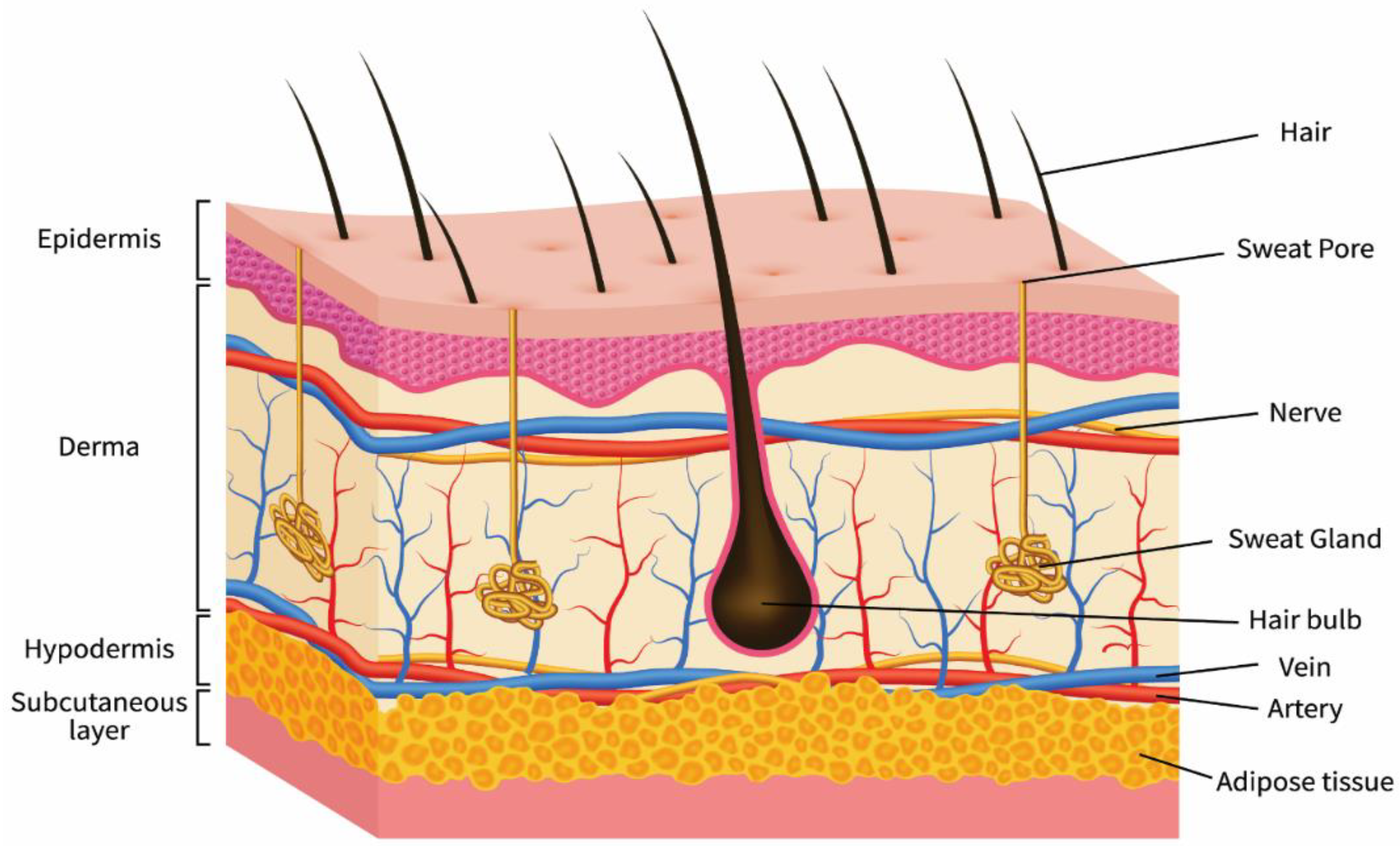
2. Structure of the Skin

The skin is a semipermeable membrane that serves as a defence against harm and protects the structural integrity of the human body. It is the most crucial multifunctional organ in the human body, performing critical tasks while defending the body’s internal tissues against pathogens and excessive water loss [16,17]. Figure 2 depicts a generic skin schematic. The epidermis, dermis, and hypodermis are the three primary layers of the skin. The significant functions of the epidermis are to prevent pathogenic microorganisms from entering the body and to keep the body hydrated. The stratum comprises five layers: basale, spinosum, granulosum, lucidum, and corneum, forming the skin epidermal layer. These layers include a variety of non-immune and immune cells (such as Merkel cells, melanocytes, keratinocytes, Langerhans cells, and stem cells). Keratinocytes, which contain spinous, granular, and outermost stratum corneum layers, are produced by the proliferative component of the epidermis. Some regions of the body, such as the foot soles and skin palms, contain an extra stratum lucidum. The stem cells in the basal epidermis regulate the regeneration of injured skin and the restoration of keratinocytes lost by exfoliation. The dermis layer is placed between the epidermis and hypodermis and comprises blood vessels, nerve cells, collagen, hair roots, mesenchymal stem cells, and lymphatic vessels. The dermis’ primary job is to provide structural hardness to the skin. The hypodermis is the subcutaneous fat layer that supports the epidermal and dermal layers and comprises fibroblasts, adipocytes, vasculature, macrophages, and nerves [18,19].

3. Brief Medical History of Silver Nanoparticles

Silver nanoparticles have been extensively used in various medicinal applications, as shown in (Figure 3). Silver nanoparticles are already widely used in wound dressings and burn treatment in biomedicine and also in the food and textile industries, in paints, household items, catheters, implants, and cosmetics, as well as in combination with a variety of materials to prevent infection [20,21,22,23,24,25]. During World War I, they treated soldiers’ wounds with silver leaves to stop infections and help them heal [26,27]. Silver as an ion was used in earlier civilizations, particularly in Egypt. This silver ion was used mainly in wound dressings to treat wounds that were hard to heal. In 1998, Ziehl-Abegg was the first firm to introduce AgNPs into wound dressings, resulting in the AgNPs antimicrobial dressing ActicoatTM. Silver products improve efficacy compared to standard wound dressing [28,29]. Acticoat and Actisorb, two silver-based wound dressings, are commercially available [30]. Dermatology increasingly employs metal nanoparticles to expedite wound healing and to treat and prevent bacterial infections [31].
Indeed, AgNPs’ excellent antimicrobial properties have already been tested against 650 bacterial strains [32,33,34]. Because of their extensive antimicrobial properties and ability to reduce the chance of infection from antibiotic-resistant strains, silver nanoparticles also found their application in wound management products [35,36,37,38,39]. AgNPs are utilized in wound care to prevent secondary infections since they are effective against a spectrum of microbes that can slow down the healing process [40]. Their size and charge allow them to enter microorganisms [41,42,43]. The AgNPs destroy many multi-resistant strains. It also helps rid pathogenic microbes that can slow or stop the typical stages of wound healing [44,45].
The biosynthesis of AgNP utilizing aqueous Bryonia laciniosa leaf extract resulted in rapid wound epithelialization and scarless wound repair without significant inflammation due to effective cytokine modulation. It was used as a wound healing agent due to its outstanding anti-inflammatory and antibacterial properties [46]. Biosynthesized AgNPs are made from Delonix elata leaf aqueous extract for wound healing in human patients who have had anorectal surgery [47]. Due to their sustained ability to release silver ions, which exhibit concentration-dependent toxicity in HaCaT cells, these nanoparticles have a high potential for usage in dermatology and wound healing [48]. Because AgNPs are biocompatible and can avoid this involuntary inflammatory action, they are not nullified by the immune system; thus, they could be used as anti-inflammatory agents [49]. Because of their anti-inflammatory properties, topical application of AgNPs at the wound site minimizes the release of inflammatory cytokines, lymphocytes, and mast cell infiltration, promoting wound healing with minimal scarring. A. Hebeish et al. also evaluated the anti-inflammatory effects of Ag by comparing them to indomethacin. This commercially available anti-inflammatory drug revealed a dose-dependent reduction in inflammation inside the rat bow edema model [50].
A study showed that AgNPs eradicate 44 strains of six fungi [51]. Gajbhiye et al. discovered that biogenic AgNPs were effective against Fusarium semitectum, Pleospora herbarum, Trichoderma spp, Phoma glomerata, and Candida albicans. They also reported a synergic effect of AgNPs in conjunction with fluconazole [52,53]. When AgNPs were added, they changed the growth rates of all tested fungi except Mortierella spp, meaning that Chaetomium and Stachybotrys could not grow on gypsum products.
J.L. Speshock et al. investigated AgNPs’ potential in prokaryotic and eukaryotic organisms, and AgNPs at about 25 nm or even less were found to have exceptional potential for viral infection suppression [54]. AgNPs inhibited virus attachment, cell penetration, and the cell’s ability to propagate the virus [55]. As an HIV-1 antiviral nanohybrid and in the deactivation of SARS-Cov-2 spike proteins, TPU-Ag worked better than PVA-Ag. TPU-Ag and PVA-Ag nanofibrous membranes displayed increased antibacterial activity by increasing Ag content from 2 to 4 wt. Additionally, the developed membranes showed good mechanical and physical properties and antiviral and antibacterial activities [56].
Silver synthesized from M. Domestica extracts significantly affected breast cancer MCF-7 cells. In contrast, silver synthesized from O. Vulgare aqueous extracts had a dose-dependent effect on the A549 cell line [57,58]. Moringa olifera stem bark extract was used to produce AgNPs. K. Vasanth et al. investigated the anticancer properties of these biosynthesized AgNPs. The flow cytometry analysis indicated that ROS generation caused apoptosis in HeLa cells [59]. According to the findings, AgNPs effectively prevent the development of HepG2 cells by inducing apoptosis [60]. Venkatesan et al. found that the human breast cancer cells MDA-MB-231 were killed by chitosan-alginate-biosynthesized AgNPs that were highly permeable (IC50 = 4.6 mg) [61]. A recent study found that packed quinazolinone polypyrrole with chitosan silver chloride nanocomposite was active against Ehrlich ascites carcinoma cells [62]. I.M. El-Sherbiny et al. found that Chitosan-silver hybrid nanoparticles cause HepG2 cells to die by turning down the BCL2 gene and the P53 gene [63]. A significant decrease in cyclobutene-pyrimidine-dimer creation demonstrated their chemo-preventive efficacy in HaCaT cells after UVB-induced DNA damage, which has a good potential for avoiding skin cancer [64]. The UVB-protective effectiveness of AgNPs in human keratinocytes is proportional to their size [65]. As a result, pre-treating HaCaT cells with smaller AgNPs (10–40 nm) helped shield skin cells from UVB-induced DNA damage and UV-induced apoptosis. Using 60 and 100 nm AgNPs, no prevention was obtained. AgNPs are increasingly used in healthcare and consumer products, so many commercial products now include these nanoparticles for topical administration to human skin.
Over one million people die from malaria yearly, caused by protozoal vector-borne diseases, the most prevalent and dangerous infections in wealthy nations [66]. Z. Jiang et al. are creating novel antimalarial strategies to control the malaria vector. AgNPs were tested against Plasmodium falciparum malarial parasites and other antimalarial medications [67]. The bio-reduction of AgNPs was 5%. The malaria vector Anopheles stephensi and chloroquine-sensitive and resistant P. falciparum strains were all treated with Cassia occidentalis leaf broth [68].

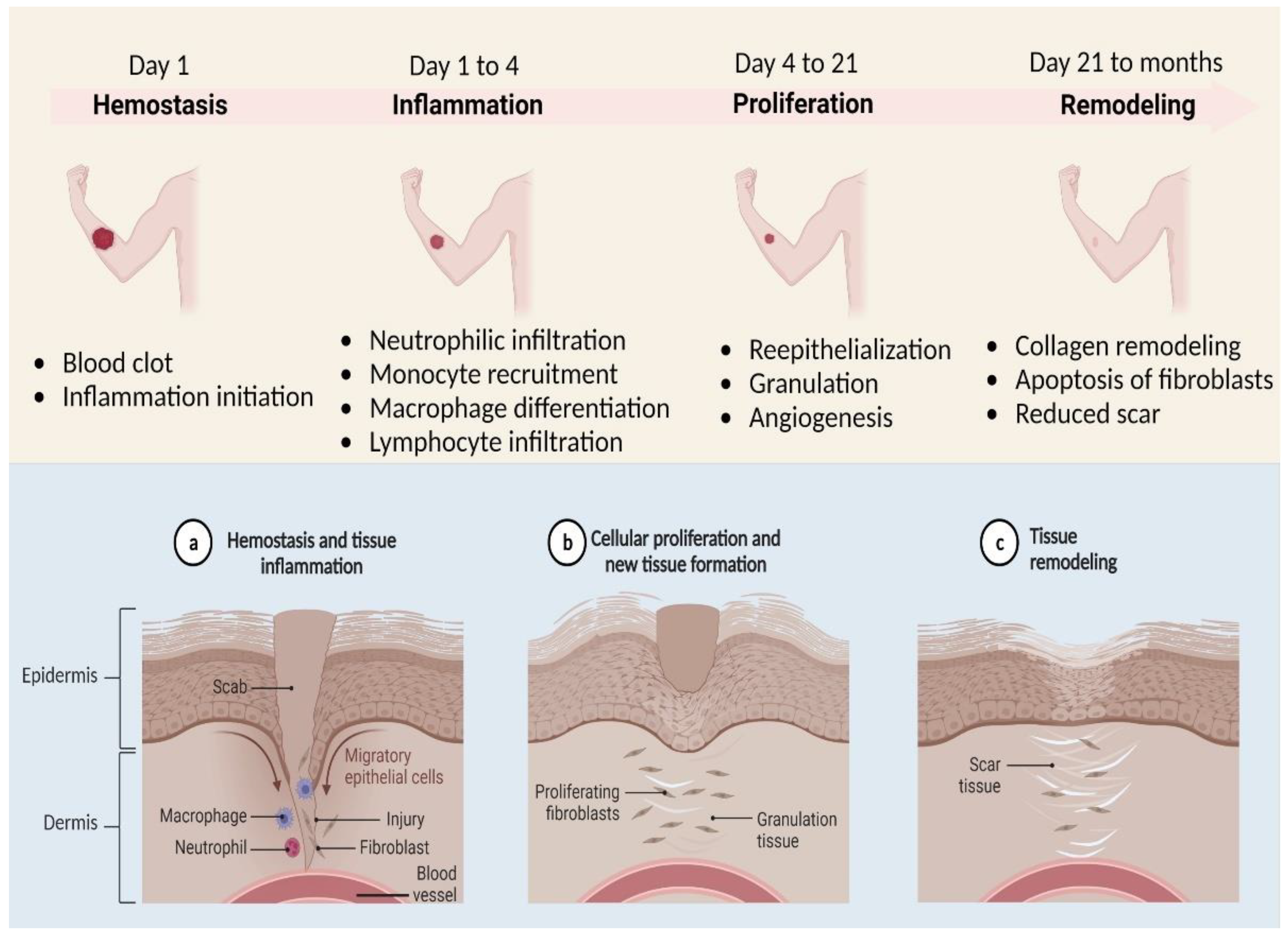
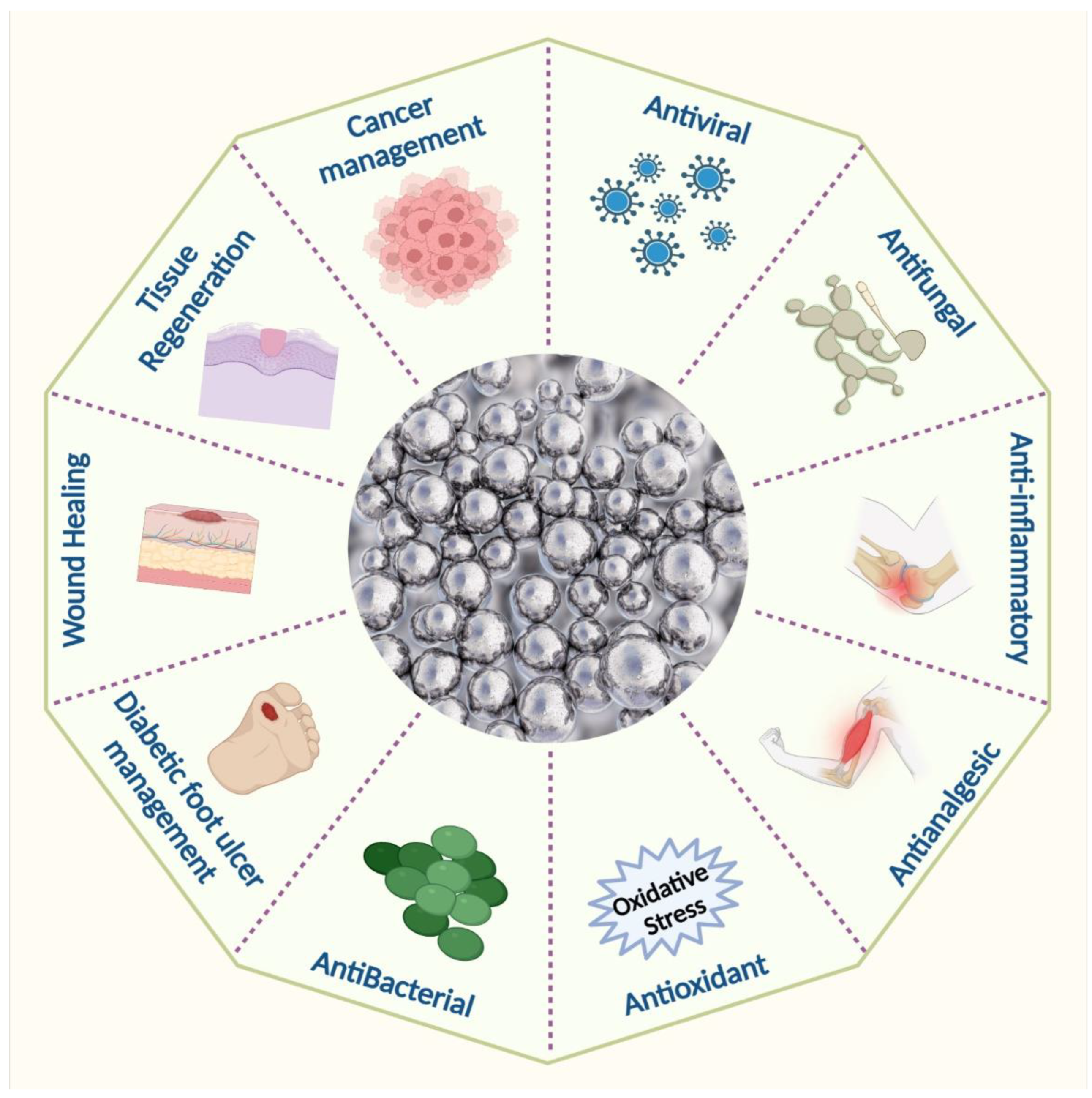
4. Wound Healing Properties of Silver Nanoparticles

Compounds of silver, such as silver nitrate and silver sulfadiazine, are often used to treat infections in chronic wounds and burns [69]. AgNPs help fibroblasts change into myofibroblasts, which makes wounds tighter and speeds up the healing of diabetic wounds. AgNPs accelerate wound healing by enhancing keratinocyte proliferation and migration [70,71]. AgNPs may engage with sulphur-containing proteins in bacterial membrane cells and, ideally, attack the respiratory chain, resulting in apoptosis [72]. When it comes into contact with the injured region, they cause neutrophil apoptosis by lowering mitochondrial function, which reduces cytokine production. As a result, the inflammatory response is modulated or reduced, resulting in faster healing [73,74]. However, because of their small size, AgNPs can easily penetrate biofilms and cell membranes, causing DNA damage, inhibiting cell proliferation, and inhibiting cellular ATP production [75]. The silver nanoparticles change the amount of m-RNA in the wound environment. Aside from antibacterial activities, silver surgical textiles exhibit an increase in healing properties; as an outcome, silver exploitation has an optimistic effect on cell migration and proliferation quality [76,77,78]. Cytokine modulation is mediated by silver nanoparticles’ anti-inflammatory activity [79]. As stated in the preceding section, Cytokines can stimulate fibroblasts and chondrocytes to generate ROS [80]. Thus, silver nanoparticle modulation of cytokine production can reduce ROS levels to avoid severe cellular damage and lag wound healing [81]. Silver has many antibacterial effects, making it less likely that bacteria will become resistant and more effective against microorganisms resistant to multiple drugs. When the amount of AgNPs in the dressing increases, the wound area becomes smaller and more collagen is deposited, which is linked to macrophage and fibroblast migration [82,83]. Sustained release mechanisms can decrease silver ion toxicity and stimulate local antibacterial activity [84,85].

5. Mechanistic Understanding of Silver Nanoparticles (AgNPs)

The usual quantity of silver in human plasma is less than 2 µg/mL, and this concentration comes from diet and particulate matter inhalation. Oral exposure to silver can also come via dietary supplements, contaminated water, or from eating fish and other aquatic species [86]. Ionic silver can be ingested orally, inhaled, or absorbed through wounds to enter the body. AgNPs are believed to be transported inside the body by two processes: pinocytosis and endocytosis. The development of a revolutionary medication delivery method was prompted by the discovery that nanoscale particles penetrate far deeper than bulk particles. Although the precise mode of action of AgNPs is not yet known, numerous ideas for their antibacterial qualities have been put forth. Its antibacterial effect is thought to solely be caused by the ongoing release of silver in its ionic state [87,88,89]. Silver ions cling to the cytoplasmic membrane and cell wall because of the sulphur protein affinity and electrostatic attraction. This increases the permeability of the membrane and causes the bacterial cell to rupture and degenerate. Reactive oxygen species are produced, and the respiratory enzymes are essentially deactivated when the silver ion enters the bacterial cell. Reactive oxygen species, a critical element in the mechanism of action for silver, contribute significantly to the disruption of the cell membrane and DNA damage (by interacting with sulphur and phosphorus in the DNA molecule), which hinder replication and reproduction and ultimately lead to microbe death. By denaturing ribosomes, silver ions also prevent the synthesis of ATP and hinder the formation of proteins. After anchoring and observing the cell’s surface, silver nanoparticles build up in the cellular wall pits of microorganisms, causing the denaturation and degeneration of the cell membrane. Due to their micro size, they can easily enter cells, rupturing cell organelles and even causing cell lysis. They interfere with the phosphorylation of protein substrates, which can cause cell death and proliferation, which has an impact on the bacterial transduction process as well. Due to their cellular walls being shorter than those of Gram-positive bacteria, Gram-negative bacterial strains are more susceptible to the effects of AgNPs [86,89]. Silver nanoparticles have a significant downside in that bacterial biofilms make them less effective and penetrating. Due to their intricate structure, biofilms typically change the transport chain to shield the membrane from both silver ions and nanoparticles. The nanoparticle size, which is around 50 nm, severely obstructs the path of penetration that is currently being used. Additionally, it has been observed that silver nanoparticle adsorption and deposition on bacterial biofilms reduces the nanoparticles’ ability to diffuse into bacterial cells (Figure 4). Silver’s interaction with a molecule containing a thiol group in bacterial, fungal, and fungus cells provides the basis for silver nanoparticles’ antibacterial effect (Figure 4). It has been observed that bacterial and fungal cells undergo structural changes after coming into touch with silver nanoparticles, albeit the precise process is yet unclear. Silver nanoparticles have higher antibacterial and antifungal characteristics than normal silver particles because of their extensive surface area, which enables better interaction with bacterial and fungal pathogens. Additionally, the gel made of silver nanoparticles penetrates bacteria and fungi in addition to attaching to cell membranes. Silver penetrates cells and connects to the cell membrane and wall, which prevents the cell from respiring [90,91]. When silver is present, Escherichia coli is prevented from absorbing phosphate and from releasing mannitol, succinate, proline, and glutamine. As a result, silver nanoparticles can be used as effective growth inhibitors in a wide range of microbes and are helpful in many antibacterial control systems [92,93].

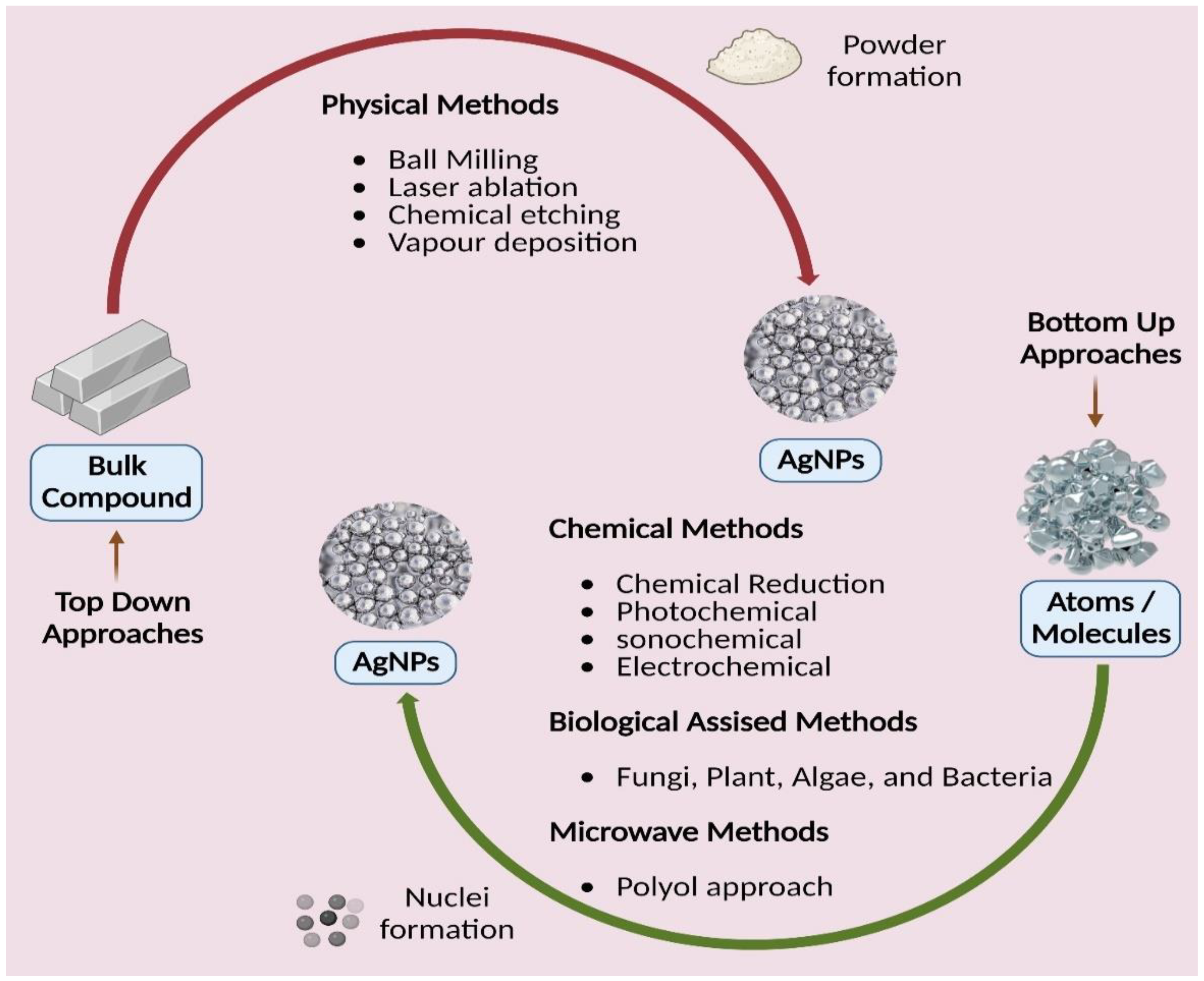
6. Silver Nanoparticles and Their Synthesis
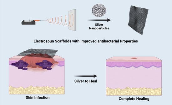
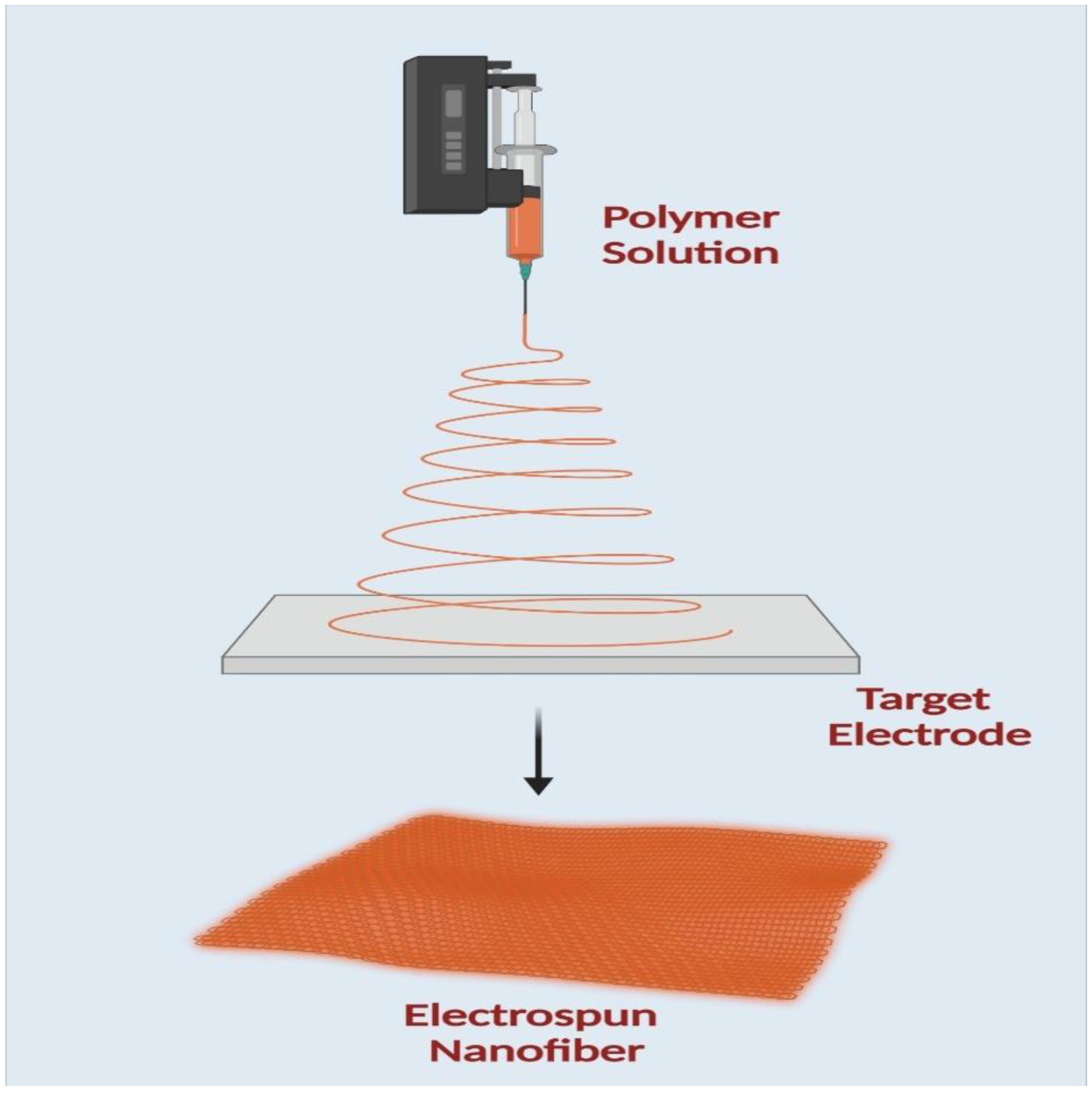
AgNP is synthesized using either a top-down or bottom-up method, as is typical for most nanomaterials, as illustrated in (Figure 5) [94]. Physically breaking down a large complex into smaller parts is a critical component of the top-down strategy. Physical and mechanical procedures such as UV irradiation, lithography, laser ablation, ultrasonic sounds, and photochemical reduction create a strong energy/force to compress the macromolecule into nanoparticles [95]. The physical reduction procedures generally have quick processing durations, yielding AgNPs with a restricted size distribution range. However, they have several drawbacks, including the tremendous amount of equipment space needed, the extended time required to obtain thermal stability, the energy-intensive nature of the methods, and the difficulty in easily using them for scaling-up reasons [96]. When creating nanoparticles from the bottom up, tiny particles such as atoms and molecules are used as the building blocks. These smaller particles come together to form a complex nanoparticle by self-assembly or aided assembly. Chemical, microwave, and biological approaches are subcategories of the bottom-up strategy. For the manufacture of AgNPs in solution, chemical techniques are often utilized. They are produced in water or other organic solvents [97]. Chemical processes are quick and practical but represent a severe environmental risk because they use hazardous substances as reducing agents and create dangerous byproducts.
New alternative techniques that are cost-efficient, energy-efficient, and environmentally benign are swiftly becoming available to counteract these negative environmental consequences. There has been a thorough literature assessment on the development of green or biological synthesis [98,99,100,101]. In green synthesis, an ecologically friendly substance such as microbial (fungal and bacterial) enzymes and phytochemicals from plant extracts are used instead of the capping/stabilizing and reducing agents used in chemical reduction procedures (leaves, roots, barks, flowers, fruits, peels, and seeds). These biological processes generate biocompatible nanoparticles suitable for pharmaceutical and biomedical applications [102,103]. While utilizing microbes as reducing agents in nanoparticle manufacturing is quite tricky and involves several methods, it is more advantageous than chemical and physical techniques. Because they are widely accessible, simple to extract, and do not need laborious processes, plant extracts are considered a solution to the issues mentioned above in the manufacturing of AgNP. For the most part, because they are so readily available, plant extracts are employed to make nanoparticles. Manufacturing AgNPs with regular shapes and sizes is the main difficulty with plant-based synthesis, and the reduction processes of biological approaches are not well understood [104,105,106,107,108].

7. Electrospinning

An electrospinning machine consists of four main parts: a high-power source, a syringe pump, a syringe needle carrying solutions, and a fiber deposition collector, as shown in (Figure 6). A positive electrode is attached to the needle, and a negative electrode is connected to the collector to create the applied electric field. As a result, the repulsion charge accumulates near the hemispherical needle tip when a voltage is applied. A Taylor cone is made when the repulsive charge surpasses the surface tension. The negative electrode, which serves as the collector in this procedure, is where the polymer solution is directed to create fibers. The polymer solution evaporates, leaving behind dry fibers ranging in size from nanometers to micrometers deposited on the collector [109,110].
As shown in (Figure 7 and Figure 8), electrospun nanofiber mats are an excellent choice for chronic wound healing because of their numerous advantages and inherent properties. In multiple medical applications, nanofiber composite materials are widely used. Silver nanoparticle-loaded electrospun nanofiber scaffolds showed exceptional antibacterial activity, high porosity, non-toxicity, and biodegradability. Due to their hydrophilic qualities and prolonged release pattern, these nanofiber scaffolds have become more and more well-liked on a global scale. With the help of this delivery method, a better dressing and therapy for ulcers and wounds in diabetic patients would be made available clinically. Silver nanoparticles have been extensively used in various medicinal applications. Encapsulating or coating nanofibrous scaffolds with metal-based nanoparticles can boost their therapeutic efficacy in wound healing applications [111,112]. Synthetic polymers, particularly those with biodegradable and biocompatible properties, may provide excellent treatment options for severe wounds and burn injuries.

8. Cytoprotective Effect of Silver Nanoparticle-Loaded Nanofibers

AgNPs have the potential to spread throughout the body, build up in many organs, and cause significant damage. AgNP bystander effects are being reduced by the development of new fabrication techniquesas shown in (Table 1). To minimize environmental impact, disposal options must be investigated. High nanoparticle loading capacity and controllable release mechanisms are two significant advantages that silver nanoparticle-loaded nanofibers are putting forth. Due to the increased surface area and short diffusion length of electrospun nanofibers, a more excellent Ag+ release is accomplished compared to traditional wound dressing materials.
Additionally, regulated release mechanisms can improve local antibacterial effectiveness while decreasing the dose-related systemic toxicity of silver ions. Silver ions can be released from polymeric nanofibers in a variety of ways, including diffusion caused by the polymer matrix swelling (a swelling-controlled system), polymer breakdown, or a combination of the two. Long-lasting antibacterial activity can be attributed in part to controlled release performance. It will be more effective to incorporate AgNPs into the wound dressings rather than placing them directly on the wound bed. This layer also worked as a sieve to prevent cytotoxicity brought on by the extensive release of AgNPs in the area close to the wound [113,114].
Table 1. Various forms of fabricated biomaterials for wound healing applications.
S. No.Wound Dressing MaterialsFabrication Techniques and OutcomesRef.
1Polyurethane/keratin/AgNP biocomposite matsElectrospinning method
The material’s keratin content increased fibroblast cell proliferation while also having strong antibacterial properties. A histological analysis showed that the created biocomposite mat could promote wound healing.
[115]
2Hyaluronan and PVA embedded-AgNP HydrogelFreeze-thawing method
The hydrogel’s semi-interpenetrating network aided in the AgNPs’ uniform dispersion. The hydrogel may be used as a wound dressing since it had strong antibacterial activity, was biocompatible, had a low swelling index, and was nontoxic.
[116]
3Genipin-crosslinked chitosan/poly(ethylene glycol)ZnO/AgFilm casting method
The created nanocomposites showed improved mechanical characteristics and pH-sensitive swelling behaviour, and they were successfully used as a material for wound dressings.
[117]
4AgNP-Calcium alginate beads in gelatin scaffoldsFreeze-drying method
Due to their favourable swelling qualities and non-toxic behaviour against human dermal fibroblasts, they are recommended as acceptable wound dressings.
[118]
5Chitosan-hyaluronan nano composite spongesIonic cross-linking followed by freeze drying
The material had adequate porosity for applications involving wound healing, good biodegradation, and improved swelling properties.
[119]
6Methoxy poly (ethylene glycol)-graft-chitosan composite film Casting/solvent evaporation method
The substance that was manufactured showed that the medication curcumin had been loaded successfully. The film had an uneven surface without any pores. The produced film has a significant deal of potential for use in wound healing applications, according to an in vitro cytotoxicity research, antioxidant effectiveness assessments, and animal trials (histological study).
[120]
7Tannic acid/chitosan/pullulan composite nanofibersForce spinning method
It has the potential to be used in the treatment of intricate and deep wounds since it replicates a 3D environment, exhibits good water absorption, and encourages fibroblast cell adhesion.
[121]
8Ag/ZnO nanocompositesDeposition precipitation method
The porosity of composites, which ranged from 81 to 88%, the swelling ratios, which ranged from 21 to 24, and the moisture retention period, which ranged from 13 to 14 days, all demonstrated good results in various experiments. These characteristics are all crucial for expediting wound healing.
[122]
9Silver/hyaluronan bio-nanocomposite fabricsWet-dry-spinning technique
According to in vivo research, fabrics improved the material’s mechanical qualities and increased wound healing effectiveness.
[123]
10Chitosan-Ag/ZnO composite dressingLyophilisation and immersion method
In many tests, composites performed well in terms of porosity (81–88%), swelling ratios (21–24%), and moisture retention period (13–14 days), all of which are critical elements in improving wound healing.
[124]
11Starch-AgNPsNanoprecipitation method
By using an ecologically friendly process, alkali-dissolved starch served as a reducing and stabilising agent to create AgNPs, and this strategy may be used for applications in the treatment of wounds.
[125]
12Cellulose/Polypyrrole/AgNPs/ Ionic liquid composite films Simple chemical polymerization method
Composite films demonstrated effective antibacterial action and may be applied as patches to help heal wounds.
[126]
13Fibrin nanoconstructsWater-in-oil emulsification diffusion technique
It served as a reliable carrier molecule for tacrolimus, an immunosuppressant.
[127]

9. Advantages of Silver and Fibre Platforms

The ideal wound dressing should fulfill a number of criteria, including those listed below: (i) representing a physical barrier that is permeable to oxygen while also maintaining or providing a moist environment; (ii) being sterile, non-toxic, and protective against microorganism infections; (iii) providing an appropriate tissue temperature to favour epidermal migration and promote angiogenesis; and (iv) being non-adherent to prevent traumatic removal after healing. An ideal wound dressing should have all the qualities listed above, but it is challenging for one kind of dressing to meet every one of these needs. Creating a moist wound environment reduces dehydration and cell death. It facilitates angiogenesis and epidermal migration. It preserves moisture at the site of the wound. Excess exudate must be removed for the wound to heal, but it can also cause healthy tissue to macerate, creating a persistent wound. It enables gaseous exchange. Oxygenation regulates exudate levels and promotes fibroblast and epithelial growth. It prevents infection by prolonging the inflammatory phase and preventing epidermal migration and collagen formation; microbial infections slow the healing of wounds. Low adherence and painless removal of adherent dressings can be uncomfortable and can worsen existing granulation tissue damage. The cost-effective and optimal dressing should promote wound healing while remaining reasonably priced [128,129,130]. The main categories of wound-dressing materials are fibers, gels, membranes, films, sponges, and hydrocolloids, as shown in (Table 2). Nanofiber mats are a superior option for drug delivery compared to all other biomaterials because of their numerous advantages and inherent properties, as shown in (Table 2). The incorporation of nanoparticles into nanofiber scaffolds constitutes a novel approach to “nanoparticle dressing” that has acquired significant popularity for wound healing (Table 2). Due to their remarkable antibacterial capabilities, silver nanoparticles are attractive materials for wound healing. Numerous wound-dressing materials have been created in this area (Table 3), based either on synthetic or natural polymers.

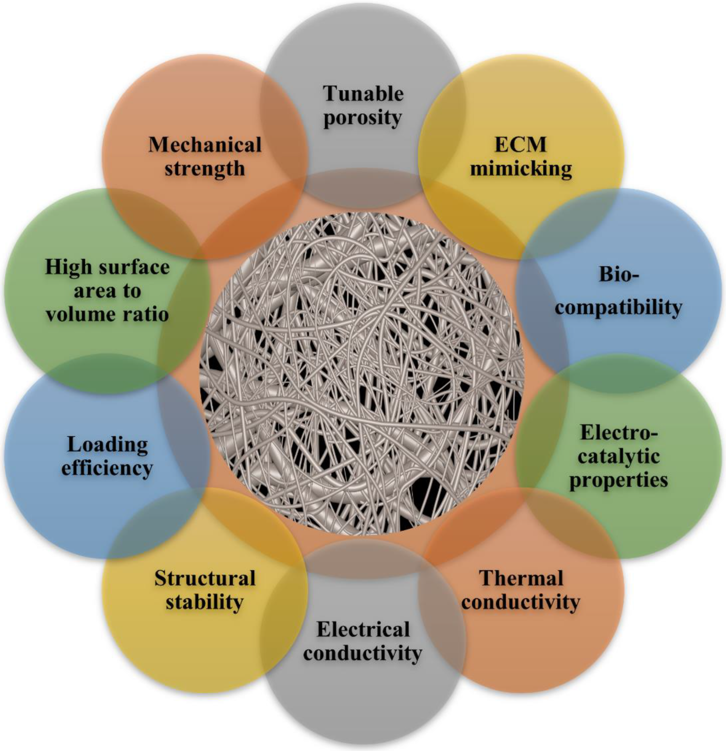
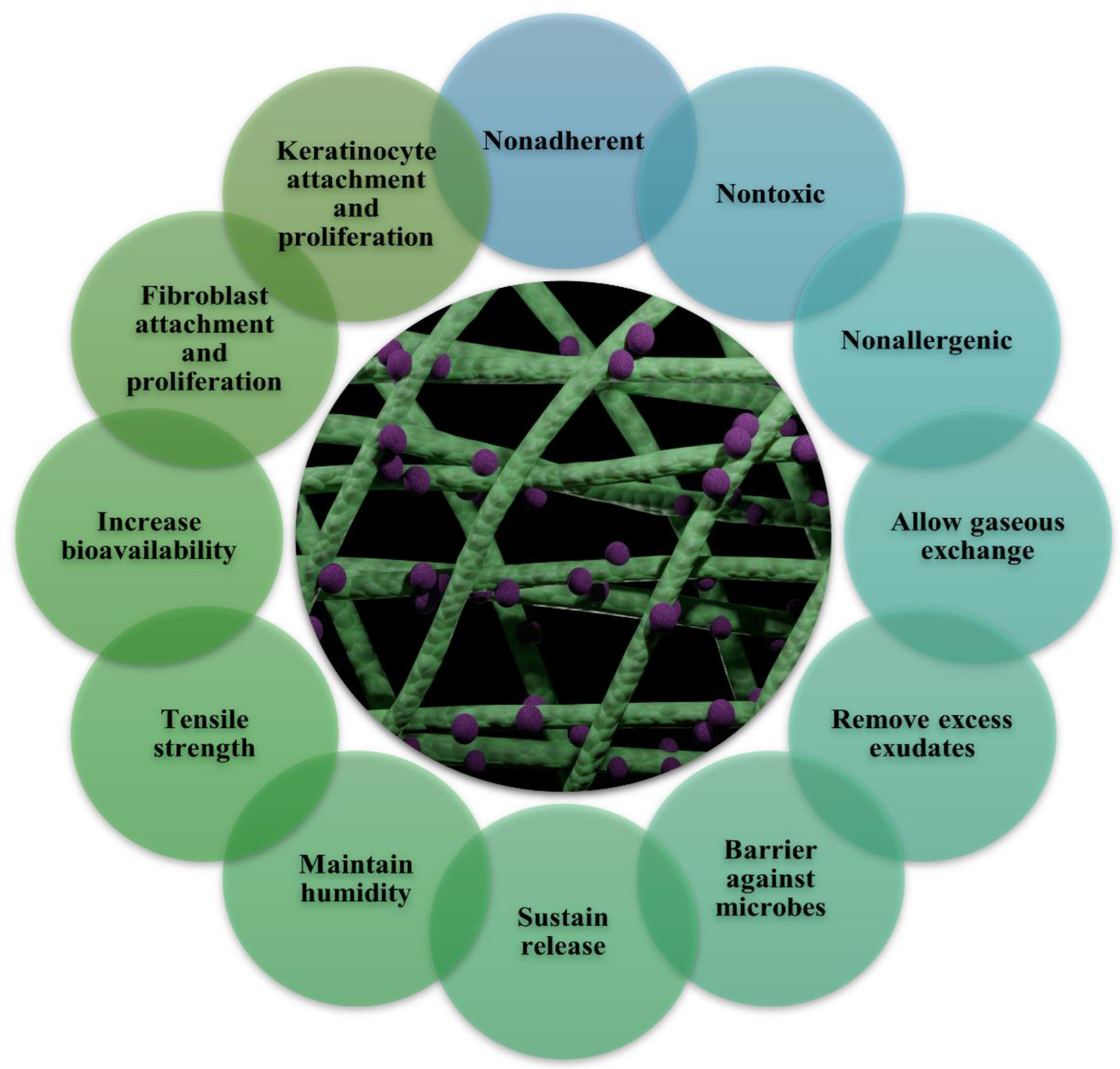
10. Silver Nanoparticles Containing Nanofibers for Wound Healing

Ag nanoparticles can generally be incorporated into polymeric nanofibers in various ways (Table 4). Electrospinning a Ag nanoparticle dispersion in a polymer solution is one method. Such a strategy has already been demonstrated to be problematic since the nanoparticles begin to aggregate and lose their effectiveness [159,160]. However, the process of forming Ag nanoparticles onto polymeric nanofibers appears to be advantageous in terms of maintaining the antibacterial activity of the nanoparticles. Table 5 depicts some of the clinical transformation status of silver nanoparticles. Sol-gel and surface functionalization are two approaches that have shown promising relevance in this respect. Similar to the first procedure, adding a precursor salt to the polymer solution before electrospinning results in uniform surface decorating of the nanofibers by the nanoparticles, followed by hydrothermal treatment of the produced nanocomposite nanofibers [161]. An efficient method to produce polymeric nanofibers embellished with metal nanoparticles is surface functionalization by proteins and other substances, such as polydopamine. The functional polymers, such as poly(acrylonitrile-co-glycidyl methacrylate) (PANGMA), can easily link with inexpensive serum albumin proteins such as bovine serum albumin (BSA), for example, by an amine-epoxy process. When submerged in a metal-containing aqueous dispersion, this biofunctionalized nanofiber system can subsequently collect noble metal nanoparticles via a metal-protein interaction. In this circumstance, the protein changes shape from an alpha helix to a beta sheet (Figure 9a), exposing functional groups that can grab biomolecules, metal nanoparticles, etc. (Figure 9b) [160]. Thus, as seen in Figure 9c, the final nanocomposite nanofibers have nanoparticles evenly coated on their surface. Polydopamine created by the self-polymerization of dopamine in an alkaline environment has also provided intriguing prospects for surface functionalization (Figure 9d) [162]. This work was motivated by mussel adhesion onto various surfaces in nature. Through the formation of metal nanoparticles such as Ag, this coating material can diminish metal cations. The long-lasting antibacterial action of the as-synthesized Ag nanoparticles is made possible by their insensitivity to oxidation. The problematic control of coating thickness and surface shape is one of the main problems with polydopamine-based coatings. This flaw prevents the Ag nanoparticles from being distributed uniformly because it causes the surface to become rougher due to the unwanted aggregation of polydopamine in random places [163]. In order to overcome this issue, nanofibers made of poly(dopamine methacrylamide-co-methyl methacrylate), a copolymer inspired by mussels, were electrospun. As a result, Ag nanoparticles can be formed on the surface of these catalytic nanofibers [13].
The silver loaded nanofiber network has shown notable characteristics in wound healing applications (Figure 7 and Figure 8). There are increasing studies on using AgNPs in wound dressings, and all outcomes promoting wound healing are positive [165,166,167]. C. He et al. prepared antibacterial wound dressings whose outermost layer was hydrophobic, which inhibited external microbe adhesion and invasion. Invading microorganisms could be hampered by a specially engineered intermediate region with a high concentration of AgNPs. The polycaprolactone and gelatin hydrophilic surface was employed as the inner layer adhered to the wound bed. The antibacterial activity of the nanofibers containing 10% silver nanoparticles was much higher than that of the dressings containing 1% and 5% silver nanoparticles (Figure 10a). Furthermore, the biocompatibility was far better than that of commercial silver sulfadiazine. This layer also worked as a screen to prevent cytotoxicity from the abundant discharge of AgNPs in the wound’s immediate vicinity. According to the results, the wound dressing’s sandwich structure provides good antibacterial activity and cytocompatibility. The findings pave the path for developing more clinically-relevant wound closure and healing dressings (Figure 10b) [168] AgNPs were evenly distributed throughout the fibers. The XRD patterns of AgNPs and PCL/Gel-AgNPs are depicted in Figure 11. The characteristic peaks at approximately 38° and 44° corresponded to the (1 1 1) and (2 0 0) planes of AgNPs, respectively, proving that AgNPs were successfully doped.
Dalong Li et al. used a combination of sol-gel processing and electrospinning to create Ag-doped mesoporous silica fibers. The main precursors used to make composite fibers were tetraethylorthosilicate, polyvinyl alcohol, and silver nitrate. Water-soluble PVA dispersed silver nitrate better than other polymers used in the preparation of silica composite fiber. After that, the electrospun fibers were heat treated to produce AgNPs via AgNO3 reduction and pyrolyze the PVA component. XPS was performed on the composite nanofiber and pure silica fiber to further investigate the chemical states of the Ag species in the composite nanofiber. Compared to the XPS spectrum of pure silica fiber, composite nanofiber exhibited new peaks attributed to Ag3d (Figure 12A). The typical fully-scanned spectra revealed the presence of Ag, Si, O, and C in the AgNPs/silica composite nanofiber. The C1 peak binding energy of 284.6 eV is used as the calibration standard. The two peaks at binding energies of 368.5 and 374.5 eV, assigned to Ag0 3d5/2 and Ag0 3d3/2, respectively, in the high-resolution XPS spectra (Figure 12B) demonstrate the metallic nature of AgNPs [169].
Suxia Ren et al. created nanofibers with high activity in surface-enhanced Raman scattering (SERS) by electrospinning precursor suspensions of polyacrylonitrile, silver nanoparticles, silicon nanoparticles, and cellulose nanocrystals. Figure 13 displays the FTIR spectra of the electrospun nanostructures. For pure PAN nanofibers, separate peaks at 2244, 1451, and 1096 cm−1 correlated with, respectively, the skeletal vibration of the PAN chemical chain, CH wagging vibration, CN stretching vibration, and CH2 bending vibration. The O-H bending of adsorbed water is thought to be responsible for the peaks of about 1658 cm−1. The aliphatic CH group vibrations of CH2 are attributed to the peaks at 1340 and 1380. The spectra of PAN/CNC/Ag and PAN/CNC/Ag/Si nanofibers, when compared to those of pure PAN, exhibit new peaks at 1034 cm−1 and 824 cm−1, respectively, which correspond to the characteristic absorption bands O-H of CNCs and C-H rock, respectively, indicating the presence of CNC in the electrospun nanofibers. It is possible that there were no chemical connections or interactions between the -CN- groups in the PAN and AgNPs because there was no detectable -CN- bond vibration shift [170].
M.R. EI-Aassar et al. constructed a nanofiber scaffold composed of naturally bioabsorbable components, such as hyaluronic and polygalacturonic acid, and embedded with silver nanoparticles for use in vivo. In the interim, silver nanoparticles in this formulation will function as an antioxidant, as antibacterial, and as anti-inflammatory, protecting cells from the harmful effects of high ROS and accelerating wound healing. The Ag nanoparticle of the nanofiber membrane exhibited strong antibacterial zone inhibition efficacy against Gram (+) and Gram (−) bacteria. The increased hydrophilicity and strain activities were the result of the hyaluronic acid component. In addition, the in vivo study on albino rats demonstrated that wound epithelization and collagen deposition were at their highest 14 days following nanofiber delivery. Therefore, it is essential to provide effective nanofibers to heal infected wounds rapidly [171].
J. Shao et al. produced nanofiber membranes comprised of chitosan and silver nanoparticles. In vitro, membranes containing AgNP exhibited antibacterial activities commensurate with the silver release profile. The fibrous membranes with different amounts of AgNPs were fabricated by electrospinning, and SEM and TEM characterized their morphologies. SEM images (Figure 14A–C) showed a uniform fibrous structure for all groups with a similar average fiber diameter of ~200 nm (p > 0.05). The TEM image (Figure 14D) demonstrated that the electron-dense AgNPs were formed within the fibers. Figure 14E shows the morphological changes after immersion in PBS or FBS. The membranes kept their fibrous structure after immersion in PBS, while more and more white spots appeared on the fibers with the increase in silver amount and immersion time. Further identification using EDS revealed that the white spots had a high amount of silver and chloride. For the FBS-immersed membranes, a thick layer of protein was observed on the fibrous membranes since day 4. The study utilized an intra-operative contamination model using rat subcutaneous tissue to further evaluate antibacterial efficacy in vivo. The capacity for wound healing was examined using a rat excisional wound splinting model. Researcher findings indicated that AgNP inclusion can improve the antibacterial activity of biomaterials without impairing wound healing.
H.S. Sofi et al. developed wound dressing made of polyurethane incorporated lavender oil and AgNP. Compositional nanofiber dressings offer much potential for use as multipurpose wound dressings since they can both protect from external irritants and promote tissue regeneration. Through diffusion and penetration, lavender oil lowered the stiffness of polyurethane fibers and changed their hydrophobicity. These composite nanofibers dramatically improved the growth and proliferation of CEFs. Studies on cell fixation have shown that fibroblasts grew on fiber mats containing lavender oil and AgNPs in their natural structure [172].
N. Eghbalifam et al. aimed to fabricate a Gum Arabic-based electrospun nanofiber membrane with the ideal porosity, water absorption, and water vapor permeability. It was demonstrated that mat has antibacterial activity against E. coli, Candida albicans, S. aureus, and P. aeruginosa. AgNPs nanofibers were coated with PCL using a stable and electrospinnable solution by combining Gum Arabic and PVA. The PCL-coated mat exhibited a high-water absorption capacity and was water resistant. Gram (+) and Gram (−) bacteria and a fungal strain were all suppressed from growing on the composite nanofiber [173].
B. Salesa et al. prepared Carbon nanofibers that are one-dimensional nanomaterials with superior physical and broad-spectrum antibacterial characteristics resistant to antimicrobials. AgNPs are already used in various industrial applications. In HaCaT cells, AgNPs and carbon nanofibers were tested for cytotoxicity, proliferation, and gene expression. AgNPs are smaller and have a completely different morphology than filamentous carbon nanofibers. From pH 5–12, AgNPs displayed a more substantial negative zeta potential than CNFs and equal time-dependent cytotoxicity. These findings hold great potential since they allow AgNPs to be used in various tissue engineering and wound healing applications [174].
G. Sandri et al. aimed to create an electrospun chitosan and glycosaminoglycan membrane incorporated with AgNPs to control bacterial infections during wound healing. The heating method crosslinked the scaffolds to create water-resistant structures, which may also be termed a sterilization process. The systems’ preliminary enzymatic breakdown was tested using lysozyme, which is generally secreted by white cells (macrophages and neutrophils during the inflammatory phase). Nanofibers with a regular uniform shape, a smooth surface, and AgNPs implanted into the polymeric mat creating the fibers were all characteristics of AgNP scaffolds. Antimicrobial activity, primarily against S. aureus, was a feature of these systems. The performance of the membrane makes it a viable tool for treating persistent skin lesions [175].
M. Mohseni et al. prepared PCL and PVA nanofibers loaded with various quantities of silver sulfadiazine (SSD) and silver nanoparticles. Wound dressings have sufficient flexibility and hydrophilicity, resulting in satisfactory wound closure adhesion. Silver sulfadiazine is one of the most common antibacterial medicines in wound healing. Antibacterial mats are resistant to S. aureus, and increasing the concentration of silver sulfadiazine or AgNPs improves action. The flexibility and hydrophilicity of PCL and PVA nanofibers allowed the wound closure to be moistened with wound fluid loaded with cytokines and growth factors. On the other hand, SSD was more hazardous to fibroblast cells than AgNPs [176].
Z.W. Li et al. synthesized polyvinyl alcohol, Chitosan oligosaccharides, and AgNP nanofibers to stimulate HSF adhesion, proliferation, and cell cycle transition from the quiescent G0/G1 phase to the active S DNA synthesis and the active G2/M phase of division. Using RT-PCR, immunofluorescent labelling, and Western blot analysis, investigators demonstrated that the nanofibers upregulated molecules implicated in the TGF-β1/Smad signaling pathway, hence boosting collagen synthesis and improving wound healing by upregulating TGF-β1 secretion and activating the TGF-β1/Smad signaling pathway during the early stages of wound healing, boosting the adhesion and proliferation of fibroblasts. This occurrence is followed by an acceleration in the proliferation and differentiation of keratinocytes, an increase in the synthesis of collagen and the extracellular matrix, the facilitation of granulation tissue development and angiogenesis, and finally, the promotion of re-epithelialization. This research offers a significant step forward in creating novel, enhanced drug delivery vehicles for therapeutic wound-healing medicines [177].
M. Mostafa et al. used an electrospinning approach to manufacture AgNPs loaded in polystyrene nanofiber scaffold. In biomedical fields, this can be used as a potent bioactive substance. Furthermore, AgNPs-polystyrene nanofibers have improved their antibacterial effect against both S. aureus and E. coli. Various electrospinning settings were investigated, with this nanofiber proving the most effective. DMF can be used as a reducing agent to make Ag nanoparticles with a diameter of 21–40 nm [178].
E. Esmaeili et al. designed nanofibrous scaffolds from polyurethane and cellulose acetate. Due to their significant antibacterial action, reduced graphene oxide/silver was also employed in the mats. By coming into direct contact with bacteria, scaffolds could inhibit them. Curcumin has the most significant impact on wound healing and can speed up the healing of artificial wounds. Curcumin and graphene oxide/silver nanocomposites can be combined to create ultrafine, bead-free nanofibers with a porous structure, making them suitable for biomedical applications. On direct contact with microbial cells, their antibacterial activities result in a 100% inactivation rate for Pseudomonas bacteria and a 95% inactivation rate for S. aureus bacteria. In this study, in vivo histopathological studies revealed that adding curcumin can considerably improve wound healing and epidermal layer regeneration [179].
H. Bardania et al. synthesized silver nanoparticles biogenically produced using Teucrium polium extract and implanted in PLA and PEG mat to offer an absorbable wound dressing with antibacterial and antioxidant properties. Antibacterial tests revealed that S. aureus and P. aeruginosa were sensitive to biosynthesized AgNPs at different concentrations and had a good safety profile in human macrophage cells. This green synthesis method has proven to be a quick, cost-effective, and efficient way to make AgNPs without needing external stabilizers or reducing agents [180].
S.M. Hong et al. developed antibacterial polyurethane nanofiber textiles with Ag nanoparticles based on polycarbonate diol/isosorbide. The materials were pliable, with breaking strains ranging from 355 percent to 950 percent under a tensile stress of 7.28 to 23.1 MPa. Cell proliferation was performed using the HaCaT cell line, which exhibited cytocompatibility and no toxicity. Researchers also tested antimicrobial capabilities against S. aureus and methicillin-resistant MRSA. Adding AgNPs to the polyurethane nanofiber membrane enhances physicochemical parameters such as mechanical, thermal, and biological properties [181].
M.A. Mohamady et al. aimed to create core-shell electrospun membranes that could promote cell proliferation and act as antibacterial agents. A polycaprolactone shell membrane was used to load phenytoin. Silver-chitosan nanoparticles were placed in a PVA core layer as biocidal agents. Researchers employed coaxial techniques to create dual-drug delivery systems suitable for the model pharmaceuticals. The use of phenytoin to improve the cytocompatibility of scaffolds was proposed. In the core, biocides such as CS-coated silver nanoparticles were used as biocides, while the shell contained phenytoin, a healing-promoting substance [182].
R. Liu et al. designed a novel hydrogel dressing created by crosslinking cellulose fibers with gelatin and aminated AgNPs (Ag-NH2 NPs). The use of multiple components significantly enhanced the mechanical, self-recovery, hemostatic (gelation), antibacterial, and fluid balancing capabilities of the wound bed. In vitro and in vivo assessment of the mat wound healing model revealed excellent biocompatibility and healing performance (90 percent healed and 83.3 percent survival after 14 days) [183].
M. Hu et al. synthesized electrospun nanofibers of ZnO, Ag, PVP, and PCL. ZnONPs had an average diameter of nanofibers of 40.07 ± 9.70 nm and AgNPs’ diameter of 37.46 ± 12.02 nm, respectively. The antibacterial effects of the single metal material embedded scaffold were far superior to those of the single metal material-loaded nanofibers against S. aureus and E. coli. Adding ZnO and Ag to these nanofibers lowered their cytotoxicity against fibroblasts [184].
J.P. Ye et al. created soluble keratin from wool using keratinase and raised the molecular weight of the keratin to 120 kDa using TGase. In situ bio-reduction was used to create Ag nanoparticles on nanofibers using the keratinase stated earlier as the reducing agent. High molecular weight keratin had improved mechanical and hydrophilic qualities when co-electrospun with Poly(3-hydroxybutyrate-co-3-hydroxy valerate). E. coli and S. aureus were utilized to test the antibacterial effectiveness. The researcher demonstrated that they both possessed potent antibacterial properties. The mats dramatically accelerated the healing of skin wounds [185].
M.K. Ahmed et al. designed the doped antimicrobial silver nanoparticles incorporated into nanofibrous polymeric scaffolds. Ag concentration in the scaffolds’ magnetite phase increased the human melanocyte survival and antibacterial activity against E. coli and S. aureus, preventing dermal and epidermal abnormalities on day 10. In rats, Ag exposure accelerated wound healing. Large pores in this porous structure capture nanoparticles, possibly increasing the material’s properties. Morphological studies show that Ag reduces nanoparticle agglomerates. All compositions were highly biocompatible when tested against a human skin melanocyte, with viability rising as Ag ions in their magnetite phase increased [186].
C. Tonda-Turo et al. prepared antibacterial medication gentamicin sulfate or silver nanoparticles placed into gelatin nanofibrous matrices in this study to elicit a significant antibacterial activity against Gram (+) and Gram (−) microorganisms. Only water was employed as a solvent throughout the process, resulting in gelatin-crosslinked nanofibers doped with antibacterial compounds in an environmentally and cell-friendly approach to green electrospinning. Because of their dual qualities of tissue development support and antibacterial capabilities, the discovered matrices are potential membranes for use in wound healing. Biomolecules (e.g., growth factors) capable of aiding the regeneration process will be loaded into them with a minimum risk of denaturation, the green electrospun approach [187].
M. Bagheri et al. designed silver and zinc oxide nanoparticles embedded in chitosan nanofibers. Nanocomposites’ antibacterial and antioxidant activities against E. coli, S. aureus, and P. aeruginosa were discovered. After 24 h of treatment, the scratches significantly improved. According to the impact of coagulation time, the nano-scaffold also exhibited good blood compatibility. In the wound-healing experiment, there was also a lot of movement and growth of fibroblasts at the wound edge [188].
A.M. Abdel Mohsen et al. synthesized hyaluronan/silver bio-nano composite textiles are used to create a novel material in which Ag-NPs are generated in situ. In-situ produced hyaluronan and AgNPs were used to prepare fibers for the first time. Fibers were tested against E. coli K12 and were found to have significant bactericidal activity. The fibers have no cytotoxicity when tested against a HaCaT. The in vivo study revealed that the produced fibers have a high healing efficacy and significantly speed up the healing process.
R. Ashraf et al. investigated a unique technique for fabricating nanofiber scaffolds made of cellulose assimilated with TiO2 and AgNPs. The biocompatibility and bioactivity of the produced nanofibers were determined using cell viability experiments using chicken embryo fibroblasts. Model microorganisms were used to evaluate the antibacterial characteristics of these scaffolds (e.g., E. coli and S. aureus). Electrospun nanofibers are an effective scaffold for cell proliferation. The antibacterial ability of the scaffold was conferred by in situ adsorption of AgNPs, as evidenced by disc diffusion techniques’ suppression of bacterial strains [189].
A.J. Hassiba et al. synthesized a double-layered nanocomposite nanofibrous mat made of an upper layer of PVA and chitosan loaded with AgNPs, and a bottom layer of chlorhexidine was created by electrospinning. According to thermal measurements, the PVP-drug-loaded layer showed maximum thermal stability, which justifies future investigation for various wound-healing applications. The in vitro study obtained tests against E. coli, S. aureus, C. albicans, and P. aeruginosa. In this work, researchers created two brand-new, dual-purpose nanofiber wound dressings. The identical system used two electrospun layers loaded with the same medication in both dressing designs. A barrier against microbial invasion might infect the wound into the top layer. Its ability to combat microbes results from AgNPs’ antibacterial activity [190].
Q.H. Tran et al. fabricated PU/Ag nanocomposite dressings that provide broad-spectrum antimicrobial efficacy against microorganisms that are easy to use and offer a warm, moist healing environment that protects and cleanses a wound while promoting the body’s natural healing process [191].
S. Amer et al. developed biocompatible electrospun binary nanofiber mats that were created by combining PVA and gelatin. The silver nanoparticles were added to the PVA and gelatin mixture. Before being used as wound dressings, both types of mats were studied using SEM evaluation. The researchers studied both kinds of wound healing, and both enhanced the microscopic quality of the healed skin, though not at a faster rate. Both membranes with and without AgNPs successfully combated microbial invasion into the wound bed. Both mats increased the quality of the restored skin [192].
D. Yan et al. was the first attempt at creating optimal ocular bandages with the ability to promote cell growth while also being antibacterial. It has the potential to rescue patients from the problems caused by the repeated application of antibiotics to the skin to prevent infections. Fungal keratitis is one of the most common causes of blindness, and typical medical treatment is ineffective. In this study, investigators produced surface-modified PLA electrospun fibers with AgNPs and cellulose nanofibrils for cell proliferation and antimicrobial application. The scaffold had a surface modification that has excellent biocompatibility and antibacterial characteristics. The application of membrane to their surface wettability, which caused the value of WCA to drop substantially from 130.4 to 0°, increased cell proliferation, which was necessary for wound healing [193].
G. Rath et al. reported silver nanoparticle-composite collagen nanofibers, and the histological investigation revealed increased collagen formation, re-epithelization, and better wound contraction. These findings were attributed to silver nanoparticles’ anti-inflammatory and aseptic properties, resulting in less reactive cell infiltration and encouraging fibrous connective tissue proliferation and successive keratin layer regeneration [194].
Table 4. Incorporation of silver nanoparticles into electrospun nanofibers for wound healing.
S. No.AgNPs
with Polymers
SolventsVoltage, Distance, Flow Rate Diameter (nm)Antibacterial Efficiency (ZOI (mm), MIC or %)Bio-
Compatability
Ref.
(kV)(cm)(mL/h)
1PLAMethylene chloride, DMF141031.44 ± 0.32 μmS. aureus—6.5 mm
P. aeruginosa—9.3 mm
CjECs
CECs
[85]
2PCL-GelatinAcetone15151830–920E. coli—1.53 ± 0.32 mmHDF[168]
3PolyurethaneTHF15150.5200–2000E. coli—16.2 ± 0.8 mm
S. aureus—8.7 ± 1.2 mm
CEFs[172]
4Gum Arabic-PVA-PCLDMF
DI-water
18150.5150–250E. coli—2.5 mm
S. aureus—2.9 mm
MEF[173]
5PCL-PVACHCl3, CH3OH, H2O27-3-S. aureus—90 mmHDF[176]
6PolystyreneDMF2–3-1–396–471E. coli—11 mm
S. aureus—4.0 mm
-[178]
7PolyurethaneHFP17201.5500S. aureus—20.41 mm
MRSA—18.24 mm
HaCaT[181]
8PVA-PCLCHCl3, CH3OH, Water23150.0270 nmE. coli—14 mm
S. aureus—18 mm
NIH3T3[182]
9PCLwater181610.38 μmS. aureus—79.2 ± 4.5 %
E. coli—80.1 ± 4.9%
HFB4[186]
10PVA-TPUWater: DMF25101230–280S. aureus—50 μg/mL
E. coli—25 μg/ml
-[188]
11Chitosan-
PEO
Acetone2014-100–300E. coli—20 ± 2 nmHDF[123]
12PCL-Cellulose acetateAcetone: DCM221612–6.3 μmS. aureus—18 mm
P. aeruginosa—10 mm
HOB, HFB4[190]
13CollagenHFIP181025 mL/min300–700S. aureus—3.2 cm
P. aeruginosa—2.3 cm
No toxic on rat skin [194]
14PLA-PVPDCM20–30152500–650E. coli—96.7 %
S. aureus—96.9 %
[195]
15Cellulose acetate-PVAcAcetone
water
25100.81.33 ± 0.63µmS. aureus—9.2 ± 1.6 mm
E. coli—8.2 ± 0.9 mm
CEFs[196]
Table 5. Clinical transformation status of some silver nanoparticles-based products.
S. No.Study Title (ClinicalTrial Identifier ID)Status of Clinical Trails
1Topical Application of Silver Nanoparticles and Oral Pathogens in Ill Patients (NCT02761525)Completed
2Topical Silver Nanoparticles for Microbial Activity (NCT03752424)Unknown
3Silver Nanoparticles in Multidrug-Resistant Bacteria (NCT04431440)Completed
4Efficacy of Silver Nanoparticle Gel Versus a Common Antibacterial Hand Gel (NCT00659204)Unknown
5P11-4 and Nanosilver Fluoride Varnish in Treatment of White Spot Carious Lesions (NCT04929509) Recruiting
6Evaluation of Diabetic Foot Wound Healing Using Hydrogel/ Nano Silver-based Dressing vs. Traditional Dressing (NCT04834245)Completed

11. Conclusions Challenges and Perspective

Wound healing with nanofibrous platforms loaded with silver nanoparticles has been studied biologically in vivo and in vitro as well as mechanically in this review. Because of their unique physicochemical and biological characteristics, AgNPs have drawn significant attention from researchers working on applications for wound healing. Ag nanoparticle-loaded electrospun nanofiber scaffolds also showed exceptional antibacterial activity, high porosity, non-toxicity, and biodegradability. Due to their hydrophilic qualities and prolonged release pattern, these nanofiber scaffolds have become more and more well-liked on a global scale. By promoting and hastening the healing process, they contribute significantly to wound dressing. Additionally, silver nanoparticles and other antibacterial substances together showed synergistic antibacterial properties. The effectiveness of silver nanoparticles in wound healing and skin regeneration has been established in numerous papers from various researchers. Without a doubt, additional research will produce scaffolds with unique properties beneficial for treating chronic wounds. However, since it avoids hazardous chemicals, manufacturing silver nanoparticles using green chemistry is an intelligent strategy. Silver nanoparticles can be produced through green chemistry and used for wound dressings to make them non-toxic and compatible with the body. Much research has been conducted on silver nanoparticles to improve the antibacterial capability of medical products such as wound dressings. Nonwoven mats made of electrospun nanofibers offer a great deal of potential for use in wound healing since they structurally resemble native extracellular matrix.
Various biomedical applications, including leading-edge research aimed at healing chronic diabetic wounds, have investigated potential methods of nanotechnology-based medication delivery. In addition to their nanometric size, AgNPs were discovered to have possible use in treating diabetic wounds to lessen the likelihood of limb amputation. Their excellent antibacterial activity, anti-inflammatory response, and non-toxic nature make them an ideal and suitable alternative to other nanomaterials for wound dressing. AgNPs’ advantageous physicochemical characteristics support antibacterial effectiveness, and their surface charge also enables surface functionalization by coordinating particular ligands on the surface to try target-specific delivery. Consequential research has demonstrated the biocompatible AgNPs’ promise for treating diabetic wounds effectively, and a handful of the compounds have already been approved for commercialization. Several might be available soon with improved efficacy as an optimal dressing for successful wound healing in diabetes patients, according to a concurrent clinical study in human subjects.
Additionally, those investigators found a combination of AgNPs and biopolymers to be more effective, and the inclusion of growth factors or phytochemicals may hasten wound healing by correcting any tissue damage. The exponential rise in research papers on green synthesis, when considering the AgNPs synthesis methods, is a drawback since it demonstrates the value and interest of plant materials in the production process. The synthesis rate of AgNPs is increased by using this economical and ecologically beneficial technique. However, morphological traits play a significant role in how effective AgNPs are. For the AgNPs to have the desired features, such as superior wound healing properties, it is necessary to standardize the optimization of the plant extracts and other reactive product characteristics.
Additional research is required to link the physiological attributes of AgNPs with the physiological milieu in which they act. Before widespread usage in wound care products, careful consideration of their toxicity must be made because silver nanoparticles are incredibly active relative to their bulk. Extensive research into short- and long-term toxicity studies should be necessary to ascertain the underlying mechanism, and it should take thorough in vivo investigations into consideration as one of the future possibilities in developing AgNPs for wound healing. Additionally, special care must be given to the ideal AgNP dosage in formulations and suitable pairings to achieve a superior response in the diabetic wound. In recent years, nanotechnology has enabled the fabrication of various forms of AgNPs. However, AgNPs’ efficacy is hindered by their propensity for aggregation; surface passivator reagents are usually required to avoid accumulation. Further, silver oxidation may produce reactive oxygen species and radicals that can harm intracellular micro-organelles (such as mitochondria, ribosomes, and vacuoles) and macromolecules such as DNA, protein, and lipids. AgNPs are biocompatible but can occasionally result in argyria, according to research on how risk-free they are for people with DFU infections. However, most current studies on electrospun nanofibers throughout wound healing have been limited to pharmacodynamic assessments. As a result, the precise mechanism underlying nanofiber-assisted wound healing is unknown. Despite their increasing applications, comprehensive biological information still requires additional research due to several controversial results published on their safety. Researchers used chemical reduction methods to create a stable and colloidal dispersion of AgNPs using borohydride and hydrazine as reducing agents. Whereas these reduce the agent’s hyperactivity, they are toxic to the environment, limiting their applications.

Author Contributions

Investigation, formal analysis, and writing—original draft, G.S. and G.P.T.; conceptualization, supervision, investigation, formal analysis, and writing—original draft, V.A. and V.R.S.; supervision and writing—review and editing, V.V. and V.R.S.; investigation, G.S. and G.P.T. All authors have read and agreed to the published version of the manuscript.

Funding

This research received no external funding.

Institutional Review Board Statement

Not applicable.

Informed Consent Statement

Not applicable.

Data Availability Statement

The data presented in this study are available on request from the corresponding author.

Acknowledgments

We acknowledge SRM College of Pharmacy for their support.

Conflicts of Interest

The authors declare no conflict of interest.

References

  1. Sharifi, S.; Hajipour, M.J.; Gould, L.; Mahmoudi, M. Nanomedicine in Healing Chronic Wounds: Opportunities and Challenges. Mol. Pharm. 2021, 18, 550–575. [Google Scholar] [CrossRef]
  2. Parani, M.; Lokhande, G.; Singh, A.; Gaharwar, A.K. Engineered Nanomaterials for Infection Control and Healing Acute and Chronic Wounds. ACS Appl. Mater. Interfaces 2016, 8, 10049–10069. [Google Scholar] [CrossRef]
  3. Ather, S.; Harding, K.G. Wound Management and Dressings. Adv. Text. Wound Care A Vol. Woodhead Publ. Ser. Text. 2009, 3–19. [Google Scholar] [CrossRef]
  4. Hampton, J. Providing Cost-Effective Treatment of Hard-to-Heal Wounds in the Community through Use of NPWT. Br. J. Community Nurs. 2015, 20, S14–S20. [Google Scholar] [CrossRef]
  5. Byrd, A.L.; Belkaid, Y.; Segre, J.A. The Human Skin Microbiome. Nat. Rev. Microbiol. 2018, 16, 143–155. [Google Scholar] [CrossRef]
  6. Mihai, M.M.; Dima, M.B.; Dima, B.; Holban, A.M. Nanomaterials for Wound Healing and Infection Control. Materials 2019, 12, 2176. [Google Scholar] [CrossRef] [PubMed]
  7. Hamdan, S.; Pastar, I.; Drakulich, S.; Dikici, E.; Tomic-Canic, M.; Deo, S.; Daunert, S. Nanotechnology-Driven Therapeutic Interventions in Wound Healing: Potential Uses and Applications. ACS Cent. Sci. 2017, 3, 163–175. [Google Scholar] [CrossRef]
  8. Vijayakumar, V.; Samal, S.K.; Mohanty, S.; Nayak, S.K. Recent Advancements in Biopolymer and Metal Nanoparticle-Based Materials in Diabetic Wound Healing Management. Int. J. Biol. Macromol. 2019, 122, 137–148. [Google Scholar] [CrossRef]
  9. Nethi, S.K.; Das, S.; Patra, C.R.; Mukherjee, S. Recent Advances in Inorganic Nanomaterials for Wound-Healing Applications. Biomater. Sci. 2019, 7, 2652–2674. [Google Scholar] [CrossRef]
  10. Sen, C.K. Human Wound and Its Burden: Updated 2020 Compendium of Estimates. Adv. Wound Care 2021, 10, 281–292. [Google Scholar] [CrossRef]
  11. Sen, C.K. Human Wounds and Its Burden: An Updated Compendium of Estimates. Adv. Wound Care 2019, 8, 39–48. [Google Scholar] [CrossRef]
  12. Cascini, S.; Agabiti, N.; Davoli, M.; Uccioli, L.; Meloni, M.; Giurato, L.; Marino, C.; Bargagli, A.M. Survival and Factors Predicting Mortality after Major and Minor Lower-Extremity Amputations among Patients with Diabetes: A Population-Based Study Using Health Information Systems. BMJ Open Diabetes Res. Care 2020, 8, e001355. [Google Scholar] [CrossRef] [PubMed]
  13. GhavamiNejad, A.; Rajan Unnithan, A.; Ramachandra Kurup Sasikala, A.; Samarikhalaj, M.; Thomas, R.G.; Jeong, Y.Y.; Nasseri, S.; Murugesan, P.; Wu, D.; Hee Park, C.; et al. Mussel-Inspired Electrospun Nanofibers Functionalized with Size-Controlled Silver Nanoparticles for Wound Dressing Application. ACS Appl. Mater. Interfaces 2015, 7, 12176–12183. [Google Scholar] [CrossRef]
  14. Konnova, S.A.; Danilushkina, A.A.; Fakhrullina, G.I.; Akhatova, F.S.; Badrutdinov, A.R.; Fakhrullin, R.F. Silver Nanoparticle-Coated “Cyborg” Microorganisms: Rapid Assembly of Polymer-Stabilised Nanoparticles on Microbial Cells. RSC Adv. 2015, 5, 13530–13537. [Google Scholar] [CrossRef]
  15. Raczkowska, J.; Stetsyshyn, Y.; Awsiuk, K.; Brzychczy-Włoch, M.; Gosiewski, T.; Jany, B.; Lishchynskyi, O.; Shymborska, Y.; Nastyshyn, S.; Bernasik, A.; et al. “Command” Surfaces with Thermo-Switchable Antibacterial Activity. Mater. Sci. Eng. C 2019, 103, 109806. [Google Scholar] [CrossRef] [PubMed]
  16. Schommer, N.N.; Gallo, R.L. Structure and Function of the Human Skin Microbiome. Trends Microbiol. 2013, 21, 660–668. [Google Scholar] [CrossRef] [PubMed]
  17. Gallo, R.L. Human Skin Is the Largest Epithelial Surface for Interaction with Microbes. J. Investig. Dermatol. 2017, 137, 1213–1214. [Google Scholar] [CrossRef] [PubMed]
  18. Wong, R.; Geyer, S.; Weninger, W.; Guimberteau, J.C.; Wong, J.K. The Dynamic Anatomy and Patterning of Skin. Exp. Dermatol. 2016, 25, 92–98. [Google Scholar] [CrossRef]
  19. Blair, M.J.; Jones, J.D.; Woessner, A.E.; Quinn, K.P. Skin Structure-Function Relationships and the Wound Healing Response to Intrinsic Aging. Adv. Wound Care 2020, 9, 127–143. [Google Scholar] [CrossRef]
  20. Pugazhendhi, A.; Prabakar, D.; Jacob, J.M.; Karuppusamy, I.; Saratale, R.G. Synthesis and Characterization of Silver Nanoparticles Using Gelidium Amansii and Its Antimicrobial Property against Various Pathogenic Bacteria. Microb. Pathog. 2018, 114, 41–45. [Google Scholar] [CrossRef] [PubMed]
  21. Lee, Y.H.; Cheng, F.Y.; Chiu, H.W.; Tsai, J.C.; Fang, C.Y.; Chen, C.W.; Wang, Y.J. Cytotoxicity, Oxidative Stress, Apoptosis and the Autophagic Effects of Silver Nanoparticles in Mouse Embryonic Fibroblasts. Biomaterials 2014, 35, 4706–4715. [Google Scholar] [CrossRef] [PubMed]
  22. Cohen, M.S.; Stern, J.M.; Vanni, A.J.; Kelley, R.S.; Baumgart, E.; Field, D.; Libertino, J.A.; Summerhayes, I.C. In Vitro Analysis of a Nanocrystalline Silver-Coated Surgical Mesh. Surg. Infect. (Larchmt) 2007, 8, 397–403. [Google Scholar] [CrossRef]
  23. Ibrahim, H.M.; Reda, M.M.; Klingner, A. Preparation and Characterization of Green Carboxymethylchitosan (CMCS)–Polyvinyl Alcohol (PVA) Electrospun Nanofibers Containing Gold Nanoparticles (AuNPs) and Its Potential Use as Biomaterials. Int. J. Biol. Macromol. 2020, 151, 821–829. [Google Scholar] [CrossRef] [PubMed]
  24. Xu, Z.; Mahalingam, S.; Rohn, J.L.; Ren, G.; Edirisinghe, M. Physio-Chemical and Antibacterial Characteristics of Pressure Spun Nylon Nanofibres Embedded with Functional Silver Nanoparticles. Mater. Sci. Eng. C 2015, 56, 195–204. [Google Scholar] [CrossRef]
  25. Munteanu, B.S.; Aytac, Z.; Pricope, G.M.; Uyar, T.; Vasile, C. Polylactic Acid (PLA)/Silver-NP/VitaminE Bionanocomposite Electrospun Nanofibers with Antibacterial and Antioxidant Activity. J. Nanoparticle Res. 2014, 16, 2643. [Google Scholar] [CrossRef]
  26. Medici, S.; Peana, M.; Nurchi, V.M.; Zoroddu, M.A. Medical Uses of Silver: History, Myths, and Scientific Evidence. J. Med. Chem. 2019, 62, 5923–5943. [Google Scholar] [CrossRef]
  27. Barillo, D.J.; Marx, D.E. Silver in Medicine: A Brief History BC 335 to Present. Burns 2014, 40, S3–S8. [Google Scholar] [CrossRef]
  28. Franková, J.; Pivodová, V.; Vágnerová, H.; Juráňová, J.; Ulrichová, J. Effects of Silver Nanoparticles on Primary Cell Cultures of Fibroblasts and Keratinocytes in a Wound-Healing Model. J. Appl. Biomater. Funct. Mater. 2016, 14, e137–e142. [Google Scholar] [CrossRef]
  29. You, C.; Li, Q.; Wang, X.; Wu, P.; Ho, J.K.; Jin, R.; Zhang, L.; Shao, H.; Han, C. Silver Nanoparticle Loaded Collagen/Chitosan Scaffolds Promote Wound Healing via Regulating Fibroblast Migration and Macrophage Activation. Sci. Rep. 2017, 7, 10489. [Google Scholar] [CrossRef]
  30. Atiyeh, B.S.; Costagliola, M.; Hayek, S.N.; Dibo, S.A. Effect of Silver on Burn Wound Infection Control and Healing: Review of the Literature. Burns 2007, 33, 139–148. [Google Scholar] [CrossRef]
  31. Rosli, N.A.; Teow, Y.H.; Mahmoudi, E. Current Approaches for the Exploration of Antimicrobial Activities of Nanoparticles. Sci. Technol. Adv. Mater. 2021, 22, 885–907. [Google Scholar] [CrossRef]
  32. Gul, A.; Gallus, I.; Tegginamath, A.; Maryska, J.; Yalcinkaya, F. Electrospun Antibacterial Nanomaterials for Wound Dressings Applications. Membranes 2021, 11, 908. [Google Scholar] [CrossRef] [PubMed]
  33. Wilkinson, L.J.; White, R.J.; Chipman, J.K. Silver and Nanoparticles of Silver in Wound Dressings: A Review of Efficacy and Safety. J. Wound Care 2011, 20, 543–549. [Google Scholar] [CrossRef]
  34. Song, J.; Birbach, N.L.; Hinestroza, J.P. Deposition of Silver Nanoparticles on Cellulosic Fibers via Stabilization of Carboxymethyl Groups. Cellulose 2012, 19, 411–424. [Google Scholar] [CrossRef]
  35. Lok, C.N.; Ho, C.M.; Chen, R.; He, Q.Y.; Yu, W.Y.; Sun, H.; Tam, P.K.H.; Chiu, J.F.; Che, C.M. Silver Nanoparticles: Partial Oxidation and Antibacterial Activities. J. Biol. Inorg. Chem. 2007, 12, 527–534. [Google Scholar] [CrossRef] [PubMed]
  36. Kim, J.S.; Kuk, E.; Yu, K.N.; Kim, J.H.; Park, S.J.; Lee, H.J.; Kim, S.H.; Park, Y.K.; Park, Y.H.; Hwang, C.Y.; et al. Antimicrobial Effects of Silver Nanoparticles. Nanomed. Nanotechnol. Biol. Med. 2007, 3, 95–101. [Google Scholar] [CrossRef]
  37. Lara, H.H.; Garza-Treviño, E.N.; Ixtepan-Turrent, L.; Singh, D.K. Silver Nanoparticles Are Broad-Spectrum Bactericidal and Virucidal Compounds. J. Nanobiotechnol. 2011, 9, 30. [Google Scholar] [CrossRef] [PubMed]
  38. Galdiero, S.; Falanga, A.; Vitiello, M.; Cantisani, M.; Marra, V.; Galdiero, M. Silver Nanoparticles as Potential Antiviral Agents. Molecules 2011, 16, 8894–8918. [Google Scholar] [CrossRef]
  39. Zorraquín-Peña, I.; Cueva, C.; de Llano, D.G.; Bartolomé, B.; Moreno-Arribas, M.V. Glutathione-Stabilized Silver Nanoparticles: Antibacterial Activity against Periodontal Bacteria, and Cytotoxicity and Inflammatory Response in Oral Cells. Biomedicines 2020, 8, 375. [Google Scholar] [CrossRef]
  40. Pal, S.; Nisi, R.; Stoppa, M.; Licciulli, A. Silver-Functionalized Bacterial Cellulose as Antibacterial Membrane for Wound-Healing Applications. ACS Omega 2017, 2, 3632–3639. [Google Scholar] [CrossRef] [PubMed] [Green Version]
  41. Konop, M.; Damps, T.; Misicka, A.; Rudnicka, L. Certain Aspects of Silver and Silver Nanoparticles in Wound Care: A Minireview. J. Nanomater. 2016, 2016, 7614753. [Google Scholar] [CrossRef]
  42. You, C.; Han, C.; Wang, X.; Zheng, Y.; Li, Q.; Hu, X.; Sun, H. The Progress of Silver Nanoparticles in the Antibacterial Mechanism, Clinical Application and Cytotoxicity. Mol. Biol. Rep. 2012, 39, 9193–9201. [Google Scholar] [CrossRef]
  43. Shanmuganathan, R.; Karuppusamy, I.; Saravanan, M.; Muthukumar, H.; Ponnuchamy, K.; Ramkumar, V.S.; Pugazhendhi, A. Synthesis of Silver Nanoparticles and Their Biomedical Applications-A Comprehensive Review. Curr. Pharm. Des. 2019, 25, 2650–2660. [Google Scholar] [CrossRef]
  44. Srikar, S.K.; Giri, D.D.; Pal, D.B.; Mishra, P.K.; Upadhyay, S.N. Green Synthesis of Silver Nanoparticles: A Review. Green Sustain. Chem. 2016, 6, 34–56. [Google Scholar] [CrossRef]
  45. Rajendran, N.K.; Kumar, S.S.D.; Houreld, N.N.; Abrahamse, H. A Review on Nanoparticle Based Treatment for Wound Healing. J. Drug Deliv. Sci. Technol. 2018, 44, 421–430. [Google Scholar] [CrossRef]
  46. Berthet, M.; Gauthier, Y.; Lacroix, C.; Verrier, B.; Monge, C. Nanoparticle-Based Dressing: The Future of Wound Treatment? Trends Biotechnol. 2017, 35, 770–784. [Google Scholar] [CrossRef]
  47. Wang, Y.; Qiao, Y.; Wang, P.; Li, Q.; Xia, C.; Ju, M. Bio Fabrication of Silver Nanoparticles as an Effective Wound Healing Agent in the Wound Care after Anorectal Surgery. J. Photochem. Photobiol. B Biol. 2018, 178, 457–462. [Google Scholar] [CrossRef]
  48. Ahlberg, S.; Meinke, M.C.; Werner, L.; Epple, M.; Diendorf, J.; Blume-Peytavi, U.; Lademann, J.; Vogt, A.; Rancan, F. Comparison of Silver Nanoparticles Stored under Air or Argon with Respect to the Induction of Intracellular Free Radicals and Toxic Effects toward Keratinocytes. Eur. J. Pharm. Biopharm. 2014, 88, 651–657. [Google Scholar] [CrossRef] [PubMed]
  49. Zhang, X.F.; Liu, Z.G.; Shen, W.; Gurunathan, S. Silver Nanoparticles: Synthesis, Characterization, Properties, Applications, and Therapeutic Approaches. Int. J. Mol. Sci. 2016, 17, 1534. [Google Scholar] [CrossRef]
  50. Hebeish, A.; El-Rafie, M.H.; EL-Sheikh, M.A.; Seleem, A.A.; El-Naggar, M.E. Antimicrobial Wound Dressing and Anti-Inflammatory Efficacy of Silver Nanoparticles. Int. J. Biol. Macromol. 2014, 65, 509–515. [Google Scholar] [CrossRef]
  51. Kim, K.J.; Sung, W.S.; Suh, B.K.; Moon, S.K.; Choi, J.S.; Kim, J.G.; Lee, D.G. Antifungal Activity and Mode of Action of Silver Nano-Particles on Candida Albicans. BioMetals 2009, 22, 235–242. [Google Scholar] [CrossRef]
  52. Gajbhiye, M.; Kesharwani, J.; Ingle, A.; Gade, A.; Rai, M. Fungus-Mediated Synthesis of Silver Nanoparticles and Their Activity against Pathogenic Fungi in Combination with Fluconazole. Nanomed. Nanotechnol. Biol. Med. 2009, 5, 382–386. [Google Scholar] [CrossRef]
  53. Jo, Y.K.; Kim, B.H.; Jung, G. Antifungal Activity of Silver Ions and Nanoparticles on Phytopathogenic Fungi. Plant Dis. 2009, 93, 1037–1043. [Google Scholar] [CrossRef]
  54. Speshock, J.L.; Murdock, R.C.; Braydich-Stolle, L.K.; Schrand, A.M.; Hussain, S.M. Interaction of Silver Nanoparticles with Tacaribe Virus. J. Nanobiotechnol. 2010, 8, 19. [Google Scholar] [CrossRef]
  55. Narasimha, G. Antiviral Activity of Silver Nanoparticles Synthesized by Fungal Strain Aspergillus Niger. J. Nanosci. Nanotechnol. 2012, 6, 18–20. [Google Scholar]
  56. Alshabanah, L.A.; Hagar, M.; Al-Mutabagani, L.A.; Abozaid, G.M.; Abdallah, S.M.; Shehata, N.; Ahmed, H.; Hassanin, A.H. Hybrid Nanofibrous Membranes as a Promising Functional Layer for Personal Protection Equipment: Manufacturing and Antiviral/Antibacterial Assessments. Polymers 2021, 13, 1776. [Google Scholar] [CrossRef]
  57. Lokina, S.; Stephen, A.; Kaviyarasan, V.; Arulvasu, C.; Narayanan, V. Cytotoxicity and Antimicrobial Activities of Green Synthesized Silver Nanoparticles. Eur. J. Med. Chem. 2014, 76, 256–263. [Google Scholar] [CrossRef]
  58. Sankar, R.; Karthik, A.; Prabu, A.; Karthik, S.; Shivashangari, K.S.; Ravikumar, V. Origanum Vulgare Mediated Biosynthesis of Silver Nanoparticles for Its Antibacterial and Anticancer Activity. Colloids Surf. B Biointerfaces 2013, 108, 80–84. [Google Scholar] [CrossRef]
  59. Vasanth, K.; Ilango, K.; MohanKumar, R.; Agrawal, A.; Dubey, G.P. Anticancer Activity of Moringa Oleifera Mediated Silver Nanoparticles on Human Cervical Carcinoma Cells by Apoptosis Induction. Colloids Surf. B Biointerfaces 2014, 117, 354–359. [Google Scholar] [CrossRef]
  60. Guo, D.; Dou, D.; Ge, L.; Huang, Z.; Wang, L.; Gu, N. A Caffeic Acid Mediated Facile Synthesis of Silver Nanoparticles with Powerful Anti-Cancer Activity. Colloids Surf.B Biointerfaces 2015, 134, 229–234. [Google Scholar] [CrossRef]
  61. Venkatesan, J.; Lee, J.Y.; Kang, D.S.; Anil, S.; Kim, S.K.; Shim, M.S.; Kim, D.G. Antimicrobial and Anticancer Activities of Porous Chitosan-Alginate Biosynthesized Silver Nanoparticles. Int. J. Biol. Macromol. 2017, 98, 515–525. [Google Scholar] [CrossRef]
  62. Salahuddin, N.; Elbarbary, A.A.; Alkabes, H.A. Antibacterial and Anticancer Activity of Loaded Quinazolinone Polypyrrole/Chitosan Silver Chloride Nanocomposite. Int. J. Polym. Mater. Polym. Biomater. 2017, 66, 307–316. [Google Scholar] [CrossRef]
  63. El-Sherbiny, I.M.; Salih, E.; Yassin, A.M.; Hafez, E.E. Newly Developed Chitosan-Silver Hybrid Nanoparticles: Biosafety and Apoptosis Induction in HepG2 Cells. J. Nanoparticle Res. 2016, 18, 172. [Google Scholar] [CrossRef]
  64. Lu, W.; Senapati, D.; Wang, S.; Tovmachenko, O.; Singh, A.K.; Yu, H.; Ray, P.C. Effect of Surface Coating on the Toxicity of Silver Nanomaterials on Human Skin Keratinocytes. Chem. Phys. Lett. 2010, 487, 92–96. [Google Scholar] [CrossRef]
  65. Palanki, R.; Arora, S.; Tyagi, N.; Rusu, L.; Singh, A.P.; Palanki, S.; Carter, J.E.; Singh, S. Size Is an Essential Parameter in Governing the UVB-Protective Efficacy of Silver Nanoparticles in Human Keratinocytes. BMC Cancer 2015, 15, 636. [Google Scholar] [CrossRef]
  66. Kamareddine, L. The Biological Control of the Malaria Vector. Toxins 2012, 4, 748–767. [Google Scholar] [CrossRef]
  67. Jiang, Z.; Jiang, D.; Showkot Hossain, A.M.; Qian, K.; Xie, J. In Situ Synthesis of Silver Supported Nanoporous Iron Oxide Microbox Hybrids from Metal-Organic Frameworks and Their Catalytic Application in p-Nitrophenol Reduction. Phys. Chem. Chem. Phys. 2015, 17, 2550–2559. [Google Scholar] [CrossRef]
  68. Ajitha, B.; Ashok Kumar Reddy, Y.; Shameer, S.; Rajesh, K.M.; Suneetha, Y.; Sreedhara Reddy, P. Lantana Camara Leaf Extract Mediated Silver Nanoparticles: Antibacterial, Green Catalyst. J. Photochem. Photobiol. B Biol. 2015, 149, 84–92. [Google Scholar] [CrossRef]
  69. Bergin, S.; Wraight, P. Silver Based Wound Dressings and Topical Agents for Treating Diabetic Foot Ulcers. Cochrane Database Syst. Rev. 2006, CD005082. [Google Scholar] [CrossRef]
  70. Liu, X.; Lee, P.Y.; Ho, C.M.; Lui, V.C.H.; Chen, Y.; Che, C.M.; Tam, P.K.H.; Wong, K.K.Y. Silver Nanoparticles Mediate Differential Responses in Keratinocytes and Fibroblasts during Skin Wound Healing. ChemMedChem 2010, 5, 468–475. [Google Scholar] [CrossRef]
  71. Nam, G.; Rangasamy, S.; Purushothaman, B.; Song, J.M. The Application of Bactericidal Silver Nanoparticles in Wound Treatment. Nanomater. Nanotechnol. 2015, 5, 5–23. [Google Scholar] [CrossRef]
  72. Ai , J.; Biazar, E.; Jafarpour, M.; Montazeri, M.; Majdi, A.; Aminifard, S.; Zafari, M.; Akbari, H.R.; Rad, H.G. Nanotoxicology and Nanoparticle Safety in Biomedical Designs. Int. J. Nanomed. 2011, 6, 1117. [Google Scholar] [CrossRef]
  73. Chakrabarti, S.; Chattopadhyay, P.; Islam, J.; Ray, S.; Raju, P.S.; Mazumder, B. Aspects of Nanomaterials in Wound Healing. Curr. Drug Deliv. 2018, 16, 26–41. [Google Scholar] [CrossRef]
  74. Tyavambiza, C.; Elbagory, A.M.; Madiehe, A.M.; Meyer, M.; Meyer, S. The Antimicrobial and Anti-Inflammatory Effects of Silver Nanoparticles Synthesised from Cotyledon Orbiculata Aqueous Extract. Nanomaterials 2021, 11, 1343. [Google Scholar] [CrossRef]
  75. Jura, J.; Szmyd, R.; Goralczyk, A.G.; Skalniak, L.; Cierniak, A.; Lipert, B.; Filon, F.L.; Crosera, M.; Borowczyk, J.; Laczna, E.; et al. Effect of Silver Nanoparticles on Human Primary Keratinocytes. Biol. Chem. 2013, 394, 113–123. [Google Scholar] [CrossRef]
  76. Gallo, A.L.; Paladini, F.; Romano, A.; Verri, T.; Quattrini, A.; Sannino, A.; Pollini, M. Efficacy of Silver Coated Surgical Sutures on Bacterial Contamination, Cellular Response and Wound Healing. Mater. Sci. Eng. C 2016, 69, 884–893. [Google Scholar] [CrossRef]
  77. Paladini, F.; Picca, R.A.; Sportelli, M.C.; Cioffi, N.; Sannino, A.; Pollini, M. Surface Chemical and Biological Characterization of Flax Fabrics Modified with Silver Nanoparticles for Biomedical Applications. Mater. Sci. Eng. C 2015, 52, 1–10. [Google Scholar] [CrossRef]
  78. Paladini, F.; De Simone, S.; Sannino, A.; Pollini, M. Antibacterial and Antifungal Dressings Obtained by Photochemical Deposition of Silver Nanoparticles. J. Appl. Polym. Sci. 2014, 131, 40326. [Google Scholar] [CrossRef]
  79. Tian, J.; Wong, K.K.Y.; Ho, C.M.; Lok, C.N.; Yu, W.Y.; Che, C.M.; Chiu, J.F.; Tam, P.K.H. Topical Delivery of Silver Nanoparticles Promotes Wound Healing. ChemMedChem 2007, 2, 129–136. [Google Scholar] [CrossRef]
  80. Meier, B.; Radeke, H.H.; Selle, S.; Younes, M.; Sies, H.; Resch, K.; Habermehl, G.G. Human Fibroblasts Release Reactive Oxygen Species in Response to Interleukin-1 or Tumour Necrosis Factor-α. Biochem. J. 1989, 263, 539–545. [Google Scholar] [CrossRef]
  81. Yu, K.; Zhou, X.; Zhu, T.; Wu, T.; Wang, J.; Fang, J.; El-Aassar, M.R.; El-Hamshary, H.; El-Newehy, M.; Mo, X. Fabrication of Poly(Ester-Urethane)Urea Elastomer/Gelatin Electrospun Nanofibrous Membranes for Potential Applications in Skin Tissue Engineering. RSC Adv. 2016, 6, 73636–73644. [Google Scholar] [CrossRef]
  82. Burdușel, A.C.; Gherasim, O.; Grumezescu, A.M.; Mogoantă, L.; Ficai, A.; Andronescu, E. Biomedical Applications of Silver Nanoparticles: An up-to-Date Overview. Nanomaterials 2018, 8, 681. [Google Scholar] [CrossRef]
  83. Baygar, T.; Sarac, N.; Ugur, A.; Karaca, I.R. Antimicrobial Characteristics and Biocompatibility of the Surgical Sutures Coated with Biosynthesized Silver Nanoparticles. Bioorg. Chem. 2019, 86, 254–258. [Google Scholar] [CrossRef]
  84. Prabhu, S.; Poulose, E.K. Silver Nanoparticles: Mechanism of Antimicrobial Action, Synthesis, Medical Applications, and Toxicity Effects. Int. Nano Lett. 2012, 2, 32. [Google Scholar] [CrossRef]
  85. Calamak, S.; Aksoy, E.A.; Ertas, N.; Erdogdu, C.; Sagıroglu, M.; Ulubayram, K. Ag/Silk Fibroin Nanofibers: Effect of Fibroin Morphology on Ag+ Release and Antibacterial Activity. Eur. Polym. J. 2015, 67, 99–112. [Google Scholar] [CrossRef]
  86. Gaillet, S.; Rouanet, J.M. Silver Nanoparticles: Their Potential Toxic Effects after Oral Exposure and Underlying Mechanisms-A Review. Food Chem. Toxicol. 2015, 77, 58–63. [Google Scholar] [CrossRef]
  87. Lansdown, A.B.G. A Pharmacological and Toxicological Profile of Silver as an Antimicrobial Agent in Medical Devices. Adv. Pharmacol. Sci. 2010, 2010, 910686. [Google Scholar] [CrossRef]
  88. Patra, J.K.; Das, G.; Fraceto, L.F.; Campos, E.V.R.; Rodriguez-Torres, M.D.P.; Acosta-Torres, L.S.; Diaz-Torres, L.A.; Grillo, R.; Swamy, M.K.; Sharma, S.; et al. Nano Based Drug Delivery Systems: Recent Developments and Future Prospects 10 Technology 1007 Nanotechnology 03 Chemical Sciences 0306 Physical Chemistry (Incl. Structural) 03 Chemical Sciences 0303 Macromolecular and Materials Chemistry 11 Medical and He. J. Nanobiotechnol. 2018, 16, 71. [Google Scholar] [CrossRef]
  89. Yin, I.X.; Zhang, J.; Zhao, I.S.; Mei, M.L.; Li, Q.; Chu, C.H. The Antibacterial Mechanism of Silver Nanoparticles and Its Application in Dentistry. Int. J. Nanomed. 2020, 15, 2555–2562. [Google Scholar] [CrossRef]
  90. Dai, X.; Guo, Q.; Zhao, Y.; Zhang, P.; Zhang, T.; Zhang, X.; Li, C. Functional Silver Nanoparticle as a Benign Antimicrobial Agent That Eradicates Antibiotic-Resistant Bacteria and Promotes Wound Healing. ACS Appl. Mater. Interfaces 2016, 8, 25798–25807. [Google Scholar] [CrossRef]
  91. Deng, H.; McShan, D.; Zhang, Y.; Sinha, S.S.; Arslan, Z.; Ray, P.C.; Yu, H. Mechanistic Study of the Synergistic Antibacterial Activity of Combined Silver Nanoparticles and Common Antibiotics. Environ. Sci. Technol. 2016, 50, 8840–8848. [Google Scholar] [CrossRef] [PubMed] [Green Version]
  92. Roy, A.; Bulut, O.; Some, S.; Mandal, A.K.; Yilmaz, M.D. Green Synthesis of Silver Nanoparticles: Biomolecule-Nanoparticle Organizations Targeting Antimicrobial Activity. RSC Adv. 2019, 9, 2673–2702. [Google Scholar] [CrossRef] [PubMed]
  93. Salleh, A.; Naomi, R.; Utami, N.D.; Mohammad, A.W.; Mahmoudi, E.; Mustafa, N.; Fauzi, M.B. The Potential of Silver Nanoparticles for Antiviral and Antibacterial Applications: A Mechanism of Action. Nanomaterials 2020, 10, 1556. [Google Scholar] [CrossRef] [PubMed]
  94. Rosa, P.d.F.; Cirqueira, S.S.R.; Aguiar, M.L.; Bernardo, A. Synthesis and Characterization of Silver Nanoparticles. Mater. Sci. Forum 2014, 802, 135–139. [Google Scholar] [CrossRef]
  95. Paramasivam, G.; Palem, V.V.; Sundaram, T.; Sundaram, V.; Kishore, S.C.; Bellucci, S. Nanomaterials: Synthesis and Applications in Theranostics. Nanomaterials 2021, 11, 3228. [Google Scholar] [CrossRef]
  96. Syafiuddin, A.; Salmiati; Salim, M.R.; Beng Hong Kueh, A.; Hadibarata, T.; Nur, H. A Review of Silver Nanoparticles: Research Trends, Global Consumption, Synthesis, Properties, and Future Challenges. J. Chinese Chem. Soc. 2017, 64, 732–756. [Google Scholar] [CrossRef]
  97. Zhang, X.; Servos, M.R.; Liu, J. Ultrahigh Nanoparticle Stability against Salt, PH, and Solvent with Retained Surface Accessibility via Depletion Stabilization. J. Am. Chem. Soc. 2012, 134, 9910–9913. [Google Scholar] [CrossRef]
  98. Hasan, S. A Review on Nanoparticles : Their Synthesis and Types. Res. J. Recent Sci. Res. J. Recent. Sci. Uttar Pradesh (Lucknow Campus) 2014, 4, 1–3. [Google Scholar]
  99. Khan, A.U.; Malik, N.; Khan, M.; Cho, M.H.; Khan, M.M. Fungi-Assisted Silver Nanoparticle Synthesis and Their Applications. Bioprocess Biosyst. Eng. 2018, 41, 1–20. [Google Scholar] [CrossRef]
  100. Sintubin, L.; Verstraete, W.; Boon, N. Biologically Produced Nanosilver: Current State and Future Perspectives. Biotechnol. Bioeng. 2012, 109, 2422–2436. [Google Scholar] [CrossRef]
  101. Singh, P.; Kim, Y.J.; Zhang, D.; Yang, D.C. Biological Synthesis of Nanoparticles from Plants and Microorganisms. Trends Biotechnol. 2016, 34, 588–599. [Google Scholar] [CrossRef] [PubMed]
  102. Aboyewa, J.A.; Sibuyi, N.R.S.; Meyer, M.; Oguntibeju, O.O. Green Synthesis of Metallic Nanoparticles Using Some Selected Medicinal Plants from Southern Africa and Their Biological Applications. Plants 2021, 10, 1929. [Google Scholar] [CrossRef] [PubMed]
  103. Simon, S.; Sibuyi, N.R.S.; Fadaka, A.O.; Meyer, M.; Madiehe, A.M.; du Preez, M.G. The Antimicrobial Activity of Biogenic Silver Nanoparticles Synthesized from Extracts of Red and Green European Pear Cultivars. Artif. Cells 2021, 49, 614–625. [Google Scholar] [CrossRef] [PubMed]
  104. Rajan, R.; Chandran, K.; Harper, S.L.; Yun, S.-I.; Kalaichelvan, P.T. Plant Extract Synthesized Silver Nanoparticles: An Ongoing Source of Novel Biocompatible Materials. Ind. Crops Prod. 2015, 70, 356–373. [Google Scholar] [CrossRef]
  105. Ahmed, S.; Ahmad, M.; Swami, B.L.; Ikram, S. A Review on Plants Extract Mediated Synthesis of Silver Nanoparticles for Antimicrobial Applications: A Green Expertise. J. Adv. Res. 2016, 7, 17–28. [Google Scholar] [CrossRef] [PubMed]
  106. Kumar, S.; Basumatary, I.B.; Sudhani, H.P.K.; Bajpai, V.K.; Chen, L.; Shukla, S.; Mukherjee, A. Plant Extract Mediated Silver Nanoparticles and Their Applications as Antimicrobials and in Sustainable Food Packaging: A State-of-the-Art Review. Trends Food Sci. Technol. 2021, 112, 651–666. [Google Scholar] [CrossRef]
  107. Patel, P.; Agarwal, P.; Kanawaria, S.; Kachhwaha, S.; Kothari, S.L. Plant-Based Synthesis of Silver Nanoparticles and Their Characterization. Nanotechnol. Plant Sci. Nanoparticles Their Impact Plants 2015, 271–288. [Google Scholar] [CrossRef]
  108. Agarwal, Y.; Rajinikanth, P.S.; Ranjan, S.; Tiwari, U.; Balasubramnaiam, J.; Pandey, P.; Arya, D.K.; Anand, S.; Deepak, P. Curcumin Loaded Polycaprolactone-/Polyvinyl Alcohol-Silk Fibroin Based Electrospun Nanofibrous Mat for Rapid Healing of Diabetic Wound: An in-Vitro and in-Vivo Studies. Int. J. Biol. Macromol. 2021, 176, 376–386. [Google Scholar] [CrossRef]
  109. Greiner, A.; Wendorff, J.H. Electrospinning: A Fascinating Method for the Preparation of Ultrathin Fibers. Angew. Chemie-Int. Ed. 2007, 46, 5670–5703. [Google Scholar] [CrossRef]
  110. Kadavil, H.; Zagho, M.; Elzatahry, A.; Altahtamouni, T. Sputtering of Electrospun Polymer-Based Nanofibers for Biomedical Applications: A Perspective. Nanomaterials 2019, 9, 77. [Google Scholar] [CrossRef]
  111. Porrelli, D.; Mardirossian, M.; Musciacchio, L.; Pacor, M.; Berton, F.; Crosera, M.; Turco, G. Antibacterial Electrospun Polycaprolactone Membranes Coated with Polysaccharides and Silver Nanoparticles for Guided Bone and Tissue Regeneration. ACS Appl. Mater. Interfaces 2021, 13, 17255–17267. [Google Scholar] [CrossRef] [PubMed]
  112. Kumar, S.; Majhi, R.K.; Singh, A.; Mishra, M.; Tiwari, A.; Chawla, S.; Guha, P.; Satpati, B.; Mohapatra, H.; Goswami, L.; et al. Carbohydrate-Coated Gold-Silver Nanoparticles for Efficient Elimination of Multidrug Resistant Bacteria and in Vivo Wound Healing. ACS Appl. Mater. Interfaces 2019, 11, 42998–43017. [Google Scholar] [CrossRef] [PubMed]
  113. Miguel, S.P.; Sequeira, R.S.; Moreira, A.F.; Cabral, C.S.D.; Mendonça, A.G.; Ferreira, P.; Correia, I.J. An Overview of Electrospun Membranes Loaded with Bioactive Molecules for Improving the Wound Healing Process. Eur. J. Pharm. Biopharm. 2019, 139, 1–22. [Google Scholar] [CrossRef]
  114. Alven, S.; Buyana, B.; Feketshane, Z.; Aderibigbe, B.A. Electrospun Nanofibers/Nanofibrous Scaffolds Loaded with Silver Nanoparticles as Effective Antibacterial Wound Dressing Materials. Pharmaceutics 2021, 13, 964. [Google Scholar] [CrossRef] [PubMed]
  115. Wang, Y.; Li, P.; Xiang, P.; Lu, J.; Yuan, J.; Shen, J. Electrospun Polyurethane/Keratin/AgNP Biocomposite Mats for Biocompatible and Antibacterial Wound Dressings. J. Mater. Chem. B 2016, 4, 635–648. [Google Scholar] [CrossRef]
  116. Zhang, F.; Wu, J.; Kang, D.; Zhang, H. Development of a Complex Hydrogel of Hyaluronan and PVA Embedded with Silver Nanoparticles and Its Facile Studies on Escherichia Coli. J. Biomater. Sci. Polym. Ed. 2013, 24, 1410–1425. [Google Scholar] [CrossRef]
  117. Liu, Y.; Kim, H. Il Characterization and Antibacterial Properties of Genipin-Crosslinked Chitosan/Poly(Ethylene Glycol)/ZnO/Ag Nanocomposites. Carbohydr. Polym. 2012, 89, 111–116. [Google Scholar] [CrossRef]
  118. Pankongadisak, P.; Ruktanonchai, U.R.; Supaphol, P.; Suwantong, O. Preparation and Characterization of Silver Nanoparticles-Loaded Calcium Alginate Beads Embedded in Gelatin Scaffolds. AAPS PharmSciTech 2014, 15, 1105–1115. [Google Scholar] [CrossRef]
  119. Anisha, B.S.; Sankar, D.; Mohandas, A.; Chennazhi, K.P.; Nair, S.V.; Jayakumar, R. Chitosan-Hyaluronan/Nano Chondroitin Sulfate Ternary Composite Sponges for Medical Use. Carbohydr. Polym. 2013, 92, 1470–1476. [Google Scholar] [CrossRef]
  120. Li, X.Y.; Kong, X.Y.; Shi, S.; Gu, Y.C.; Yang, L.; Guo, G.; Luo, F.; Zhao, X.; Wei, Y.Q.; Qian, Z.Y. Biodegradable MPEG-g-Chitosan and Methoxy Poly(Ethylene Glycol)-b-Poly(ε-Caprolactone) Composite Films: Part 1. Preparation and Characterization. Carbohydr. Polym. 2010, 79, 429–436. [Google Scholar] [CrossRef]
  121. Xu, F.; Weng, B.; Gilkerson, R.; Materon, L.A.; Lozano, K. Development of Tannic Acid/Chitosan/Pullulan Composite Nanofibers from Aqueous Solution for Potential Applications as Wound Dressing. Carbohydr. Polym. 2015, 115, 16–24. [Google Scholar] [CrossRef] [PubMed]
  122. Wang, S.; Wu, J.; Yang, H.; Liu, X.; Huang, Q.; Lu, Z. Antibacterial Activity and Mechanism of Ag/ZnO Nanocomposite against Anaerobic Oral Pathogen Streptococcus Mutans. J. Mater. Sci. Mater. Med. 2017, 28, 23. [Google Scholar] [CrossRef] [PubMed]
  123. Abdel-Mohsen, A.M.; Jancar, J.; Abdel-Rahman, R.M.; Vojtek, L.; Hyršl, P.; Dušková, M.; Nejezchlebová, H. A Novel in Situ Silver/Hyaluronan Bio-Nanocomposite Fabrics for Wound and Chronic Ulcer Dressing: In Vitro and in Vivo Evaluations. Int. J. Pharm. 2017, 520, 241–253. [Google Scholar] [CrossRef] [PubMed]
  124. Lu, Z.; Gao, J.; He, Q.; Wu, J.; Liang, D.; Yang, H.; Chen, R. Enhanced Antibacterial and Wound Healing Activities of Microporous Chitosan-Ag/ZnO Composite Dressing. Carbohydr. Polym. 2017, 156, 460–469. [Google Scholar] [CrossRef]
  125. Kumar, S.S.D.; Rajendran, N.K.; Houreld, N.N.; Abrahamse, H. Recent Advances on Silver Nanoparticle and Biopolymer-Based Biomaterials for Wound Healing Applications. Int. J. Biol. Macromol. 2018, 115, 165–175. [Google Scholar] [CrossRef]
  126. Chowdhury, N.A.; Al-Jumaily, A.M. Regenerated Cellulose/Polypyrrole/Silver Nanoparticles/Ionic Liquid Composite Films for Potential Wound Healing Applications. Wound Med. 2016, 14, 16–18. [Google Scholar] [CrossRef]
  127. Praveen, G.; Sreerekha, P.R.; Menon, D.; Nair, S.V.; Chennazhi, K.P. Fibrin Nanoconstructs: A Novel Processing Method and Their Use as Controlled Delivery Agents. Nanotechnology 2012, 23, 095102. [Google Scholar] [CrossRef]
  128. Zhang, Y.; Li, S.; Xu, Y.; Shi, X.; Zhang, M.; Huang, Y.; Liang, Y.; Chen, Y.; Ji, W.; Kim, J.R.; et al. Engineering of Hollow Polymeric Nanosphere-Supported Imidazolium-Based Ionic Liquids with Enhanced Antimicrobial Activities. Nano Res. 2022, 15, 5556–5568. [Google Scholar] [CrossRef]
  129. Tang, Y.; Varyambath, A.; Ding, Y.; Chen, B.; Huang, X.; Zhang, Y.; Yu, D.G.; Kim, I.; Song, W. Porous Organic Polymers for Drug Delivery: Hierarchical Pore Structures, Variable Morphologies, and Biological Properties. Biomater. Sci. 2022. [Google Scholar] [CrossRef]
  130. Zhang, Y.; Kim, I.; Lu, Y.; Xu, Y.; Yu, D.G.; Song, W. Intelligent Poly(L-Histidine)-Based Nanovehicles for Controlled Drug Delivery. J. Control. Release 2022, 349, 963–982. [Google Scholar] [CrossRef]
  131. Kenry; Lim, C.T. Nanofiber Technology: Current Status and Emerging Developments. Prog. Polym. Sci. 2017, 70, 1–17. [Google Scholar] [CrossRef]
  132. Morgado, P.I.; Aguiar-Ricardo, A.; Correia, I.J. Asymmetric Membranes as Ideal Wound Dressings: An Overview on Production Methods, Structure, Properties and Performance Relationship. J. Memb. Sci. 2015, 490, 139–151. [Google Scholar] [CrossRef]
  133. Souto, E.B.; Yoshida, C.M.P.; Leonardi, G.R.; Cano, A.; Sanchez-Lopez, E.; Zielinska, A.; Viseras, C.; Severino, P.; da Silva, C.F.; Barbosa, R.d.M. Lipid-Polymeric Films: Composition, Production and Applications in Wound Healing and Skin Repair. Pharmaceutics 2021, 13, 1199. [Google Scholar] [CrossRef] [PubMed]
  134. Dumville, J.C.; Deshpande, S.; O’Meara, S.; Speak, K. Hydrocolloid Dressings for Healing Diabetic Foot Ulcers. Cochrane Database Syst. Rev. 2013, 2013, CD009099. [Google Scholar] [CrossRef]
  135. Liang, Y.; He, J.; Guo, B. Functional Hydrogels as Wound Dressing to Enhance Wound Healing. ACS Nano 2021, 15, 12687–12722. [Google Scholar] [CrossRef] [PubMed]
  136. Henry, L.A.; Hart, M. Regeneration from Injury and Resource Allocation in Sponges and Corals-A Review. Int. Rev. Hydrobiol. 2005, 90, 125–158. [Google Scholar] [CrossRef]
  137. Archana, D.; Singh, B.K.; Dutta, J.; Dutta, P.K. Chitosan-PVP-Nano Silver Oxide Wound Dressing: In Vitro and in Vivo Evaluation. Int. J. Biol. Macromol. 2015, 73, 49–57. [Google Scholar] [CrossRef]
  138. Duraipandy, N.; Lakra, R.; Vinjimur Srivatsan, K.; Ramamoorthy, U.; Korrapati, P.S.; Kiran, M.S. Plumbagin Caged Silver Nanoparticle Stabilized Collagen Scaffold for Wound Dressing. J. Mater. Chem. B 2015, 3, 1415–1425. [Google Scholar] [CrossRef] [PubMed]
  139. An, J.; Zhang, H.; Zhang, J.; Zhao, Y.; Yuan, X. Preparation and Antibacterial Activity of Electrospun Chitosan/ Poly(Ethylene Oxide) Membranes Containing Silver Nanoparticles. Colloid Polym. Sci. 2009, 287, 1425–1434. [Google Scholar] [CrossRef]
  140. Madhumathi, K.; Sudheesh Kumar, P.T.; Abhilash, S.; Sreeja, V.; Tamura, H.; Manzoor, K.; Nair, S.V.; Jayakumar, R. Development of Novel Chitin/Nanosilver Composite Scaffolds for Wound Dressing Applications. J. Mater. Sci. Mater. Med. 2010, 21, 807–813. [Google Scholar] [CrossRef]
  141. Kim, M.H.; Cho, D.; Kwon, O.H.; Park, W.H. Thermal Fabrication and Characterization of Ag Nanoparticle-Activated Carbon Composites for Functional Wound-Dressing Additives. J. Alloys Compd. 2018, 735, 2670–2674. [Google Scholar] [CrossRef]
  142. Paladini, F.; Di Franco, C.; Panico, A.; Scamarcio, G.; Sannino, A.; Pollini, M. In Vitro Assessment of the Antibacterial Potential of Silver Nano-Coatings on Cotton Gauzes for Prevention of Wound Infections. Materials 2016, 9, 411. [Google Scholar] [CrossRef] [PubMed] [Green Version]
  143. Namviriyachote, N.; Lipipun, V.; Akkhawattanangkul, Y.; Charoonrut, P.; Ritthidej, G.C. Development of Polyurethane Foam Dressing Containing Silver and Asiaticoside for Healing of Dermal Wound. Asian J. Pharm. Sci. 2019, 14, 63–77. [Google Scholar] [CrossRef] [PubMed]
  144. Rattanaruengsrikul, V.; Pimpha, N.; Supaphol, P. In Vitro Efficacy and Toxicology Evaluation of Silver Nanoparticle-Loaded Gelatin Hydrogel Pads as Antibacterial Wound Dressings. J. Appl. Polym. Sci. 2012, 124, 1668–1682. [Google Scholar] [CrossRef]
  145. Masood, N.; Ahmed, R.; Tariq, M.; Ahmed, Z.; Masoud, M.S.; Ali, I.; Asghar, R.; Andleeb, A.; Hasan, A. Silver Nanoparticle Impregnated Chitosan-PEG Hydrogel Enhances Wound Healing in Diabetes Induced Rabbits. Int. J. Pharm. 2019, 559, 23–36. [Google Scholar] [CrossRef]
  146. Chen, M.; Yang, Z.; Wu, H.; Pan, X.; Xie, X.; Wu, C. Antimicrobial Activity and the Mechanism of Silver Nanoparticle Thermosensitive Gel. Int. J. Nanomed. 2011, 6, 2873–2877. [Google Scholar] [CrossRef]
  147. Shao, J.; Wang, B.; Li, J.; Jansen, J.A.; Walboomers, X.F.; Yang, F. Antibacterial Effect and Wound Healing Ability of Silver Nanoparticles Incorporation into Chitosan-Based Nanofibrous Membranes. Mater. Sci. Eng. C 2019, 98, 1053–1063. [Google Scholar] [CrossRef]
  148. Liang, D.; Lu, Z.; Yang, H.; Gao, J.; Chen, R. Novel Asymmetric Wettable AgNPs/Chitosan Wound Dressing: In Vitro and in Vivo Evaluation. ACS Appl. Mater. Interfaces 2016, 8, 3958–3968. [Google Scholar] [CrossRef]
  149. Ye, D.; Zhong, Z.; Xu, H.; Chang, C.; Yang, Z.; Wang, Y.; Ye, Q.; Zhang, L. Construction of Cellulose/Nanosilver Sponge Materials and Their Antibacterial Activities for Infected Wounds Healing. Cellulose 2016, 23, 749–763. [Google Scholar] [CrossRef]
  150. Pérez-Díaz, M.; Alvarado-Gomez, E.; Magaña-Aquino, M.; Sánchez-Sánchez, R.; Velasquillo, C.; Gonzalez, C.; Ganem-Rondero, A.; Martínez-Castañon, G.; Zavala-Alonso, N.; Martinez-Gutierrez, F. Anti-Biofilm Activity of Chitosan Gels Formulated with Silver Nanoparticles and Their Cytotoxic Effect on Human Fibroblasts. Mater. Sci. Eng. C 2016, 60, 317–323. [Google Scholar] [CrossRef]
  151. Pei, Z.; Sun, Q.; Sun, X.; Wang, Y.; Zhao, P. Preparation and Characterization of Silver Nanoparticles on Silk Fibroin/Carboxymethy Lchitosan Composite Sponge as Anti-Bacterial Wound Dressing. Biomed. Mater. Eng. 2015, 26, S111–S118. [Google Scholar] [CrossRef] [PubMed]
  152. Ding, L.; Shan, X.; Zhao, X.; Zha, H.; Chen, X.; Wang, J.; Cai, C.; Wang, X.; Li, G.; Hao, J.; et al. Spongy Bilayer Dressing Composed of Chitosan–Ag Nanoparticles and Chitosan–Bletilla Striata Polysaccharide for Wound Healing Applications. Carbohydr. Polym. 2017, 157, 1538–1547. [Google Scholar] [CrossRef] [PubMed]
  153. Wu, J.; Zheng, Y.; Song, W.; Luan, J.; Wen, X.; Wu, Z.; Chen, X.; Wang, Q.; Guo, S. In Situ Synthesis of Silver-Nanoparticles/Bacterial Cellulose Composites for Slow-Released Antimicrobial Wound Dressing. Carbohydr. Polym. 2014, 102, 762–771. [Google Scholar] [CrossRef]
  154. Wu, J.; Zheng, Y.; Wen, X.; Lin, Q.; Chen, X.; Wu, Z. Silver Nanoparticle/Bacterial Cellulose Gel Membranes for Antibacterial Wound Dressing: Investigation in Vitro and in Vivo. Biomed. Mater. 2014, 9, 035005. [Google Scholar] [CrossRef]
  155. Kumar, P.T.S.; Abhilash, S.; Manzoor, K.; Nair, S.V.; Tamura, H.; Jayakumar, R. Preparation and Characterization of Novel β-Chitin/Nanosilver Composite Scaffolds for Wound Dressing Applications. Carbohydr. Polym. 2010, 80, 761–767. [Google Scholar] [CrossRef]
  156. Montaser, A.S.; Abdel-Mohsen, A.M.; Ramadan, M.A.; Sleem, A.A.; Sahffie, N.M.; Jancar, J.; Hebeish, A. Preparation and Characterization of Alginate/Silver/Nicotinamide Nanocomposites for Treating Diabetic Wounds. Int. J. Biol. Macromol. 2016, 92, 739–747. [Google Scholar] [CrossRef]
  157. Abdel-Mohsen, A.M.; Hrdina, R.; Burgert, L.; Abdel-Rahman, R.M.; Hašová, M.; Šmejkalová, D.; Kolář, M.; Pekar, M.; Aly, A.S. Antibacterial Activity and Cell Viability of Hyaluronan Fiber with Silver Nanoparticles. Carbohydr. Polym. 2013, 92, 1177–1187. [Google Scholar] [CrossRef]
  158. Verma, J.; Kanoujia, J.; Parashar, P.; Tripathi, C.B.; Saraf, S.A. Wound Healing Applications of Sericin/Chitosan-Capped Silver Nanoparticles Incorporated Hydrogel. Drug Deliv. Transl. Res. 2017, 7, 77–88. [Google Scholar] [CrossRef]
  159. Homaeigohar, S.S.; Mahdavi, H.; Elbahri, M. Extraordinarily Water Permeable Sol-Gel Formed Nanocomposite Nanofibrous Membranes. J. Colloid Interface Sci. 2012, 366, 51–56. [Google Scholar] [CrossRef]
  160. Elbahri, M.; Homaeigohar, S.; Abdelaziz, R.; Dai, T.; Khalil, R.; Zillohu, A.U. Smart Metal-Polymer Bionanocomposites as Omnidirectional Plasmonic Black Absorber Formed by Nanofluid Filtration. Adv. Funct. Mater. 2012, 22, 4771–4777. [Google Scholar] [CrossRef]
  161. Homaeigohar, S.; Botcha, N.K.; Zarie, E.S.; Elbahri, M. Ups and Downs of Water Photodecolorization by Nanocomposite Polymer Nanofibers. Nanomaterials 2019, 9, 250. [Google Scholar] [CrossRef] [PubMed]
  162. Homaeigohar, S.; Davoudpour, Y.; Habibi, Y.; Elbahri, M. The Electrospun Ceramic Hollow Nanofibers. Nanomaterials 2017, 7, 383. [Google Scholar] [CrossRef] [PubMed] [Green Version]
  163. Wang, J.-l.; Li, B.-c.; Li, Z.-j.; Ren, K.-f.; Jin, L.j.; Zhang, S.-m.; Chang, H.; Sun, Y.-x.; Ji, J. Electropolymerization of Dopamine for Surface Modification of Complex-Shaped Cardiovascular Stents. Biomaterials 2014, 35, 7679–7689. [Google Scholar] [CrossRef] [PubMed]
  164. Lee, H.; Dellatore, S.M.; Miller, W.M.; Messersmith, P.B. Mussel-Inspired Surface Chemistry for Multifunctional Coatings. Science 2007, 318, 426–430. [Google Scholar] [CrossRef] [PubMed]
  165. Liu, M.; Luo, G.; Wang, Y.; Xu, R.; Wang, Y.; He, W.; Tan, J.; Xing, M.; Wu, J. Nano-Silver-Decorated Microfibrous Eggshell Membrane: Processing, Cytotoxicity Assessment and Optimization, Antibacterial Activity and Wound Healing. Sci. Rep. 2017, 7, 436. [Google Scholar] [CrossRef]
  166. Singla, R.; Soni, S.; Patial, V.; Kulurkar, P.M.; Kumari, A.; Mahesh, S.; Padwad, Y.S.; Yadav, S.K. Cytocompatible Anti-Microbial Dressings of Syzygium Cumini Cellulose Nanocrystals Decorated with Silver Nanoparticles Accelerate Acute and Diabetic Wound Healing. Sci. Rep. 2017, 7, 10457. [Google Scholar] [CrossRef]
  167. Yu, K.; Lu, F.; Li, Q.; Chen, H.; Lu, B.; Liu, J.; Li, Z.; Dai, F.; Wu, D.; Lan, G. In Situ Assembly of Ag Nanoparticles (AgNPs) on Porous Silkworm Cocoon-Based Would Film: Enhanced Antimicrobial and Wound Healing Activity. Sci. Rep. 2017, 7, 2107. [Google Scholar] [CrossRef]
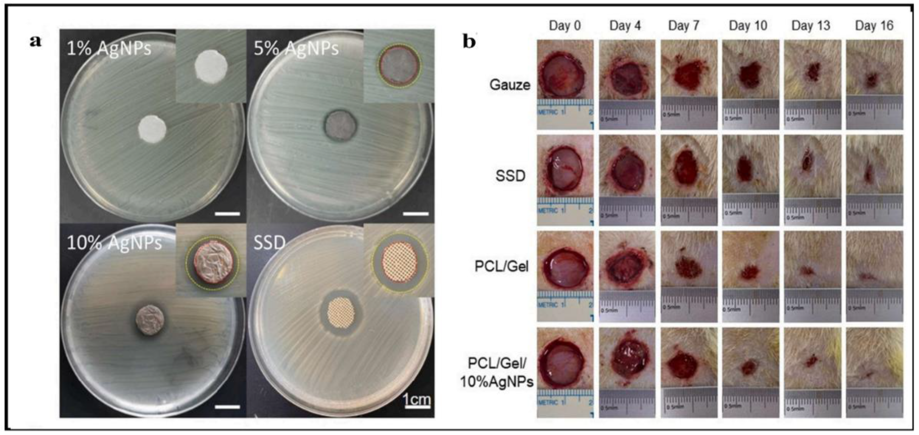
  168. He, C.; Liu, X.; Zhou, Z.; Liu, N.; Ning, X.; Miao, Y.; Long, Y.; Wu, T.; Leng, X. Harnessing Biocompatible Nanofibers and Silver Nanoparticles for Wound Healing: Sandwich Wound Dressing versus Commercial Silver Sulfadiazine Dressing. Mater. Sci. Eng. C 2021, 128, 112342. [Google Scholar] [CrossRef]
  169. Li, D.; Dong, M.; Besenbacher, F.; Huang, Y.; Chen, M. The Preparation of a Recyclable Catalyst of Silver Nanoparticles Dispersed in a Mesoporous Silica Nanofiber Matrix. RSC Adv. 2016, 6, 65613–65618. [Google Scholar] [CrossRef]
  170. Ren, S.; Dong, L.; Zhang, X.; Lei, T.; Ehrenhauser, F.; Song, K.; Li, M.; Sun, X.; Wu, Q. Electrospun Nanofibers Made of Silver Nanoparticles, Cellulose Nanocrystals, and Polyacrylonitrile as Substrates for Surface-Enhanced Raman Scattering. Materials 2017, 10, 68. [Google Scholar] [CrossRef]
  171. El-Aassar, M.R.; Ibrahim, O.M.; Fouda, M.M.G.; El-Beheri, N.G.; Agwa, M.M. Wound Healing of Nanofiber Comprising Polygalacturonic/Hyaluronic Acid Embedded Silver Nanoparticles: In-Vitro and in-Vivo Studies. Carbohydr. Polym. 2020, 238, 116175. [Google Scholar] [CrossRef] [PubMed]
  172. Sofi, H.S.; Akram, T.; Tamboli, A.H.; Majeed, A.; Shabir, N.; Sheikh, F.A. Novel Lavender Oil and Silver Nanoparticles Simultaneously Loaded onto Polyurethane Nanofibers for Wound-Healing Applications. Int. J. Pharm. 2019, 569, 118590. [Google Scholar] [CrossRef]
  173. Eghbalifam, N.; Shojaosadati, S.A.; Hashemi-Najafabadi, S.; Khorasani, A.C. Synthesis and Characterization of Antimicrobial Wound Dressing Material Based on Silver Nanoparticles Loaded Gum Arabic Nanofibers. Int. J. Biol. Macromol. 2020, 155, 119–130. [Google Scholar] [CrossRef] [PubMed]
  174. Salesa, B.; Assis, M.; Andrés, J.; Serrano-Aroca, Á. Carbon Nanofibers versus Silver Nanoparticles: Time-Dependent Cytotoxicity, Proliferation, and Gene Expression. Biomedicines 2021, 9, 1155. [Google Scholar] [CrossRef] [PubMed]
  175. Sandri, G.; Miele, D.; Faccendini, A.; Bonferoni, M.C.; Rossi, S.; Grisoli, P.; Taglietti, A.; Ruggeri, M.; Bruni, G.; Vigani, B.; et al. Chitosan/Glycosaminoglycan Scaffolds: The Role of Silver Nanoparticles to Control Microbial Infections in Wound Healing. Polymers 2019, 11, 1207. [Google Scholar] [CrossRef] [PubMed]
  176. Mohseni, M.; Shamloo, A.; Aghababaie, Z.; Afjoul, H.; Abdi, S.; Moravvej, H.; Vossoughi, M. A Comparative Study of Wound Dressings Loaded with Silver Sulfadiazine and Silver Nanoparticles: In Vitro and in Vivo Evaluation. Int. J. Pharm. 2019, 564, 350–358. [Google Scholar] [CrossRef] [PubMed]
  177. Li, Z.W.; Li, C.W.; Wang, Q.; Shi, S.J.; Hu, M.; Zhang, Q.; Cui, H.H.; Sun, J.B.; Zhou, M.; Wu, G.L.; et al. The Cellular and Molecular Mechanisms Underlying Silver Nanoparticle/Chitosan Oligosaccharide/Poly(Vinyl Alcohol) Nanofiber-Mediated Wound Healing. J. Biomed. Nanotechnol. 2017, 13, 17–34. [Google Scholar] [CrossRef]
  178. Mostafa, M.; Kandile, N.G.; Mahmoud, M.K.; Ibrahim, H.M. Synthesis and Characterization of Polystyrene with Embedded Silver Nanoparticle Nanofibers to Utilize as Antibacterial and Wound Healing Biomaterial. Heliyon 2022, 8, e08772. [Google Scholar] [CrossRef]
  179. Esmaeili, E.; Eslami-Arshaghi, T.; Hosseinzadeh, S.; Elahirad, E.; Jamalpoor, Z.; Hatamie, S.; Soleimani, M. The Biomedical Potential of Cellulose Acetate/Polyurethane Nanofibrous Mats Containing Reduced Graphene Oxide/Silver Nanocomposites and Curcumin: Antimicrobial Performance and Cutaneous Wound Healing. Int. J. Biol. Macromol. 2020, 152, 418–427. [Google Scholar] [CrossRef]
  180. Bardania, H.; Mahmoudi, R.; Bagheri, H.; Salehpour, Z.; Fouani, M.H.; Darabian, B.; Khoramrooz, S.S.; Mousavizadeh, A.; Kowsari, M.; Moosavifard, S.E.; et al. Facile Preparation of a Novel Biogenic Silver-Loaded Nanofilm with Intrinsic Anti-Bacterial and Oxidant Scavenging Activities for Wound Healing. Sci. Rep. 2020, 10, 6129. [Google Scholar] [CrossRef]
  181. Hong, S.M.; Kim, J.W.; Knowles, J.C.; Gong, M.S. Facile Preparation of Antibacterial, Highly Elastic Silvered Polyurethane Nanofiber Fabrics Using Silver Carbamate and Their Dermal Wound Healing Properties. J. Biomater. Appl. 2017, 31, 1026–1038. [Google Scholar] [CrossRef] [PubMed]
  182. Mohamady Hussein, M.A.; Guler, E.; Rayaman, E.; Cam, M.E.; Sahin, A.; Grinholc, M.; Sezgin Mansuroglu, D.; Sahin, Y.M.; Gunduz, O.; Muhammed, M.; et al. Dual-Drug Delivery of Ag-Chitosan Nanoparticles and Phenytoin via Core-Shell PVA/PCL Electrospun Nanofibers. Carbohydr. Polym. 2021, 270, 118373. [Google Scholar] [CrossRef] [PubMed]
  183. Liu, R.; Dai, L.; Si, C.; Zeng, Z. Antibacterial and Hemostatic Hydrogel via Nanocomposite from Cellulose Nanofibers. Carbohydr. Polym. 2018, 195, 63–70. [Google Scholar] [CrossRef] [PubMed]
  184. Hu, M.; Li, C.; Li, X.; Zhou, M.; Sun, J.; Sheng, F.; Shi, S.; Lu, L. Zinc Oxide/Silver Bimetallic Nanoencapsulated in PVP/PCL Nanofibres for Improved Antibacterial Activity. Artif. Cells Nanomed. Biotechnol. 2018, 46, 1248–1257. [Google Scholar] [CrossRef]
  185. Ye, J.P.; Gong, J.S.; Su, C.; Liu, Y.G.; Jiang, M.; Pan, H.; Li, R.Y.; Geng, Y.; Xu, Z.H.; Shi, J.S. Fabrication and Characterization of High Molecular Keratin Based Nanofibrous Membranes for Wound Healing. Colloids Surf. B Biointerfaces 2020, 194, 111158. [Google Scholar] [CrossRef] [PubMed]
  186. Ahmed, M.K.; Zayed, M.A.; El-dek, S.I.; Hady, M.A.; El Sherbiny, D.H.; Uskoković, V. Nanofibrous ε-Polycaprolactone Scaffolds Containing Ag-Doped Magnetite Nanoparticles: Physicochemical Characterization and Biological Testing for Wound Dressing Applications in Vitro and in Vivo. Bioact. Mater. 2021, 6, 2070–2088. [Google Scholar] [CrossRef] [PubMed]
  187. Tonda-Turo, C.; Ruini, F.; Ceresa, C.; Gentile, P.; Varela, P.; Ferreira, A.M.; Fracchia, L.; Ciardelli, G. Nanostructured Scaffold with Biomimetic and Antibacterial Properties for Wound Healing Produced by ‘Green Electrospinning. ’ Colloids Surfaces B Biointerfaces 2018, 172, 233–243. [Google Scholar] [CrossRef]
  188. Bagheri, M.; Validi, M.; Gholipour, A.; Makvandi, P.; Sharifi, E. Chitosan Nanofiber Biocomposites for Potential Wound Healing Applications: Antioxidant Activity with Synergic Antibacterial Effect. Bioeng. Transl. Med. 2022, 7, 1–15. [Google Scholar] [CrossRef]
  189. Ashraf, R.; Sofi, H.S.; Akram, T.; Rather, H.A.; Abdal-hay, A.; Shabir, N.; Vasita, R.; Alrokayan, S.H.; Khan, H.A.; Sheikh, F.A. Fabrication of Multifunctional Cellulose/TiO2/Ag Composite Nanofibers Scaffold with Antibacterial and Bioactivity Properties for Future Tissue Engineering Applications. J. Biomed. Mater. Res.-Part A 2020, 108, 947–962. [Google Scholar] [CrossRef]
  190. Hassiba, A.J.; El Zowalaty, M.E.; Webster, T.J.; Abdullah, A.M.; Nasrallah, G.K.; Khalil, K.A.; Luyt, A.S.; Elzatahry, A.A. Synthesis, Characterization, and Antimicrobial Properties of Novel Double Layer Nanocomposite Electrospun Fibers for Wound Dressing Applications. Int. J. Nanomed. 2017, 12, 2205–2213. [Google Scholar] [CrossRef]
  191. Tran, Q.H.; Nguyen, V.Q.; Le, A.T. Silver Nanoparticles: Synthesis, Properties, Toxicology, Applications and Perspectives. Adv. Nat. Sci. Nanosci. Nanotechnol. 2013, 4, 033001. [Google Scholar] [CrossRef]
  192. Amer, S.; Attia, N.; Nouh, S.; El-Kammar, M.; Korittum, A.; Abu-Ahmed, H. Fabrication of Sliver Nanoparticles/Polyvinyl Alcohol/Gelatin Ternary Nanofiber Mats for Wound Healing Application. J. Biomater. Appl. 2020, 35, 287–298. [Google Scholar] [CrossRef] [PubMed]
  193. Yan, D.; Yao, Q.; Yu, F.; Chen, L.; Zhang, S.; Sun, H.; Lin, J.; Fu, Y. Surface Modified Electrospun Poly(Lactic Acid) Fibrous Scaffold with Cellulose Nanofibrils and Ag Nanoparticles for Ocular Cell Proliferation and Antimicrobial Application. Mater. Sci. Eng. C 2020, 111, 110767. [Google Scholar] [CrossRef] [PubMed]
  194. Rath, G.; Hussain, T.; Chauhan, G.; Garg, T.; Goyal, A.K. Collagen Nanofiber Containing Silver Nanoparticles for Improved Wound-Healing Applications. J. Drug Target. 2016, 24, 520–529. [Google Scholar] [CrossRef]
  195. Zhang, S.; Ye, J.; Sun, Y.; Kang, J.; Liu, J.; Wang, Y.; Li, Y.; Zhang, L.; Ning, G. Electrospun Fibrous Mat Based on Silver (I) Metal-Organic Frameworks-Polylactic Acid for Bacterial Killing and Antibiotic-Free Wound Dressing. Chem. Eng. J. 2020, 390, 124523. [Google Scholar] [CrossRef]
  196. Ahmed, M.K.; Menazea, A.A.; Abdelghany, A.M. Blend Biopolymeric Nanofibrous Scaffolds of Cellulose Acetate/ε-Polycaprolactone Containing Metallic Nanoparticles Prepared by Laser Ablation for Wound Disinfection Applications. Int. J. Biol. Macromol. 2020, 155, 636–644. [Google Scholar] [CrossRef]
Figure 1. A diagrammatic illustration of the basic steps of cutaneous wound healing.
Polymers 14 03994 g001
Figure 2. A diagrammatic illustration of a three-dimensional (3D) structure of the skin.
Polymers 14 03994 g002
Figure 3. Biomedical applications of silver nanoparticles.
Polymers 14 03994 g003
Figure 4. Antibacterial mechanism of silver nanoparticles.
Polymers 14 03994 g004
Figure 5. Different approaches of silver nanoparticles synthesis.
Polymers 14 03994 g005
Figure 6. A schematic diagram of electrospinning apparatus.
Polymers 14 03994 g006
Figure 7. Key properties of electrospun nanofibers.
Polymers 14 03994 g007
Figure 8. Characteristics of AgNPs containing nanofibers on wound healing.
Polymers 14 03994 g008
Figure 9. (a) ATR-FTIR spectra for the BSA functionalized PANGMA nanofibers in different hydration states (of dry and wet represented by the solid and dashed lines, respectively). (b) Schematic illustration of the capturing process of the metal nanoparticle by the swollen functionalized nanofibers). (c) SEM image shows uniform distribution of metal nanoparticles across the nanofibers. Reproduced with permission [160]. Copyright 2012, Wiley-VCH. (d) Camera image of a mussel stuck onto a polymeric surface along with a simple representation of the amine and catechol groups of the dopamine building block for surface coating. Reproduced with permission [164]. Copyright 2007, Science AAAS.
Polymers 14 03994 g009
Figure 10. (a) Representative photograph showing the antibacterial zones of E. coli growth inhibition by the different dressings. (b) Photographs show the wound healing process at different time points; images reproduced with permission from [168] ©2021 Materials Science & Engineering C. Published by Elsevier Ltd.
Polymers 14 03994 g010
Figure 11. XRD patterns of AgNPs and PCL/Gel-AgNPs. Images reproduced with permission from [168] ©2021 Materials Science & Engineering C. Published by Elsevier Ltd. (Amsterdam, The Netherlands).
Polymers 14 03994 g011
Figure 12. XPS spectra of composite nanofiber: (A) full XPS spectrum of AgNPs/silica nanofiber and pure silica nanofiber, (B) Ag3d of AgNPs/silica nanofiber. Reproduced with permission [169]. Copyright 2016, Royal Society of Chemistry.
Polymers 14 03994 g012
Figure 13. FTIR spectra of the electrospun nanofibers: (I) PAN; (II) PAN/CNC/Ag; (III) PAN/CNC/Ag/Si; and (IV) p-PAN/CNC/Ag/Si. Reproduced from [170] Materials (MDPI).
Polymers 14 03994 g013
Figure 14. The SEM images of the electrospun membranes of (A) Ag0, (B) Ag1, and (C) Ag5 groups. (D) The TEM image of a single fiber from the Ag5 group, where the electron-dense areas were silver nanoparticles. In vitro interaction with PBS and FBS. (E) The SEM micro-images of membranes after being immersed in PBS and FBS. Scale bar: 1 μm; images reproduced with permission from [147] ©2019 Materials Science & Engineering C. Published by Elsevier Ltd.
Polymers 14 03994 g014
Table 2. The benefits and drawbacks of the various types of nanomaterials.
S. No.Wound Dressing TypeAdvantagesDisadvantagesRef.
1Fibers
  • nonadherent, nontoxic, nonallergenic
  • allow gaseous exchange
  • remove excess exudates
  • barrier against microbes
  • sustain release
  • maintain humidity
  • tensile strength
  • increase bioavailability
  • fibroblast attachment and proliferation
  • keratinocyte attachment and proliferation
  • tunable porosity
  • ECM mimicking
  • bio-compatibility
  • electro-catalytic properties
  • thermal conductivity
  • electrical conductivity
  • structural stability
  • loading efficiency
  • high surface area to volume ratio
  • mechanical strength
  • unsuitable for third degree, eschar, and dry wounds
  • if the wound is highly exudative, need a secondary dressing
[131]
2Membranes
  • act as physical barriers
  • membranes simulate extracellular matrix (ECM) structure
  • assure gas exchange, cell proliferation, and nutrient supply
  • the materials and solvents used in the production process may be harmful
[132]
3Films
  • impermeable to bacteria
  • allows the healing process to be monitored
  • painless removal
  • hard to handle
  • non-absorbent
  • adhere to the wound bed and cause exudate accumulation
[133]
4Hydrocolloids
  • non-adherent
  • high density
  • painless removal
  • high absorption properties
  • can be cytotoxic
  • have an unpleasant odor
  • low mechanical stability
  • maintain acidic pH at the wound site
[134]
5Hydrogels
  • high absorption properties
  • provide a moist environment at the wound site
  • water retention
  • oxygen permeability
  • ensure the solubility of growth factor/antimicrobial agents
  • weak mechanical properties
  • need a secondary dressing
[135]
6Sponges
  • high porosity
  • thermal insulation
  • sustain a moist environment
  • absorb wound exudates
  • enhance tissue regeneration
  • mechanically weak
  • may provoke skin maceration
  • unsuitable for third degree burn treatment or wounds with dry eschar
[136]
Table 3. A summary of available AgNP-based wound dressing products and their benefits.
S. No.Wound Dressing MaterialsSize of AgNPs (nm)Target MicrobeIn Vivo/In Vitro ModelAdvantage of NanocoatingRef.
1Chitosan-Poly Vinyl Pyrrolidone (PVP) composite 10–30E. coli and S. aureus L929 cell lineCompared to the control sample, silver nanocomposite reduced the amount of inflammatory cells by 99.[137]
2Plumbagin caged AgNP-collagen scaffolds60 nmE. coli and B. subtiliswistar rat/Swiss 3T6The antibacterial and wound-healing capabilities of silver and plumbagin in the PCSN cross-linked collagen scaffold showed the importance of nano-biotechnology.[138]
3Chitosan/Poly (Ethylene Oxide) matrix5E. coli-AgNPs, because of their size and structure, were found to increase antibacterial activity when introduced.[139]
4Chitin/nanosilver composite scaffolds 5 nmE. coli and S. aureusL929The scaffolds are antibacterial and have excellent blood clotting capabilities, which will help with wound healing. These scaffolds were hazardous to mouse fibroblasts in vitro. Whether in vitro cytotoxicity affects in vivo wound healing is unknown.[140]
5Activated Carbon coated silver nanocomposite50–400S. aureus, Klebsiella pneumoniae and P. aeruginosa-When compared to plain activated carbon, the Ag composites’ antibacterial activity was significantly higher.[141]
6Silver nano-coatings on cotton gauzes 100–300 nmS. aureusHaCaT/3T3The developed textile materials show promise as an alternative to traditional wound dressings due to their antimicrobial properties and biocompatibility.[142]
7Polyurethane Foam mixed Ag-NPs Dressing 100E. coli, P. aeruginosa and S. aureusHuman fibroblastWound healing was enhanced by the use of the foam dressing.[143]
8AgNP gelatin hydrogel pads7.7–10.8 nmE. coli, S. aureus P. aeruginosaHuman’s normal skin fibroblastsGelatin hydrogel pads infused with silver nanoparticles have shown promise as antibacterial wound dressings.[144]
9Chitosan-PEG hydrogel75E. coli, P. aeruginosa and S. aureusRabbitOn day 14, the dermal layer of skin and the collagen pattern were both healthy in the Ag-NPs impregnated chitosan-PEG hydrogel group.[145]
10AgNPs incorporated Pluronic F127 and Pluronic F68 thermosensitive gel -E. coli, S. aureus and P. aeruginosa-Gel may disrupt the structure of bacterial cell membranes, allowing the substance to enter the cell, where it can condense DNA, combine and coagulate with the cytoplasm, and ultimately kill the bacteria by causing the cytoplasmic component to leak out.[146]
11Chitosan nanofiber25S. aureusWistar Hannover ratsBiological media had a substantial impact on the release of silver; proteins blocked the release of the metal, whereas inorganic ions slowed it down. As a result, to elicit in vivo antibacterial activities, a high concentration of AgNPs was required.[147]
12Asymmetric Wettable Chitosan nanocomposite25E. coli, P. aeruginosa and S. aureus HEK293 cell lineThe dressing has been shown to encourage cell growth in an in vitro cytocompatibility study.[148]
13Cellulose hydrogel5–50E. coli and S. aureusNew Zealand rabbitThree days faster wound healing was seen using nanohydrogel compared to the control group.[149]
14Chitosan gels15P. aeruginosaHuman dermal fibroblastsChitosan gels containing AgNPs showed improvement in biocompatibility tests on primary fibroblasts.[150]
15Silk fibroin/ carboxymethyl chitosan composite sponge4.9 ± 1.9 nmS. aureus and P. aeruginosa-This AgNP-loaded SF/CMC sponge shows promise as a potential antimicrobial wound dressing.[151]
16Chitosan cross-linked bilayer nanocomposite45E. coli, P. aeruginosa and S. aureusL929 cell lineThe treated group’s organized and developed epithelium was a marked improvement over that of the control group.[152]
17AgNPs/Bacterial cellulose composites10–30 nmE. coli, S. aureus and P. aeruginosaEpidermal cellsIn vitro studies show that a nanostructured AgNP-BC gel-membrane has the potential to be an effective antimicrobial wound dressing with good biocompatibility for the expedited healing of scald wounds.[153]
18Silver NPs embedded bacterial cellulose gel membranes30S. aureusWestar ratsA significant amount of healing (85.92%) occurred after 14 days of treatment.[154]
19β-chitin-based hydrogels 5E. coli and S. aureusERO cell lineManufactured scaffolds showed improved whole-blood clotting ability.[155]
20Silver Alginate/Nicotinamide Nanocomposites 20–80E. coli and S. aureusMiceSignificant wound healing had occurred by the fourth day of treatment.[156]
21Hyaluronan Nanofiber25E. coli and S. aureusCell line (NIH 3T3)Since nanoparticles are so much smaller than typical particles, they are able to exert a far stronger effect on microbes.[157]
22Chitosan-Ag/ZnO composite dressing10–30 nmDrug sensitive E. coli, S. aureus and P. aeruginosaBALB/c mice /L02 cellsThese findings support the feasibility of using the prepared chitosan-Ag/ZnO composite dressing in wound care.[124]
23Chitosan-based multifunctional hydrogel250E. coli and S. aureusRat modelFollowing 14 days of therapy, the test organism showed the slowest rate of re-epithelialization.[158]
Publisher’s Note: MDPI stays neutral with regard to jurisdictional claims in published maps and institutional affiliations.

