
Development and Characterization of Novel Cellulose Composites Obtained in 1-Ethyl-3-methylimidazolium Chloride Used as Drug Delivery Systems

 , and
, and
Abstract
:1. Introduction
2. Materials and Methods
2.1. Materials
2.2. Material Manufacturing
2.3. Methods
2.3.1. FTIR
2.3.2. Mechanical Testing
2.3.3. Bioadhesion and Mucoadhesion Properties
2.3.4. Scanning Electron Micrography (SEM)
2.3.5. In Vitro Release
2.3.6. Evaluation of Antimicrobial Properties
3. Results and Discussions
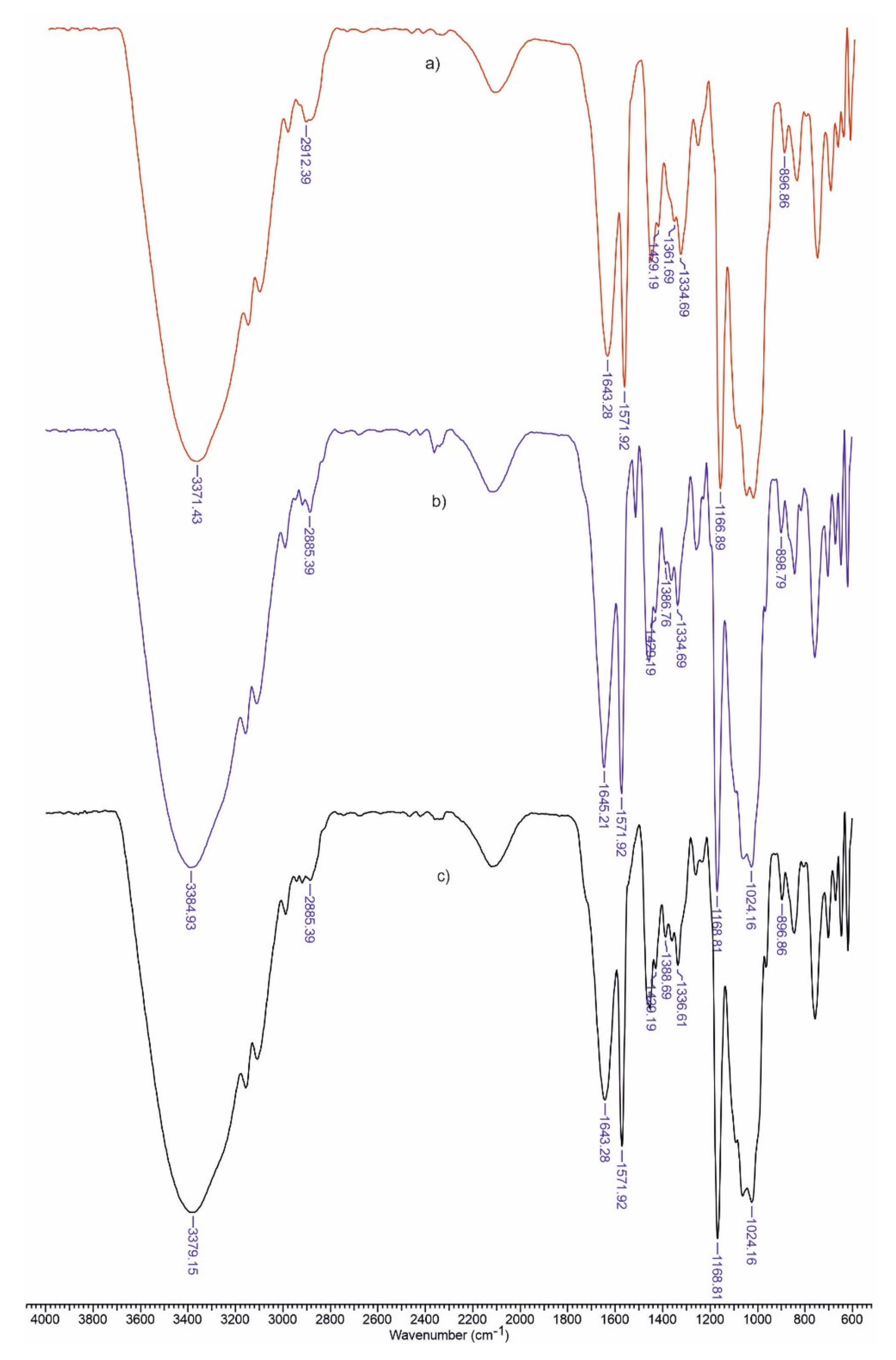
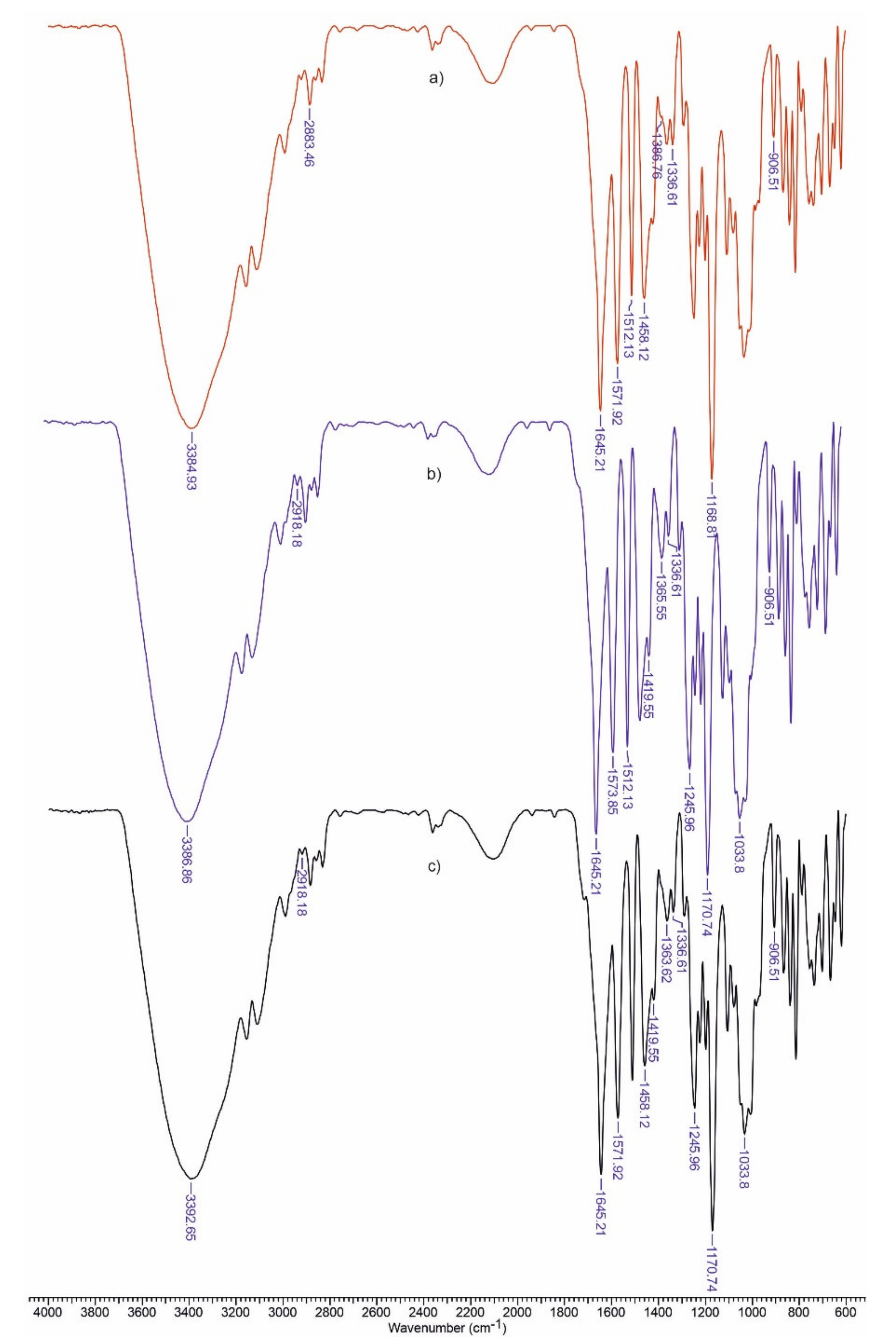
3.1. FTIR Spectra
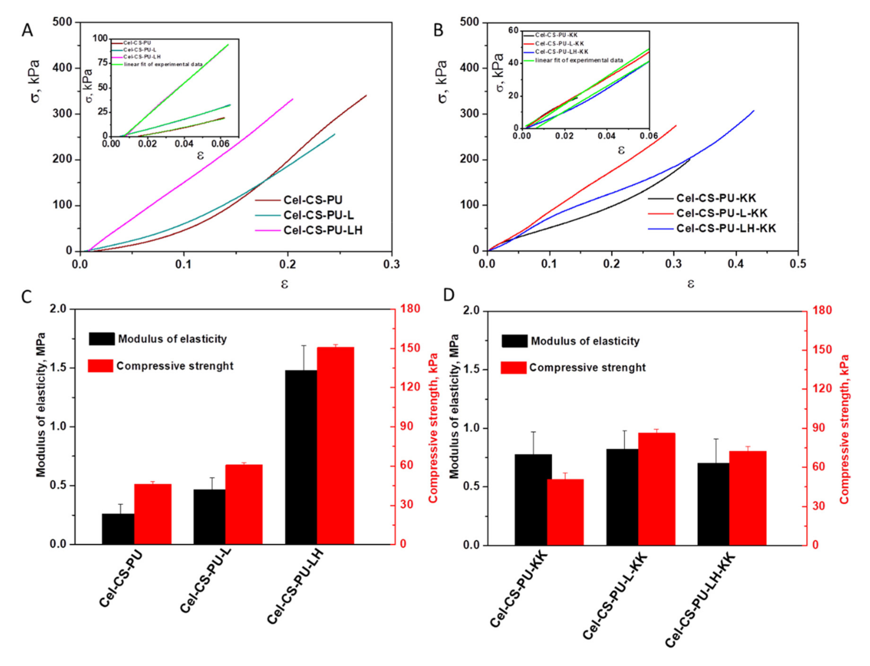
3.2. Mechanical Properties
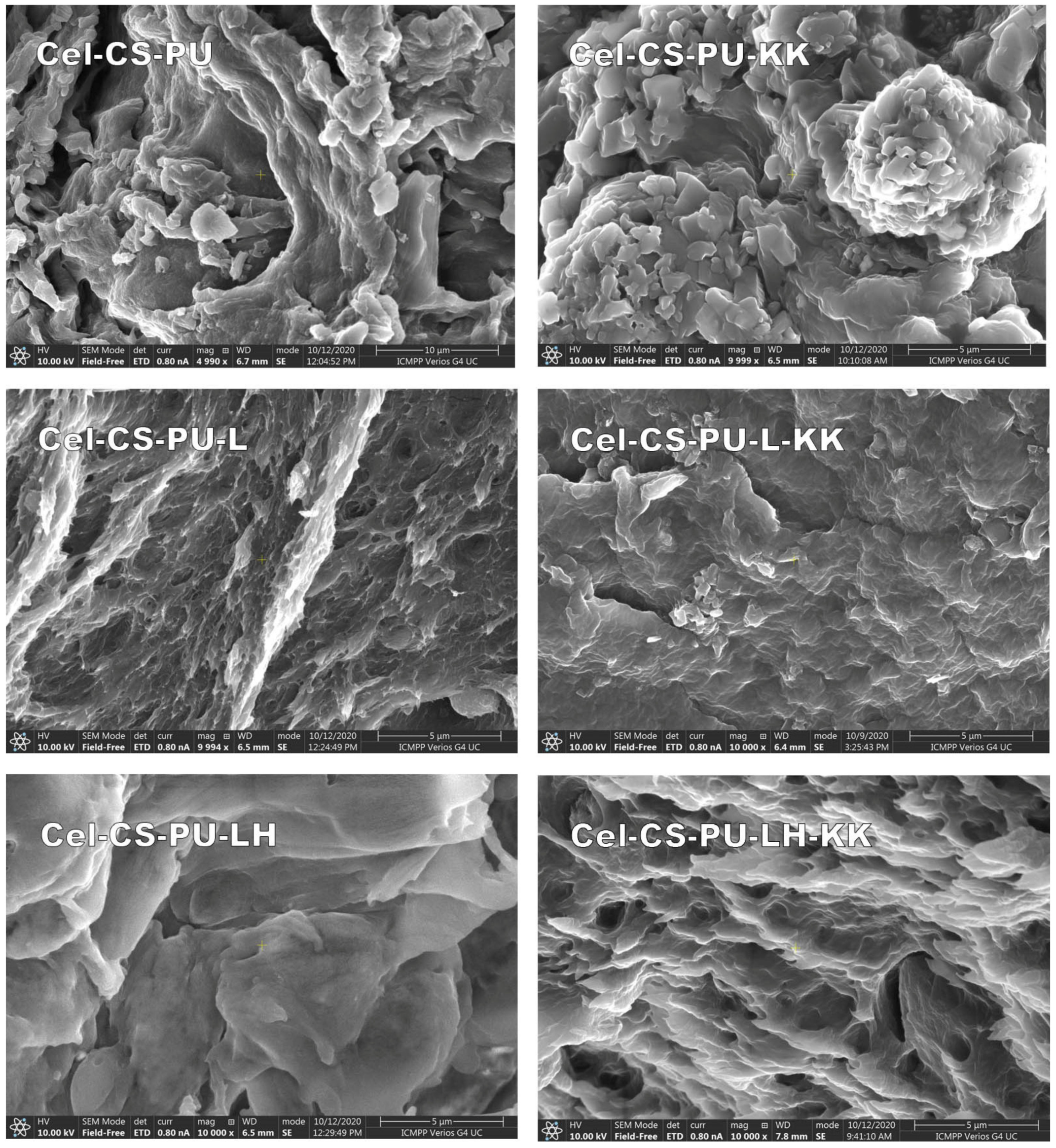
3.3. Scanning Electron Micrography (SEM)
3.4. Bioadhesivity
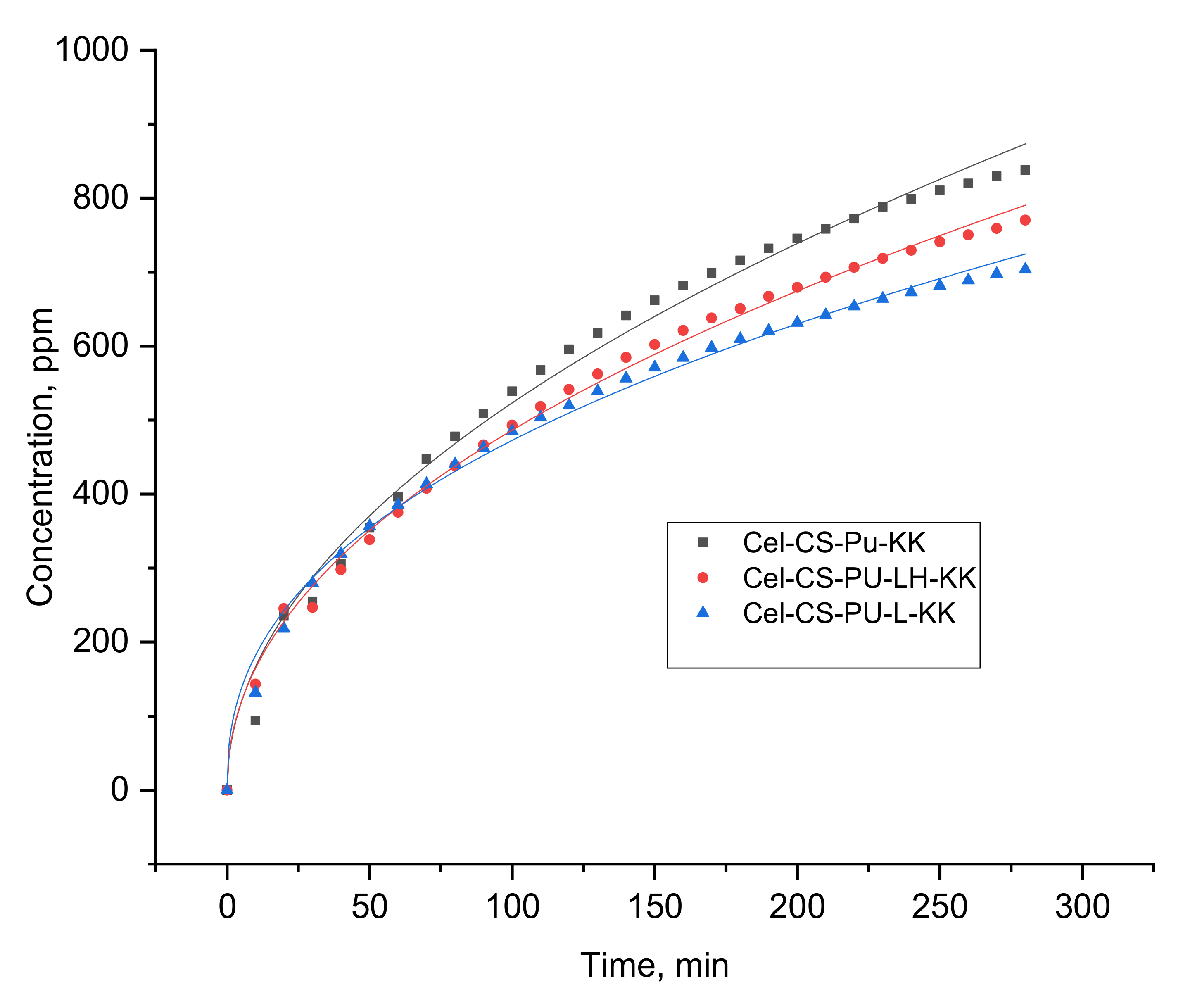
3.5. In Vitro Release
3.6. Antimicrobial Activity of the Studied Materials
4. Conclusions
Author Contributions
Funding
Institutional Review Board Statement
Informed Consent Statement
Data Availability Statement
Conflicts of Interest
References
- Michalik, R.; Wandzik, I. A mini-review on chitosan-based hydrogels with potential for sustainable agricultural applications. Polymers 2020, 12, 2425. [Google Scholar] [CrossRef] [PubMed]
- Pan, H.; Fu, C.; Huang, L.; Jiang, Y. Anti-obesity effect of chitosan oligosaccharide capsules (COSCs) in obese rats by ameliorating leptin resistance and adipogenesis. Mar. Drugs 2018, 16, 198. [Google Scholar] [CrossRef] [Green Version]
- Tapdiqov, S.Z. A drug-loaded gel based on graft radical co-polymerization of n-vinylpyrrolidone and 4-vinylpyridine with chitosan. Cellul. Chem. Technol. 2020, 54, 429–438. [Google Scholar] [CrossRef]
- Svirshchevskaya, E.V.; Zubareva, A.A.; Boyko, A.A.; Shustova, O.A.; Grechikhina, M.V.; Shagdarova, B.T.; Varlamov, V.P. Analysis of toxicity and biocompatibility of chitosan derivatives with different physico-chemical properties. Appl. Biochem. Microbiol. 2016, 52, 483–490. [Google Scholar] [CrossRef]
- Stefanescu, C.; Daly, W.H.; Negulescu, I.I. Functionalization of cellulose and chitosan in ionic liquids. Cellul. Chem. Technol. 2020, 54, 857–868. [Google Scholar] [CrossRef]
- Santamaria-Echart, A.; Fernandes, I.; Barreiro, F.; Corcuera, M.A.; Eceiza, A. Advances in waterborne polyurethane and polyurethane-urea dispersions and their eco-friendly derivatives: A review. Polymers 2021, 13, 409. [Google Scholar] [CrossRef] [PubMed]
- Zaltariov, M.F.; Filip, D.; Macocinschi, D.; Spiridon, I. Hydrohypropyl cellulose/polyurethane blends. The behavior after accelerated ageing. A FTIR study. Cellul. Chem. Technol. 2020, 54, 903–914. [Google Scholar] [CrossRef]
- Sanches, A.O.; Ricco, L.H.S.; Malmonge, L.F.; Da Silva, M.J.; Sakamoto, W.K.; Malmonge, J.A. Influence of cellulose nanofibrils onsoft and hard segments of polyurethane/cellulose nanocomposites and effect of humidity on their mechanical properties. Polym. Test. 2014, 40, 99–105. [Google Scholar] [CrossRef]
- Otto, G.P.; Moises, M.P.; Carvalho, G.; Rinaldi, A.W.; Garcia, J.C.; Radovanovic, E.; Favaro, S.L. Mechanical properties of a polyurethane hybrid composite with natural lignocellulosic fibers. Compos. Part B Eng. 2017, 110, 459–465. [Google Scholar] [CrossRef]
- Anghel, N.C.; Dinu, M.V.; Verestiuc, L.; Spiridon, I.A. Transcutaneous drug delivery systems based on collgen/polyurethane composites reinforced with cellulose. Polymers 2021, 13, 1845. [Google Scholar] [CrossRef]
- Spiridon, I. Biological and pharmaceutical applications of lignin and its derivatives: A mini-review. Cellul. Chem. Technol. 2018, 52, 543–550. [Google Scholar]
- Pishnamazi, M.; Hafizi, H.; Shirazian, S.; Culebras, M.; Walker, G.M.; Collins, M.N. Design of controlled release system for paracetamol based on modified lignin. Polymers 2019, 11, 1059. [Google Scholar] [CrossRef] [Green Version]
- Spiridon, I. Extraction of lignin and therapeutic applications of lignin-derived compounds: A review. Environ. Chem. Lett. 2020, 18, 771–785. [Google Scholar] [CrossRef]
- Figueiredo, P.; Lintinen, K.; Kiriazis, A.; Hynninen, V.; Liu, Z.; Bauleth-Ramos, T.; Rahikkala, A.; Correia, A.; Kohout, T.; Sarmento, B.; et al. In vitro evaluation of biodegradable lignin-based nanoparticles fordrug delivery and enhanced antiproliferation effect in cancer cells. Biomaterials 2017, 121, 97–108. [Google Scholar] [CrossRef]
- Erakovic, S.; Jankovic, A.; Tsui, G.C.P.; Tang, C.Y.; Miskovic-Stankovic, V.; Stevanovic, T. Novel bioactive antimicrobial lignin containing coatings on titanium obtained by electrophoretic deposition. Int. J. Mol. Sci. 2014, 15, 12294–12322. [Google Scholar] [CrossRef] [Green Version]
- Quraishi, S.; Martins, M.; Barros, A.A.; Gurikov, P.; Ramana, S.P.; Smirnova, I.; Duarte, A.R.C.; Reis, L.R. Novel non-cytotoxic alginate–lignin hybrid aerogels as scaffolds for tissue engineering. J. Supercruit. Fluids 2015, 105, 1–8. [Google Scholar] [CrossRef]
- Chieu, D.T.; Duria, S.; Delneri, A.; Franko, M. Chitosan-cellulose composite materials: Preparation, Characterization andapplication for removal of microcystin. J. Hazard. Mater. 2013, 252–253, 355–366. [Google Scholar]
- Spiridon, I.; Anghel, N.; Dinu, M.V.; Vlad, S.; Bele, A.; Ciubotaru, B.I.; Verestiuc, L.; Pamfil, D. Development and performance of bioactive compounds-loaded cellulose/collagen/polyurethane materials. Polymers 2020, 12, 1191. [Google Scholar] [CrossRef]
- Anghel, N.; Dinu, M.V.; Zaltariov, M.; Pamfil, D.; Spiridon, I. New cellulose-collagen-alginate materials incorporated with quercetin, anthocyanins and lipoic acid. Int. J. Biol. Macromol. 2021, 18, 130–140. [Google Scholar]
- Colom, X.; Carrillo, F. Crystallinity changes in lyocell and viscose-type fibres by caustic treatment. Eur. Polym. J. 2002, 38, 2225–2230. [Google Scholar] [CrossRef]
- Smart, J.D. The basics and underlying mechanisms of mucoadhesion. Adv. Drug Deliv. Rev. 2005, 57, 1556–1568. [Google Scholar] [CrossRef]
- Modi, J.; Joshi, G.; Sawant, K. Chitosan based mucoadhesive nanoparticles of ketoconazole for bioavailability enhancement: Formulation, optimization, in vitro and ex vivo evaluation. Drug Dev. Ind. Pharm. 2013, 39, 540–547. [Google Scholar] [CrossRef]
- Mandapalli, P.K.; Venuganti, V.V.K. Layer-by-layer microcapsules for pH-controlled delivery of small molecules. J. Pharm. Investig. 2015, 45, 131–141. [Google Scholar] [CrossRef]
- Alzagameem, A.; Klein, S.E.; Bergs, M.; Do, X.T.; Korte, I.; Dohlen, S.; Hüwe, C.; Kreyenschmidt, J.; Kamm, B.; Larkins, M.; et al. Antimicrobial activity of lignin and lignin-derived cellulose and chitosan composites against selected pathogenic and spoilage microorganisms. Polymers 2019, 11, 670. [Google Scholar] [CrossRef] [PubMed] [Green Version]
- Hajipour, M.J.; Fromm, K.M.; Akbar Ashkarran, A.; Aberasturi, J.D.; Larramendi, I.R.; Rojo, T.; Serpooshan, V.; Parak, W.J.; Mahmoudi, M. Antibacterial properties of nanoparticles. Trends Biotechnol. 2012, 30, 499–511. [Google Scholar] [CrossRef] [Green Version]
- Bhushan, M.; Kumar, Y.; Periyasamy, L.; Viswanath, A.K. Antibacterial applications of α-Fe2O3/Co3O4 nanocomposites and study of their structural, optical, magnetic and cytotoxic characteristics. Appl. Nanosci. 2018, 8, 137–153. [Google Scholar] [CrossRef] [Green Version]


| Code | Component (wt%) | |||||
|---|---|---|---|---|---|---|
| Cel | CS | PU | KK | L | LH | |
| Cel-CS-PU | 66.6 | 16.7 | 16.7 | - | - | - |
| Cel-CS-PU-L | 64.5 | 16.1 | 16.1 | - | 3.2 | - |
| Cel-CS-PU-LH | 64.5 | 16.1 | 16.1 | - | - | 3.2 |
| Cel-CS-PU- KK | 65.6 | 16.4 | 16.4 | 1.6 | - | - |
| Cel-CS-PU-KK-L | 63.5 | 15.8 | 15.8 | 1.6 | 3.3 | - |
| Cel-CS-PU-LH-KK | 63.5 | 15.8 | 15.8 | 1.6 | - | 3.3 |
| Sample | TCI (A1376/A2902) | LOI (A1437/A899) | HBI (A3336/A1336) |
|---|---|---|---|
| Cellulose | 1.844 | 2.174 | 5.14 |
| Cel-CS-PU | 2.349 | 1.765 | 3.161 |
| Cel-CS-PU- L | 1.159 | 1.959 | 4.511 |
| Cel-CS-PU-LH | 1.886 | 1.933 | 4.685 |
| Cel-CS-PU-KK | 1.562 | 3.093 | 5.835 |
| Cel-CS-PU-L-KK | 1.134 | 2.523 | 5.946 |
| Cel-CS-PU-LH-KK | 1.554 | 2.71 | 6.19 |
| Sample | Bioadhesivity | Mucoadhesivity | ||
|---|---|---|---|---|
| Adhesion Force (N) | Work of Adhesion (N × s) | Adhesion Force (N) | Work of Adhesion (N × s) | |
| Cel-CS-PU | 0.060433 ± 0.00247 | 0.005033 ± 0.000231 | 0.053577 ± 0.005577 | 0.009333 ± 0.001002 |
| Cel-CS-PU-L | 0.046067 ± 0.003435 | 0.003567 ± 0.000321 | 0.054833 ± 0.002589 | 0.0054 ± 0.000624 |
| Cel-CS-PU-LH | 0.04508 ± 0.003583 | 0.0026 ± 0.000721 | 0.055533 ± 0.003988 | 0.004433 ± 0.001595 |
| Cel-CS-PU-KK | 0.061767 ± 0.002589 | 0.0062 ± 0.000436 | 0.055533 ± 0.004561 | 0.0082 ± 0.000917 |
| Cel-CS-PU-L-KK | 0.063033 ± 0.001155 | 0.0057 ± 0.000361 | 0.0624 ± 0.00956 | 0.005733 ± 0.000651 |
| Cel-CS-PU-LH-KK | 0.059767 ± 0.00195 | 0.006333 ± 0.000153 | 0.050643 ± 0.009454 | 0.007867 ± 0.001739 |
| Samples | n | k, min–n | R2 |
|---|---|---|---|
| Cel-CS-PU-KK | 0.49 | 52.6 | 0.990 |
| Cel-CS-PU-LH-KK | 0.47 | 55.1 | 0.996 |
| Cel-CS-PU-L-KK | 0.41 | 69.5 | 0.993 |
| Sample | Growth Rate Inhibition (%) | ||
|---|---|---|---|
| E. coli ATCC 25922 | S. aureus ATTC 25923 | C. albicans | |
| Cel-CS-PU | 97 | 96 | 75 |
| Cel-CS-PU-L | 98 | 84 | 87 |
| Cel-CS-PU-LH | 70 | 83 | 65 |
| Cel-CS-PU-KK | 100 | 97 | 100 |
| Cel-CS-PU-L-KK | 100 | 100 | 100 |
| Cel-CS-PU-LH-KK | 93 | 97 | 94 |
Publisher’s Note: MDPI stays neutral with regard to jurisdictional claims in published maps and institutional affiliations. |
© 2021 by the authors. Licensee MDPI, Basel, Switzerland. This article is an open access article distributed under the terms and conditions of the Creative Commons Attribution (CC BY) license (https://creativecommons.org/licenses/by/4.0/).
Share and Cite
Spiridon, I.; Andrei, I.-M.; Anghel, N.; Dinu, M.V.; Ciubotaru, B.-I. Development and Characterization of Novel Cellulose Composites Obtained in 1-Ethyl-3-methylimidazolium Chloride Used as Drug Delivery Systems. Polymers 2021, 13, 2176. https://doi.org/10.3390/polym13132176
Spiridon I, Andrei I-M, Anghel N, Dinu MV, Ciubotaru B-I. Development and Characterization of Novel Cellulose Composites Obtained in 1-Ethyl-3-methylimidazolium Chloride Used as Drug Delivery Systems. Polymers. 2021; 13(13):2176. https://doi.org/10.3390/polym13132176
Chicago/Turabian StyleSpiridon, Iuliana, Iuliana-Marilena Andrei, Narcis Anghel, Maria Valentina Dinu, and Bianca-Iulia Ciubotaru. 2021. "Development and Characterization of Novel Cellulose Composites Obtained in 1-Ethyl-3-methylimidazolium Chloride Used as Drug Delivery Systems" Polymers 13, no. 13: 2176. https://doi.org/10.3390/polym13132176