Article

Antiinflammatory Medicinal Plants from the Ugandan Greater Mpigi Region Act as Potent Inhibitors in the COX-2/PGH2 Pathway

1
Institute of Biotechnology, Faculty III—Process Sciences, Technical University of Berlin, Gustav-Meyer-Allee 25, 13355 Berlin, Germany
2
Department of Agriculture and Food Sciences, Neubrandenburg University of Applied Sciences, Brodaer Str. 2, 17033 Neubrandenburg, Germany
3
Department of Plant Sciences, Microbiology and Biotechnology, Makerere University, P.O. Box 7062 Kampala, Uganda
4
ZELT—Neubrandenburg Center for Nutrition and Food Technology gGmbH, Seestraße 7A, 17033 Neubrandenburg, Germany
*
Author to whom correspondence should be addressed.
Plants 2021, 10(2), 351; https://doi.org/10.3390/plants10020351
Submission received: 31 December 2020 / Revised: 28 January 2021 / Accepted: 9 February 2021 / Published: 12 February 2021
(This article belongs to the Special Issue Research of Bioactive Substances in Plant Extracts)

Abstract

:
Our study investigates 16 medicinal plants via assessment of inhibition of proinflammatory enzymes such as cyclooxygenases (COX). The plants are used by traditional healers in the Greater Mpigi region in Uganda to treat inflammation and related disorders. We present results of diverse in vitro experiments performed with 76 different plant extracts, namely, (1) selective COX-2 and COX-1 inhibitor screening; (2) 15-LOX inhibition screening; (3) antibacterial resazurin assay against multidrug-resistant Staphylococcus aureus, Listeria innocua, Listeria monocytogenes, and Escherichia coli K12; (4) DPPH assay for antioxidant activity; and (5) determination of the total phenolic content (TPC). Results showed a high correlation between traditional use and pharmacological activity, e.g., extracts of 15 out of the 16 plant species displayed significant selective COX-2 inhibition activity in the PGH2 pathway. The most active COX-2 inhibitors (IC50 < 20 µg/mL) were nine extracts from Leucas calostachys, Solanum aculeastrum, Sesamum calycinum subsp. angustifolium, Plectranthus hadiensis, Morella kandtiana, Zanthoxylum chalybeum, and Warburgia ugandensis. There was no counteractivity between COX-2 and 15-LOX inhibition in these nine extracts. The ethyl acetate extract of Leucas calostachys showed the lowest IC50 value with 0.66 µg/mL (COX-2), as well as the most promising selectivity ratio with 0.1 (COX-2/COX-1). The TPCs and the EC50 values for DPPH radical scavenging activity showed no correlation with COX-2 inhibitory activity. This led to the assumption that the mechanisms of action are most likely not based on scavenging of reactive oxygen species and antioxidant activities. The diethyl ether extract of Harungana madagascariensis stem bark displayed the highest growth inhibition activity against S. aureus (MIC value: 13 µg/mL), L. innocua (MIC value: 40 µg/mL), and L. monocytogenes (MIC value: 150 µg/mL). This study provides further evidence for the therapeutic use of the previously identified plants used medicinally in the Greater Mpigi region.

1. Introduction

Approximately 80% of Africa’s population relies almost entirely on plants for medication [1,2,3]. The knowledge of plants in Uganda and their medicinal uses is mainly transferred orally from one generation to the next by traditional healers, leading to the potential for loss of vital information due to lack of records [4,5]. A previous ethnopharmacological study from the Greater Mpigi region documented the traditional use of 39 healers [5]. In this study, Schultz et al. described the medicinal uses of 16 plant species used in treatment of diverse medical disorders. The 16 Ugandan medicinal plant species were Albizia coriaria, Cassine buchananii, Combretum molle, Erythrina abyssinica, Ficus saussureana, Harungana madagascariensis, Leucas calostachys, Microgramma lycopodioides, Morella kandtiana, Plectranthus hadiensis, Securidaca longipedunculata, Sesamum calycinum subsp. angustifolium, Solanum aculeastrum, Toddalia asiatica, Warburgia ugandensis, and Zanthoxylum chalybeum. Another study applying the Degrees of Publication (DoP) method as a tool for literature assessment in ethnopharmacological research classified six of these 16 plant species as being “highly understudied” and three species as “understudied” [6]. This DoP analysis further strengthened the justification for conducting pharmacological lab studies, investigating these select medicinal plant species from the Greater Mpigi region. The ethnobotanical survey specifically sought to investigate the treatment of cardinal signs of acute inflammation, which is relevant to the present study. Uses documented for each species include the treatment of pain, fever, redness, heat, wounds, cancer, and general infections [5]. Figure 1 depicts the relative frequencies of citation (RFCs, n = 39) for these use reports.
Inflammation is the reaction of the immune system to injury and invading pathogens and can be considered one of the most important human host defense mechanisms [5,7,8]. The scientific pursuit of novel antiinflammatory therapeutics and drug leads, e.g., for treatment of pain, is complex and challenging [9,10]. Inflammation has also been implicated in the pathogeneses of diverse medical disorders, and over- or persistent inflammation can cause tissue damage, failure of vital organs, and death [8,11,12]. Its mediators are involved in diverse biochemical signaling pathways. One of these pathways is the cyclooxygenase-1 (COX-1) and cyclooxygenase-2 (COX-2) pathway, which plays a key role in the production of eicosanoids (Figure 2). It is also known as the prostaglandin H2 (PGH2) pathway, named after the resulting prostaglandin precursor of the COX-catalyzed reaction of arachidonic acid in the human body [7,13]. The main human cyclooxygenases, COX-1 and COX-2, are prostaglandin (PG) endoperoxide synthases (E.C.1.14.99.1) that catalyze the metabolic biosynthesis of arachidonic acid to prostanoids, encompassing potent proinflammatory signaling molecules such as prostaglandin F and prostaglandin E2 [14,15,16,17]. Each of these COX isoforms catalyzes the reaction of individual prostanoids, whereas products of COX-1 catalysis are involved in normal, homeostatic functions, such as cytoprotection of gastric mucosa, renal blood flow, macrophage differentiation, and hemostasis. These prostaglandins are also involved in regulating normal cells in general, which is why COX-1 is constitutively present in human cells. The concentration in the body generally remains stable [15,18,19,20]. The isoform COX-2, however, plays a major role in inflammatory response. While underexpressed in cells under normal conditions, COX-2 expression is upregulated during inflammation as part of the immune response, rapidly displaying elevated levels. Stimuli that induce COX-2 expression in cells can include proinflammatory cytokines (TNFα, IL-1) or growth factors [8,15,17,18,19,20]. Proinflammatory prostaglandins produced through the COX-2 pathway contribute to or induce pain, fever, and swelling, and are even implicated with types of cancer, allergy, asthma, arthritis, stroke, and Alzheimer’s disease [7,13,19,21,22,23,24,25,26,27,28,29,30,31,32].
Large-scale applied nonsteroidal antiinflammatory drugs (NSAIDs), such as ibuprofen, Paracetamol, or Aspirin, share the capacity for COX/PGH2 inhibition, thereby reducing pain, fever, and inflammation. Yet the vast majority of the NSAIDs on the market exhibit no selectivity to COX-1 and COX-2, leading to various side effects caused by inhibition of COX-1 regulated “housekeeping” functions in the body (such as ulceration and gastrointestinal bleeding) [7,20,33,34,35,36,37,38]. In the past, a few selective COX-2 inhibitors were discovered and marketed, e.g., celecoxib and rofecoxib. These drug molecules selectively inhibited COX-2 catalyzed biosynthesis of proinflammatory prostaglandins and were tremendously more potent at inhibiting COX-2 than COX-1 [38,39,40,41]. However, these selective COX-2 inhibitors (members of the diaryl heterocycle group of drug molecules) exhibited severe skin-related and cardiovascular toxicities, including myocardial infarction, leading to partial withdrawal of this class of compounds from the market [8,38,42,43]. As a result, medicinal plants have regained momentum for treatment of inflammatory diseases, making research in this particular field a hot topic [8,17,44,45,46].
Another pathway of inflammatory response in the human body is the lipoxygenase (LOX) pathway. Here, long-chain polyunsaturated fatty acids, such as arachidonic acid, are enzymatically peroxidized by lipoxygenases to the corresponding hydroperoxyl derivatives to produce eicosanoid signaling metabolites (Figure 3) [47].
While COX-2- and 5-LOX-mediated reactions generally produce prostaglandins and leukotrienes that act as proinflammatory mediators involved in pathogenesis, 12/15-LOX generates protectins and resolvins derived from n-3 polyunsaturated fatty acids, as well as lipoxins derived from arachidonic acid (n-6) [48]. A hydroperoxy cis-trans-1,3-conjugated pentadienyl moiety within the unsaturated fatty acid is the initial, unstable product of the LOX reaction [49,50,51]. Protectins, resolvins, and lipoxins are considered antiinflammatory mediators involved in the regulation of inflammatory responses and resolution of acute inflammation. Therefore, they are required in order to maintain homeostasis [48,52,53]. With regard to lipoxin, there is typically an inverse concentration between antiinflammatory lipoxin and proinflammatory leukotriene present at the site of inflammation [54]. 15-LOX is also involved in the development and progression of cancer, yet its role is complex and still controversial [52]. Due to the presence of two 15-LOX isoforms in human tumor biopsies and its implication in carcinogenesis of some cancers, the literature suggests procarcinogenic as well as anticarcinogenic roles [52,55,56,57,58,59]. Thus, discovery of 15-LOX inhibitors derived from medicinal plants may provide promising, novel, and selective therapies for certain cancers [48,56,57].
We screened up to 76 different extracts derived from these 16 medicinal plants from the Greater Mpigi region in Uganda for antiinflammatory, antioxidant, and antibacterial activity associated with the traditional use of medical disorders described in Figure 1. The main objectives of the study were (a) the pharmacological evaluation of traditional use and (b) contributing to drug discovery. Specifically, the study aims were to investigate the potential in vitro (1) human recombinant COX-2 inhibition activity; (2) human recombinant COX-1 inhibition activity; (3) 15-LOX inhibition activity; (4) free radical scavenging activity; (5) growth inhibitory activity against multidrug-resistant Listeria innocua, Listeria monocytogenes, Escherichia coli K12 and Staphylococcus aureus; and (6) to determine the total phenolic content (TPC) of the plant extracts.

2. Results

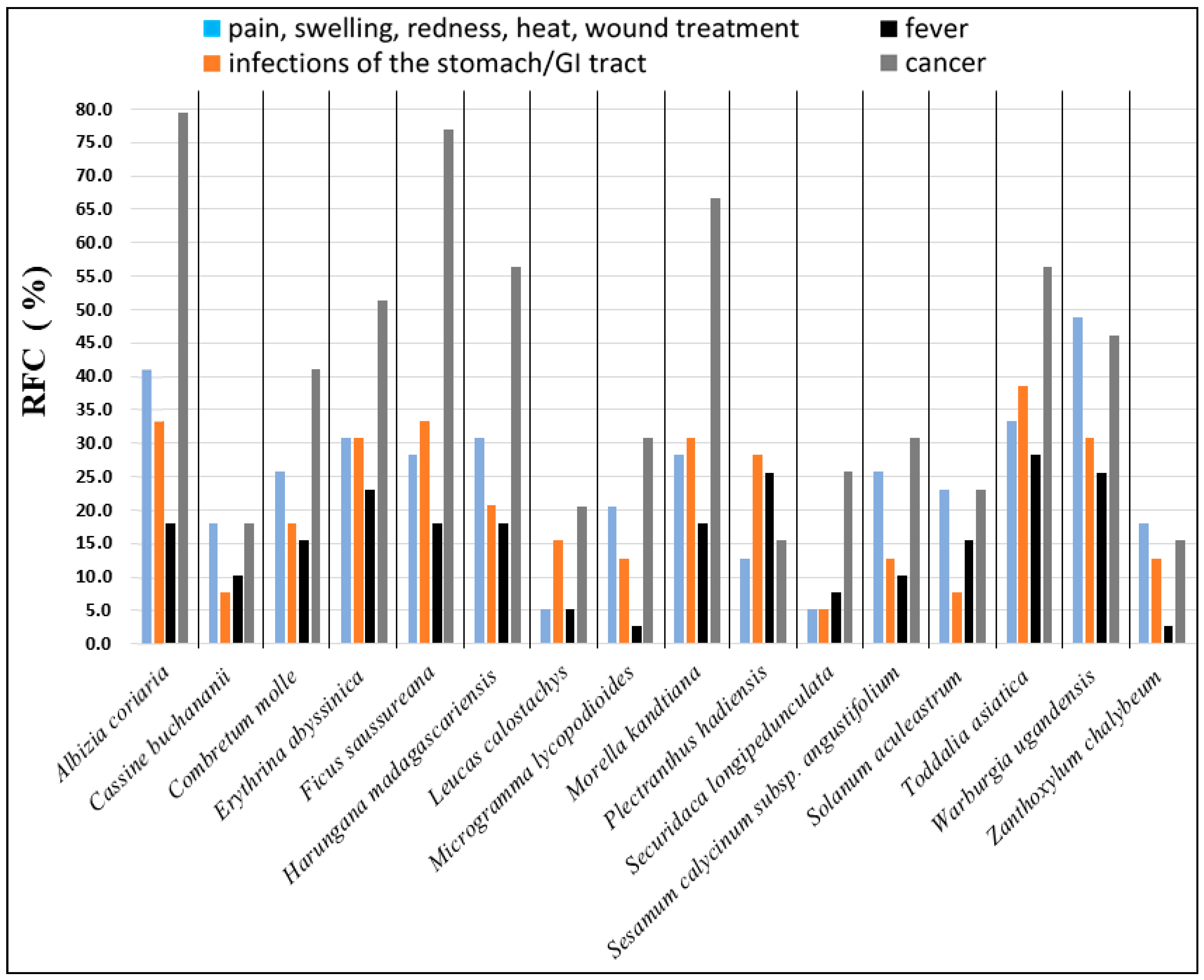
2.1. Information on Plant Species and Extractions

Table 1 shows taxonomic information on the 16 medicinal plant species studied, extract identification numbers (extract IDs), extraction solvents used, local names in the Luganda language, plant parts selected for investigation, and herbarium voucher specimen numbers and locations. Extracts were produced through different methods: (a) maceration in either methanol, ethanol, ethyl acetate or diethyl ether, (b) Soxhlet extraction using n-hexane and successively methanol, and (c) aqueous decoction, which simulated the original methods of traditional preparation [5].

2.2. Selective COX-2 Inhibition Library Screen

The plant extract library was initially screened for COX-2 inhibition activity at a concentration of 50 µg/mL. Extracts displaying a COX-2 inhibition percentage above 80 were further investigated by dose-response experiments in order to obtain IC50 values. The COX (human) inhibition assay has two steps involving a COX reaction and a PG-acetylcholinesterase (AChE) competitive ELISA for direct spectrophotometric quantification of PGF by Tin(II) chloride reduction of the PGH2 output produced in the COX reaction (Figure 4). The two distinct COX isoforms are bifunctional enzymes, displaying both COX and peroxidase activity [60]. Thus, arachidonic acid is first converted by the prostaglandin synthase active site to a hydroperoxyl endoperoxide (PGG2), and then further reduced by the peroxidase synthase active site to the corresponding alcohol (PGH2), which is the precursor for PG mediator molecules. The ELISA utilizes a broadly specific antiserum capable of binding to all major PG compounds. It is based on a PG tracer (PG-AChE conjugate) and PGs present in the sample that compete for a limited amount of PG antiserum. Since the PG concentration varies depending on the COX inhibitory activity of plant extracts tested, while the concentration of PG-AChE conjugate is constant, the concentration of PG in the sample is inversely proportional to the amount of PG-AChE conjugate that can bind to the PG antiserum.
In this initial library screen, extracts of 15 out of 16 species inhibited COX-2 at 50 µg/mL. The only exception was extracts of C. molle, which did not display any inhibitory activity on COX-2 (I = 0%). Details of the results of the prescreen are given in Supplementary Data Table S1. In total, out of 58 extracts screened, 19 extracts from nine species did not show COX-2 inhibition activity, 15 extracts from 10 species resulted in percentage inhibition of 0–40, and 15 extracts from 10 species exhibited percentage inhibition values between 40 and 80. Nine extracts from seven species were identified as particularly promising due to their high percent inhibition values (%I > 80). These were the ethyl acetate and the n-hexane extract of S. calycinum subsp. angustifolium leaves (eE004, hE004), the ethyl acetate and the n-hexane extract of S. aculeastrum root (eE006, hE006), the diethyl ether extract of W. ugandensis stem bark (dietE014), the ethyl acetate extract of L. calostachys leaves (eE005), the diethyl ether extract of Morella kandtiana root (dietE012), the diethyl ether extract of P. hadiensis leaves (dietE016), and the ethanolic extract of Z. chalybeum stem bark (etE009). These nine extracts were selected for the next stage of COX experiments and subsequently introduced to the dose-response COX-2 and COX-1 inhibition studies.

2.3. Dose-Response COX-2 Inhibition Experiments

The results of the dose-response COX-2 inhibition experiments, further investigating the most promising nine extracts from seven species identified in the library screen, are reported in Table 2.
Calculated IC50 values for these nine extracts ranged from 0.66 to 17.24 μg/mL. The ethyl acetate extract of L. calostachys leaves (eE005) displayed the highest inhibitory activity against human recombinant COX-2 in the study (IC50: 0.66 μg/mL). The second most active extract in inhibiting COX-2 was the ethyl acetate extract of S. aculeastrum root (eE006), reaching an IC50 value of 1.74 μg/mL. Further, high COX-2 inhibition activity can be reported for the n-hexane extract of S. aculeastrum root (hE006; IC50: 3.19 μg/mL) and the n-hexane extract of S. calycinum subsp. angustifolium leaves (hE004; IC50: 3.65 μg/mL). There was only one extract among the most active nine extracts that was produced using a polar extraction solvent (ethanol, etE009, Z. chalybeum stem bark), meaning that most of the extracts were apolar (extraction solvent: n-hexane) or somewhat apolar extracts (extraction solvents: diethyl ether, ethyl acetate).

2.4. COX-1 Inhibition Analysis and Selectivity Ratio Determination

The nine most active plant extracts, selected in the initial COX-2 inhibition library screen and followed up on via dose-response COX-2 inhibition studies, were further assayed to assess their potential inhibition activity against human recombinant COX-1. The calculation of the COX-2/COX-1 selectivity ratio for balance of inhibition can be used for the assessment of side effects and efficacy [61,62]. Results are given in Table 2.
All nine extracts inhibited COX-1 enzyme activity and their IC50 values ranged from 3.99 to 24.89 μg/mL. Extract eE005, which was previously identified as the strongest COX-2 inhibitor in the extract library, showed a COX-1 inhibition IC50 value of 7.76 μg/mL, leading to a calculated COX-2/COX-1 selectivity ratio of 0.1. The second most active COX-2 inhibitor, eE006, displayed moderate COX-1 inhibition activity (IC50: 7.76 μg/mL) and a COX-2/COX-1 selectivity ratio of 0.2. The analysis of extract hE004, which was previously highly active against COX-2, resulted in an IC50 value of 8.57 μg/mL and a selectivity ratio of 0.4. The most active COX-1 inhibitors among the nine extracts were hE006 (n-hexane extract of S. aculeastrum root; IC50: 3.99 μg/mL; selectivity ratio: 0.8) and dietE016 (diethyl ether extract of P. hadiensis leaves; IC50: 5.83 μg/mL; selectivity ratio: 0.8). Two extracts exhibited stronger COX-1 than COX-2 inhibitory effects. These were the diethyl ether extracts of W. ugandensis stem bark (dietE014; selectivity ratio: 1.2) and M. kandtiana root (dietE012; selectivity ratio: 1.2).

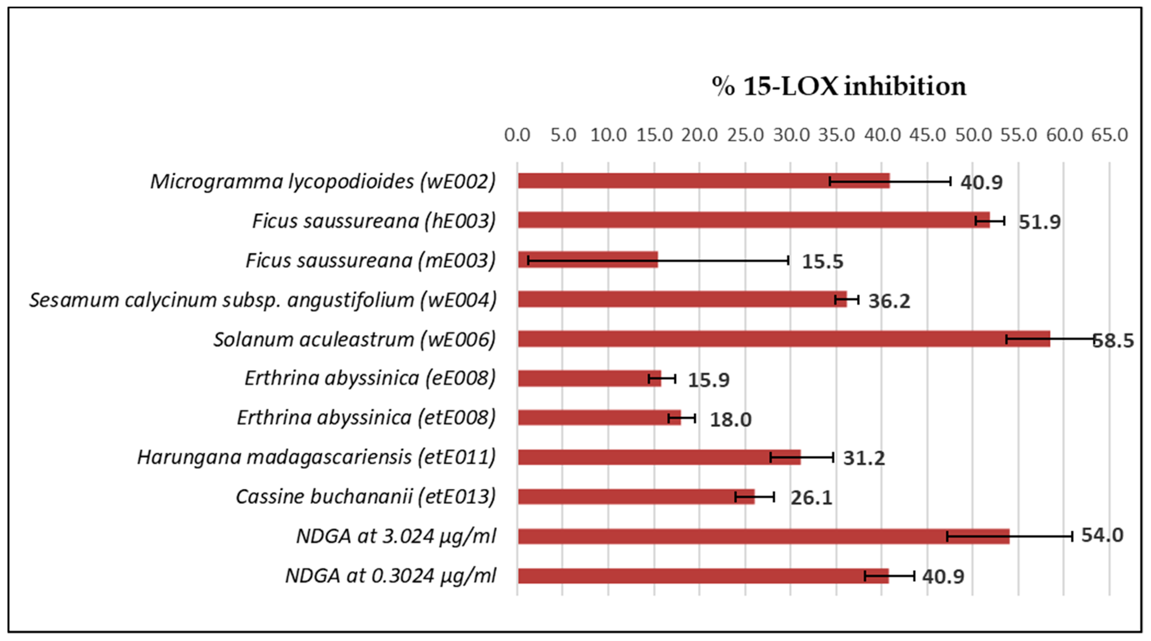
2.5. 15-LOX Inhibition Counterscreen

In an effort to estimate the 15-LOX counteractivity, the extract library, containing 58 plant extracts previously investigated for COX-2 inhibition activity, was screened at a concentration of 10 μg/mL. The results of extracts inhibiting 15-LOX enzyme activity are reported in Figure 5.
In total, only nine extracts from six plant species exhibited 15-LOX inhibition activity at 10 μg/mL, whereas 49 extracts from 16 species did not display inhibitory activity (I = 0%). These nine extracts were wE002 (aqueous extract, M. lycopodioides roots/rhizomes), hE003 and mE003 (n-hexane and methanolic extracts, F. saussureana stems), wE004 (aqueous extract, S. calycinum subsp. angustifolium leaves), wE006 (aqueous extract, S. aculeastrum roots), eE008 and etE008 (ethyl acetate and ethanolic extracts, E. abyssinica stem bark), etE011 (ethanolic extract, H. madagascariensis stem bark), and etE013 (ethanolic extract, C. buchananii stem bark). Interestingly, except for extracts hE003 and eE008, these active extracts were all polar extracts (extraction solvents: water, methanol, ethanol). Extracts with the highest 15-LOX activity at 10 μg/mL were the aqueous root extract from S. aculeastrum (I: 58.5%) and the n-hexane stem extract from F. saussureana (I: 51.9%).

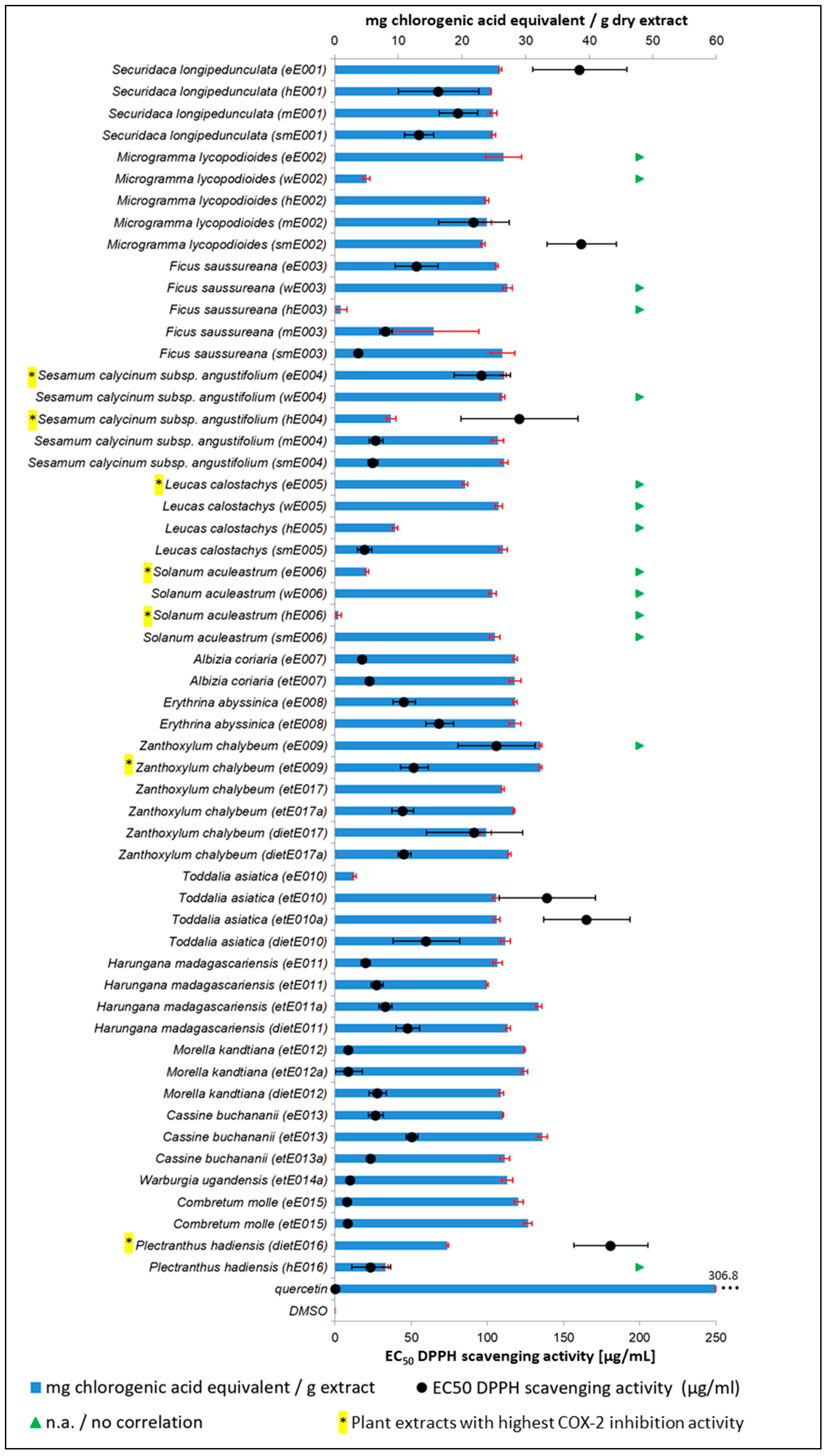
2.6. DPPH Assay for Antioxidant Activity and TPC Determination

The plant extract library was further screened for free radical scavenging potential (antioxidant activity) and the total phenolic content (TPC) was determined. Both assays were conducted to rule out a potential mechanism of action for the COX-2/1 and 15-LOX inhibition due increased presence of free radical scavenging compounds in highly active plant extracts. Many phenolic compounds, such as tannins or flavonoids, are considered to act via their free radical scavenging activities, facilitating the inhibition of proinflammatory enzymes, e.g., COX and LOX, during host immune response [63,64]. For example, reactivity with the radical trap DPPH (1,1-diphenyl-2-picrylhydrazyl radical) in the presence of each plant extract was evaluated to elucidate the potential of lipid-derived radical scavenging in the mechanism of the 15-LOX enzyme inhibition previously assessed. Results are summarized in Figure 6 and absolute values are reported in Supplementary Table S2.
The results show that there is poor correlation between the TPC and the corresponding EC50 values (antioxidant activity), which is further addressed in the Discussion section. Extracts containing the highest TPC in the extract library were etE013 (C. buchananii; 32.69 mg chlorogenic acid equivalent/g dry extract), eE009 and etE009 (Z. chalybeum; 32.29 mg chlorogenic acid equivalent/g dry extract), and etE011a (H. madagascariensis; 32.09 mg chlorogenic acid equivalent/g dry extract). Analysis of hE006 (S. aculeastrum; 0.61 mg chlorogenic acid equivalent/g dry extract), hE003 (F. saussureana; 1.01 mg chlorogenic acid equivalent/g dry extract), and eE010 (T. asiatica; 3.00 mg chlorogenic acid equivalent/g dry extract) resulted in the lowest TPCs in the library.
Interestingly, the two extracts from C. molle (the only species inactive in the initial COX-2 inhibition library screen) exhibited the lowest EC50 values for free radical scavenging activity in the library, resulting in 8.26 µg/mL (eE015) and 8.73 µg/mL (etE015). Other extracts in a similar EC50 range were etE012a and etE012 (M. kandtiana; EC50: 8.97 µg/mL and 9.03 µg/mL). The highest EC50 value for free radical scavenging potential in the extract library was recorded for COX-2 inhibitor dietE016 (P. hadiensis; EC50: 181.00 µg/mL). A total of 15 extracts did not reach an EC50 value in the tested concentration range, which included the top three performing COX-2 inhibitors eE005 (L. calostachys), eE006 and hE006 (S. aculeastrum), identified above. Except for etE009 (relatively high TPC), none of the nine extracts that exhibited high COX-2 inhibition activity showed an increased antioxidant activity or TPC compared to the other extracts in the library.

2.7. Antibacterial Resazurin Bioassay

The plant extract library was further screened for growth inhibition activity against multidrug-resistant strains of S. aureus and E. coli K12, and L. innocua (no resistances reported) to further evaluate their potential for treatment of wounds and infections of the stomach/GI tract. Extracts that were active against the non-pathogenic L. innocua were subsequently tested against multidrug-resistant pathogenic L. monocytogenes. Minimal inhibition concentration (MIC) values are reported in Table 3.
Compared to the other bacteria tested, the extracts were significantly more active against the Gram-positive S. aureus (ATCC 25923). Here, the majority of the plant extracts showed at least a minor inhibitory impact on S. aureus growth at the highest concentration tested (500 µg/mL). Thirty-one of 75 plant extracts (41.3%) displayed MIC values below 125 µg/mL. The highest growth inhibitory activity (<50 µg/mL) was recorded for 11 extracts from seven plant species: (1) the methanolic Soxhlet extract of S. aculeastrum roots (smE006, MIC: 11.72 µg/mL); (2) the diethyl ether, n-hexane, and ethanolic extracts of H. madagascariensis stem bark (dietE011, MIC: 13.02 µg/mL; hE011-18 and etE011-18, MIC: 31.25 µg/mL); (3) the methanolic extract of F. saussureana stems (mE003, MIC: 20.83 µg/mL); (4) the methanolic Soxhlet extract of M. lycopodioides roots/rhizomes (smE002, MIC: 26.04 µg/mL); (5) the n-hexane extract of S. calycinum subsp. angustifolium leaves (hE004-18, MIC: 31.25 µg/mL); (6) ethyl acetate and ethanolic extracts of T. asiatica leaves and stem bark (eE010 and etE010, MIC: 31.25 µg/mL); and (7) the diethyl ether and ethanolic extracts of W. ugandensis stem bark (dietE014 and eE014-18: 31.25 µg/mL).
The experiments screening the extract library against multidrug-resistant E. coli K12 (ATCC 23716) generally resulted in low growth inhibitory activity. None of the extracts reached a MIC below 250 µg/mL. The antibiotic screen against L. innocua (ATCC 33090) resulted in low growth inhibition activities of tested extracts. The exception was extract dietE011 (MIC: 41.67 µg/mL), which is the diethyl ether extract of H. madagascariensis stem bark and which was also the second most active extract in the S. aureus inhibition bioassays reported above. Seven extracts from four plant species were further investigated for growth inhibition activity against L. monocytogenes. The results indicate that extract dietE011 is less effective against this pathogenic strain of Listeria than against L. innocua (MIC: 125 µg/mL). Extract dietE14 (W. ugandensis stem bark extract; also one of the high-performing S. aureus growth inhibitors) showed similar activity on L. monocytogenes, reaching a MIC of 125 µg/mL.

3. Discussion and Conclusions

The results of this study provide scientific evidence for the therapeutic use of medicinal plants from the Ugandan Greater Mpigi region in treatment of inflammatory disorders and infections. Antiinflammatory (COX-2 inhibition) and antibacterial (growth inhibition of S. aureus) effects were recorded for most plant species, successfully validating traditional use in 15 out of 16 medicinal plant species investigated in the in vitro studies. The only species exhibiting no COX-2 inhibition activity in the experiments was C. molle. All 16 species displayed at least low inhibitory effects on S. aureus growth. The determination of the TPC and the assessment of the DPPH radical scavenging activity of the strongest COX-2 inhibitors led to the assumption that a high concentration of phenols and free radical scavenging seem not to play the crucial role in the mechanism of action of the most active plant extracts.
Extracts of the same species distinguished themselves in terms of method of extraction and polarity of extraction solvent used (“pre-fractionation strategy”). The most active COX-2 inhibitors in the extract library were extracts from L. calostachys (eE005), S. aculeastrum (eE006, hE006), S. calycinum subsp. angustifolium (eE004, hE004), P. hadiensis (dietE016), M. kandtiana (dietE012), Z. chalybeum (etE009), and W. ugandensis (dietE014). There was no counteractivity between COX-2 and 15-LOX inhibition in these nine extracts from seven plant species. Except for the ethanolic extract of Z. chalybeum stem bark, all of these highly active extracts were produced using an apolar or somewhat apolar extraction solvent, namely n-hexane, ethyl acetate, or diethyl ether. In general, aqueous extracts, whose lab preparation simulated the traditional methods of preparation in the Greater Mpigi region [5], often failed to exhibit bioactive effects in our antiinflammatory and antibacterial in vitro models. The present result is similar to our previous findings that investigated the extract library with other pharmacological test methods [65]. This phenomenon might be explained by the fact that the lab-produced extracts are filtered prior to solvent evaporation as part of extract standardization procedures. This led to the removal of tiny solids present in the traditional preparations, which are normally swallowed by patients along with the infused water. This way, apolar pharmacologically active secondary plant metabolites may remain in the traditional herbal remedy, but only occur in the apolar extracts in our plant extract library.
The ethyl acetate extract of L. calostachys leaves (eE005) showed the lowest IC50 value for COX-2 inhibition (0.66 µg/mL). With 0.1, this extract also displayed the most promising selectivity ratio (COX-2/COX-1). The IC50 value for COX-1 inhibition was 7.76 µg/mL. As a comparison, the IC50 values of Aspirin and ibuprofen in the literature are 210 µg/mL and 46 µg/mL (COX-2), and 5 µg/mL and 1 µg/mL (COX-1) [39,40], respectively. This leads to a poor selectivity ratio of 42 (Aspirin) and 46 (ibuprofen), which is characteristic for most commercial NSAIDs [7,36,39,40,41]. Thus, extract eE005 seems to be a much more potent COX-2 inhibitor than Aspirin or ibuprofen, while also displaying much higher selectivity for COX-2 in contrast to COX-1, thereby potentially generating fewer side effects due to decreased COX-1 and increased COX-2 inhibition. The in vitro performance of eE005 can be further highlighted by the fact that it is a crude extract containing a complex mixture of hundreds or thousands of compounds, whereas Aspirin and ibuprofen are pure substances. The TPC content of extract eE005 was not significantly higher than those of the inactive extracts in the library and no EC50 value was reached in the DPPH assay at the highest test concentration, indicating low antioxidant activity and eliminating free radical scavenging as a potential mechanism of action. Moreover, extract eE005 only exhibited low growth inhibition activity against S. aureus (MIC: 500 µg/mL) and no antibacterial effects on L. innocua and E. coli (MICs: >500 µg/mL). L. calostachys is an aromatic herb occurring in some parts of Uganda. It was recently identified by the DoP method as a “highly understudied” species [6]. In fact, not much research has been done on this species so far. Three studies reported moderate to low antiplasmodial activity of L. calostachys crude extracts [66,67,68]. The n-hexane extract of the leaves (hE005) displayed significant quorum sensing inhibition activity against the accessory gene regulator (agr) system in S. aureus [65]. Thus, our study provides the first report of strong in vitro antiinflammatory activity of L. calostachys. Other publications in the literature describe the traditional uses of L. calostachys in Kenya, which include use for the treatment of ulcers [69,70,71], colic pain in infants, cancer, skin diseases, headache, arthritis, heart diseases [69], malaria [72,73], gastrointestinal disorders [69,71,74,75,76], flu [76,77], and stomach ache [70,76]. Our data provides further evidence for some of these traditional therapeutic uses. According to the authors’ knowledge, there have been no articles published so far reporting isolation and identification of bioactive natural products from L. calostachys.
Other strong COX-2 inhibitors identified were the ethyl acetate extract of S. aculeastrum root (eE006; COX-2 IC50: 1.74 µg/mL; COX-2 IC50: 9.72 µg/mL; selectivity ratio: 0.2), the n-hexane extract of S. aculeastrum root (hE006; COX-2 IC50: 3.19 µg/mL; COX-2 IC50: 3.99 µg/mL; selectivity ratio: 0.8), and the n-hexane extract of S. calycinum subsp. angustifolium leaves (hE004; COX-2 IC50: 3.65 µg/mL; COX-2 IC50: 8.57 µg/mL; selectivity ratio: 0.4).
S. aculeastrum is a small tree or large shrub with branchlets covered in dense woolly hairs and sharp, curved thorns [6,78]. In our assessment of antioxidant activity, the extracts eE006 and hE006 displayed significantly lower TPCs than other extracts of the same extraction solvent in the extract library (5.06 and 0.61 mg chlorogenic acid equivalent/g extract), as well as no EC50 value reached in the DPPH assay. We therefore hypothesize that the mechanism of action for the COX-2 inhibition is not due to free radical scavenging and high phenol content, as often proposed for antiinflammatory medicinal plants [79,80,81,82]. Extract hE006 exhibited moderate antibacterial activity against multidrug-resistant S. aureus (MIC: 125 µg/mL) and no inhibitory activity against L. innocua (MIC: >500 µg/mL), whereas extract eE006 showed low antibacterial activity against S. aureus and L. innocua (MIC: 500 µg/mL). Interestingly, a previous study by Schultz et al. [65] identified extract eE006 as one of the two most active extracts in the library for quorum sensing inhibition (agr system in S. aureus), exhibiting reporter gene subtype-dependent IC50 values of 4, 1, 16, and 64 µg/mL (agr I-IV). This antivirulence activity was successfully confirmed via a direct protein output assessment (δ-toxin). S. aculeastrum is one of the Ugandan species that were recently classified via the DoP method as being “understudied” [6]. Published studies focus on documentation of traditional use, and pharmacological and phytochemical investigation of the berries and the leaves [6], not the roots that were investigated in this study. For instance, these include reports of low antioxidant and antimicrobial activity of the berries and leaves [83,84,85]; antiproliferative activity against human HeLa, MCF7, and HT29 tumor cell lines of methanolic berry extracts [86]; and toxicity studies of berry extracts in Wistar rats [87,88], mainly published by the Afolayan research group at Fort Hare University, South Africa. The new steroidal alkaloids solamargine, β-solamargine, solasodine and tomatidine were isolated from S. aculeastrum root bark and berries [89,90,91,92], and solamargine induced P-glycoprotein inhibition and non-selective cytotoxicity [93]. Apart from traditional uses reported from the Greater Mpigi region [5], few other publications also mentioned traditional uses of S. aculeastrum, e.g., use of the roots, berries, leaves, and bark to treat cancer in Kenya and South Africa [94,95]; use of the roots to treat stomach ache in South Africa [96]; use of the berry juice to treat ditlapedi (a facial skin condition) in South Africa [97]; and use of the berries and leaves to treat lymphatic filariasis in South Africa [98].
S. calycinum subsp. angustifolium leaves displayed high COX-2 inhibition activity in this study. This species is an erect, annual to perennial herb with spotted pink or purple flowers, reaching a height of 0.4–2.0 m. It can often be seen in Uganda along the roadside [6,99]. Yet it has also been classified as a “highly understudied” species with regard to ethnopharmacological research [6]. This is because only four publications mention the traditional use of the herb (excluding the study from the Greater Mpigi region [5]) [6]. To briefly summarize, S. calycinum subsp. angustifolium is used in the treatment of burns, wounds, eye infections, and diarrhea, and as a contraceptive and emetic in Tanzania [100]. In Uganda, it was reported to be used to treat hernias [101], to induce vomiting [102], and to treat hypertension in combination with other herbs [103]. There have been no pharmacological investigations published on this species so far, except for the antibiotic, cytotoxicity, and antivirulence study investigating the same extract library [65]. Here, extract hE004, just as extract eE006, was among the two most active agr system quorum sensing inhibitors (IC50 values: 2, 2, 16, and 32 µg/mL (agr I-IV)). The antiinflammatory activity of the n-hexane extract of S. calycinum subsp. angustifolium leaves (hE004) described in this study is the first report in the literature to date, as well as the first scientific evidence for its therapeutic use in the Greater Mpigi region of Uganda.
Phenolic compounds are often thought to possess antiinflammatory properties. The mechanisms of action of many phenolic compounds are most likely associated with their inhibition of proinflammatory enzymes in the arachidonic acid pathway (e.g., COX-2 and 5-LOX) or with their free radical scavenging activity [79,80,81,82]. Except for extract etE009 (relatively high TPC), none of the nine extracts that exhibited high COX-2 inhibition activity showed an increased antioxidant activity or calculated TPC compared to the other extracts in the library. Interestingly, the lowest EC50 value for free radical scavenging/antioxidant activity was recorded for the two extracts of C. molle (eE015, EC50: 8.26 µg/mL; etE015, EC50: 8.73 µg/mL), the only species that did not display any inhibitory activity on COX-2 in the initial library screen. These findings suggest that free radical scavenging and a high concentration of phenols in general seem not to be involved in the mechanism of action of the most active COX-2 inhibiting plant extracts. In theory, there is a direct relationship between the TPC and the free radical scavenging activity because phenols significantly contribute to the antiradical activity [104]. The poor correlation between high TPC and low EC50 values reported for some samples may be attributed to the different quality of phenols present in the samples, resulting in varying antioxidant activities. Constituents other than phenols, such as carbohydrates, fatty acids, phospholipids, etc., may also play a role in the antioxidant activity of the sample. Correlations between the phytochemical composition of the plant species and their bioactive properties should be further investigated by more advanced methods of analytical chemistry, such as LC-MS-MS profiling, enabling identification of specific sets of molecules present in the extract.
In the antibiotic resazurin bioassay, the diethyl ether extract of H. madagascariensis stem bark (dietE11) displayed high growth inhibition activity against S. aureus (MIC value: 13 µg/mL), L. innocua (MIC value: 42 µg/mL), and L. monocytogenes (MIC value: 125 µg/mL). H. madagascariensis, the “orange-milk tree,” is an evergreen shrub or tree whose sap is orange and turns blood-red upon exposure [105,106,107]. It is not considered an understudied species because it has been extensively studied in the past [6]. Traditional use has been reported in many regions of the African continent [73,108,109,110,111,112].
Another plant extract exhibiting strong growth inhibitory effects against S. aureus was the diethyl ether extract of Z. chalybeum stem bark (dietE017a; MIC: 13 µg/mL). This species is a spiny deciduous tree or shrub, reaching heights of about 8 m. It occurs in dry woodland, bushland, or grassland in medium to low altitudes throughout Uganda (up to 1500 m.a.s.l.) [105,106,107]. According to the DoP analysis [6], Z. chalybeum is regarded a “moderately studied species.” None of the traditional healers were cited to use this medicinal plant as a remedy for skin infections. However, 18% of the survey participants stated that it is used for disinfection of wounds, as well as treatment of sore throat (8%) and disorders of the stomach/GI tract (13%) (Figure 1 and [5,65]). The present data, therefore, supports the traditional use of Z. chalybeum stem bark in the study area.
A polar extract of S. aculeastrum root (smE006; methanolic Soxhlet extraction) displayed the highest antibacterial activity against S. aureus with a MIC of 12 µg/mL (species discussed above). The results of the antibiotic resazurin assay against multidrug-resistant S. aureus strongly support the traditional medicinal use of S. aculeastrum, H. madagascariensis, and Z. chalybeum in treatment of wounds and infections.
Another bioactive medicinal plant that inhibited the growth of S. aureus, L. innocua, and L. monocytogenes is S. longipedunculata. This species is mentioned in the literature in connection with treatment of measles [113]. In Uganda, measles is one of the major diseases responsible for fatalities in children. Pneumonia is the most common severe complication, leading to the most measles-associated deaths. It can be caused by the measles virus alone, secondary viral infection, or also secondary bacterial infections. Here, S. aureus is the most abundant organism of secondary bacterial infections [114]. In parts of Uganda, the roots of S. longipedunculata are prescribed by traditional healers to control secondary bacterial and measles infections [115].
The same plant extract library was previously screened in another study against a panel of multidrug-resistant ESKAPE pathogens, in which we used a different method to test antibacterial growth inhibition [65]. The bacterial species and strains tested also varied, however, the S. aureus (UAMS-1 strain) can be compared to the S. aureus strain (ATCC 25923) used in this study. Generally, some MIC values differed. However, many values were confirmed by the resazurin bioassay in our study, e.g.:
  • extract hE005-18 (L. calostachys, MICUAMS-1: >256 µg/mL, MIC25923: 500 µg/mL);
  • extract hE006 (S. aculeastrum, MICUAMS-1: 128 µg/mL, MIC25923: 125 µg/mL);
  • extract etE008 (E. abyssinica, MICUAMS-1: 64 µg/mL, MIC25923: 63 µg/mL);
  • extract etE011-18 (H. madagascariensis, MICUAMS-1: 32 µg/mL, MIC25923: 31 µg/mL);
  • extract hE011-18 (H. madagascariensis, MICUAMS-1: 32 µg/mL, MIC25923: 31 µg/mL); and
  • extract etE013 (C. buchananii, MICUAMS-1: >256 µg/mL, MIC25923: 500 µg/mL).
For some extracts, approximately one additional two-fold dilution could be reported as MIC value against S. aureus:
  • extract dietE011 (H. madagascariensis, MICUAMS-1: 32 µg/mL, MIC25923: 13 µg/mL);
  • extract dietE017a (Z. chalybeum, MICUAMS-1: 32 µg/mL, MIC25923: 13 µg/mL);
  • extract dietE014-18 (W. ugandensis, MICUAMS-1: 64 µg/mL, MIC25923: 31 µg/mL); and
  • extract hE014-18 (W. ugandensis, MICUAMS-1: 64 µg/mL, MIC25923: 31 µg/mL).
Extract smE006, which was the strongest S. aureus growth inhibitor in this study, did not display antibiotic effects against the UAMS-1 strain [65]. A similar result was obtained for extract dietE10 (T. asiatica). This significant discrepancy can most likely be explained by the different resistance profiles of the two strains investigated. The results of this study also showed that the antibacterial potential of medicinal plants depends on the species, which plant part is used, the time and location of harvest, and on the solvents used for extraction.
The results of this study, reporting pharmacological effects of medicinal plants on inflammatory enzyme cascades and growth of bacterial pathogens, could be the starting point for subsequent studies to investigate potential leads for the development of potent antiinflammatory drugs or antibiotics. Further work is required to characterize the extracts phytochemically in order to identify compounds responsible for the antiinflammatory, antioxidant, and antibacterial properties of the plant species. This could be achieved, for instance, via bioassay-guided fractionation experiments and investigation of the mechanisms of action. However, these effects might also be due synergistic relationships of multiple active ingredients within the plant extracts. To assess the plant species’ potential for drug discovery endeavors, future research should also include evaluation of toxicity (e.g., cytotoxicity and genotoxicity) and in vivo studies. Regarding future in vivo studies and for more accurate validation of traditional use, it will also be essential to continue including the original preparation cited by the traditional healers in the experimental setup, as well as their route of administration and dose. Extracts that displayed strong antibacterial activity need to be further investigated regarding their ability to limit the severity of disease, as well as their potential of increasing the efficacy of conventional antibiotics (that pathogens may have acquired resistance to already). Future studies should therefore also focus on the deactivation of other virulence pathways, such as secretion systems and biofilm formation.

4. Materials and Methods

4.1. Ethnobotanical Data

Ethnobotanical information on the traditional use of the 16 plant species for treatment of inflammatory disorders was obtained from a previously published survey among 39 traditional healers in the Greater Mpigi region in Uganda [5]. Traditional use reports from this study served as the basis for antiinflammatory, antioxidant, and antibacterial experiments.

4.2. Collection and Identification of Plant Material

Following standard collection procedures [116,117,118] and under guidance of the traditional healers, plant specimens were collected during fieldwork in 2015, 2016, and 2017. For all collected samples of species, voucher specimens were prepared and deposited at the Makerere University Herbarium in Kampala, Uganda. Additional select specimens were deposited at the Emory University Herbarium (GEO) in Atlanta, GA, USA, which are also digitally available on the SERNEC portal [119]. Voucher specimen numbers are given in the Table 1. Plant identification and assignment of scientific names (cross-checked with http://www.theplantlist.org) (accessed on 11 August 2019) were conducted following the best practice in the field of ethnopharmacology [120]. Assignments of plant family correspond to The Angiosperm Phylogeny Group IV guidance [121].

4.3. Extractions

Collected plant samples were dried in the shade, taken to the laboratory, and ground. Extractions were performed as described in detail in the flow sheet of the Supplementary Material Figure S2 of a previous publication [65]. Briefly, methods applied to extract plant samples were either maceration, aqueous decoction, or Soxhlet extraction. Extraction procedures were conducted using different solvents, aiming to achieve selective extraction of biomolecules of different polarities from the samples. Individual crude extract samples were labeled according to their extraction solvent and the collection number (EXXX) assigned to a plant species during the field studies, ranging from E001 to E017. Regarding the maceration procedure, the extraction solvents used were (a) methanol (mEXXX), (b) ethanol (etEXXX), (c) ethyl acetate (eEXXX), and (d) diethyl ether (dietEXXX). According to the ethnobotanical survey [5], traditional healers usually prepare their herbal drugs as aqueous decoctions. In order to simulate this original method of preparation, plant samples were also boiled in (e) water at 95 °C for 30 min while being stirred (wEXXX). Soxhlet extraction crude extracts were extracted using (f) n-hexane (hEXXX) or (g) methanol (smEXXX; successive extraction of corresponding EXXX material). Some of the plant species had to be collected again in 2018 due to the requirement to obtain higher amounts of extract, e.g., for future bioassay-guided fractionation strategies. Their resulting extracts were additionally labeled with “−18” in their sample ID.

4.4. Sample Preparation

Crude extracts were dissolved in DMSO (Carl Roth) at 10 mg/mL. Sonication and temperature increase up to 55 °C were applied for some samples with moderate or low solubility experienced at RT. Extract solutions were stored at −20 °C until assaying.

4.5. COX-1/2 Inhibition Screening Assays

First, an initial COX-2 inhibition library screen was performed at 50 μg/mL. Extracts exhibiting a COX-2 inhibition value above 80% were introduced to the COX-2 and COX-1 dose-response studies.
Materials and chemicals for the COX inhibition screening assays were sourced from Cayman Chemical, Ann Arbor, MI, USA (Cayman Item No. 701070-96 and 701080-96). This assay for assessment of human recombinant cyclooxygenase inhibitory potential of plant extracts was divided into two steps (described in the following paragraphs): (1) the COX reaction step and (2) the ELISA step. The ELISA step was performed to quantify the prostaglandin product generated in the COX reaction step.
COX reaction step: 5 mL reaction buffer (Cayman Item No. 460104) was mixed with 45 mL ultrapure water (COX buffer). 80 μL human recombinant COX-2 (Cayman Item No. 460121) or COX-1 (Cayman Item No. 460108) solution were diluted with 320 μL of COX buffer and stored on ice, resulting in a quantity sufficient for 40 COX reactions (COX solution). To obtain a heme solution necessary for the reaction, a commercial heme in DMSO solution (Cayman Item No. 460102) was further diluted (40 μL with 960 μL of COX buffer; stable at room temperature for 12 h). 50 μL of arachidonic acid in ethanol (Cayman Item No. 460103) were mixed with 50 μL of 0.1 M potassium hydroxide, vortexed, and further diluted with 400 μL of ultrapure water (final concentration of substrate solution: 2 mM; to be used within 1 hour if kept on ice). 5 mL of 1 M hydrochloric acid were added to a crystalline stannous chloride vial (Cayman Item No. 460107) and vortexed to obtain a saturated solution, which was stable for 8 hours at RT. The selective COX-2 inhibitor compound DuP-769 was used as a positive control. The COX reaction procedure is described in Supplementary Table S4.
Briefly, the background tubes were used to generate the background values. These two tubes contained an inactivated COX solution (10 μL; produced by placing a 500 μL microfuge tube containing 20 μL COX solution in boiling water for 3 min), 160 μL of COX buffer, and 10 μL of heme solution. The sample (or positive control) tubes and two COX 100% initial activity tubes were prepared by adding 160 μL of COX buffer, 10 μL of heme solution, and 10 μL COX solution. 10 μL of DMSO (sample vehicle) were added to each background and COX 100% tube. 10 μL of sample or positive control solution were then transferred to each sample tube. All tubes were incubated for 10 min at 37 °C. Initiation of the COX reaction was completed by adding 10 μL of arachidonic acid substrate solution and incubating for exactly 2.00 min at 37 °C (final substrate concentration in the reaction: 100 μM). The enzymatic reaction was stopped through the addition of 30 μL of saturated stannous chloride solution and the tubes were further incubated for 5 min at RT. The tubes were then tightly capped and stored at 4 °C for up to 3 days (produced PG F is stable for one week). A more detailed description of the assay procedure is available [122].
ELISA step: PG F was then quantified via Cayman Chemical’s Prostaglandin Screening AChE ELISA kit (Cayman Item No. 514012). The AChE competitive ELISA procedure was performed according to the manufacturer’s instructions [122,123]. Briefly, the COX reaction tubes, containing prostanoids, were diluted in ELISA assay buffer (1:2000 and 1:400 dilutions were run in the ELISA). A prostaglandin standard screening solution was freshly prepared and diluted (twofold), ranging from 2000.0–15.6 pg/mL. Each plate setup contained a minimum of two blanks, two non-specific binding wells, two maximum binding wells, one total activity well, an eight-point standard curve run in duplicate, and the COX 100% activity and COX reaction samples at 1:2000 and 1:4000 dilution in duplicate, respectively. Samples and controls were transferred to pre-coated mouse monoclonal anti-rabbit IgG antibodies. After the addition of a PG-AChE tracer to each well (except for the total activity and the blank wells) and a specific PG antiserum, the plate was incubated for 18 hours at room temperature on a rotary microtiter plate shaker. After incubation, the wells were emptied and rinsed five times with wash buffer in order to remove all unbound reagents. The plate was developed by adding Ellmann’s reagent (AChE substrate) to all wells, as well as addition of the tracer to the total activity wells, and shaking on a rotary microtiter plate shaker in the dark for 75 min. The yellow color of the reaction product of AChE was then measured spectrophotometrically at 412 nm. The intensity is proportional to the amount of PG tracer bound to the well (determined from the PG standard curve from the plate), thus inversely proportional to the quantity of free PG present in the well (Figure 4).
The calculation of % COX inhibition values was performed using the Cayman Chemical’s Workshop Sheets Excel template (Version 11 October 2011). IC50 values were calculated using the GraphPad Prism 9.0.0 software and a log(inhibitor) vs. response—variable slope (four parameters) model with duplicate and triplicate determinations. Error propagation was performed as described by the software manufacturer [124] and in the literature [125].

4.6. 15-LOX Inhibition Assay

The extract library previously screened for COX-2 inhibition activity was subsequently counterscreened for 15-LOX inhibition activity at 10 μg/mL. The 15-LOX inhibition assay was performed using a lipoxygenase inhibitor screening assay kit, manufactured by Cayman Chemical, Ann Arbor, MI, USA (Cayman Item No. 760700). This 96-well microtiter plate-based method utilizes the 15-LOX catalyzed enzymatic reaction between a polyunsaturated free fatty acids with a cis,cis-1,4-pentadiene-type structure and molecular oxygen (Figure 7). In the assay, the hydroperoxides, namely 12(S)-hydroxyeicosatetraenoic acid (12(S)-HpETE) and 15(S)-hydroxyeicosatetraenoic acid (15(S)-HpETE), are detected and measured.
Purified soybean 15-LOX (Cayman Item No. 760714) was used to facilitate lipoxygenation (final concentration in each well: 200 U/mL). Arachidonic acid was selected as the substrate (final concentration in each well: 125 mM). Nordihydroguaiaretic acid (NDGA), a non-selective LOX inhibitor, was used as a positive control (Cayman Item No. 760717). The assay was performed according to the manufacturer’s instructions [126] and as previously described [127,128,129,130]. Briefly, individual plant extract solutions were incubated with LOX assay buffer containing 15-LOX for 5 min at RT. Three blank control wells, three 100% initial activity wells, three negative control wells, and NDGA positive control wells were also included in each plate setup. After the addition of the substrate and initiation of the reaction, the uncovered plate was placed on a shaker at 500 rpm for 20 min. After incubation, chromogen was rapidly added to stop enzyme catalysis and the plates were covered and placed on a shaker at 500 rpm for 5 min to develop the reaction. The absorbance at 495 nm was then read using a plate reader.
All experiments were conducted in triplicate. During data analysis, the average absorbance of the blank, 100% initial activity, positive control, and sample wells were determined. After subtraction of the average blank absorbance from the average 100% initial activity and sample wells, the % inhibition for each sample was calculated using the following equation:
% inhibition = ((100% initial activity − sample)/100% initial activity) * 100

4.7. DPPH Assay

A quantitative DPPH assay for free radical scavenging activity (antioxidant potential) of plant extracts was conducted as previously described [131,132,133]. The 96-well microtiter plates were incubated in the dark for 30 min at RT. Absorbance was measured at 517 nm via UV-vis spectrophotometer. All plant extracts were assayed at 500 µg/mL in their initial well and double-fold diluted down to 0.24 µg/mL. Quercetin was used as a positive control and DMSO as the vehicle control. All experiments were performed in triplicate. The % inhibition was calculated with the following equation:
% inhibition = ((absorbanceblank − absorbancesample)/absorbanceblank) * 100
The EC50 values were calculated via linear regression using Microsoft Excel®.

4.8. TPC Determination

Determination of the total phenolic content (TPC) of plant extracts was performed in 96-well microtiter plates as previously described [134]. Briefly, Folin–Ciocalteu reagent was used and a standard curve with chlorogenic acid (CHA) was prepared (serial dilution ranging from 100 µg/mL (or 282.24 µM) to 0.049 µg/mL (or 0.1383 µM)). Spectrophotometric measurement was conducted at 765 nm. Quercetin was used as a positive control and DMSO as a vehicle control. All experiments were performed in triplicate. For data analysis, the CHA standard curve was plotted and linear regression applied using the software GraphPad Prism 9.0.0. After subtracting plant extract blank absorbances from the sample absorbance, the results were interpolated in the standard curve to determine the equivalent CHA concentration for each extract.

4.9. Bacterial Strains

In order to realistically evaluate the extracts’ potential for future drug discovery advances for antimicrobial resistance threats, multidrug-resistant isolates of three bacterial species, (a) E. coli K12 (ATCC 23716), (b) S. aureus (ATCC 25923), and (c) L. monocytogenes (ATCC 15313), were selected for experiments. As a prescreen, a non-pathogenic L. innocua strain (ATCC 33090) was used as a substitute (no resistances reported), and hits were followed up with the human pathogen L. monocytogenes. Antibiotic resistance profiles, strain numbers and characteristics, and sources are reported in Supplementary Table S3. All strains were streaked from freezer stock and maintained on tryptic soy agar (TSA) via overnight incubation at 37 °C. Overnight liquid cultures were prepared in Mueller-Hinton (MHB, S. aureus, E. coli) or brain heart infusion (BHI) broth at 37 °C with constant shaking at 200 rpm. For S. aureus, a bacterial growth curve was generated, allowing for determination of the growth phase and CFU/mL for standardization. After incubation of the overnight culture, 1.0 mL of the bacteria culture was taken and pipetted into a sterile flask containing 30 mL of broth. After 5–6 h of incubation, the bacteria were in the exponential phase according to the growth curve. In order to get rid of the preculture broth medium, 20 mL of the bacterial culture were centrifuged at 4000 rpm for 5 min. The supernatant was discarded, and the bacterial pellet was resuspended in 20 mL of sterile saline. After vortexing, the suspension was centrifuged again under the same conditions as stated above. Those steps were repeated three times until the supernatant was clear. The pellet was once again resuspended in 20 mL sterile saline and the optical density (OD) was measured at 600 nm. The OD of the culture and the growth curve were used to calculate the dilution factor to achieve a final concentration of 5 × 10⁶ CFU/mL for the resazurin bioassay. As a control and to ensure correct bacterial concentration in the assay, the bacterial suspension was additionally spread onto TSA plates, incubated overnight at 37 °C, and colonies were counted. The other three strains were standardized via cell counting in a Thoma chamber, followed by calculation of the cell concentration, calculation of the correct dilution factor, and dilution.

4.10. Resazurin Bioassay

An in vitro 96-well microtiter plate-based antibacterial bioassay, incorporating resazurin as a colorimetric indicator of cell growth, was used to assess the growth inhibitory effects of plant extracts against tested bacterial pathogens. The method was previously described by Sarker et al. [131,135]. The plates were labelled as shown in Supplementary Material Figure S1. Sterility and vehicle/growth controls, as well as a positive control (ciprofloxacin), were incorporated on each microtiter plate of the bioassay. A schematic description of the bioassay procedure is given in Supplementary Material Figure S2. Briefly, 50 µL of sterile saline was pipetted in all wells, except for the first row (20 µL of sterile saline). Next, (a) in column 1 to 4 80 µL of extract solution, (b) in the two X columns 80 µL of sterile saline (sterility control), (c) in column Y 80 µL of DMSO (vehicle control), and (d) in column Z 80 µL of ciprofloxacin (10 µg/mL) as a positive control were added. A serial dilution from row 1 to row 12 was performed. After each dilution step, the pipette tips were discarded. A total of 30 µL of 3.3 x strength MHB and 10 µL of resazurin solution (0.05% (w/v) resazurin sodium salt in sterile ultrapure water) were added to all wells. Except for column X, all wells were inoculated with 10 µL of standardized bacterial suspension, resulting in a final bacterial concentration of 5 × 10⁵ CFU/mL in the wells. An amount of 10 µL of sterile saline was added to the sterility control (column X). The plate was sealed with a microtiter plate foil to prevent draining and the plate was shaken on an orbital shaker at 500 rpm for 5 min. The plate was then incubated in the dark for 18 h at 37 °C. After incubation, the MIC was determined by visual assessment of the color change. Any color change from blue to pink was recorded as negative, indicating bacterial growth. Blue was interpreted as inhibition of growth by the individual plant extract (Supplementary Material Figure S1). The lowest concentration at which color change occurred was taken as the MIC value. All bacterial experiments were performed as biological triplicates.

Supplementary Materials

The following are available online at https://www.mdpi.com/2223-7747/10/2/351/s1, Table S1: Results of the initial COX-2 extract library screen at 50 µg/mL, Table S2: Detailed data of the TPC determination and the DPPH assay, Table S3: Information on bacterial strains used in the study, Table S4: Procedure of the COX reaction step of the COX inhibition library screening assay, Figure S1: Plate layout and setup during the resazurin bioassay, Figure S2: Schematic description of the resazurin bioassay for growth inhibition, and references cited in the supplementary files.

Author Contributions

F.S. and G.A. collected and processed the plant material. G.A. prepared herbarium voucher specimens and taxonomically identified the plant species. F.S. and O.F.O. prepared the extracts and built up the extract library. F.S. and B.W. performed the COX-2 and COX-1 inhibition experiments. F.S. and O.F.O. conducted the 15-LOX inhibition assays, the DPPH assays, and the TPC determination experiments. F.S., O.F.O., and B.W. performed the antibacterial experiments. F.S., O.F.O., and B.W. analyzed the data. F.S. and B.W. wrote the manuscript. L.-A.G. provided oversight of lab work and directed the study. All authors have read and agreed to the published version of the manuscript.

Funding

This work was supported by two grants from the BMBF—German Federal Ministry of Education and Research (13FH026IX5, 13FH066PX5; PI: L.A.G. and Co-I: F.S.). We acknowledge support for the article processing charge from the German Research Foundation (DFG, 414051096) and the Open Access Publication Fund of Neubrandenburg University of Applied Sciences (HSNB). The content is solely the responsibility of the authors and does not necessarily reflect the official view of the DFG, HSNB, or BMBF.

Institutional Review Board Statement

Not applicable.

Informed Consent Statement

Not applicable.

Data Availability Statement

The data presented in this study are available in this article and in the Supplementary Material. Ethnobotanical data are available in Schultz, F.; Anywar, G.; Wack, B.; Quave, C.L.; Garbe, L.-A., Ethnobotanical study of selected medicinal plants traditionally used in the rural Greater Mpigi region of Uganda. J. Ethnopharmacol. 2020, 256, 112742.

Acknowledgments

Greatest thanks to the Ugandan traditional healers in the Greater Mpigi region and neighboring regions who provided ethnobotanical information as a foundation for this study, and who provided guidance during the collection of plant materials. Thanks to student assistant Kristine Kossol, and research assistants Tidjani Cisse, and Tina Seehafer for assisting during the extractions of plant material. Thanks to student assistant Kristine Kossol and to Inken Dworak-Schultz for assistance in graphic design. Thanks to Logan Penniket for proofreading the manuscript.

Conflicts of Interest

The authors declare no conflict of interest. The funding agencies had no role in the study design, data collection and analysis, decision to publish, or preparation of the manuscript.

References

  1. Ghorbanpour, M.; Varma, A. Medicinal Plants and Environmental Challenges; Springer Publishing: Berlin, Germany, 2017. [Google Scholar]
  2. Ekor, M. The growing use of herbal medicines: Issues relating to adverse reactions and challenges in monitoring safety. Front. Pharm. 2014, 4, 177. [Google Scholar] [CrossRef] [PubMed] [Green Version]
  3. WHO. WHO Traditional Medicine Strategie 2014–2023; WHO: Geneva, Switzerland, 2013. [Google Scholar]
  4. Tugume, P.; Nyakoojo, C. Ethno-pharmacological survey of herbal remedies used in the treatment of paediatric diseases in Buhunga parish, Rukungiri District, Uganda. BMC Complement. Altern. Med. 2019, 19, 353. [Google Scholar] [CrossRef] [Green Version]
  5. Schultz, F.; Anywar, G.; Wack, B.; Quave, C.L.; Garbe, L.-A. Ethnobotanical study of selected medicinal plants traditionally used in the rural Greater Mpigi region of Uganda. J. Ethnopharmacol. 2020, 256, 112742. [Google Scholar] [CrossRef] [PubMed]
  6. Schultz, F.; Anywar, G.; Quave, C.L.; Garbe, L.-A. A Bibliographic Assessment Using the Degrees of Publication Method: Medicinal Plants from the Rural Greater Mpigi Region (Uganda). Evid. Based Complement. Alternat. Med. 2021, 6661565. [Google Scholar]
  7. Ricciotti, E.; FitzGerald, G.A. Prostaglandins and inflammation. Arterioscler. Thromb. Vasc. Biol. 2011, 31, 986–1000. [Google Scholar] [CrossRef]
  8. George, A.; Chinnappan, S.; Chintamaneni, M.; Kotak, C.V.; Choudhary, Y.; Kueper, T.; Radhakrishnan, A.K. Anti-inflammatory effects of Polygonum minus (Huds) extract (Lineminus) in in-vitro enzyme assays and carrageenan induced paw edema. BMC Complement. Altern. Med. 2014, 14, 355. [Google Scholar] [CrossRef] [PubMed] [Green Version]
  9. Tarselli, M.A.; Raehal, K.M.; Brasher, A.K.; Streicher, J.M.; Groer, C.E.; Cameron, M.D.; Bohn, L.M.; Micalizio, G.C. Synthesis of conolidine, a potent non-opioid analgesic for tonic and persistent pain. Nat. Chem. 2011, 3, 449–453. [Google Scholar] [CrossRef] [PubMed]
  10. Davis, K.D.; Aghaeepour, N.; Ahn, A.H.; Angst, M.S.; Borsook, D.; Brenton, A.; Burczynski, M.E.; Crean, C.; Edwards, R.; Gaudilliere, B.; et al. Discovery and validation of biomarkers to aid the development of safe and effective pain therapeutics: Challenges and opportunities. Nat. Rev. Neurol. 2020, 16, 381–400. [Google Scholar] [CrossRef] [PubMed]
  11. Stuhlmüller, B.; Ungethüm, U.; Scholze, S.; Martinez, L.; Backhaus, M.; Kraetsch, H.-G.; Kinne, R.W.; Burmester, G.-R. Identification of known and novel genes in activated monocytes from patients with rheumatoid arthritis. Arthritis Rheum. 2000, 43, 775–790. [Google Scholar] [CrossRef]
  12. Michaëlsson, E.; Holmdahl, M.; Engström, A.; Burkhardt, H.; Scheynius, A.; Holmdahl, R. Macrophages, but not dendritic cells, present collagen to T cells. Eur. J. Immunol. 1995, 25, 2234–2241. [Google Scholar] [CrossRef] [PubMed]
  13. Williams, C.S.; Mann, M.; DuBois, R.N. The role of cyclooxygenases in inflammation, cancer, and development. Oncogene 1999, 18, 7908–7916. [Google Scholar] [CrossRef] [PubMed] [Green Version]
  14. Limongelli, V.; Bonomi, M.; Marinelli, L.; Gervasio, F.L.; Cavalli, A.; Novellino, E.; Parrinello, M. Molecular basis of cyclooxygenase enzymes (COXs) selective inhibition. Proc. Natl. Acad. Sci. USA 2010, 107, 5411–5416. [Google Scholar] [CrossRef] [PubMed] [Green Version]
  15. Cao, H.; Yu, R.; Choi, Y.; Ma, Z.Z.; Zhang, H.; Xiang, W.; Lee, D.Y.; Berman, B.M.; Moudgil, K.D.; Fong, H.H.; et al. Discovery of cyclooxygenase inhibitors from medicinal plants used to treat inflammation. Pharmacol. Res. 2010, 61, 519–524. [Google Scholar] [CrossRef] [PubMed] [Green Version]
  16. Gorwitz, R.J. A Review of Community-Associated Methicillin-Resistant Staphylococcus aureus Skin and Soft Tissue Infections. Pediatric Infect. Dis. J. 2008, 27, 1–7. [Google Scholar] [CrossRef] [PubMed]
  17. Shaikh, R.U.; Pund, M.M.; Gacche, R.N. Evaluation of anti-inflammatory activity of selected medicinal plants used in Indian traditional medication system in vitro as well as in vivo. J. Tradit. Complementary Med. 2016, 6, 355–361. [Google Scholar] [CrossRef] [PubMed] [Green Version]
  18. Kurumbail, R.G.; Kiefer, J.R.; Marnett, L.J. Cyclooxygenase enzymes: Catalysis and inhibition. Curr. Opin. Struct. Biol. 2001, 11, 752–760. [Google Scholar] [CrossRef]
  19. Zarghi, A.; Arfaei, S. Selective COX-2 Inhibitors: A Review of Their Structure-Activity Relationships. Iran J. Pharm. Res. 2011, 10, 655–683. [Google Scholar] [PubMed]
  20. Botting, R.M. Inhibitors of cyclooxygenases: Mechanisms, selectivity and uses. J. Physiol. Pharmacol. 2006, 57 (Suppl. S5), 113–124. [Google Scholar]
  21. Konturek, P.C.; Kania, J.; Burnat, G.; Hahn, E.G.; Konturek, S.J. Prostaglandins as mediators of COX-2 derived carcinogenesis in gastrointestinal tract. J. Physiol. Pharmacol. 2005, 56 (Suppl. S5), 57–73. [Google Scholar] [PubMed]
  22. Hashemi Goradel, N.; Najafi, M.; Salehi, E.; Farhood, B.; Mortezaee, K. Cyclooxygenase-2 in cancer: A review. J. Cell. Physiol. 2019, 234, 5683–5699. [Google Scholar] [CrossRef] [PubMed]
  23. Young, J.L.; Jazaeri, A.A.; Darus, C.J.; Modesitt, S.C. Cyclooxygenase-2 in cervical neoplasia: A review. Gynecol. Oncol. 2008, 109, 140–145. [Google Scholar] [CrossRef]
  24. Menter, D.G.; Schilsky, R.L.; DuBois, R.N. Cyclooxygenase-2 and Cancer Treatment: Understanding the Risk Should Be Worth the Reward. Clin. Cancer Res. 2010, 16, 1384–1390. [Google Scholar] [CrossRef] [PubMed] [Green Version]
  25. Sheng, J.; Sun, H.; Yu, F.-B.; Li, B.; Zhang, Y.; Zhu, Y.-T. The Role of Cyclooxygenase-2 in Colorectal Cancer. Int. J. Med. Sci. 2020, 17, 1095–1101. [Google Scholar] [CrossRef] [PubMed]
  26. Saba, N.F.; Choi, M.; Muller, S.; Shin, H.J.C.; Tighiouart, M.; Papadimitrakopoulou, V.A.; El-Naggar, A.K.; Khuri, F.R.; Chen, Z.G.; Shin, D.M. Role of cyclooxygenase-2 in tumor progression and survival of head and neck squamous cell carcinoma. Cancer Prev. Res. 2009, 2, 823–829. [Google Scholar] [CrossRef] [Green Version]
  27. Kuźbicki, Ł.; Brożyna, A.A. Expression of Cyclooxygenase-2 in Human Epithelial Skin Lesions: A Systematic Review of Immunohistochemical Studies. Appl. Immunohistochem. Mol. Morphol. 2020. [Google Scholar] [CrossRef]
  28. AlFadly, E.D.; Elzahhar, P.A.; Tramarin, A.; Elkazaz, S.; Shaltout, H.; Abu-Serie, M.M.; Janockova, J.; Soukup, O.; Ghareeb, D.A.; El-Yazbi, A.F.; et al. Tackling neuroinflammation and cholinergic deficit in Alzheimer’s disease: Multi-target inhibitors of cholinesterases, cyclooxygenase-2 and 15-lipoxygenase. Eur. J. Med. Chem. 2019, 167, 161–186. [Google Scholar] [CrossRef] [PubMed]
  29. Bitto, A.; Giuliani, D.; Pallio, G.; Irrera, N.; Vandini, E.; Canalini, F.; Zaffe, D.; Ottani, A.; Minutoli, L.; Rinaldi, M.; et al. Effects of COX1-2/5-LOX blockade in Alzheimer transgenic 3xTg-AD mice. Inflamm. Res. 2017, 66, 389–398. [Google Scholar] [CrossRef]
  30. Mattmiller, S.A.; Carlson, B.A.; Sordillo, L.M. Regulation of inflammation by selenium and selenoproteins: Impact on eicosanoid biosynthesis. J. Nutr. Sci. 2013, 2, e28. [Google Scholar] [CrossRef] [PubMed] [Green Version]
  31. Amtul, Z.; Attaur, R. Chapter 11—Nutraceuticals Neuroprotect Naturally: Alzheimer’s disease, Parkinson’s disease, Stroke and Major Depressive Disorder. In Studies in Natural Products Chemistry; Attaur, R., Ed.; Elsevier: Amsterdam, The Netherlands, 2016; Volume 50, pp. 373–397. [Google Scholar]
  32. Schneider, C.; Pozzi, A. Cyclooxygenases and lipoxygenases in cancer. Cancer Metastasis Rev. 2011, 30, 277–294. [Google Scholar] [CrossRef] [PubMed] [Green Version]
  33. Green, G.A. Understanding NSAIDs: From aspirin to COX-2. Clin. Cornerstone 2001, 3, 50–59. [Google Scholar] [CrossRef]
  34. Crofford, L.J. Use of NSAIDs in treating patients with arthritis. Arthritis Res. Ther. 2013, 15, S2. [Google Scholar] [CrossRef] [PubMed] [Green Version]
  35. Pirlamarla, P.; Bond, R.M. FDA labeling of NSAIDs: Review of nonsteroidal anti-inflammatory drugs in cardiovascular disease. Trends Cardiovasc. Med. 2016, 26, 675–680. [Google Scholar] [CrossRef] [PubMed]
  36. Varrassi, G.; Pergolizzi, J.V.; Dowling, P.; Paladini, A. Ibuprofen Safety at the Golden Anniversary: Are all NSAIDs the Same? A Narrative Review. Adv. Ther. 2020, 37, 61–82. [Google Scholar] [CrossRef] [Green Version]
  37. Wongrakpanich, S.; Wongrakpanich, A.; Melhado, K.; Rangaswami, J. A Comprehensive Review of Non-Steroidal Anti-Inflammatory Drug Use in The Elderly. Aging Dis. 2018, 9, 143–150. [Google Scholar] [CrossRef] [PubMed] [Green Version]
  38. Ho, K.Y.; Gwee, K.A.; Cheng, Y.K.; Yoon, K.H.; Hee, H.T.; Omar, A.R. Nonsteroidal anti-inflammatory drugs in chronic pain: Implications of new data for clinical practice. J. Pain Res. 2018, 11, 1937–1948. [Google Scholar] [CrossRef] [Green Version]
  39. Mitchell, J.A.; Warner, T.D. Cyclo-oxygenase-2: Pharmacology, physiology, biochemistry and relevance to NSAID therapy. Br. J. Pharmacol. 1999, 128, 1121–1132. [Google Scholar] [CrossRef] [PubMed]
  40. Mitchell, J.A.; Akarasereenont, P.; Thiemermann, C.; Flower, R.J.; Vane, J.R. Selectivity of nonsteroidal antiinflammatory drugs as inhibitors of constitutive and inducible cyclooxygenase. Proc. Natl. Acad. Sci. USA 1993, 90, 11693–11697. [Google Scholar] [CrossRef] [PubMed] [Green Version]
  41. Mitchell, J.A.; Evans, T.W. Cyclooxygenase-2 as a therapeutic target. Inflamm. Res. 1998, 47 (Suppl. S2), S88–S92. [Google Scholar] [CrossRef] [PubMed]
  42. Naesdal, J.; Brown, K. NSAID-associated adverse effects and acid control aids to prevent them: A review of current treatment options. Drug Saf. 2006, 29, 119–132. [Google Scholar] [CrossRef] [PubMed]
  43. Solomon, S.D. Cyclooxygenase-2 inhibitors and cardiovascular risk. Curr. Opin. Cardiol. 2006, 21, 613–617. [Google Scholar] [CrossRef] [PubMed]
  44. Chrubasik, J.E.; Roufogalis, B.D.; Chrubasik, S. Evidence of effectiveness of herbal antiinflammatory drugs in the treatment of painful osteoarthritis and chronic low back pain. Phytother. Res. 2007, 21, 675–683. [Google Scholar] [CrossRef] [PubMed]
  45. Ghasemian, M.; Owlia, S.; Owlia, M.B. Review of Anti-Inflammatory Herbal Medicines. Adv. Pharmacol. Sci. 2016, 2016, 9130979. [Google Scholar] [CrossRef] [PubMed] [Green Version]
  46. Taylor, J.L.S.; van Staden, J.; Jäger, A.K. COX-1 and COX-2 inhibitory activity in extracts prepared from Eucomis species, with further reference to extracts from E. autumnalis autumnalis. S. A. J. Botany 2002, 68, 80–85. [Google Scholar]
  47. Snodgrass, R.G.; Brüne, B. Regulation and Functions of 15-Lipoxygenases in Human Macrophages. Front. Pharmacol. 2019, 10, 719. [Google Scholar] [CrossRef] [PubMed] [Green Version]
  48. Arita, M. Eosinophil polyunsaturated fatty acid metabolism and its potential control of inflammation and allergy. Allergol. Int. 2016, 65, S2–S5. [Google Scholar] [CrossRef] [PubMed] [Green Version]
  49. Gaffney, B.J. Lipoxygenases: Structural principles and spectroscopy. Annu. Rev. Biophys. Biomol. Struct. 1996, 25, 431–459. [Google Scholar] [CrossRef]
  50. Yamamoto, S. Mammalian lipoxygenases: Molecular structures and functions. Biochim. Biophys. Acta 1992, 1128, 117–131. [Google Scholar] [CrossRef]
  51. Kuhn, H.; Humeniuk, L.; Kozlov, N.; Roigas, S.; Adel, S.; Heydeck, D. The evolutionary hypothesis of reaction specificity of mammalian ALOX15 orthologs. Prog. Lipid Res. 2018, 72, 55–74. [Google Scholar] [CrossRef]
  52. Chedea, V.; Jisaka, M. Inhibition of Soybean Lipoxygenases Structural and Activity Models for the Lipoxygenase Isoenzymes Family. In Recent Trends for Enhancing the Diversity and Quality of Soybean Products; IntechOpen Limited: London, UK, 2011. [Google Scholar]
  53. Serhan, C.N.; Brain, S.D.; Buckley, C.D.; Gilroy, D.W.; Haslett, C.; O’Neill, L.A.J.; Perretti, M.; Rossi, A.G.; Wallace, J.L. Resolution of inflammation: State of the art, definitions and terms. FASEB J. Off. Publ. Fed. Am. Soc. Exp. Biol. 2007, 21, 325–332. [Google Scholar] [CrossRef]
  54. Golan, D.E.; Armstrong, E.; Armstrong, A. Principles of Pharmacology: The Pathophysiologic Basis of Drug Therapy, 4th ed.; Lippincott Williams & Wilkins: Philadelphia, PA, USA, 2016; pp. 1–1024. [Google Scholar]
  55. Bhattacharya, S.; Mathew, G.; Jayne, D.G.; Pelengaris, S.; Khan, M. 15-Lipoxygenase-1 in Colorectal Cancer: A Review. Tumour Biol. 2009, 30, 185–199. [Google Scholar] [CrossRef] [PubMed]
  56. Pidgeon, G.P.; Lysaght, J.; Krishnamoorthy, S.; Reynolds, J.V.; O’Byrne, K.; Nie, D.; Honn, K.V. Lipoxygenase metabolism: Roles in tumor progression and survival. Cancer Metastasis Rev. 2007, 26, 503–524. [Google Scholar] [CrossRef]
  57. Orafaie, A.; Matin, M.M.; Sadeghian, H. The importance of 15-lipoxygenase inhibitors in cancer treatment. Cancer Metastasis Rev. 2018, 37, 397–408. [Google Scholar] [CrossRef] [PubMed]
  58. Barooni, A.B.; Ghorbani, M.; Salimi, V.; Alimohammadi, A.; Khamseh, M.E.; Akbari, H.; Imani, M.; Nourbakhsh, M.; Sheikhi, A.; Shirian, F.I.; et al. Up-regulation of 15-lipoxygenase enzymes and products in functional and non-functional pituitary adenomas. Lipids Health Dis. 2019, 18, 152. [Google Scholar] [CrossRef] [Green Version]
  59. Il Lee, S.; Zuo, X.; Shureiqi, I. 15-Lipoxygenase-1 as a tumor suppressor gene in colon cancer: Is the verdict in? Cancer Metastasis Rev. 2011, 30, 481–491. [Google Scholar] [CrossRef] [PubMed] [Green Version]
  60. Rouzer, C.A.; Marnett, L.J. Cyclooxygenases: Structural and functional insights. J. Lipid Res. 2009, 50, S29–S34. [Google Scholar] [CrossRef] [Green Version]
  61. Knights, K.M.; Mangoni, A.A.; Miners, J.O. Defining the COX inhibitor selectivity of NSAIDs: Implications for understanding toxicity. Expert Rev. Clin. Pharmacol. 2010, 3, 769–776. [Google Scholar] [CrossRef] [PubMed]
  62. Hawash, M.; Jaradat, N.; Hameedi, S.; Mousa, A. Design, synthesis and biological evaluation of novel benzodioxole derivatives as COX inhibitors and cytotoxic agents. BMC Chem. 2020, 14, 54. [Google Scholar] [CrossRef] [PubMed]
  63. Soberón, J.R.; Sgariglia, M.A.; Sampietro, D.A.; Quiroga, E.N.; Vattuone, M.A. Free radical scavenging activities and inhibition of inflammatory enzymes of phenolics isolated from Tripodanthus acutifolius. J. Ethnopharmacol. 2010, 130, 329–333. [Google Scholar] [CrossRef] [PubMed]
  64. Yang, M.H.; Yoon, K.D.; Chin, Y.-W.; Park, J.H.; Kim, J. Phenolic compounds with radical scavenging and cyclooxygenase-2 (COX-2) inhibitory activities from Dioscorea opposita. Bioorg. Med. Chem. 2009, 17, 2689–2694. [Google Scholar] [CrossRef]
  65. Schultz, F.; Anywar, G.; Tang, H.; Chassagne, F.; Lyles, J.T.; Garbe, L.A.; Quave, C.L. Targeting ESKAPE pathogens with anti-infective medicinal plants from the Greater Mpigi region in Uganda. Nat. Sci. Rep. 2020, 10, 11935. [Google Scholar] [CrossRef]
  66. Jeruto, P.; Nyangacha, R.; Mutai, C. In Vitro and In Vivo Antiplasmodial Activity of Extracts of Selected Kenyan Medicinal Plants. Afr. J. Pharm. Pharmacol. 2015, 9, 505. [Google Scholar]
  67. Nyambati, G.; Osiemo, Z.; Maranga, R.; Samuel, M.A.; Ozwara, H. In Vitro Antiplasmodial Activity of Rubia Cardifolia, Harizonnia Abyssinica, Leucas Calostachys Olive, Sanchus Schweinfurthii Medicinal Plants. J. Appl. Pharm. Sci. 2013, 3, 57–62. [Google Scholar]
  68. Muregi, F.W.; Chhabra, S.C.; Njagi, E.N.; Lang’at-Thoruwa, C.C.; Njue, W.M.; Orago, A.S.; Omar, S.A.; Ndiege, I.O. Anti-plasmodial activity of some Kenyan medicinal plant extracts singly and in combination with chloroquine. Phytother. Res. 2004, 18, 379–384. [Google Scholar] [CrossRef]
  69. Kigen, G.; Some, F.; Kibosia, J.; Rono, H.; Kiprop, E.; Wanjohi, B.; Kigen, P.; Kipkore, W. Ethnomedicinal Plants Traditionally Used by the Keiyo Community in Elgeyo Marakwet County, Kenya. J. Biodivers. Biopros. Dev. 2014, 1, 132. [Google Scholar]
  70. Kimutai, N.; Ariya, O.; Mutai, C.; Jeruto, P. Ethnobotanical study of selected medicinal plants used against bacterial infections in Nandi county, Kenya. J. oMed. Plants Stud. 2019, 7, 103–108. [Google Scholar]
  71. Douglas, K. Review of Traditionally Used Medicinal Plants by the Kipsigis Community in Kenya. Br. J. Pharm. Res. 2016, 12, 1–6. [Google Scholar] [CrossRef]
  72. Jeruto, P.; Lukhoba, C.; Ouma, G.; Otieno, D.; Mutai, C. An ethnobotanical study of medicinal plants used by the Nandi people in Kenya. J. Ethnopharmacol. 2008, 116, 370–376. [Google Scholar] [CrossRef] [PubMed]
  73. Mukungu, N.; Abuga, K.; Okalebo, F.; Ingwela, R.; Mwangi, J. Medicinal plants used for management of malaria among the Luhya community of Kakamega East sub-County, Kenya. J. Ethnopharmacol. 2016, 194, 98–107. [Google Scholar] [CrossRef] [PubMed] [Green Version]
  74. Johns, T.; Faubert, G.M.; Kokwaro, J.O.; Mahunnah, R.L.A.; Kimanani, E.K. Anti-giardial activity of gastrointestinal remedies of the Luo of East Africa. J. Ethnopharmacol. 1995, 46, 17–23. [Google Scholar] [CrossRef]
  75. Analo, C. Local indigenous knowledge about some medicinal plants in and around Kakamega forest in western Kenya. F1000Research 2012, 1. [Google Scholar] [CrossRef]
  76. Okach, D.O.; Nyunja, A.R.O.; Opande, G. Phytochemical screening of some wild plants from Lamiaceae and their role in traditional medicine in Uriri District—Kenya. Int. J. Herb. Med. 2013, 5, 135–143. [Google Scholar]
  77. Kigen, G.; Maritim, A.; Some, F.; Kibosia, J.; Rono, H.; Chepkwony, S.; Kipkore, W.; Wanjohi, B. Ethnopharmacological survey of the medicinal plants used in Tindiret, Nandi County, Kenya. Afr. J. Tradit. Complementary Altern. Med. 2016, 13, 156. [Google Scholar] [CrossRef] [Green Version]
  78. Jaeger, P.M.L. Systematic Studies in the Genus Solanum in Africa. Ph.D. Thesis, University of Birmingham, Birmingham, UK, 1985. [Google Scholar]
  79. Priya, T.T.; Sabu, M.C.; Jolly, C.I. Free radical scavenging and anti-inflammatory properties of Lagerstroemia speciosa (L). Inflammopharmacology 2008, 16, 182–187. [Google Scholar] [CrossRef] [PubMed]
  80. Schinella, G.R.; Tournier, H.A.; Prieto, J.M.; Mordujovich de Buschiazzo, P.; Ríos, J.L. Antioxidant activity of anti-inflammatory plant extracts. Life Sci. 2002, 70, 1023–1033. [Google Scholar] [CrossRef]
  81. Eom, T.; Kim, E.; Kim, J.S. In Vitro Antioxidant, Antiinflammation, and Anticancer Activities and Anthraquinone Content from Rumex crispus Root Extract and Fractions. Antioxidants 2020, 9, 726. [Google Scholar] [CrossRef] [PubMed]
  82. Allegra, M. Antioxidant and Anti-Inflammatory Properties of Plants Extract. Antioxidants 2019, 8, 549. [Google Scholar] [CrossRef] [PubMed] [Green Version]
  83. Koduru, S.; Grierson, D.S.; Aderogba, M.; Eloff, J.; Afolayan, A. Antioxidant Activity of Solanum aculeastrum (Solanaceae) berries. Int. J. Pharmacol. 2006, 2, 262–264. [Google Scholar] [CrossRef] [Green Version]
  84. Koduru, S.; Grierson, D.S.; Afolayan, A. Antimicrobial Activity of Solanum aculeastrum. Pharm. Biol. 2008, 44, 283–286. [Google Scholar] [CrossRef]
  85. Steenkamp, V.; Fernandes, A.C.; Van Rensburg, C.E.J. Screening of Venda medicinal plants for antifungal activity against Candida albicans. S. Afr. J. Bot. 2007, 73, 256–258. [Google Scholar] [CrossRef] [Green Version]
  86. Koduru, S.; Grierson, D.S.; Van de Venter, M.; Afolayan, A. In vitro Antitumour Activity of Solanum aculeastrum Berries on Three Carcinoma Cells. Int. J. Cancer Res. 2006, 2, 397–402. [Google Scholar]
  87. Aboyade, O.; Yakubu, M.; Grierson, D.; Afolayan, A. Safety evaluation of aqueous extract of unripe berries of Solanum aculeastrum in male Wistar rats. Afr. J. Pharm. Pharmacol. 2010, 4, 90–97. [Google Scholar]
  88. Aboyade, O.; Yakubu, M.; Grierson, D.; Afolayan, A. Studies on the toxicological effect of the aqueous extract of the fresh, dried and boiled berries of Solanum aculeastrum Dunal in male Wistar rats. Hum. Exp. Toxicol. 2009, 28, 765–775. [Google Scholar] [CrossRef] [PubMed]
  89. Koduru, S.; Grierson, D.S.; Van de Venter, M.; Afolayan, A. Anticancer Activity of Steroid Alkaloids Isolated from Solanum aculeastrum. Pharm. Biol. 2008, 45, 613–618. [Google Scholar] [CrossRef]
  90. Koduru, S.; Jimoh, F.; Grierson, D.S.; Afolayan, A. Antioxidant Activity of two Steroid Alkaloids extracted from Solanum aculeastrum. J. Pharmacol. Toxicol. 2007, 2, 160–167. [Google Scholar] [CrossRef] [Green Version]
  91. Wanyonyi, A.W.; Chhabra, S.C.; Mkoji, G.; Eilert, U.; Njue, W.M. Bioactive steroidal alkaloid glycosides from Solanum aculeastrum. Phytochemistry 2002, 59, 79–84. [Google Scholar] [CrossRef]
  92. Wanyonyi, A.W.; Chhabra, S.C.; Mkoji, G.; Njue, W.; Tarus, P.K. Molluscicidal and antimicrobial activity of Solanum aculeastrum. Fitoterapia 2003, 74, 298–301. [Google Scholar] [CrossRef]
  93. Burger, T.; Mokoka, T.; Fouche, G.; Steenkamp, P.; Steenkamp, V.; Cordier, W. Solamargine, a bioactive steroidal alkaloid isolated from Solanum aculeastrum induces non-selective cytotoxicity and P-glycoprotein inhibition. BMC Complement. Altern. Med. 2018, 18, 137. [Google Scholar] [CrossRef] [PubMed] [Green Version]
  94. Ochwang’i, D.O.; Kimwele, C.N.; Oduma, J.A.; Gathumbi, P.K.; Mbaria, J.M.; Kiama, S.G. Medicinal plants used in treatment and management of cancer in Kakamega County, Kenya. J. Ethnopharmacol. 2014, 151, 1040–1055. [Google Scholar] [CrossRef]
  95. Koduru, S.; Grierson, D.; Afolayan, A. Ethnobotanical information of medicinal plants used for treatment of cancer in the Eastern Cape Province, South Africa. Curr. Sci. 2007, 92, 906–908. [Google Scholar]
  96. Mongalo, N.I.; Makhafola, T.J. Ethnobotanical knowledge of the lay people of Blouberg area (Pedi tribe), Limpopo Province, South Africa. J. Ethnobiol. Ethnomed. 2018, 14, 46. [Google Scholar] [CrossRef] [PubMed] [Green Version]
  97. Mogale, M.M.P.; Raimondo, D.C.; VanWyk, B.E. The ethnobotany of Central Sekhukhuneland, South Africa. S. Afr. J. Bot. 2019, 122, 90–119. [Google Scholar] [CrossRef]
  98. Komoreng, L.; Thekisoe, O.; Lehasa, S.; Tiwani, T.; Mzizi, N.; Mokoena, N.; Khambule, N.; Ndebele, S.; Mdletshe, N. An ethnobotanical survey of traditional medicinal plants used against lymphatic filariasis in South Africa. S. Afr. J. Bot. 2017, 111, 12–16. [Google Scholar] [CrossRef]
  99. Ihlenfeldt, H.D. Flora Zambesiaca—FZ Volume:8 Part:3. Available online: http://apps.kew.org/efloras/namedetail.do?qry=namelist&flora=fz&taxon=6482&nameid=16417 (accessed on 15 August 2020).
  100. Chhabra, S.C.; Mahunnah, R.L.A. Plants used in traditional medicine by hayas of the Kagera region, Tanzania. Econ. Bot. 1994, 48, 121–129. [Google Scholar] [CrossRef]
  101. Kibuuka, M.S.; Anywar, G. Medicinal Plant Species Used in the Management of Hernia by Traditional Medicine Practitioners in Central Uganda. Ethnobot. Res. Appl. 2015, 14, 289–298. [Google Scholar] [CrossRef] [Green Version]
  102. Hamill, F.A.; Apio, S.; Mubiru, N.K.; Bukenya-Ziraba, R.; Mosango, M.; Maganyi, O.W.; Soejarto, D.D. Traditional herbal drugs of Southern Uganda, II: Literature analysis and antimicrobial assays. J. Ethnopharmacol. 2003, 84, 57–78. [Google Scholar] [CrossRef]
  103. Tabuti, J.R.; Lye, K.A.; Dhillion, S.S. Traditional herbal drugs of Bulamogi, Uganda: Plants, use and administration. J. Ethnopharmacol. 2003, 88, 19–44. [Google Scholar] [CrossRef]
  104. Zargoosh, Z.; Ghavam, M.; Bacchetta, G.; Tavili, A. Effects of ecological factors on the antioxidant potential and total phenol content of Scrophularia striata Boiss. Sci. Rep. 2019, 9, 16021. [Google Scholar] [CrossRef] [PubMed] [Green Version]
  105. Dharani, N.; Yenesew, A. Medicinal Plants of East Africa; Drongo Editing & Publishing: Nairobi, Kenya, 2010. [Google Scholar]
  106. Katende, A.B.; Birnie, A.; Tengnas, B. Useful Trees and Shrubs for Uganda—Identification, Propagation, and Management for Agricultural and Pastoral Communities; Nairobi (Kenya) Regional Soil Conservation Unit: Nairobi, Kenya, 1995. [Google Scholar]
  107. Neuwinger, H.D. African Ethnobotany: Poisons and Drugs: Chemistry, Pharmacology, Toxicology; CRC Press: Boca Raton, FL, USA, 1996. [Google Scholar]
  108. Erinoso, S.; Aworinde, D. Ethnobotanical survey of some medicinal plants used in traditional health care in Abeokuta areas of Ogun State, Nigeria. Afr. J. Pharm. Pharmacol. 2012, 6, 1352–1362. [Google Scholar] [CrossRef]
  109. Tjeck, O.P.; Souza, A.; Mickala, P.; Lepengue, A.N.; M’Batchi, B. Bio-efficacy of medicinal plants used for the management of diabetes mellitus in Gabon: An ethnopharmacological approach. J. Intercult. Ethnopharmacol. 2017, 6, 206–217. [Google Scholar] [CrossRef]
  110. Rakotondrafara, A.; Rakotondrajaona, R.; Rakotoarisoa, M.; Ratsimbason, M.; Rasamison, V.; Rakotonandrasana, S.R. Ethnobotany of medicinal plants used by the Zafimaniry clan in Madagascar. J. Phytopharmacol. 2018, 7, 483–494. [Google Scholar]
  111. Malan, D.F.; Neuba, D.F.; Kouakou, K.L. Medicinal plants and traditional healing practices in Ehotile people, around the Aby Lagoon (eastern littoral of Cote d’Ivoire). J. Ethnobiol. Ethnomed. 2015, 11, 21. [Google Scholar] [CrossRef] [PubMed] [Green Version]
  112. Nyamukuru, A.; Tabuti, J.R.S.; Lamorde, M.; Kato, B.; Sekagya, Y.; Aduma, P.R. Medicinal plants and traditional treatment practices used in the management of HIV/AIDS clients in Mpigi District, Uganda. J. Herb. Med. 2017, 7, 51–58. [Google Scholar] [CrossRef]
  113. Philip, K.; Mwangi, E.; Cheplogoi, P.; Samuel, K. Ethnobotanical Survey of Antimalarial Medicinal Plants Used in Butebo County, Eastern Uganda. Eur. J. Med. Plants 2017, 21, 1–22. [Google Scholar] [CrossRef]
  114. Perry, R.T.; Halsey, N.A. The Clinical Significance of Measles: A Review. J. Infect. Dis. 2004, 189 (Suppl. S1), S4–S16. [Google Scholar] [PubMed] [Green Version]
  115. Olila, D.; Olwa, O.; Opuda-Asibo, J. Screening extracts of Zanthoxylum chalybeum and Warburgia ugandensis for activity against measles virus (Swartz and Edmonston strains) in vitro. Afr. Health Sci. 2002, 2, 2–10. [Google Scholar] [PubMed]
  116. Martin, J.G. Ethnobotany: A Methods Manual; Springer: London, UK, 2019. [Google Scholar]
  117. Alexiades, M.N.; Sheldon, J.W. Selected Guidelines for Ethnobotanical Research: A Field Manual; New York Botanical Garden: The Bronx, NY, USA, 1996; Volume 10. [Google Scholar]
  118. Heinrich, M.; Lardos, A.; Leonti, M.; Weckerle, C.; Willcox, M.; Applequist, W.; Ladio, A.; Lin Long, C.; Mukherjee, P.; Stafford, G. Best practice in research: Consensus Statement on Ethnopharmacological Field Studies—ConSEFS. J. Ethnopharmacol. 2018, 211, 329–339. [Google Scholar] [CrossRef] [PubMed] [Green Version]
  119. SERNEC. SouthEast Regional Network of Expertise and Collections. Available online: http://sernecportal.org/portal/index.php (accessed on 25 January 2021).
  120. Weckerle, C.S.; de Boer, H.J.; Puri, R.K.; van Andel, T.; Bussmann, R.W.; Leonti, M. Recommended standards for conducting and reporting ethnopharmacological field studies. J. Ethnopharmacol. 2018, 210, 125–132. [Google Scholar] [CrossRef] [PubMed]
  121. The Angiosperm Phylogeny Group. An update of the Angiosperm Phylogeny Group classification for the orders and families of flowering plants: APG IV. Bot. J. Linn. Soc. 2016, 181, 1–20. [Google Scholar] [CrossRef] [Green Version]
  122. Cayman COX-2 ((Human) Inhibitor Screening Assay. Available online: https://www.caymanchem.com/pdfs/701080.pdf (accessed on 21 December 2020).
  123. Cayman Prostaglandin Screening ELISA Kit. Available online: https://www.caymanchem.com/pdfs/514012.pdf (accessed on 21 December 2020).
  124. GraphPad KNOWLEDGEBASE-ARTICLE #34 Error Propagandation. Available online: https://www.graphpad.com/support/faq/why-does-prism-report-the-standard-error-for-logec50-but-not-for-the-ec50-itself-when-it-fits-dose-response-curves/ (accessed on 15 October 2020).
  125. Gierlinski, M. Understanding Statistical Errors—A Primer for Biologists; John Wiley & Sons: Chichester, UK, 2016. [Google Scholar]
  126. Cayman Lipoxygenase Inhibitor Screening Assay Kit Item No. 760700. Available online: https://www.caymanchem.com/pdfs/760700.pdf (accessed on 5 January 2020).
  127. Alferez, F.; Singh, S.; Umbach, A.L.; Hockema, B.; Burns, J.K. Citrus abscission and Arabidopsis plant decline in response to 5-chloro-3-methyl-4-nitro-1H-pyrazole are mediated by lipid signalling. Plant Cell Environ. 2005, 28, 1436–1449. [Google Scholar] [CrossRef]
  128. Jacob, J.; Kumar, P. Dual COX/LOX inhibition: Screening and evaluation of effect of medicinal plants of Kerala as Anti-inflammatory agents. J. Pharmacogn. Phytochem. 2015, 3, 62–66. [Google Scholar]
  129. Muruganantham, N.; Basavaraj, K.H.; Dhanabal, S.P.; Praveen, T.K.; Shamasundar, N.M.; Rao, K.S. Screening of Caesalpinia bonduc leaves for antipsoriatic activity. J. Ethnopharmacol. 2011, 133, 897–901. [Google Scholar] [CrossRef] [PubMed]
  130. Kumar, P.; Jacob, J.; Augustine, J. Dual Inhibition of Lipoxygenase/Cyclooxygenase by An Ayurvedic Herb, Tinospora cordifolia (WILLD.); Possible Explanation For Anti-inflammatory Activity. J. Pharm. Res. 2011, 4, 1779–1780. [Google Scholar]
  131. Sarker, S.D.; Nahar, L. An introduction to natural products isolation. In Natural Products Isolation; Springer: Berlin/Heidelberg, Germany, 2012; pp. 1–25. [Google Scholar]
  132. Takao, T.; Kitatani, F.; Watanabe, N.; Yagi, A.; Sakata, K. A Simple Screening Method for Antioxidants and Isolation of Several Antioxidants Produced by Marine Bacteria from Fish and Shellfish. Biosci. Biotechnol. Biochem. 1994, 58, 1780–1783. [Google Scholar] [CrossRef] [Green Version]
  133. Kumarasamy, Y.; Fergusson, M.E.; Nahar, L.; Sarker, S.D. Bioactivity of Moschamindole from Centaurea moschata. Pharm. Biol. 2002, 40, 307–310. [Google Scholar] [CrossRef]
  134. Sánchez-Rangel, J.C.; Benavides, J.; Heredia, J.B.; Cisneros-Zevallos, L.; Jacobo-Velázquez, D.A. The Folin–Ciocalteu assay revisited: Improvement of its specificity for total phenolic content determination. Anal. Methods 2013, 5, 5990–5999. [Google Scholar] [CrossRef]
  135. Sarker, S.D.; Nahar, L.; Kumarasamy, Y. Microtitre plate-based antibacterial assay incorporating resazurin as an indicator of cell growth, and its application in the in vitro antibacterial screening of phytochemicals. Methods 2007, 42, 321–324. [Google Scholar] [CrossRef] [PubMed]
Figure 1. Ethnopharmacological information, describing the traditional use of 16 medicinal plants from the Greater Mpigi region in Uganda (with emphasis on the treatment of cardinal signs of acute inflammation, cancer, and stomach and gastrointestinal (GI) tract infections). The histogram shows the relative frequencies of citation (RFCs) in %, a field assessment index that was calculated from an ethnobotanical survey of 39 traditional healers. The RFC describes the use of plants to treat a specific medical condition relative to the total number of interviewees in the study, assessing the significance of a plant species in the local traditional medicine system (y-axis). This ethnobotanical index can vary from 0% (no survey participant uses this plant in treatment of a specific medical condition) to 100% (all survey participants use this plant in treatment of a specific medical condition) [5].
Plants 10 00351 g001
Figure 2. Cyclooxygenase-1/2 pathway and its physiological functions; COX, cyclooxygenase; NSAIDs, nonsteroidal antiinflammatory drugs; IL-1, interleukin 1; IL-4, interleukin 4; TNF, tumor necrosis factor; PGH2, prostaglandin H2; PGE2, prostaglandin E2; PGF, prostaglandin F; PGD2, prostaglandin D2; PGI2, prostaglandin I2; TX, thromboxane.
Plants 10 00351 g002
Figure 3. Overview of the 12/15-lipoxygenase (12/15-LOX) pathway, highlighting biosynthesis of the antiinflammatory mediator lipoxin via a 15-S-hydroxyeicosatetraenoic acid (15(S)-HpETE) precursor with relevance to the 15-LOX inhibition assay presented in this study [48].
Plants 10 00351 g003
Figure 4. Scheme describing the COX inhibition assay used for screening plant extracts for antiinflammatory activity.
Plants 10 00351 g004
Figure 5. Results of the 15-LOX inhibition extract library counterscreen at 10 μg/mL; positive control tested at 3.024 μg/mL and 0.3024 μg/mL.
Plants 10 00351 g005
Figure 6. Results of the in vitro investigation of antioxidant activity (free radical scavenging activity) and determination of TPC for assessment of potential mechanism of action of the COX-2/1 and 15-LOX inhibition activity; plant extracts identified in the initial library COX-2 screen for COX-2/COX-1 dose-response inhibition experiments are marked with *; n.a. = not available.
Plants 10 00351 g006
Figure 7. LOX-15 inhibition assay flow sheet depicting the experimental procedure.
Plants 10 00351 g007
Table 1. Description of collected plant species and different extracts investigated in this study.
Scientific NameFamilyLocal Name in LugandaPlant PartVoucher Specimen Number and LocationExtraction SolventExtract ID
Securidaca longipedunculata Fresen.PolygalaceaeMukondwestemAG196
(Makerere University herbarium, Uganda)
ethyl acetate 1eE001
water 1wE001
n-hexane
(sox.) 1
hE001
methanol 1mE001
methanol
(sox. succ.) 1
smE001
Microgramma lycopodioides
(L.) Copel.
PolypodiaceaeKukumbaroot (rhizomes)AG639
(Makerere University herbarium, Uganda)
ethyl acetate 1eE002
aqueous 1wE002
n-hexane
(sox.) 1
hE002
methanol 1mE002
methanol 1
(sox. succ.)
smE002
Ficus saussureana DC. MoraceaeMuwostemAG219
(Makerere University herbarium, Uganda)
ethyl acetate 1eE003
aqueous 1wE003
n-hexane
(sox.) 1
hE003
methanol 1mE003
methanol
(sox. succ.) 1
smE003
Sesamum calycinum subsp. angustifolium (Oliv.) Ihlenf. & Seidenst. PedaliaceaeLutungotungoleavesAG205
(Makerere University herbarium, Uganda)
23173 *
(Emory University herbarium, USA)
ethyl acetate1eE004
water 1wE004
n-hexane
(sox.) 1
hE004
methanol 1mE004
methanol
(sox. succ.) 1
smE004
ethyl acetate5eE004-18
n-hexane
(sox.) 5
hE004-18
Leucas calostachys Oliv.LamiaceaeKakuba musuloleavesAG195
(Makerere University herbarium, Uganda)
23175 *
(Emory University herbarium, USA)
ethyl acetate 1eE005
water 1wE005
n-hexane
(sox.)1
hE005
methanol
(sox. succ.) 1
smE005
ethyl acetate 5eE005-18
n-hexane
(sox.) 5
hE005-18
methanol 5mE005-18
methanol
(sox. succ.) 5
smE005-18
Solanum aculeastrum DunalSolanaceaeKitengorootAG193
(Makerere University herbarium, Uganda)
ethyl acetate 1eE006
water 1wE006
n-hexane
(sox.) 1
hE006
methanol
(sox. succ.) 1
smE006
Albizia coriaria Oliv. FabaceaeMugavustem barkAG203
(Makerere University herbarium, Uganda)
ethyl acetate 2eE007
ethanol 2etE007
Erythrina abyssinica DC. FabaceaeJjirikitistem barkAG199
(Makerere University herbarium, Uganda)
ethyl acetate 2eE008
ethanol 2etE008
Zanthoxylum chalybeum Engl.RutaceaeNtaleyaddungustem barkAG204
(Makerere University herbarium, Uganda)
ethyl acetate 2eE009
ethanol 2etE009
ethanol 3etE017
diethyl ether 3dietE017
ethanol 4etE017a
diethyl ether 4dietE017a
Toddalia asiatica
(L.) Lam.
RutaceaeKawuleleaves, barkAG190
(Makerere University herbarium, Uganda)
ethyl acetate 2eE010
ethanol2etE010
diethyl ether 4dietE010
ethanol 4etE010a
Harungana madagascariensis
Lam. ex Poir.
HypericaceaeMukabiiransikostem barkAG230
(Makerere University herbarium, Uganda)
23180 *
(Emory University herbarium, USA)
ethyl acetate 2eE011
ethanol 2etE011
diethyl ether 4dietE011
ethanol 4etE011a
ethyl acetate 5eE011-18
n-hexane 5hE011-18
ethanol 5etE011-18
diethyl ether 5dietE011-18
Morella kandtiana (Engl.) Verdc. & PolhillMyricaeaeMukikimborootAG201
(Makerere University herbarium, Uganda)
23174 *
(Emory University herbarium, USA)
ethyl acetate 2eE012
ethanol 2etE012
ethanol 4etE012a
diethyl ether 4dietE012
ethyl acetate 5eE012-18
diethyl ether 5dietE012-18
Cassine buchananii Loes.CelastraceaeMbalukastem barkAG198
(Makerere University herbarium, Uganda)
ethyl acetate 2eE013
ethanol 2etE013
ethanol 4etE013a
Warburgia ugandensis SpragueCanellaceaeAbasistem barkAG220
(Makerere University herbarium, Uganda)
23181 *
(Emory University herbarium, USA)
ethanol 4etE014a
diethyl ether 4dietE014
ethyl acetate 5eE014-18
waterwE014-18
diethyl ether 5dietE014-18
n-hexane
(sox.) 5
hE014-18
ethanoletE014-18
Combretum molle
R.Br. ex G.Don
CombretaceaeNdagistem barkAG191
(Makerere University herbarium, Uganda)
ethyl acetate 2eE015
ethanol 2etE015
Plectranthus hadiensis (Forssk.) Schweinf. ex SprengerLamiaceaeKibwankulataleavesAG210
(Makerere University herbarium, Uganda)
diethyl ether 4dietE016
n-hexane 4hE016
* Specimens have been digitized and are available for viewing at http://sernecportal.org/portal/ (accessed on 30 December 2017); 1 collected in Apr. 2016; 2 collected in Oct. 2015; 3 collected in Sep. 2013; 4 collected in Sep. 2016; 5 collected in Dec. 2017; sox. = Soxhlet extraction; sox. succ. = successive Soxhlet extraction.
Table 2. Results of COX-2 and COX-1 inhibition by medicinal plant samples from the Greater Mpigi region in Uganda; extracts are sorted from highest to lowest COX-2 sensitivity; IC50 values are given in µg/mL (positive control: ng/mL); SEM = standard error of the mean.
Extract IDPlant SpeciesType of ExtractIC50 ± SEMRatio
COX-2
COX-1
COX-2COX-1
eE005Leucas calostachysethyl acetate0.66 ± 0.667.76 ± 1.580.1
eE006Solanum aculeastrumethyl acetate1.74 ± 0.289.72 ± 0.280.2
hE006Solanum aculeastrumn-hexane3.19 ± 0.433.99 ± 3.920.8
hE004Sesamum calycinum subsp. angustifoliumn-hexane3.65 ± 0.568.57 ± 2.030.4
dietE016Plectranthus hadiensisdiethyl ether4.55 ± 0.765.83 ± 3.790.8
eE004Sesamum calycinum subsp. angustifoliumethyl acetate6.05 ± 0.2011.47 ± 2.890.5
dietE014Warburgia ugandensisdiethyl ether13.33 ± 4.3611.05 ± 1.431.2
etE009Zanthoxylum chalybeumethnanol16.07 ± 2.2924.89 ± 4.160.7
dietE012Morella kandtianadiethyl ether17.24 ± 2.7915.01 ± 1.141.2
positive controlDuP-769- (pure compound)0.93 ± 0.20>100.0>0.001
Table 3. Resazurin bioassay growth inhibition results of medicinal plants from the Greater Mpigi region; MIC values with standard deviations are expressed as concentration (μg/mL). The maximum concentration at which extracts were tested was 500 μg/mL. Dashes indicate that a sample was not tested.
Scientific NameExtract IDS. aureus
ATCC 25923
E. coli K12
ATCC 23716
L. innocua
ATCC 33090
L. monocytogenes
ATCC 15313
Securidaca longipedunculataeE001104.17 ± 29.46>500500.00 ± 0-
wE001>500>500>500-
hE001125.00 ± 0>500250.00 ± 0250.00 ± 0
mE00183.33 ± 29.46>500>500-
smE001104.17 ± 29.46>500>500-
Microgramma lycopodioideseE002500.00 ± 0>500>500-
wE002>500>500>500-
hE002->500>500-
mE002250.00 ± 0500.00 ± 0500.00 ± 0250.00 ± 0
smE00226.04 ± 7.37>500>500-
Ficus saussureanaeE003500 ± 0500.00 ± 0>500-
wE003>500>500>500-
hE003500.00 ± 0500.00 ± 0>500-
mE00320.83 ± 7.37500.00 ± 0>500-
smE003500 ± 0500.00 ± 0>500-
Sesamum calycinum subsp. angustifoliumeE004125.00 ± 0500.00 ± 0>500-
wE004500 ± 0>500>500-
hE004125.00 ± 0500.00 ± 0>500-
mE004250.00 ± 0500.00 ± 0>500-
smE004250.00 ± 0>500>500-
eE004-18250.00 ± 0---
hE004-1831.25 ± 0---
Leucas calostachyseE005500.00 ± 0>500>500-
wE005->500>500-
hE00562.50 ± 0500.00 ± 0>500-
smE005500 ± 0>500>500-
eE005-18500.00 ± 0---
hE005-18104.17 ± 29.46---
mE005-18500 ± 0---
smE005-18104.17 ± 29.46---
Solanum aculeastrumeE006500.00 ± 0500.00 ± 0>500-
wE006500.00 ± 0>500>500-
hE006125.00 ± 0>500>500-
smE00611.72 ± 5.52>500>500-
Albizia coriariaeE007250.00 ± 0250.00 ± 0>500-
etE007500.00 ± 0500.00 ± 0>500-
Erythrina abyssinicaeE00883.33 ± 29.46500.00 ± 0>500-
etE00862.50 ± 0>500>500-
Zanthoxylum chalybeumeE00931.25 ± 0->500-
etE009500.00 ± 0>500>500-
etE017500.00 ± 0>500>500-
dietE017250.00 ± 0>500>500-
etE017a>500250.00 ± 0--
dietE017a13.02 ± 3.62>500--
Toddalia asiaticaeE01031.25 ± 0>500>500-
etE01031.25 ± 0>500>500-
dietE01020.83 ± 7.37>500>500-
etE010a83.33 ± 29.46>500>500-
Harungana madagascariensiseE011125.00 ± 0250.00 ± 0500.00 ± 0>500
etE01157.29 ± 7.37500.00 ± 0500.00 ± 0>500
dietE01113.02 ± 3.68500.00 ± 041.67 ± 14.73125 ± 0
etE011a125.00 ± 0250.00 ± 0500.00 ± 0>500
eE011-1852.08 ± 14.73---
hE011-1831.25 ± 0---
etE011-1831.25 ± 0---
dietE011-1852.08 ± 14.73---
Morella kandtianaeE012250.00 ± 0---
etE012500.00 ± 0500.00 ± 0>500-
etE012a500.00 ± 0250.00 ± 0>500-
dietE012250.00 ± 0500.00 ± 0>500-
eE012-18500.00 ± 0---
dietE012-18500.00 ± 0---
Cassine buchananiieE013500.00 ± 0500.00 ± 0>500-
etE013500.00 ± 0500.00 ± 0>500-
etE013a500.00 ± 0500.00 ± 0>500-
Warburgia ugandensisetE014a500.00 ± 0500.00 ± 0>500-
dietE01431.25 ± 0500.00 ± 0500.00 ± 0125 ± 0
eE014-1831.25 ± 0---
wE014-18>500---
dietE014-1831.25 ± 0---
hE014-1831.25 ± 0---
etE014-1841.67 ± 14.73---
Combretum molleeE015500.00 ± 0250.00 ± 0>500-
etE015500.00 ± 0500.00 ± 0>500-
Plectranthus hadiensisdietE016104.17 ± 29.46>500>500-
hE01662.50 ± 0>500>500-
ciprofloxacin-0.19 ± 0.067.81 ± 00.12 ± 0.000.12 ± 0.00
Publisher’s Note: MDPI stays neutral with regard to jurisdictional claims in published maps and institutional affiliations.

