
Biosynthesis of Silver Nanoparticles Using Commiphora mukul Extract: Evaluation of Anti-Arthritic Activity in Adjuvant-Induced Arthritis Rat Model
 , , , , and
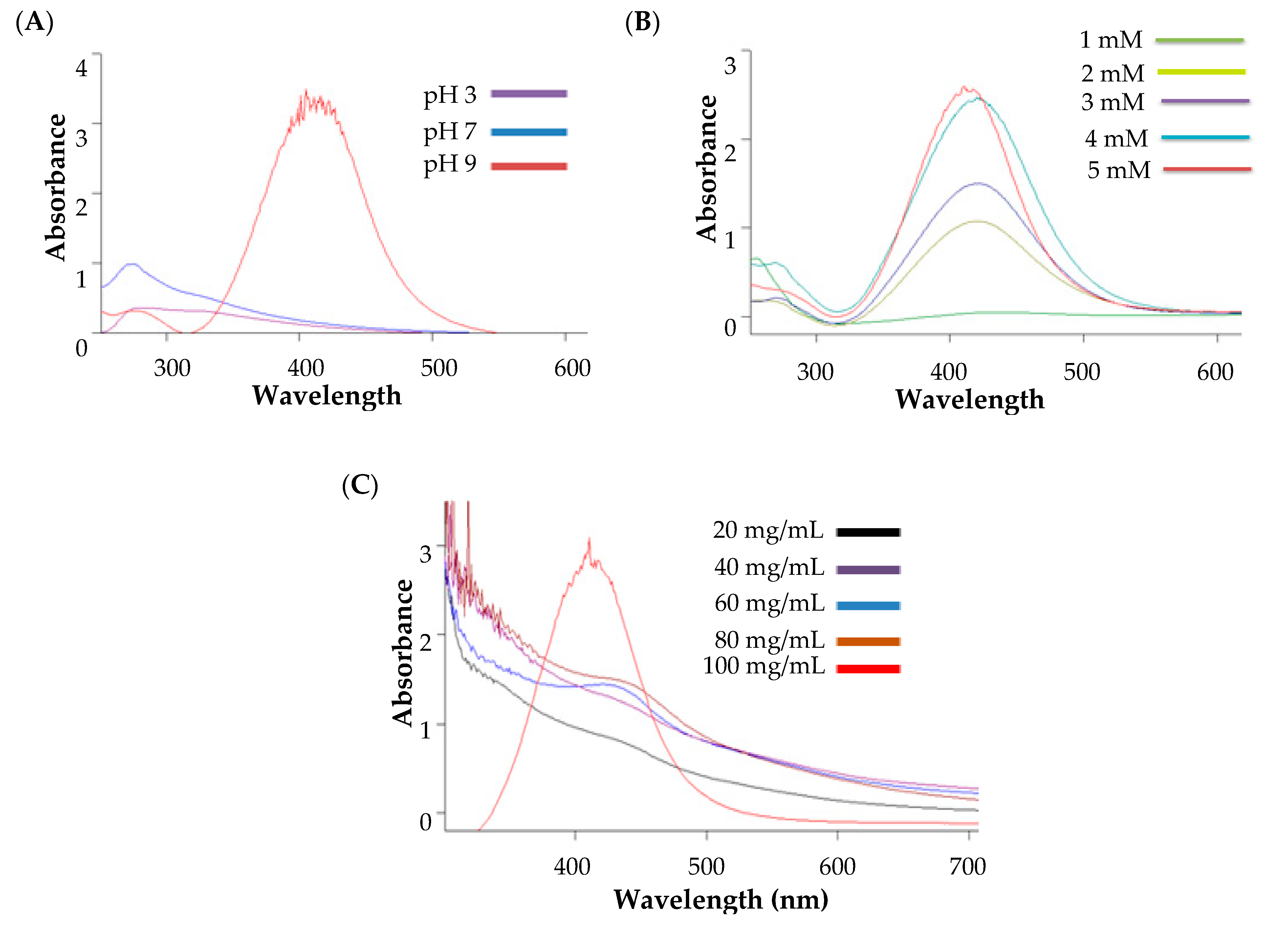
Abstract
:1. Introduction
2. Materials and Methods
2.1. Materials
2.2. Preparation of Commiphora mukul Plant (Guggul) Extract
2.3. Synthesis of Silver Nanoparticles (AgNPs)
2.4. Characterization of Silver Nanoparticles
2.4.1. UV Spectroscopy Analysis
2.4.2. Particle Size Measurement
2.4.3. Surface Charge Determination
2.4.4. Surface Morphology
2.4.5. Fourier Transform Infrared (FT-IR)
2.5. In-Vivo Anti-Arthritic Activity
2.5.1. Animal Study
2.5.2. Acute Toxicity Study
2.5.3. Adjuvant-Induced Arthritis Rodent Model
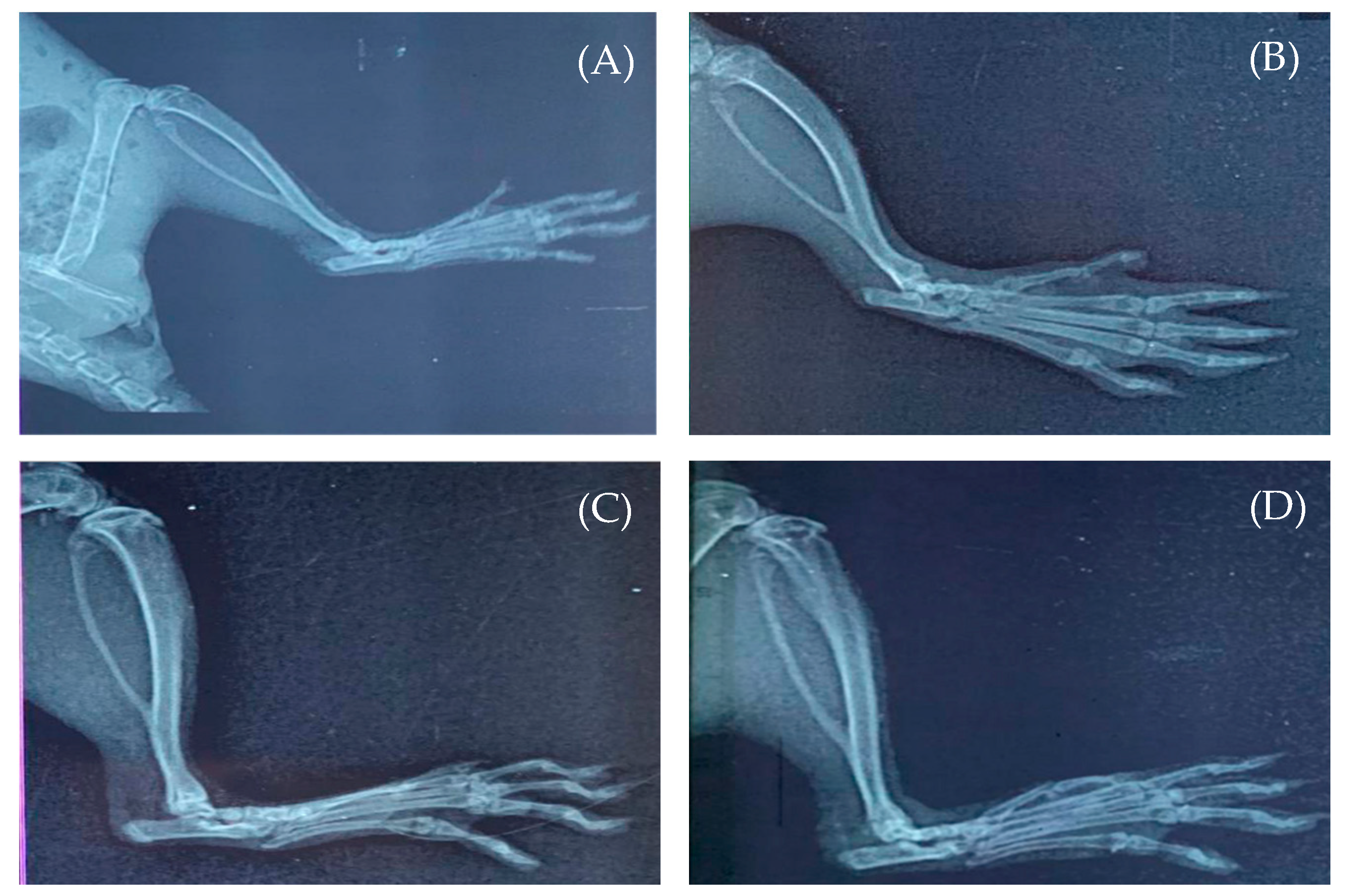
2.5.4. Radiological Assay
2.5.5. Estimation of Antioxidant Activity
Estimation of Superoxide Dismutase
Estimation of Catalase (CAT)
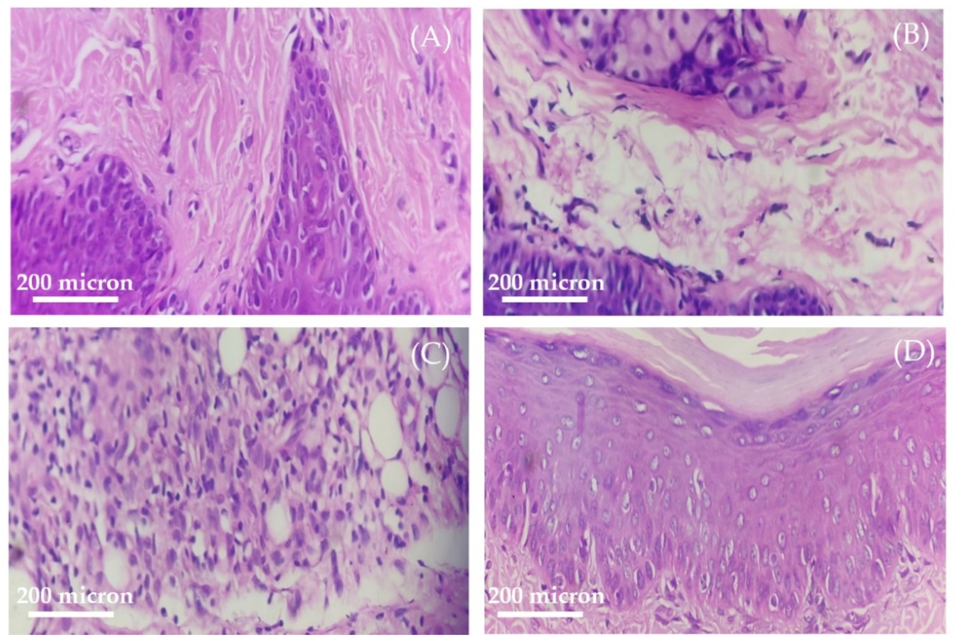
2.5.6. Histopathology Assessment
2.6. Statistical Analysis
3. Results
3.1. Phytochemical Analysis of Commiphora mukul
3.2. Synthesis of Silver Nanoparticles
3.3. Study of Process Parameters on Nanoparticle Formation
3.3.1. Influence of pH on the Nanoparticle Formation
3.3.2. Influence of Silver Salt Concentration on the Nanoparticle Formation
3.3.3. Influence of Guggul Extract on the Nanoparticle Formation
3.3.4. Optimized Process for Nanoparticle Preparation
3.4. Characterization of Silver Nanoparticles
3.4.1. UV Spectroscopy Analysis
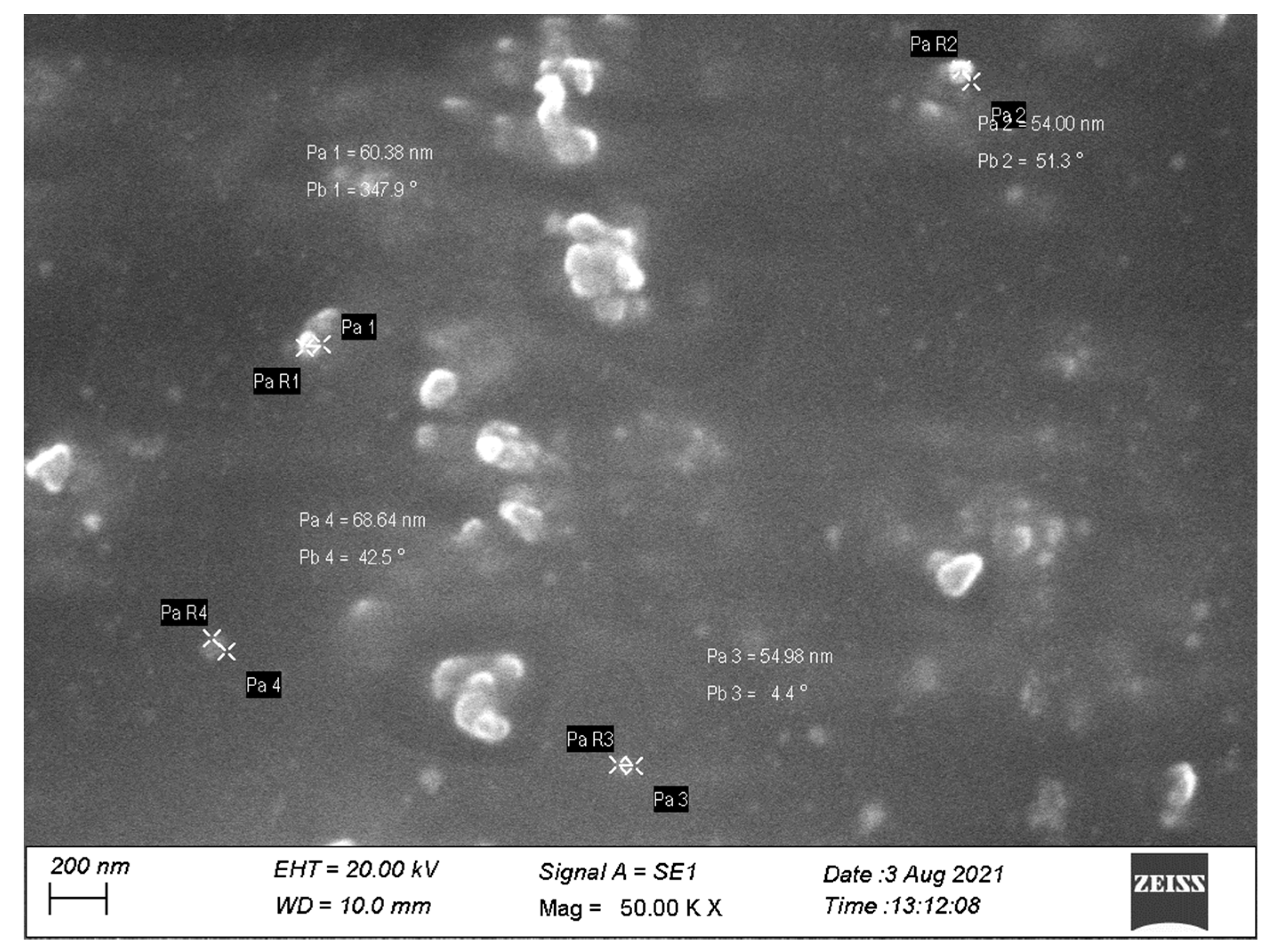
3.4.2. Surface Morphology
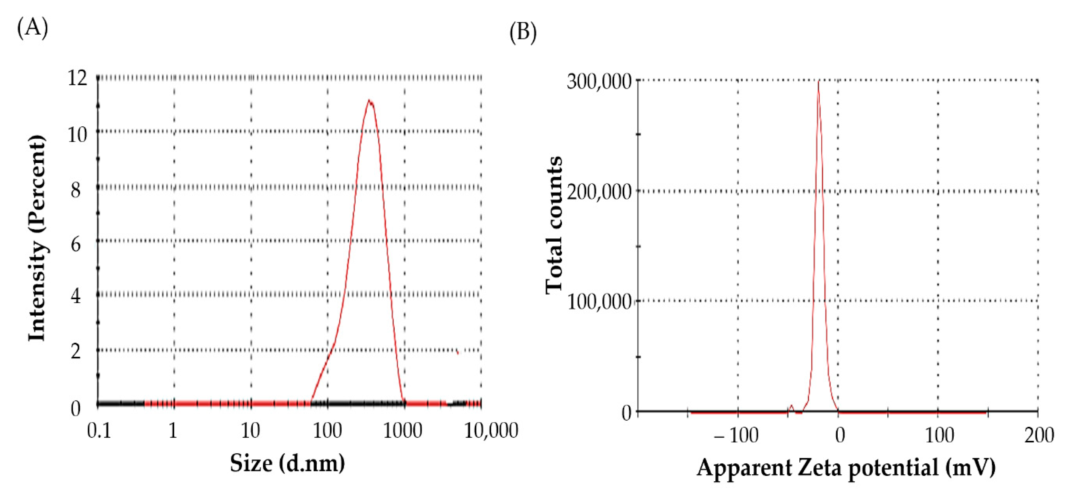
3.4.3. Particle Size Measurement
3.4.4. Fourier Transform Infrared (FT-IR) Analysis
3.5. Acute Toxicity Study
3.6. Adjuvant-Induced Arthritis Rodent Model
3.6.1. Evaluation of Anti-Arthritic Activity of AgNPs
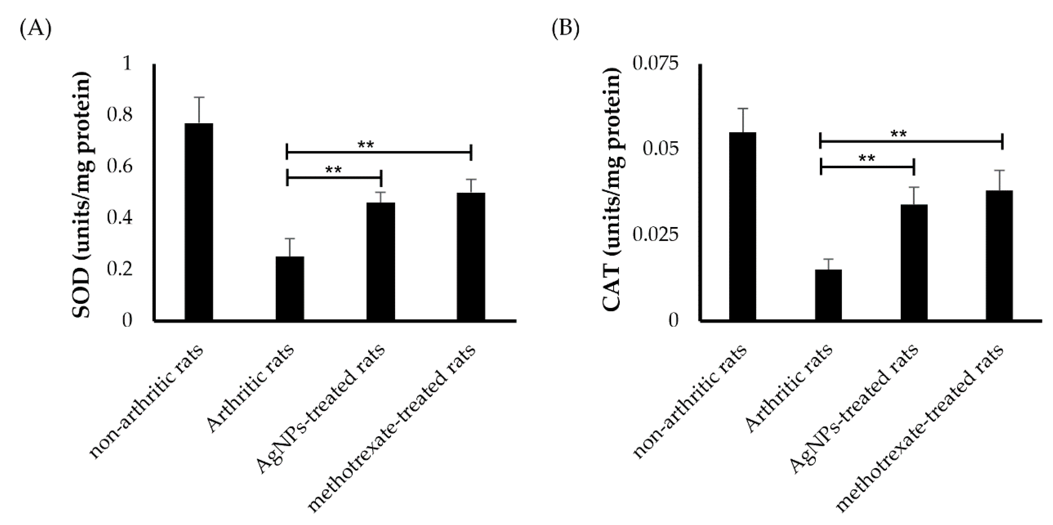
3.6.2. Estimation of Antioxidant Activity
3.6.3. Radiological Assay
3.6.4. Histopathology Assessment
4. Conclusions
Supplementary Materials
Author Contributions
Funding
Institutional Review Board Statement
Informed Consent Statement
Data Availability Statement
Acknowledgments
Conflicts of Interest
References
- Singh, E.; Osmani, R.A.M.; Banerjee, R.; Abu Lila, A.S.; Moin, A.; Almansour, K.; Arab, H.H.; Alotaibi, H.F.; Khafagy, E.S. Poly ε-Caprolactone Nanoparticles for Sustained Intra-Articular Immune Modulation in Adjuvant-Induced Arthritis Rodent Model. Pharmaceutics 2022, 14, 519. [Google Scholar] [CrossRef] [PubMed]
- Jones, G.; Nash, P.; Hall, S. Advances in rheumatoid arthritis. Med. J. Aust. 2017, 206, 221–224. [Google Scholar] [CrossRef] [PubMed]
- Hu, Y.; Cheng, W.; Cai, W.; Yue, Y.; Li, J.; Zhang, P. Advances in research on animal models of rheumatoid arthritis. Clin. Rheumatol. 2013, 32, 161–165. [Google Scholar] [CrossRef] [PubMed]
- Crowson, C.S.; Matteson, E.L.; Myasoedova, E.; Michet, C.J.; Ernste, F.C.; Warrington, K.J.; Davis, J.M., 3rd; Hunder, G.G.; Therneau, T.M.; Gabriel, S.E. The lifetime risk of adult-onset rheumatoid arthritis and other inflammatory autoimmune rheumatic diseases. Arthritis Rheum. 2011, 63, 633–639. [Google Scholar] [CrossRef] [Green Version]
- Lwin, M.N.; Serhal, L.; Holroyd, C.; Edwards, C.J. Rheumatoid Arthritis: The Impact of Mental Health on Disease: A Narrative Review. Rheumatol. Ther. 2020, 7, 457–471. [Google Scholar] [CrossRef]
- Yuan, F.; Quan, L.D.; Cui, L.; Goldring, S.R.; Wang, D. Development of macromolecular prodrug for rheumatoid arthritis. Adv. Drug Deliv. Rev. 2012, 64, 1205–1219. [Google Scholar] [CrossRef] [Green Version]
- Mansouri, S.; Cuie, Y.; Winnik, F.; Shi, Q.; Lavigne, P.; Benderdour, M.; Beaumont, E.; Fernandes, J.C. Characterization of folate-chitosan-DNA nanoparticles for gene therapy. Biomaterials 2006, 27, 2060–2065. [Google Scholar] [CrossRef]
- Emami, J.; Ansarypour, Z. Receptor targeting drug delivery strategies and prospects in the treatment of rheumatoid arthritis. Res. Pharm. Sci. 2019, 14, 471–487. [Google Scholar] [CrossRef]
- Hattori, Y.; Sakaguchi, M.; Maitani, Y. Folate-linked lipid-based nanoparticles deliver a NFkappaB decoy into activated murine macrophage-like RAW264.7 cells. Biol. Pharm. Bull. 2006, 29, 1516–1520. [Google Scholar] [CrossRef] [Green Version]
- Zhang, N.; Xu, C.; Li, N.; Zhang, S.; Fu, L.; Chu, X.; Hua, H.; Zeng, X.; Zhao, Y. Folate receptor-targeted mixed polysialic acid micelles for combating rheumatoid arthritis: In vitro and in vivo evaluation. Drug Deliv. 2018, 25, 1182–1191. [Google Scholar] [CrossRef]
- Verma, A.; Jain, A.; Tiwari, A.; Saraf, S.; Panda, P.K.; Agrawal, G.P.; Jain, S.K. Folate Conjugated Double Liposomes Bearing Prednisolone and Methotrexate for Targeting Rheumatoid Arthritis. Pharm. Res. 2019, 36, 123. [Google Scholar] [CrossRef] [PubMed]
- Chandrasekar, D.; Sistla, R.; Ahmad, F.J.; Khar, R.K.; Diwan, P.V. Folate coupled poly(ethyleneglycol) conjugates of anionic poly(amidoamine) dendrimer for inflammatory tissue specific drug delivery. J. Biomed. Mater. Res. A 2007, 82, 92–103. [Google Scholar] [CrossRef] [PubMed]
- Sharma, A.K.; Arya, A.; Sahoo, P.K.; Majumdar, D.K. Overview of biopolymers as carriers of antiphlogistic agents for treatment of diverse ocular inflammations. Mater. Sci. Eng. C Mater. Biol. Appl. 2016, 67, 779–791. [Google Scholar] [CrossRef] [PubMed]
- Berdiaki, A.; Perisynaki, E.; Stratidakis, A.; Kulikov, P.P.; Kuskov, A.N.; Stivaktakis, P.; Henrich-Noack, P.; Luss, A.L.; Shtilman, M.M.; Tzanakakis, G.N.; et al. Assessment of Amphiphilic Poly-N-vinylpyrrolidone Nanoparticles’ Biocompatibility with Endothelial Cells in Vitro and Delivery of an Anti-Inflammatory Drug. Mol. Pharm. 2020, 17, 4212–4225. [Google Scholar] [CrossRef]
- Alaaeldin, E.; Abou-Taleb, H.A.; Mohamad, S.A.; Elrehany, M.; Gaber, S.S.; Mansour, H.F. Topical Nano-Vesicular Spanlastics of Celecoxib: Enhanced Anti-Inflammatory Effect and Down-Regulation of TNF-α, NF-кB and COX-2 in Complete Freund’s Adjuvant-Induced Arthritis Model in Rats. Int. J. Nanomed. 2021, 16, 133–145. [Google Scholar] [CrossRef] [PubMed]
- Kuskov, A.; Nikitovic, D.; Berdiaki, A.; Shtilman, M.; Tsatsakis, A. Amphiphilic Poly-N-vinylpyrrolidone Nanoparticles as Carriers for Nonsteroidal, Anti-Inflammatory Drugs: Pharmacokinetic, Anti-Inflammatory, and Ulcerogenic Activity Study. Pharmaceutics 2022, 14, 925. [Google Scholar] [CrossRef]
- Al Hagbani, T.; Rizvi, S.M.D.; Hussain, T.; Mehmood, K.; Rafi, Z.; Moin, A.; Abu Lila, A.S.; Alshammari, F.; Khafagy, E.S.; Rahamathulla, M.; et al. Cefotaxime Mediated Synthesis of Gold Nanoparticles: Characterization and Antibacterial Activity. Polymers 2022, 14, 771. [Google Scholar] [CrossRef]
- Hagbani, T.A.; Yadav, H.; Moin, A.; Lila, A.S.A.; Mehmood, K.; Alshammari, F.; Khan, S.; Khafagy, E.S.; Hussain, T.; Rizvi, S.M.D.; et al. Enhancement of Vancomycin Potential against Pathogenic Bacterial Strains via Gold Nano-Formulations: A Nano-Antibiotic Approach. Materials 2022, 15, 1108. [Google Scholar] [CrossRef]
- Lila, A.S.; Kiwada, H.; Ishida, T. Selective delivery of oxaliplatin to tumor tissue by nanocarrier system enhances overall therapeutic efficacy of the encapsulated oxaliplatin. Biol. Pharm. Bull. 2014, 37, 206–211. [Google Scholar] [CrossRef] [Green Version]
- Ghiuță, I.; Cristea, D. Silver nanoparticles for delivery purposes. Nanoeng. Biomater. Adv. Drug Deliv. 2020, 2020, 347–371. [Google Scholar] [CrossRef]
- Soliman, W.E.; Khan, S.; Rizvi, S.M.D.; Moin, A.; Elsewedy, H.S.; Abulila, A.S.; Shehata, T.M. Therapeutic Applications of Biostable Silver Nanoparticles Synthesized Using Peel Extract of Benincasa hispida: Antibacterial and Anticancer Activities. Nanomaterials 2020, 10, 1954. [Google Scholar] [CrossRef] [PubMed]
- Yang, Y.; Guo, L.; Wang, Z.; Liu, P.; Liu, X.; Ding, J.; Zhou, W. Targeted silver nanoparticles for rheumatoid arthritis therapy via macrophage apoptosis and Re-polarization. Biomaterials 2021, 264, 120390. [Google Scholar] [CrossRef] [PubMed]
- Khader, S.Z.A.; Ahmed, S.S.Z.; Prabaharan, S.B.; Kumar, K.R.; David, D.; Lakshmanan, S.O.; Mahboob, M.R. In vitro anti-inflammatory, anti-arthritic and anti-proliferative activity of green synthesized silver nanoparticles—Phoenix dactylifera (Rothan dates). Braz. J. Pharm. Sci. 2022, 58, e18594. [Google Scholar] [CrossRef]
- Al Saqr, A.; Khafagy, E.S.; Alalaiwe, A.; Aldawsari, M.F.; Alshahrani, S.M.; Anwer, M.K.; Khan, S.; Lila, A.S.A.; Arab, H.H.; Hegazy, W.A.H. Synthesis of Gold Nanoparticles by Using Green Machinery: Characterization and In Vitro Toxicity. Nanomaterials 2021, 11, 808. [Google Scholar] [CrossRef] [PubMed]
- Rao, K.; Aziz, S.; Roome, T.; Razzak, A.; Sikandar, B.; Jamali, K.S.; Imran, M.; Jabri, T.; Shah, M.R. Gum acacia stabilized silver nanoparticles based nano-cargo for enhanced anti-arthritic potentials of hesperidin in adjuvant induced arthritic rats. Artif. Cells Nanomed. Biotechnol. 2018, 46, 597–607. [Google Scholar] [CrossRef] [PubMed] [Green Version]
- Zhang, D.; Ma, X.L.; Gu, Y.; Huang, H.; Zhang, G.W. Green Synthesis of Metallic Nanoparticles and Their Potential Applications to Treat Cancer. Front. Chem. 2020, 8, 799. [Google Scholar] [CrossRef] [PubMed]
- Singh, B.B.; Mishra, L.; Aquilina, N.; Kohlbeck, F. Usefulness of guggul (Commiphora mukul) for osteoarthritis of the knee: An experimental case study. Altern. Ther. Health Med. 2001, 7, 112–124. [Google Scholar]
- Sarup, P.; Bala, S.; Kamboj, S. Pharmacology and Phytochemistry of Oleo-Gum Resin of Commiphora wightii (Guggulu). Scientifica 2015, 2015, 138039. [Google Scholar] [CrossRef] [Green Version]
- Gupta, S.; Dey, Y.N.; Kannojia, P.; Halder, A.K.; Sharma, D.; Wanjari, M.M.; Chougule, S.; Pawar, S.; Kaushik, A.; Gaidhani, S.N.; et al. Analgesic and Anti-inflammatory Activities of Trayodashang Guggulu, an Ayurvedic Formulation. Phytomed. Plus 2022, 2, 100281. [Google Scholar] [CrossRef]
- Dave, V.; Yadav, R.B.; Gupta, S.; Sharma, S. Guggulosomes: A herbal approach for enhanced topical delivery of phenylbutazone. Future J. Pharm. Sci. 2017, 3, 23–32. [Google Scholar] [CrossRef]
- Raj, S.; Chand Mali, S.; Trivedi, R. Green synthesis and characterization of silver nanoparticles using Enicostemma axillare (Lam.) leaf extract. Biochem. Biophys. Res. Commun. 2018, 503, 2814–2819. [Google Scholar] [CrossRef] [PubMed]
- Abdallah, M.H.; Abu Lila, A.S.; Unissa, R.; Elsewedy, H.S.; Elghamry, H.A.; Soliman, M.S. Preparation, characterization and evaluation of anti-inflammatory and anti-nociceptive effects of brucine-loaded nanoemulgel. Colloids Surf. B Biointerfaces 2021, 205, 111868. [Google Scholar] [CrossRef] [PubMed]
- Vishwa, B.; Moin, A.; Gowda, D.V.; Rizvi, S.M.D.; Hegazy, W.A.H.; Abu Lila, A.S.; Khafagy, E.S.; Allam, A.N. Pulmonary Targeting of Inhalable Moxifloxacin Microspheres for Effective Management of Tuberculosis. Pharmaceutics 2021, 13, 79. [Google Scholar] [CrossRef] [PubMed]
- Saleem, U.; Amin, S.; Ahmad, B.; Azeem, H.; Anwar, F.; Mary, S. Acute oral toxicity evaluation of aqueous ethanolic extract of Saccharum munja Roxb. roots in albino mice as per OECD 425 TG. Toxicol. Rep. 2017, 4, 580–585. [Google Scholar] [CrossRef] [PubMed]
- Torres-Guzman, A.M.; Morado-Urbina, C.E.; Alvarado-Vazquez, P.A.; Acosta-Gonzalez, R.I.; Chávez-Piña, A.E.; Montiel-Ruiz, R.M.; Jimenez-Andrade, J.M. Chronic oral or intraarticular administration of docosahexaenoic acid reduces nociception and knee edema and improves functional outcomes in a mouse model of Complete Freund’s Adjuvant-induced knee arthritis. Arthritis Res. Ther. 2014, 16, R64. [Google Scholar] [CrossRef] [Green Version]
- Mani, A.; Vasanthi, C.; Gopal, V.; Chellathai, D. Role of phyto-stabilised silver nanoparticles in suppressing adjuvant induced arthritis in rats. Int. Immunopharmacol. 2016, 41, 17–23. [Google Scholar] [CrossRef]
- Dewangan, A.K.; Perumal, Y.; Pavurala, N.; Chopra, K.; Mazumder, S. Preparation, characterization and anti-inflammatory effects of curcumin loaded carboxymethyl cellulose acetate butyrate nanoparticles on adjuvant induced arthritis in rats. J. Drug Deliv. Sci. Technol. 2017, 41, 269–279. [Google Scholar] [CrossRef]
- Zhang, C.; Bruins, M.E.; Yang, Z.-Q.; Liu, S.-T.; Rao, P.-F. A new formula to calculate activity of superoxide dismutase in indirect assays. Anal. Biochem. 2016, 503, 65–67. [Google Scholar] [CrossRef]
- Arabsolghar, R.; Saberzadeh, J.; Khodaei, F.; Borojeni, R.A.; Khorsand, M.; Rashedinia, M. The protective effect of sodium benzoate on aluminum toxicity in PC12 cell line. Res. Pharm. Sci. 2017, 12, 391–400. [Google Scholar] [CrossRef] [Green Version]
- Ragavi, R.; Surendran, S.A. Commiphora mukul: An Overview. Res. J. Pharm. Technol. 2018, 11, 3205–3208. [Google Scholar] [CrossRef]
- Yadav, V.D.; Krishnan, R.A.; Borade, L.; Shirolikar, S.; Jain, R.; Dandekar, P. pH tunability and influence of alkali metal basicity on the plasmonic resonance of silver nanoparticles. Mater. Res. Express 2017, 4, 075021. [Google Scholar] [CrossRef]
- Jana, J.; Ganguly, M.; Pal, T. Enlightening surface plasmon resonance effect of metal nanoparticles for practical spectroscopic application. RSC Adv. 2016, 6, 86174–86211. [Google Scholar] [CrossRef]
- Ider, M.; Abderrafi, K.; Eddahbi, A.; Ouaskit, S.; Kassiba, A. Silver Metallic Nanoparticles with Surface Plasmon Resonance: Synthesis and Characterizations. J. Clust. Sci. 2017, 28, 1051–1069. [Google Scholar] [CrossRef]
- Tsuji, T.; Iryo, K.; Watanabe, N.; Tsuji, M. Preparation of silver nanoparticles by laser ablation in solution: Influence of laser wavelength on particle size. Appl. Surf. Sci. 2002, 202, 80–85. [Google Scholar] [CrossRef]
- Wilson, O.; Wilson, G.J.; Mulvaney, P. Laser Writing in Polarized Silver Nanorod Films. Adv. Mater. 2002, 14, 1000–1004. [Google Scholar] [CrossRef]
- Abu Lila, A.S.; Huwaimel, B.; Alobaida, A.; Hussain, T.; Rafi, Z.; Mehmood, K.; Abdallah, M.H.; Hagbani, T.A.; Rizvi, S.M.D.; Moin, A.; et al. Delafloxacin-Capped Gold Nanoparticles (DFX-AuNPs): An Effective Antibacterial Nano-Formulation of Fluoroquinolone Antibiotic. Materials 2022, 15, 5709. [Google Scholar] [CrossRef]
- Gabriel, S.E. The epidemiology of rheumatoid arthritis. Rheum. Dis. Clin. N. Am. 2001, 27, 269–281. [Google Scholar] [CrossRef]
- Bendele, A.M. Animal models of osteoarthritis. J. Musculoskelet. Neuronal Interact. 2001, 1, 363–376. [Google Scholar]
- Slovák, L.; Poništ, S.; Kuncírová, V.; Mihalová, D.; Fedorova, T.; Bauerová, K. Evaluation of the effect of carnosine, its novel derivative trolox-carnosine and trolox in a pre-clinical study focussing on the regulation of immunity. Eur. Pharm. J. 2016, 63, 16–19. [Google Scholar] [CrossRef] [Green Version]
- Ahmed, E.A.; Ahmed, O.M.; Fahim, H.I.; Ali, T.M.; Elesawy, B.H.; Ashour, M.B. Potency of Bone Marrow-Derived Mesenchymal Stem Cells and Indomethacin in Complete Freund’s Adjuvant-Induced Arthritic Rats: Roles of TNF-α, IL-10, iNOS, MMP-9, and TGF-β1. Stem Cells Int. 2021, 2021, 6665601. [Google Scholar] [CrossRef]
- Ahmed, O.M.; Soliman, H.A.; Mahmoud, B.; Gheryany, R.R. Ulva lactuca hydroethanolic extract suppresses experimental arthritis via its anti-inflammatory and antioxidant activities. Beni-Suef Univ. J. Basic Appl. Sci. 2017, 6, 394–408. [Google Scholar] [CrossRef]
- Quiñonez-Flores, C.M.; González-Chávez, S.A.; Del Río Nájera, D.; Pacheco-Tena, C. Oxidative Stress Relevance in the Pathogenesis of the Rheumatoid Arthritis: A Systematic Review. BioMed Res. Int. 2016, 2016, 6097417. [Google Scholar] [CrossRef] [PubMed]
- Ozkan, Y.; Yardým-Akaydýn, S.; Sepici, A.; Keskin, E.; Sepici, V.; Simsek, B. Oxidative status in rheumatoid arthritis. Clin. Rheumatol. 2007, 26, 64–68. [Google Scholar] [CrossRef] [PubMed]


Publisher’s Note: MDPI stays neutral with regard to jurisdictional claims in published maps and institutional affiliations. |
© 2022 by the authors. Licensee MDPI, Basel, Switzerland. This article is an open access article distributed under the terms and conditions of the Creative Commons Attribution (CC BY) license (https://creativecommons.org/licenses/by/4.0/).
Share and Cite
Singh, A.; Boregowda, S.S.; Moin, A.; Abu Lila, A.S.; Aldawsari, M.F.; Khafagy, E.-S.; Alotaibi, H.F.; Jayaramu, R.A. Biosynthesis of Silver Nanoparticles Using Commiphora mukul Extract: Evaluation of Anti-Arthritic Activity in Adjuvant-Induced Arthritis Rat Model. Pharmaceutics 2022, 14, 2318. https://doi.org/10.3390/pharmaceutics14112318
Singh A, Boregowda SS, Moin A, Abu Lila AS, Aldawsari MF, Khafagy E-S, Alotaibi HF, Jayaramu RA. Biosynthesis of Silver Nanoparticles Using Commiphora mukul Extract: Evaluation of Anti-Arthritic Activity in Adjuvant-Induced Arthritis Rat Model. Pharmaceutics. 2022; 14(11):2318. https://doi.org/10.3390/pharmaceutics14112318
Chicago/Turabian StyleSingh, Anupama, Sateesha Shivally Boregowda, Afrasim Moin, Amr Selim Abu Lila, Mohammed F. Aldawsari, El-Sayed Khafagy, Hadil Faris Alotaibi, and Rajamma Abburu Jayaramu. 2022. "Biosynthesis of Silver Nanoparticles Using Commiphora mukul Extract: Evaluation of Anti-Arthritic Activity in Adjuvant-Induced Arthritis Rat Model" Pharmaceutics 14, no. 11: 2318. https://doi.org/10.3390/pharmaceutics14112318