Review

Multi-Organs-on-Chips for Testing Small-Molecule Drugs: Challenges and Perspectives

1
Department of Mechanical Engineering, Rowan University, Glassboro, NJ 08028, USA
2
Molecular Biology and Genetics, Faculty of Engineering and Natural Sciences, Istinye University, Istanbul 34010, Turkey
3
Department of Pharmaceutical Technology, School of Pharmacy, Aristotle University of Thessaloniki, GR-54124 Thessaloniki, Greece
4
Department of Microbiology, Sher-i-Kashmir Institute of Medical Sciences, Srinagar 190011, India
5
Department of Biomedical Engineering, New Jersey Institute of Technology, Newark, NJ 07102, USA
6
Institute of Health Science, Department of Translational Medicine, Ankara Yildirim Beyazit University, Ankara 06800, Turkey
7
Izmir Institute of Technology, Graduate Program of Biotechnology and Bioengineering, Gulbahce Campus, Izmir 35430, Turkey
8
Izmir Institute of Technology, Department of Chemical Engineering, Gulbahce Campus, Izmir 35430, Turkey
9
Care of Elderly Program, Vocational School of Health Services, Izmir Democracy University, Izmir 35140, Turkey
10
Department of Medical Biochemistry, Faculty of Medicine, Ankara Yildirim Beyazit University, Ankara 06800, Turkey
11
Department of Mechanical and Industrial Engineering, New Jersey Institute of Technology, Newark, NJ 07102, USA
*
Author to whom correspondence should be addressed.
These authors contributed equally to this work.
Pharmaceutics 2021, 13(10), 1657; https://doi.org/10.3390/pharmaceutics13101657
Submission received: 1 September 2021 / Revised: 30 September 2021 / Accepted: 3 October 2021 / Published: 11 October 2021

Abstract

:
Organ-on-a-chip technology has been used in testing small-molecule drugs for screening potential therapeutics and regulatory protocols. The technology is expected to boost the development of novel therapies and accelerate the discovery of drug combinations in the coming years. This has led to the development of multi-organ-on-a-chip (MOC) for recapitulating various organs involved in the drug–body interactions. In this review, we discuss the current MOCs used in screening small-molecule drugs and then focus on the dynamic process of drug absorption, distribution, metabolism, and excretion. We also address appropriate materials used for MOCs at low cost and scale-up capacity suitable for high-performance analysis of drugs and commercial high-throughput screening platforms.

Graphical Abstract

1. Introduction

The failure of drug design in clinical trials is rooted in the differences between the human body and the preclinical animal models, resulting in incorrect predictions of pharmacokinetics and pharmacodynamics, such as clearance, safety margins, toxicity, and efficacy [1,2]. Clinical trials on more than 2000 drugs indicated the incompatibility of animal test results for toxic responses in the human body [3]. The growing need to develop viable in vitro alternatives to animal testing has generated the organ-on-chips technology, which combines biotechnology, cell biology, biomaterials, and biomedical sciences to recapitulate an organ or tissue microenvironment [4,5,6]. In contrast to single organ chips, which aim to recapitulate the biological function of individual organs, multi-organs-on-chips (MOCs) are introduced to integrate multiple organs on a single platform. Interconnected microchambers in organs-on-chips create dynamic drug absorption, distribution, metabolism, and excretion [4,5]. The complexity and need of multiple components, such as the liver, have led to the development of MOCs used for drug screening and toxicology testing [2,7,8,9]. The efforts associated with MOCs and how they facilitate their seamless transition from prototyping to clinical practices should be recognized to transform healthcare provision and patients’ lives.
The main motivation behind MOCs was to include a gut part for modeling drug absorption and liver part for modeling drug metabolism. A combination of these components has been tested with a kidney model for studying drug response and its metabolism [10,11]. Pires de Mello et al. designed a heart-liver-skin MOC to investigate acute and chronic drug exposure effects on tissue function. Maschmeyer et al. developed a four-part MOC integrated with sequentially connected intestine, liver, skin, and kidney parts, which provide stable homeostasis across different organ parts to investigate the systemic toxicity for drug candidates [10]. In another study, gut-liver-kidney and bone marrow-liver-kidney MOCs were developed to predict the pharmacokinetic parameters of the orally administered drug nicotine and then intravenously injected the anticancer drug cisplatin [1]. In addition, a MOC with circulating monocytic cells as a functional human immune model was developed to analyze the tissue-specific immune responses of the cardiac, skeletal, and hepatic compartments to the anti-arrhythmic drug amiodarone [12]. Gut-on-chip was used to study the impact of the coxsackievirus B serotype 1 in the intestinal cells. The study showed the destructive effect of the virus on villi and the integrity of the whole epithelium and passage through the gut lumen into the vascular channel. The detectable cytopathic results and the increase in IL-8 implied active replication and the release of infectious virions [13,14]. The study can evaluate other enteroviruses and drug development with good precision. The chances of success are higher because of the well-understood physiology in miniaturized 3-D in vitro models (see Figure 1).
Microfluidic-based devices have been developed based on drug carrier-free and drug carrier-integrated chips, which also involve a carrier loaded with the therapeutic agent, enabling spatiotemporal control over the mobility of small-molecule drugs. Compared to conventional systems, microfluidic devices provide targeted and sustained release, thus avoiding the burst distribution of drugs that improves safety and compliance through pain reduction [15]. Modern micro and nanotechnological approaches enable spatial and temporal control over the release of drugs. We can generate tunable drugs with sustained drug release properties [16] from fibrous materials [17], microgels [18], hydrogels [19], polymeric implants [16], and DNA logic gate circuits [20]. The concept of applying microdevices for regulating the release of small molecules emerged in 1998 when Santini et al. proposed a microchip demonstrating controlled and pulsatile release of single or multiple molecules [21]. The advancements in scientific knowledge and the cross-disciplinary efforts for recapitulating experiments in more controlled conditions led to MOC developments [22,23].
The recent advances in organ-on-chips and MOCs technologies have been extensively reviewed in the literature, providing a thorough insight into the development of microphysiological systems resembling the liver and heart [24], intestine [25], lung [26], kidney [27], and multi-organ [2,28,29,30,31] functions for drug discovery and drug toxicity screening applications. Driven by the momentum of MOCs technologies, the current review will cover the latest advancements and challenges encountered in the field of MOC systems and the essential parameters for their successful and timely commercialization.

2. Small Molecules: Properties and Applications
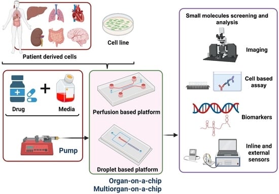
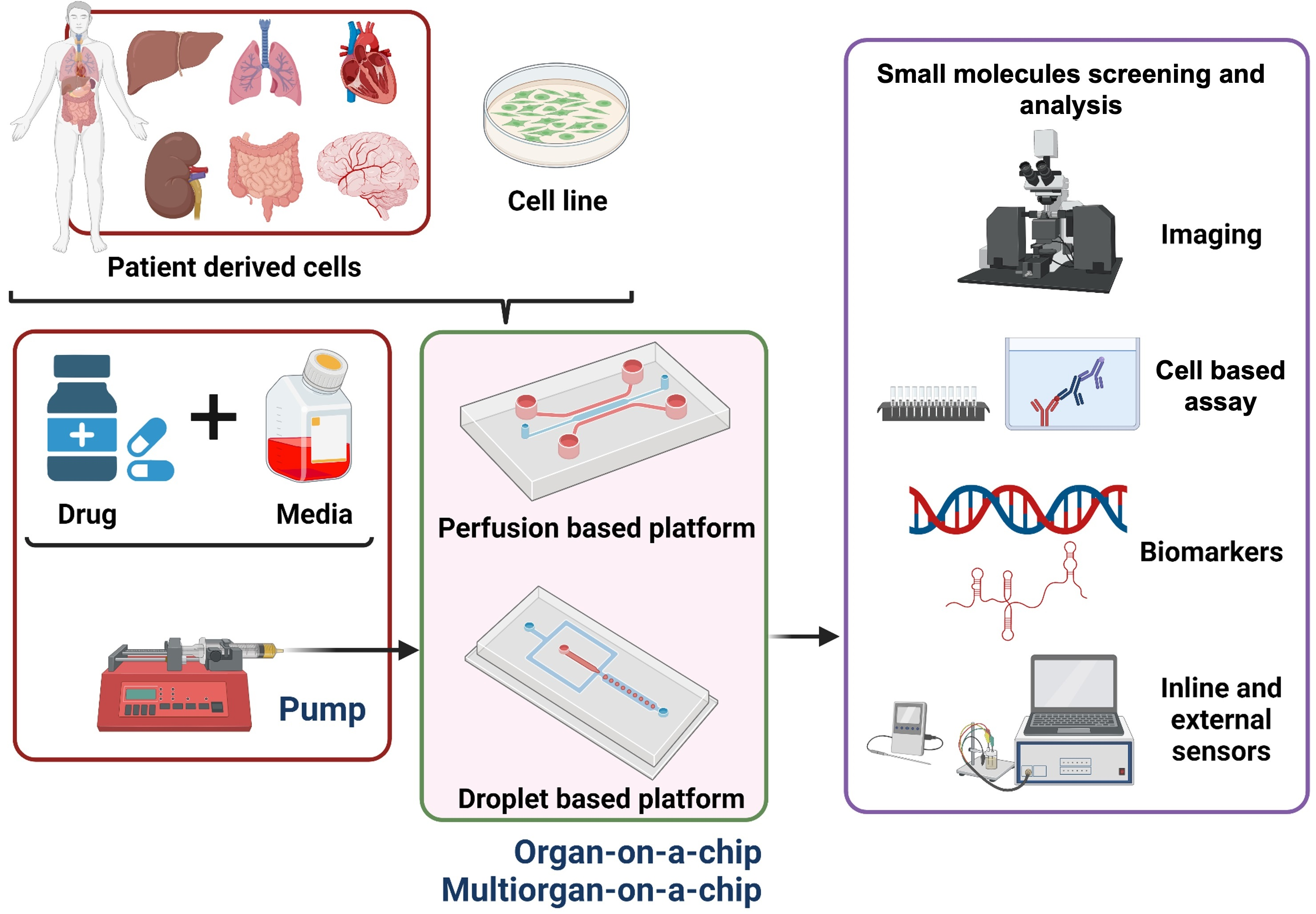
Small-molecule drugs have low molecular weights (100–1000 g/mol or 0.1–1 kDa) and include chemotherapeutics, steroids, and antibiotics. These small-sized molecules can penetrate the cell membrane and modulate intracellular signaling pathways [32,33,34,35]. They can interfere with tumor-induced cell proliferation and development via interrupting various protein pathways [36]. Off-target effects and limited efficacy of drugs make small molecules preferred for their site selectivity and sustained release [16]. Different anticancer drugs have benefited from being small molecules and are applied as protein inhibitors.
MOCs have been used to study the systemic absorption and metabolism of drugs. They are integrated with micro-pumps and channels to create multi-organ models, such as the intestine, liver, skin, and kidney [37]. Wagner et al. designed a dynamic MOC by human liver and skin coculture to investigate the toxicity of troglitazone on day 6 post-incubation. Results indicated a dose-dependent response to troglitazone after the 6-day treatment [38]. In another study, Edington et al. designed an interconnected microfluidic device made of multi-cultures for modeling the gut, endometrium, lung, liver, heart, and brain for lipophilic drugs [39].
MOC designs for the blood brain barrier have come into prominence as potential models to obtain significant predictions for the transport and efficacy of nanomedicine [40]. Miller and Shuler developed a MOC model for a 13-organ system with various cell lines by mimicking the main parenchymal organs and physiological barrier tissues in the human body to investigate the inter-organ transport of biological agents for drug response [41]. Mucus is another significant biological barrier for the uptake and absorption of particulate drug carriers. Jia et al. developed a mucus-chip model to optimize the mucosal absorption of drugs by investigating the penetration and quantifying the transport of polyethylene glycols-based nanocarriers across the mucus. This mucus-on-chip enabled effective visualization and quantitative data on the absorption of drug nanocarriers during muco-penetration [15]. Drug evaluation depends on real-time monitoring of anticancer drugs in the tumor microenvironment. Tang et al. developed a biomimetic microfluidic tumor microenvironment consisting of a coculture of tumor and endothelial cells [42]. This model included a vascular part forming a total lumen under shear flow and communicating with the 3-D solid tumor part.
Besides classical microfabrication techniques, 3-D bioprinting has a high potential in MOC technology using an integrated system in dynamic conditions. In the 3-D bioprinting technique, with bioinks and a single programmable manufacturing step, the desired porosity, interconnectivity, and pore design can be easily adjusted, which is critical for tissue remodeling of the different parts of the body [43]. This technique also has the advantage of incorporating different types of biomaterials, cells, and biomolecules in a complex structure within one controllable process step [44], which makes it easy to mimic a complex organ structure.

3. Organs-on-Chips

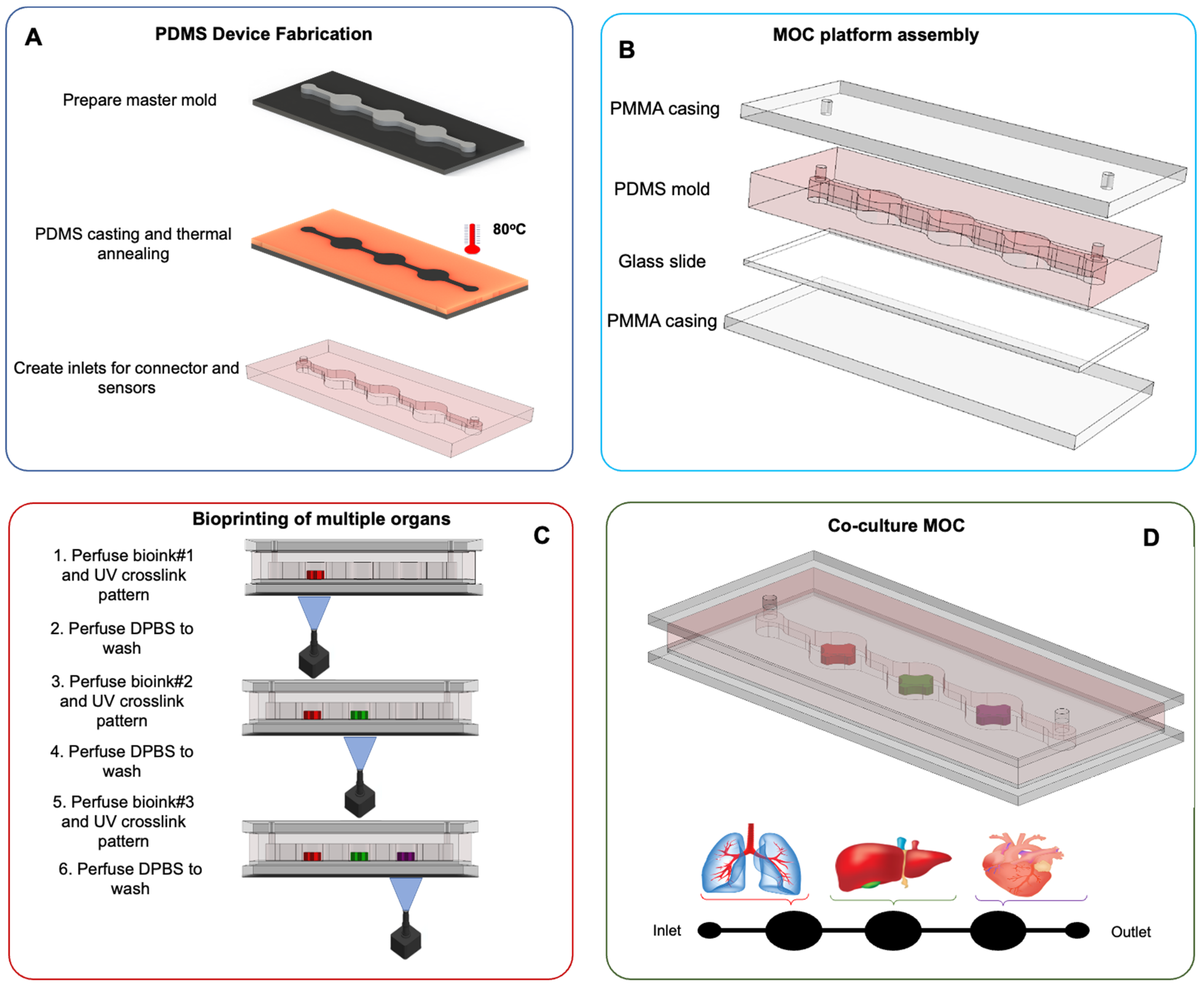
3.1. Fabrication Methods

Conventional microfluidics fabrication methods involve the use of lithography-based molding and casting processes. The processes, such as replica molding, injection molding, and embossing, assist in fabricating MOCs. Silicon, glass, and plastic materials are used in such fabrication methods [45]. One key feature is optical transparency, which restricts the available materials and manufacturing techniques [44]. The use of polydimethylsiloxane (PDMS) leads to optical transparency, biocompatibility, flexibility, and gas permeability [46].
Among the aforementioned microfluidic fabrications processes, replica molding has been used to create PDMS microdevices containing microfabricated structures that mimic the endothelial–epithelial interface [47]. The geometry of this engineered tissue interface was enhanced to resemble the liver’s blood flow rate and provide proper orientation to rat hepatocytes (liver epithelial cells) similar to their in vivo alignment along the endothelium-lined sinusoidal barrier. Simple reconstitution of the microarchitecture of this tissue–tissue interface was sufficient to prompt cultured hepatocytes to self-organize into hepatic cord-like structures and form functional bile canaliculi in vitro even in the absence of living endothelium. Laser ablation and sacrificial replica molding techniques were applied to develop microscale 3-D collagen scaffolds replicating human intestinal villi’s geometry [48]. In this work, the culture of human Caco-2 intestinal epithelial cells produced 3-D epithelial structures exhibited by villi in the human jejunum. A microfabricated breast model was created by applying a similar kind of PDMS-based replica molding [49]. Engineered automation can resolve the precision-related and repeatability limitations of in vitro models. Automated digital microfluidics proved electro-wetting-based control of hepatic organoids in a microdevice containing a set of electrodes to enumerate the behavior of 3-D hepatic platforms in media droplets and monitor the hepatic functions [50]. The highly controlled rate of material exchange and monitoring helped achieve a fast and rapid screening of the cell–drug interactions. Table 1 summarizes some key properties of materials used for the fabrication of microfluidic devices. Silicon and glass have been the ancestors of polymer and hydrogel materials widely adopted in microfluid applications. Among the polymeric materials commonly used, Teflon is a soft, inert, and optically transparent material and relatively permeable to gases [51]. Polycarbonate is a durable, transparent, low-cost material that absorbs UV and has showed low resistance against certain organic solvents [52,53]. Styrene ethylene butylene styrene is a thermoplastic elastomer optically transparent, flexible, and adhesive with low partitioning of drugs and small hydrophobic compounds [54]. Hydrogel-based materials, such as collagen [55,56] and silkworm [57,58,59], are biocompatible and bioactive, enhancing cell attachment and proliferation. However, their poor mechanical properties and low batch-to-batch consistency may compromise experimental reproducibility and cellular response [60].

3.2. Drug Assays

Patients treated with protein-based drugs frequently develop drug-specific neutralizing antibodies, rendering the drugs unavailable to the target sites [74,75]. Neutralizing antibodies can also adversely affect other organs, as demonstrated by recombinant human erythropoietin leading to anemia resulting from antibody-dependent immune responses, which destroy both extrinsic and intrinsic erythropoietin, thus causing abnormal RBC development and production [76,77].
A study found that the effects on the potency, efficiency, hepatotoxicity, and hematological toxicity of an anticancer medicine (Tegafur) channelized with a microfluidic device that were not observed in conventional tissue culture more closely mimicked the results obtained in vivo [78]. Another study observed the previously unknown cardiotoxicity of a chemotherapeutic drug (bleomycin) because of the crosstalk between the lung and the heart tissues [79].
In vitro models provide better physiology, immune status, anatomy, drug metabolism, and host–pathogen interactions [80]. Many drugs have been withdrawn from the market after obtaining approval for humans because of their toxicity, like hepatotoxicity, liver toxicity, and cardiac toxicity (Table S1). Pergolide drug products, indicated in the treatment of Parkinson’s disease, have been linked to serious damage in the heart valves of patients [81], while rofecoxib (Vioxx), a nonsteroidal anti-inflammatory drug, resulted in a higher risk of heart attack in patients undergoing long-term treatment [81]. Both drugs have been voluntarily withdrawn from the market owing to their association with cardiac toxicity. Nefazodone, an antidepressant, and troglitazone (Rezulin), an antidiabetic medication, have been associated with acute liver injury and death and were withdrawn from the market due to increased risk of liver toxicity related to their use. The economic burden, the waste of human-derived resources, and more importantly, the toxic nature of these drugs to humans had detrimental implications on both patients and pharmaceutical companies. Therefore, the strategy has been adapted to use testing methods of drug efficacy with minimal failures.
Another set of microfluidic platforms has been used for studying host–pathogen interactions to predict drug pharmacokinetic responses in patients [1]. Hepatitis B virus (HBV) is among the significant health problems that have affected millions around the globe. A study showed the significance of using the liver-on-a-chip to image HBV interaction with hepatocytes accurately. Primary rat hepatocytes and immortalized bovine-derived aortic endothelial cells were cocultured on the opposite sides of a microporous membrane in a dual microchannel under the continuous flow of culture media. The hepatocytes maintained their polygonal morphology, physiology, division, and related markers like albumin and tumor necrosis factor for more than 20 days. The primary rat hepatocytes were successfully infected with HBV-infected adenovirus. The same model was modified using the primary human hepatocytes, maintaining their morphology and physiology for up to 26 days. The secretion of HBV core antigen and HBV DNA was detected after infection with HBV without adenovirus [82].
The ongoing COVID-19 pandemic has affected billions of lives in one way or another, calling for an adequate drug or vaccine. A human lung airway chip was developed to confront the Food and Drug Administration (FDA)-approved medications against SARS-CoV-2. This platform was created to mimic the human infection by airborne SARS-CoV-2. SARS-CoV-2 pseudo particles (CoV-2pp) carrying the SARS-CoV-2 spike proteins (a vital entry ligand) were channelized into the air channel. They were then exposed to human lung epithelial cells expressing high levels of TMPRSS2 and ACE2. The study showed the impact of amodiaquine and toremifene as potential inhibitors of SARS-COV2 in lung epithelial cells [83,84].

4. High-Throughput Applications and Current Challenges

The use of physiologically relevant MOC models as a screening tool for drug processes in health and disease conditions can accelerate their clinical application and the bench-to-bedside transition. Although the fast development of high-throughput screening (HST) has shown successful R&D productivity in the pharmaceutical industry and drug screening [85,86], there have still been some challenges in the process. First, a complete system of current HST technologies, including liquid handling equipment, data acquisition, extensive robotic liquid, and plate handling equipment, is expensive. The high cost of the HST platforms restricts the screening potential of small molecular targets [87]. Second, the cost of biological reagents and drug libraries is also high, and the current approaches make it challenging to reduce reagent consumption. While the volume capacity in a 384-well plate has been reduced to 100 μL, further minimization of micro-well plates is restricted due to uncontrolled liquid evaporation. The decrease of the volumes is also limited by the difficulty of dispensing tiny volumes smaller than ~1 μL [88]. Third, the failure rate in drug development is high in the clinical phase, and clinical drug development takes approximately ~63% of the total cost [85,86]. The use of appropriate cell-based assays in an early, preclinical stage is expected to provide a more efficient way to eliminate possible false leads due to low drug efficacy or high toxicity [89]. This strategy is complicated for current HST platforms because cell-based assays are more expensive and require complex liquid handling. There is a need for technology with low sample and reagent consumption, low cost, and cell friendly environment.

4.1. Fabrication Challenges

The majority of microfluidic platforms are dominated by the application of PDMS [90]. However, PDMS-based platforms cannot mimic complex tissue and organ architecture. The fabrication of multiple PDMS layers requires sequential integration, which is time-consuming, labor-intensive, and expensive [91]. Apart from PDMS, MOCs may also be bioprinted. This fabrication technique affects the quality and speed of fabrication of micro-tissue models [92]. Fabrication based on the use of a laser is limited by pre-application of cells, inkjet printing has low printing speed and high shear force [93], and extrusion has low printing speed and resolution.
In contrast, stereolithography has long-term cell viability concerns due to toxicity issues stemming from the use of radiation due to the application of UV sources [93]. Apart from the manufacturing techniques of MOCs, the functionalization of these platforms to mimic organs’ essential functions is crucial [40]. Mimicking these functions allows us to achieve accurate and reliable preclinical analysis.
The vascular system allows nutrient and oxygen supply and removes metabolic waste products from the tissues while providing a selective drug barrier [94]. Recapitulating the in vivo dynamic conditions within the vascular microenvironment can be challenging, given that blood flow induces constant shear stress on vascular endothelial cells (Figure 2). In contrast, muscle cell stretching occurs during the cardiac cycle owing to blood vessel distension. The 3-D microenvironment and the dynamic mechanical events of the cardiac cycle are critical in maintaining proper vascular cell function. These factors should be taken into consideration when designing in vitro vascular platforms. The main challenges encountered during the fabrication of such platforms are related to the assembly, handling, and using conventional analytical methods [95]. It may also be challenging to adequately resemble the cylindrical geometry of vascular channels using lithographic techniques since these typically generate rectangular channels on flat surfaces [96]. Material selection may be an additional issue to consider regarding biocompatibility and compatibility with specific assays (Figure 3).
Engineering functional cardiac models that adequately recapitulate the biology of the heart is highly challenging compared to other tissues. An ideal in vitro model should mimic the heart’s cellular organization, mechanical contractions, electrical activity, and transport of molecules. Cardiomyocyte alignment within native heart tissue requires a proper design on the cell substrate [97]. The intrinsic contractions of the heart tissue are an additional challenge to be addressed since the simultaneous cardiomyocyte beating observed in vivo may be easily lost in an in vitro setup [98]. Equally crucial to the recreation of the microenvironmental cues in a cardiac construct would also be incorporating readout systems to record cardiac biological functions, such as contractility [99].

4.2. High-Throughput Challenges

To date, different components for cell-based microfluidic high-throughput screening (µHTS) platforms have been developed, including cell culture [88,100], introduction and transport of samples [101,102], and characterization of cell viability [103,104]. The microfluidic community has focused on the demonstration of integrating these different components into a single microfluidic device. Among current microfluidic platforms for cell-based HST, three major complementary modes of flow manipulation are perfusion flow, droplet-based, and microarray. Additionally, minimization of the well plate platform reduces reagent consumption, which is difficult due to reagent evaporation and difficulty in handling [88]. It includes expensive robotic and plate handling equipment and data acquisition systems [87], in which limitations require a microfluidic platform. The microfluidic platform provides lower reagent consumption and the ability to control cellular microenvironments [87]. The µHTS platform involves integrating numerous microfluidic components, such as valves, mixers, pumps, and sensors, to control fluid flow at the microscale [105]. The microfluidic platforms have been continuously used to detect reagents, particles, cells, or multicellular organisms for chemical and biological analysis. The detection in droplet and perfusion-based microfluidic channels is usually performed using optical, electrochemical, Raman, and mass spectrometry.
In a high-throughput screening platform, making drug gradient increases the capability of parallel screening with different drug concentrations for cell-based drug screening. The cellular response may be evaluated under multiple doses within the same platform, saving time and resources. Usually, drug gradient generators are either flow-based or diffusion-based [106]. However, flow-based gradient generators are popular among pharmaceutical evaluation. “Christmas tree structure”, a flow-based gradient generator, is well accepted for miniaturized microfluidic devices. The gradient is achieved in a miniaturized microfluidic network by sequentially diluting concentrations through a mixer [106]. The selection of the geometry for gradient generation must be considered carefully to produce an appropriate shear environment. These selections may be made by proper numerical simulation studies [107]. The simulation is helpful to determine the gradient and shear rate for individual sections with different geometry.
The optical method uses either standard microscope-based imaging or the application of fluorescence. The fluorescence allows analysis of the images, which gives an insight into biological responses. Cells can be labeled in a high-throughput screening setting, and fluorescence images may track labeled cells and micro-objects [108]. The high-throughput screening platform may integrate multiple sensors. These sensors use multiple techniques, such as conductive, resistive, capacitive, and others, to detect the presence of micro-objects and biological objects [109]. Small molecules in a microfluidic platform may be detected using Raman spectroscopy and mass spectrometry [110,111]. Raman spectroscopy uses light scattering to detect the chemical structure of the analyte, while mass spectroscopy uses the measurement of the mass-to-charge ratio of ions to detect the exact molecular weight of the analyte. The application of Raman spectroscopy is limited due to its low sensitivity; hence, an enhanced approach of surface-enhanced Raman spectroscopy may be used for the high-throughput detection of molecules [112]. Mass spectrometry is fast and has a high resolution, which is used for bioanalysis [111].
Two-dimensional culture has been widely used for high-throughput screening, but it fails to replicate the 3-D model of the liver; thus, the 3-D culture technique was developed. A liver-on-chip platform was designed to recapitulate the native hepatic microenvironment, provide physiological fluid flow and recapitulate hepatic function (Figure 4A) [113]. The organ-on-chip has been applied to mimic rat, dog, and human liver platforms, where the cells were subjected to physiological flow. This platform allows the prediction of liver toxicity detected in animal studies (Figure 4B) [114]. The platform enables real-time monitoring using an integrated 3-D bioprinted sensor that can be used in a liver-on-chip platform. The electrochemical dissolved oxygen sensor was printed inline at multiple places to monitor oxygen concentration (Figure 4C) [115]. It captured liver function in vitro, offering a desirable biomimetic microenvironment. The study applied spheroid models, which enable a high-throughput and parallel culture in a high mass transfer and low shear for long-term perfusion culture [116]. A large-scale liver-lobule-on-chip platform was constructed in a hybrid layout with a separate seed-feed network mimicking the central vein of the liver lobule. The array allowed passage to transport by diffusion-dominated mass transport (Figure 4D) [117].
The MOC high-throughput screening is performed using a continuous-flow, droplet-based, or microfluidic array-based platform [104,118]. The droplet-based platform works according to the principles of droplet generation, where the droplets act as miniaturized reaction compartments that enclose single or multiple cells [119] that may be subjected to drug screening. The droplet-based platform quickly allows a high number of cell and droplet variants quickly but has limited cell growth in droplets. The continuous-flow platform involves controlling the laminar flow of reagent/media to control the environmental condition. The control inflow regulates dissolved gas, temperature, pH, and shear stress inside the microfluidic platform [120]. The application of a continuous-flow platform is limited by swelling when exposed to strong solvents, a limited number of the tested material, and the possibility of cross-contamination. The array-based microfluidic platform is more compatible with drug screening since it enables parallel analysis [121] in a high-throughput approach. It also provides many samples to analyze the biology and small-molecule screening [122]. However, a microarray is limited by the inefficient removal of reagents within the reaction chamber and requires minimal volume detection.

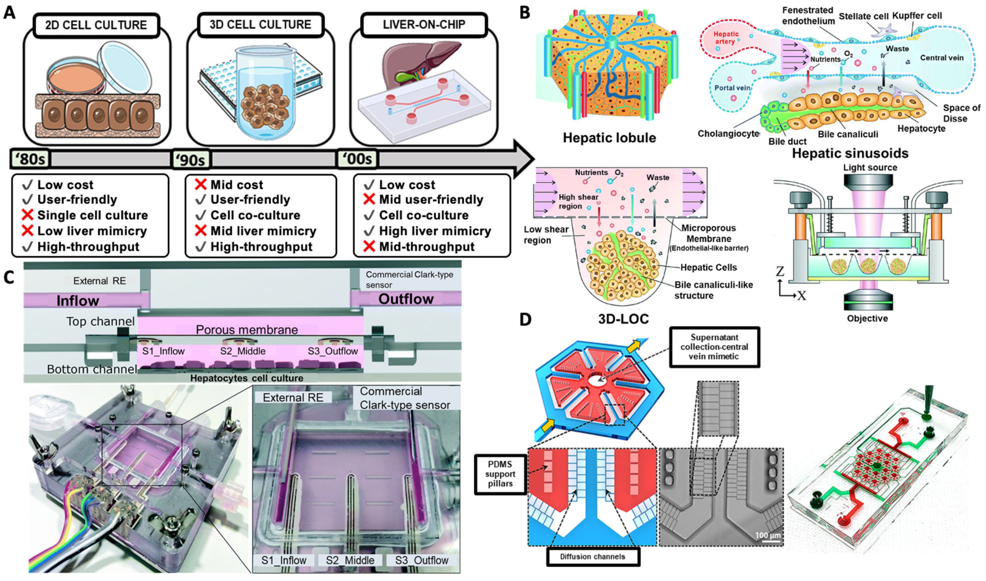
4.2.1. Liver Platforms

The liver is the largest organ in the body consisting of several cell types and the most complex function [90]. Recapitulation of the liver’s structural, cellular, and localized hemodynamic complexity is one of the major challenges in developing organotypic in vitro liver models [123,124]. Even though primary hepatocytes have been the gold standard in liver chips, their dedifferentiation in vitro has led to the use of alternative cell sources, such as mutable human embryonic stem cells and iPSCs. The first steps in attaining structural biomimicry have been made with bile tube simulation, yet functional biomimicry, such as bile acid secretion, is still in progress. The lack of testing standards, uniformity, real-time monitoring, and the relatively low throughput of liver chips are additional technical challenges to be resolved with the integration of biosensors within liver devices [125].
Drug metabolism and detoxification processes are additional challenges to be addressed in the liver. As a potential drug carrier, the metabolic fate of nanoparticles and their effect on human tissues has been intensively investigated. Within this context, Esch et al. assembled a chip based on the coculture of enterocytes, Caco-2, and mucin-producing HT29-MTX cells, and HepG2/C3A liver cells to mimic the oral uptake of carboxylated polystyrene nanoparticles (50 nm) and their effects on the liver. The Caco-2/HT29-MTX coculture posed an effective barrier to nanoparticle permeability across the cell layer, yet the nanoparticle fraction crossing this layer mediated the release of aspartate aminotransferase, an indicator of liver cell injury [126].
Liver models have been made by 2-D planar culture, matrix-less and matrix-dependent 3-D culture, layer-by-layer deposition, 3-D bioprinting, microarray, and hanging drops [125]. These fabrication strategies are viable from short- to long-term biological studies. Among these strategies, 2-D culture, 3-D-based bioprinting, and microarray strategies may be used for high-throughput screening. These strategies allow the analysis of biological responses to drugs. Still, they are limited by applying multiple cells in 2-D culture, resolution, and control over individual cells in bioprinting, and lack of spatial distribution and cellular interaction in microarray strategies.

4.2.2. Lung Platforms

Lung chips have introduced a credible alternative to 2-D cell cultures, emulating the microarchitecture and the primary physiological functions of the human lung better to understand the physiology and pathology of the human lungs and perform drug screening and toxicological studies [23]. One of the major challenges of microfluidic lung platforms in their current form is their short life span, owing to their short-term compatibility. The design of more biorelevant blood flow networks could potentially minimize the foreign body response, thus providing lung devices for more long-term applications [127].
Benam et al. engineered a human lung small airway chip to recapitulate the features of asthma and chronic obstructive pulmonary disease (COPD) in vitro and evaluate the therapeutic response in the small airway chips after drug treatment to overcome the challenge of extending the drug’s residence. The PDMS microfluidic device was constructed using soft lithography and consisted of two channels separated from a polyester membrane coated on both sides with type I collagen. Primary human airway epithelial cells and primary human lung microvascular endothelial cells were cultured on opposite sides of the membrane. Epithelium exposure to interleukin-13 and substituting human airway epithelial cells with epithelial cells from individuals with COPD generated in vitro models of asthma and COPD, respectively. This represents a challenge for the small airway chip device, providing a complementary approach to in vivo models by adequately recapitulating the in vivo organ-level therapeutic responses [128].
Aiming to simulate the lung tumor microenvironment for chemotherapeutic drug screening applications, a lung ToC device was constructed using an electrospun PLGA membrane as a substrate for cell culture [129,130]. Exposure of the coculture of human lung non-small cell lung cancer cell line A549 and human fetal lung fibroblast HFL1 cells to the epidermal growth factor receptor EGFR-targeted drug gefitinib showed A549 cell resistance. In contrast, the triple coculture of A549, HFL1, and HUVEC cells resulted in the A549 cells showing a strong invasive ability and possibly causing HUVECs’ death [129]. In a similar approach, a PLGA nanofiber/PDMS composite membrane-sandwiched microdevice enabled better simulation of the tumor lung microenvironment from a biophysical and biochemical perspective, allowing for fluid perfusion under very low shear stress and cell culture under both normoxic and hypoxic conditions.

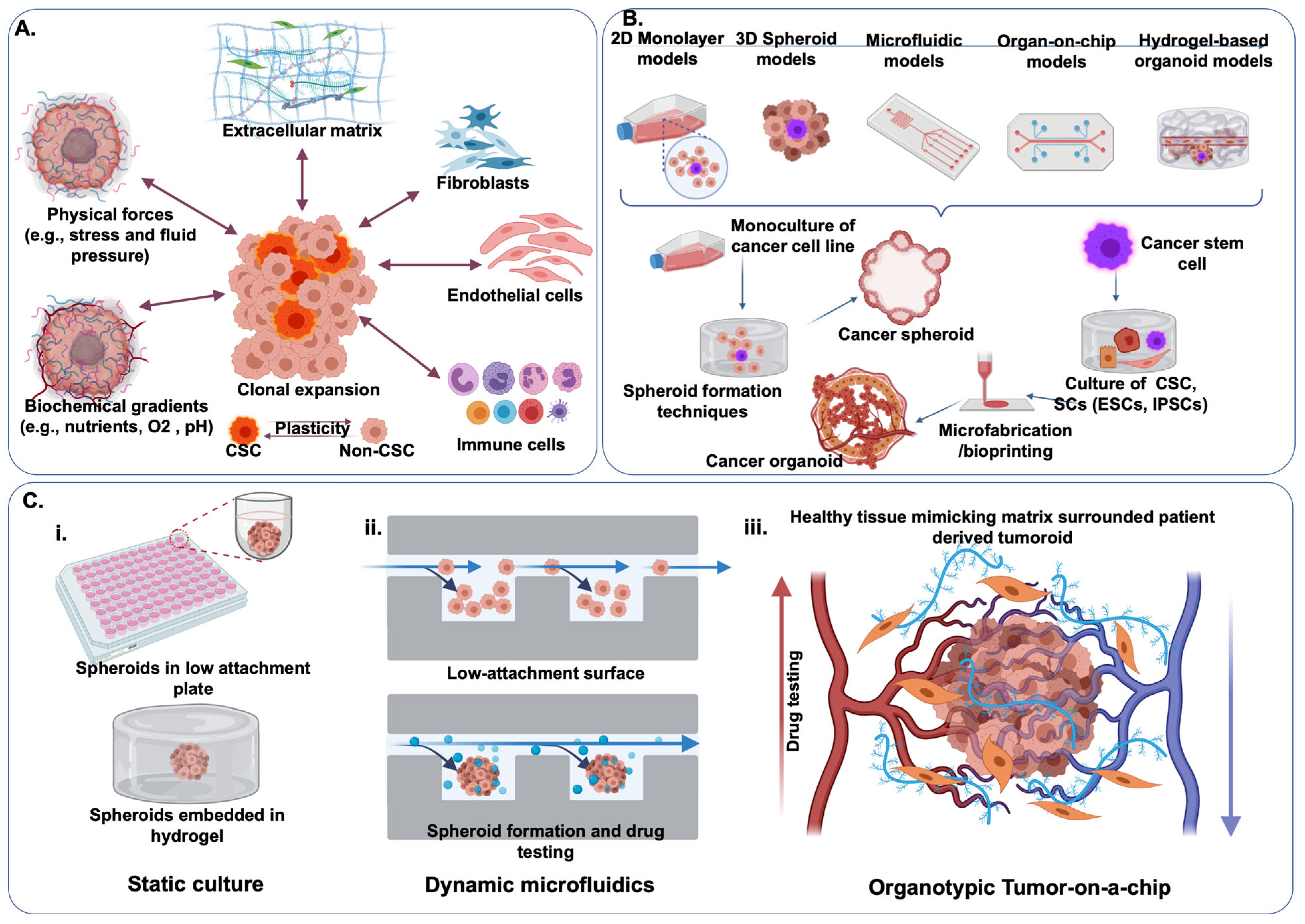
4.3. Tumor Platforms

ToC is essential for developing functional, reproducible, and robust microfluidics suitable for drug screening assays. This was exemplified in a colon cancer micro-tumor model perfused with the anticancer compounds fluorouracil, vincristine, and sorafenib compared with conventional monolayer cultures of endothelial or tumor cells. Results indicated that perfusable vascular networks are critical for drug safety evaluation, highlighting an additional challenge since this model better mimics the physiological microenvironment than 2-D cell cultures [131].
Identification of optimal chemotherapeutic drug combinations is sometimes essential to treat invasive carcinomas, such as bladder cancer. The therapeutic potential of a single or a variety of chemotherapeutic agents has been assessed in a microfluidic platform combining the coculture of the bladder carcinoma cell line T24 with HUVECs and drug transport across the endothelial monolayer. Results showed that even though complete inhibition of T24 cell dispersal was attained in monoculture and the presence of all four tested drugs at a concentration of 10 mM, the same effect was not observed in the T24 and HUVECs coculture. The microfluidic platform could help define combination chemotherapeutic strategies that present a significant challenge in vitro to treat aggressive tumors [132]. The non-specificity of current chemotherapeutic approaches is an additional challenge to be addressed. Towards this direction, a ToC device using a coculture of human breast cancer cells and hepatic cells aimed to identify optimum drug concentrations of doxorubicin, cisplatin, and paclitaxel that could increase drug efficacy against cancer cells and reduce toxicity against healthy cells [133].
Many improvements have been made in treating various tumor types by researchers and clinicians. However, most tumor models have poor approximations to patients’ tumors, and medications with promising tumor model data may still fail in clinical trials [134]. The complex tumor microenvironment (TME) includes different cell types, ECM-derived physical stresses, oxygen, nutrient, and biochemical gradients supported by a complex vascular web. Conventional 2-D models cannot mimic the interactive relationships among these complicated relationships between TME components, such as tumor cells, stromal cells, and physical and biochemical factors inside living tumors in the presence of blood vessels [92,135]. Animal tumor models have provided valuable insights into our basic understanding of tumor biology; however, these models cannot reflect pathogenic processes in humans due to genetic alterations [136,137]. We still need better tumor models to fully understand tumor behavior and the TME, including the inflammatory factors, immune system response effectors and suppressors, cell heterogeneity, and tumor vasculature, which is significantly different from healthy vasculature. Some models have been used to assess chemical cues that influence cell migration and invasion through a porous membrane that separates two chambers in Transwell containers [138]. Multicellular tumor spheroids can mimic cell–cell and cell–ECM interactions between tumor cells and the TME. Spheroids may develop oxygen and nutrition gradients, resulting in an established necrotic core similar to poorly vascularized tumor tissue [139,140]. In vitro spheroids still lack the tumor vasculature in the TME.
Since establishing the first human cancer cell line HeLa, cancer disease models clearly illustrate an evolving nature from 2-D simple models to complex hydrogel-based organoid models (Figure 5B). Microfluidics technology has been considered a gamechanger in tumor modeling because manipulating a few chemical agents has allowed researchers to preserve tumor cells in a controlled encasement while the flow of the culture medium imposes the dynamic physiological factors in the vasculature. Researchers use soft lithography or 3-D printers to create 3-D channels and chambers in PDMS or other flexible polymers. The micro-channels are connected in micro-chambers, where cells can be captured and aggregated. Hydrogels containing cells or fluid are perfused into the built-in channels and chambers to simulate the ECM cues. By continuous perfusion of nutrients and oxygen and the removal of waste products, microfluidic devices can imitate the in vivo fluid dynamics of the TME. The interstitial pressure, soluble factor gradients, and oxygen tension are all physicochemical parameters that microfluidic devices can precisely control [141]. ToC has been developed for low-cost and high-throughput anticancer drug screening in precision medicine [142].
To investigate cancer cell migration and invasion and extracellular signaling, as well as chemotherapy and immunotherapy resistance mechanisms, multiple 3-D tumor-on-a-chip models have been demonstrated to replicate diverse forms of solid and liquid TMEs, including different stromal components, immune suppressor cells, and chemokines [143,144,145,146]. Cui et al. [147] reported a glioblastoma (GBM) on-chip platform mimicking the several GBM tumor niche subtypes (proneural, classic, and mesenchymal). The chip consists of a peripheral channel. ECs formed a vascular channel, an intermediate tumor-stromal zone, where patient-derived GBM cells, tumor-derived macrophages (TAMs), and T-cells can interact with a central port. They demonstrated an immunosuppressive tumor microenvironment analyzing cytotoxic activity and significantly reducing T-cell activation and cytotoxic function in GBM tumor niche subtypes. Similarly, Jerkins et al. [147] demonstrated a tumor spheroids model to investigate the PD-1 blockade profile by recapitulating the native tumor immune microenvironment. They showed that a short-term organotypic tumor spheroid microfluidic model could recapitulate response to PD-1 blockade and identify specific interventions that counteract resistance.
Multicellular aggregates and tumor aggregates, namely spheroids, have been used as tumor models over the years under static conditions [148]. However, these models have failed to capture the effect of blood flow. Vascularized tumor models are highly promising to mimic a hostile TME that leads to cancer progression and drug resistance. Nashimoto et al. [149] demonstrated the importance of flow in a vascular network in TME to evaluate tumor activities as a drug screening platform. They fabricated a tumor-on-chip platform that enables monitoring and assessing tumor response with intraluminal flow combining the PDMS microfluidics with multicellular tumor spheroid-embedded hydrogel. They showed that drug administration under perfused conditions did not demonstrate the dose-dependent effects of anticancer drugs on tumor activities compared to the results under static conditions.

5. Conclusions

There are real grounds for optimism that MOC systems will provide a more effective, time-, and cost-efficient surrogate to current small-molecule screening methods. The potential to outgrow conventional in vitro cell culture and animal tests will significantly contribute to the ‘3Rs’ regarding the use of laboratory animals. Despite the immense potential of MOCs, this technology is still at an infant stage, facing several fundamental and translational challenges that need to be addressed. The transition from MOC to human body-on-a chip platforms is the next goal to provide even more physiologically relevant systems for drug development and screening assays. The simulation of fluid flow dynamics, selecting appropriate growth factors, and culture media are important. New culture techniques are required to support the growth and proliferation of different cell types in a single platform. Increasing the relatively low throughput and automation of current MOC platforms is also essential for accelerating the dynamics of this technology. Scaling up and simplifying the complex MOC setup would make them more user-friendly and bring them one step closer to commercialization. Towards this direction, developers should also opt for platform flexibility for MOCs to be easily integrated with different platforms and efficiently operate in various applications [151]. The establishment of specific performance standards is another aspect to be considered, as MOCs are increasingly entering the market. The lack of a universal standard is one of the main reasons pharmaceutical industries are still reluctant to introduce MOCs to their portfolios. Validation of biological, technical, and analytical aspects in the performance of MOCs is critical as they can not only reproduce the organ or tissue of interest but also consistently respond to compounds with a well-established mechanism of action or toxicity. Within this context, MOCs could prove a useful preclinical tool in screening drug toxicity and efficacy and could potentially substitute animal models and support the findings of phase I and phase II clinical trials. At the same time, the use of patient-derived cell sources could additionally introduce the element of personalization. However, it is critical to establish functional and dynamic collaborations between the academic, industrial, and regulatory sectors to bridge the existing translational chasm.
The FDA, the US National Institutes of Health (NIH), and the Defense Advanced Research Projects Agency (DARPA) have acted on this demand by providing funding opportunities for the development of organs-on-chips and the regulatory support required to set the roadmap towards market [152]. In Europe, the human Organ and Disease Model Technologies (DMT) consortium recently joined forces with 21 companies, three knowledge institutions, and two foundations to develop a flexible ‘SMART Organ-on-Chip’ under the auspice of an NWO ‘Perspective’ grant. The Asia-Pacific region is witnessing exponential market growth rates, mainly due to the government funding opportunities provided to research institutes, especially in China, and the increasing number of clinical trials based on cell studies [147]. The startup founding dynamics, especially within academic settings, is also a striking example of the continuous and comprehensive efforts to bring the MOCs technology to the market. Emulate is a microfluidics startup founded in the Wyss Institute for Biologically Inspired Engineering in Harvard that has established collaborations with AstraZeneca and Johnson & Johnson to enable more precise predictions of the effects of drugs on humans. InSphero, created at the Zurich university in Switzerland, aims to develop safer and more efficient drugs. AxoSim (Tulane University), TaraBiosystems (Columbia University, New York, NY, USA), and Nortis Bio (University of Washington, Washington, DC, USA) focus on the development of nerves-on-chip, heart-on-chip, and kidneys-on-chip devices, respectively. At the same time, TissUse (Technische University, Berlin, Germany) has recently introduced a MOC with four organs integrated on the same chip, further aiming to develop a human-on-chip with 10 organs. It, therefore, is clear that only through coordinated efforts from all stakeholders will the MOC technology flourish and find its place among well-established models and procedures for small-molecule drug testing.

Supplementary Materials

The following are available online at https://www.mdpi.com/article/10.3390/pharmaceutics13101657/s1, Table S1. Drugs that have been withdrawn from the market owing to their toxicity.

Author Contributions

Conceptualization, B.C., C.K., M.N. and A.K.M.; literature review B.C., C.K., M.N., A.B., E.D. and F.S.; writing—original draft preparation, B.C., C.K., M.N. and A.K.M.; writing—review and editing, B.C., C.K., M.N., A.B., E.D., F.S., S.T., M.B. and L.D.K.; supervision, S.T., M.B. and L.D.K.; funding acquisition, A.K.M. All authors have read and agreed to the published version of the manuscript.

Funding

This research was funded by National Institutes of Health (NIH, R21DC018818) and New Jersey Institute of Technology (NJIT, Start-Up funding) awarded to A.K.M.

Institutional Review Board Statement

Not applicable.

Informed Consent Statement

Not applicable.

Data Availability Statement

Not applicable.

Acknowledgments

A.K.M. acknowledges the receipt of DC18818 from the National Institutes of Health and Start-up Fund from NJIT.

Conflicts of Interest

The authors declare no conflict of interest.

References

  1. Herland, A.; Maoz, B.M.; Das, D.; Somayaji, M.R.; Prantil-Baun, R.; Novak, R.; Cronce, M.; Huffstater, T.; Jeanty, S.S.F.; Ingram, M.; et al. Quantitative prediction of human pharmacokinetic responses to drugs via fluidically coupled vascularized organ chips. Nat. Biomed. Eng. 2020, 4, 421–436. [Google Scholar] [CrossRef] [PubMed]
  2. Kimura, H.; Sakai, Y.; Fujii, T. Organ/body-on-a-chip based on microfluidic technology for drug discovery. Drug Metab. Pharmacokinet. 2018, 33, 43–48. [Google Scholar] [CrossRef] [PubMed]
  3. Bailey, J.; Thew, M.; Balls, M. An Analysis of the Use of Animal Models in Predicting Human Toxicology and Drug Safety. Altern. Lab. Anim. 2014, 42, 181–199. [Google Scholar] [CrossRef] [PubMed] [Green Version]
  4. Kingwell, K. 3D cell technologies head to the R&D assembly line. Nat. Rev. Drug Discov. 2017, 16, 6–7. [Google Scholar] [CrossRef]
  5. Langer, R.; Vacanti, J.P. Tissue engineering. Science 1993, 260, 920–926. [Google Scholar] [CrossRef] [PubMed] [Green Version]
  6. Barros, A.S.; Costa, A.; Sarmento, B. Building three-dimensional lung models for studying pharmacokinetics of inhaled drugs. Adv. Drug Deliv. Rev. 2021, 170, 386–395. [Google Scholar] [CrossRef] [PubMed]
  7. Skardal, A.; Shupe, T.; Atala, A. Organoid-on-a-chip and body-on-a-chip systems for drug screening and disease modeling. Drug Discov. Today 2016, 21, 1399–1411. [Google Scholar] [CrossRef]
  8. Park, D.; Lee, J.; Chung, J.J.; Jung, Y.; Kim, S.H. Integrating Organs-on-Chips: Multiplexing, Scaling, Vascularization, and Innervation. Trends Biotechnol. 2020, 38, 99–112. [Google Scholar] [CrossRef]
  9. Jalili-Firoozinezhad, S.; Miranda, C.C.; Cabral, J.M. Modeling the Human Body on Microfluidic Chips. Trends Biotechnol. 2021, 39, 838–852. [Google Scholar] [CrossRef]
  10. Maschmeyer, I.; Lorenz, A.K.; Schimek, K.; Hasenberg, T.; Ramme, A.P.; Hübner, J.; Lindner, M.; Drewell, C.; Bauer, S.; Thomas, A.; et al. A four-organ-chip for interconnected long-term co-culture of human intestine, liver, skin and kidney equivalents. Lab Chip 2015, 15, 2688–2699. [Google Scholar] [CrossRef] [Green Version]
  11. De Mello, C.P.P.; Carmona-Moran, C.A.; McAleer, C.W.; Perez, J.; Coln, E.A.; Long, C.J.; Oleaga, C.; Riu, A.; Note, R.; Teissier, S.; et al. Microphysiological heart–liver body-on-a-chip system with a skin mimic for evaluating topical drug delivery. Lab Chip 2020, 20, 749–759. [Google Scholar] [CrossRef] [PubMed]
  12. Sasserath, T.; Rumsey, J.W.; McAleer, C.W.; Bridges, L.R.; Long, C.J.; Elbrecht, D.; Schuler, F.; Roth, A.; Bertinetti-LaPatki, C.; Shuler, M.L.; et al. Differential Monocyte Actuation in a Three-Organ Functional Innate Immune System-on-a-Chip. Adv. Sci. 2020, 7, 2000323. [Google Scholar] [CrossRef]
  13. Villenave, R.; Wales, S.Q.; Hamkins-Indik, T.; Papafragkou, E.; Weaver, J.C.; Ferrante, T.C.; Bahinski, A.; Elkins, C.; Kulka, M.; Ingber, D.E. Human Gut-On-A-Chip Supports Polarized Infection of Coxsackie B1 Virus In Vitro. PLoS ONE 2017, 12, e0169412. [Google Scholar] [CrossRef] [PubMed]
  14. Cifuente, J.; Ferrer, M.F.; de Giusti, C.J.; Song, W.-C.; Romanowski, V.; Hafenstein, S.L.; Gómez, R.M. Molecular determinants of disease in coxsackievirus B1 murine infection. J. Med. Virol. 2011, 83, 1571–1581. [Google Scholar] [CrossRef] [PubMed] [Green Version]
  15. Jia, Z.; Guo, Z.; Yang, C.-T.; Prestidge, C.; Thierry, B. “Mucus-on-Chip”: A new tool to study the dynamic penetration of nanoparticulate drug carriers into mucus. Int. J. Pharm. 2021, 598, 120391. [Google Scholar] [CrossRef]
  16. Kamaly, N.; Yameen, B.; Wu, J.; Farokhzad, O.C. Degradable Controlled-Release Polymers and Polymeric Nanoparticles: Mechanisms of Controlling Drug Release. Chem. Rev. 2016, 116, 2602–2663. [Google Scholar] [CrossRef] [Green Version]
  17. Sebe, I.; Szabó, P.; Kállai-Szabó, B.; Zelkó, R. Incorporating small molecules or biologics into nanofibers for optimized drug release: A review. Int. J. Pharm. 2015, 494, 516–530. [Google Scholar] [CrossRef]
  18. Zhang, Y.; Fang, C.; Carvalho, W.S.P.; Gao, Y.; Serpe, M.J. Triggered Small-Molecule Release from Dual-Stimuli Responsive Microgels. ACS Appl. Polym. Mater. 2021, 3, 410–417. [Google Scholar] [CrossRef]
  19. Hill, L.K.; Meleties, M.; Katyal, P.; Xie, X.; Delgado-Fukushima, E.G.; Jihad, T.; Liu, C.-F.; O’Neill, S.C.; Tu, R.S.; Renfrew, P.D.; et al. Thermoresponsive Protein-Engineered Coiled-Coil Hydrogel for Sustained Small Molecule Release. Biomacromolecules 2019, 20, 3340–3351. [Google Scholar] [CrossRef]
  20. Morihiro, K.; Ankenbruck, N.; Lukasak, B.; Deiters, A. Small Molecule Release and Activation through DNA Computing. J. Am. Chem. Soc. 2017, 139, 13909–13915. [Google Scholar] [CrossRef]
  21. Santini, J.T.; Cima, M.J.; Langer, R. A controlled-release microchip. Nat. Cell Biol. 1999, 397, 335–338. [Google Scholar] [CrossRef]
  22. Zhang, B.; Korolj, A.; Lai, B.F.L.; Radisic, M. Advances in organ-on-a-chip engineering. Nat. Rev. Mater. 2018, 3, 257–278. [Google Scholar] [CrossRef]
  23. Huh, D.; Matthews, B.D.; Mammoto, A.; Montoya-Zavala, M.; Hsin, H.Y.; Ingber, D.E. Reconstituting Organ-Level Lung Functions on a Chip. Science 2010, 328, 1662–1668. [Google Scholar] [CrossRef] [Green Version]
  24. Ferrari, E.; Rasponi, M. Liver–Heart on chip models for drug safety. APL Bioeng. 2021, 5, 031505. [Google Scholar] [CrossRef]
  25. Li, X.-G.; Chen, M.-X.; Zhao, S.-Q.; Wang, X.-Q. Intestinal Models for Personalized Medicine: From Conventional Models to Microfluidic Primary Intestine-on-a-chip. Stem Cell Rev. Rep. 2021, 1–15. [Google Scholar] [CrossRef]
  26. Sun, A.M.; Hoffman, T.; Luu, B.Q.; Ashammakhi, N.; Li, S. Application of lung microphysiological systems to COVID-19 modeling and drug discovery: A review. Bio-Des. Manuf. 2021, 1–19. [Google Scholar] [CrossRef]
  27. Phillips, J.A.; Grandhi, T.S.P.; Davis, M.; Gautier, J.-C.; Hariparsad, N.; Keller, D.; Sura, R.; Van Vleet, T.R. A pharmaceutical industry perspective on microphysiological kidney systems for evaluation of safety for new therapies. Lab Chip 2020, 20, 468–476. [Google Scholar] [CrossRef] [PubMed]
  28. Wang, Y.; Oleaga, C.; Long, C.J.; Esch, M.B.; McAleer, C.W.; Miller, P.G.; Hickman, J.J.; Shuler, M.L. Self-contained, low-cost Body-on-a-Chip systems for drug development. Exp. Biol. Med. 2017, 242, 1701–1713. [Google Scholar] [CrossRef] [PubMed]
  29. Kolahchi, A.R.; Mohtaram, N.K.; Modarres, H.P.; Mohammadi, M.H.; Geraili, A.; Jafari, P.; Akbari, M.; Sanati-Nezhad, A. Microfluidic-Based Multi-Organ Platforms for Drug Discovery. Micromachines 2016, 7, 162. [Google Scholar] [CrossRef] [PubMed]
  30. Zhao, Y.; Kankala, R.K.; Wang, S.-B.; Chen, A.-Z. Multi-Organs-on-Chips: Towards Long-Term Biomedical Investigations. Molecules 2019, 24, 675. [Google Scholar] [CrossRef] [PubMed] [Green Version]
  31. Lee, J.B.; Sung, J.H. Organ-on-a-chip technology and microfluidic whole-body models for pharmacokinetic drug toxicity screening. Biotechnol. J. 2013, 8, 1258–1266. [Google Scholar] [CrossRef]
  32. Van der Zanden, S.Y.; Luimstra, J.; Neefjes, J.; Borst, J.; Ovaa, H. Opportunities for Small Molecules in Cancer Immunotherapy. Trends Immunol. 2020, 41, 493–511. [Google Scholar] [CrossRef] [PubMed]
  33. Makurvet, F.D. Biologics vs. small molecules: Drug costs and patient access. Med. Drug Discov. 2021, 9, 100075. [Google Scholar] [CrossRef]
  34. Ngo, H.X.; Garneau-Tsodikova, S. What are the drugs of the future? MedChemComm 2018, 9, 757–758. [Google Scholar] [CrossRef] [PubMed]
  35. Warner, K.D.; Hajdin, C.E.; Weeks, K.M. Principles for targeting RNA with drug-like small molecules. Nat. Rev. Drug Discov. 2018, 17, 547–558. [Google Scholar] [CrossRef]
  36. Govardhanagiri, S.; Bethi, S.; Nagaraju, G.P. Small-Molecules and Pancreatic Cancer Trials and Troubles. In Breaking Tolerance to Pancreatic Cancer Unresponsiveness to Chemotherapy; Academic Press: Cambridge, MA, USA, 2019. [Google Scholar] [CrossRef]
  37. Ma, C.; Zhao, L.; Zhou, E.-M.; Xu, J.; Shen, S.; Wang, J. On-Chip Construction of Liver Lobule-like Microtissue and Its Application for Adverse Drug Reaction Assay. Anal. Chem. 2016, 88, 1719–1727. [Google Scholar] [CrossRef] [PubMed]
  38. Wagner, I.; Materne, E.-M.; Brincker, S.; Süssbier, U.; Frädrich, C.; Busek, M.; Sonntag, F.; Sakharov, D.A.; Trushkin, E.V.; Tonevitsky, A.; et al. A dynamic multi-organ-chip for long-term cultivation and substance testing proven by 3D human liver and skin tissue co-culture. Lab Chip 2013, 13, 3538–3547. [Google Scholar] [CrossRef] [Green Version]
  39. Edington, C.D.; Chen, W.L.K.; Geishecker, E.; Kassis, T.; Soenksen, L.R.; Bhushan, B.M.; Freake, D.; Kirschner, J.; Maass, C.; Tsamandouras, N.; et al. Interconnected Microphysiological Systems for Quantitative Biology and Pharmacology Studies. Sci. Rep. 2018, 8, 4530. [Google Scholar] [CrossRef] [Green Version]
  40. Chen, X.; Zhang, Y.S.; Zhang, X.; Liu, C. Organ-on-a-chip platforms for accelerating the evaluation of nanomedicine. Bioact. Mater. 2021, 6, 1012–1027. [Google Scholar] [CrossRef]
  41. Miller, P.G.; Shuler, M.L. Design and demonstration of a pumpless 14 compartment microphysiological system. Biotechnol. Bioeng. 2016, 113, 2213–2227. [Google Scholar] [CrossRef]
  42. Tang, Y.; Soroush, F.; Sheffield, J.B.; Wang, B.; Prabhakarpandian, B.; Kiani, M.F. A Biomimetic Microfluidic Tumor Microenvironment Platform Mimicking the EPR Effect for Rapid Screening of Drug Delivery Systems. Sci. Rep. 2017, 7, 1–14. [Google Scholar] [CrossRef]
  43. Park, J.Y.; Jang, J.; Kang, H.-W. 3D Bioprinting and its application to organ-on-a-chip. Microelectron. Eng. 2018, 200, 1–11. [Google Scholar] [CrossRef]
  44. Lee, H.; Cho, D.-W. One-step fabrication of an organ-on-a-chip with spatial heterogeneity using a 3D bioprinting technology. Lab Chip 2016, 16, 2618–2625. [Google Scholar] [CrossRef] [Green Version]
  45. Convery, N.; Gadegaard, N. 30 years of microfluidics. Micro Nano Eng. 2019, 2, 76–91. [Google Scholar] [CrossRef]
  46. Spivey, E.C.; Xhemalce, B.; Shear, J.B.; Finkelstein, I.J. 3D-Printed Microfluidic Microdissector for High-Throughput Studies of Cellular Aging. Anal. Chem. 2014, 86, 7406–7412. [Google Scholar] [CrossRef] [Green Version]
  47. Nakao, Y.; Kimura, H.; Sakai, Y.; Fujii, T. Bile canaliculi formation by aligning rat primary hepatocytes in a microfluidic device. Biomicrofluidics 2011, 5, 022212. [Google Scholar] [CrossRef] [Green Version]
  48. Sung, J.H.; Yu, J.; Luo, D.; Shuler, M.L.; March, J.C. Microscale 3-D hydrogel scaffold for biomimetic gastrointestinal (GI) tract model. Lab Chip 2011, 11, 389–392. [Google Scholar] [CrossRef]
  49. Grafton, M.M.; Wang, L.; Vidi, P.-A.; Leary, J.; Lelievre, S. Breast on-a-chip: Mimicry of the channeling system of the breast for development of theranostics. Integr. Biol. 2011, 3, 451–459. [Google Scholar] [CrossRef]
  50. Au, S.; Chamberlain, M.; Mahesh, S.; Sefton, M.V.; Wheeler, A.R. Hepatic organoids for microfluidic drug screening. Lab Chip 2014, 14, 3290–3299. [Google Scholar] [CrossRef] [PubMed]
  51. Grover, W.; von Muhlen, M.; Manalis, S.R. Teflon films for chemically-inert microfluidic valves and pumps. Lab Chip 2008, 8, 913–918. [Google Scholar] [CrossRef] [PubMed]
  52. Chudobova, D.; Cihalova, K.; Skalickova, S.; Zitka, J.; Rodrigo, M.A.M.; Milosavljevic, V.; Hynek, D.; Kopel, P.; Vesely, R.; Adam, V.; et al. 3D-printed chip for detection of methicillin-resistant Staphylococcus aureuslabeled with gold nanoparticles. Electrophoresis 2015, 36, 457–466. [Google Scholar] [CrossRef]
  53. Krejcova, L.; Nejdl, L.; Rodrigo, M.A.M.; Zurek, M.; Matousek, M.; Hynek, D.; Zitka, O.; Kopel, P.; Adam, V.; Kizek, R. 3D printed chip for electrochemical detection of influenza virus labeled with CdS quantum dots. Biosens. Bioelectron. 2014, 54, 421–427. [Google Scholar] [CrossRef]
  54. Domansky, K.; Sliz, J.D.; Wen, N.; Hinojosa, C.; Thompson, G.; Fraser, J.P.; Hamkins-Indik, T.; Hamilton, G.A.; Levner, D.; Ingber, D.E. SEBS elastomers for fabrication of microfluidic devices with reduced drug absorption by injection molding and extrusion. Microfluid. Nanofluidics 2017, 21, 107. [Google Scholar] [CrossRef]
  55. Yi, H.; Wu, L.-Q.; Ghodssi, R.; Rubloff, G.; Payne, G.F.; Bentley, W.E. Signal-Directed Sequential Assembly of Biomolecules on Patterned Surfaces. Langmuir 2005, 21, 2104–2107. [Google Scholar] [CrossRef]
  56. Yi, H.; Wu, L.Q.; Bentley, W.E.; Ghodssi, R.; Rubloff, G.W.; Culver, J.N.; Payne, G.F. Biofabrication with Chitosan. Biomacromolecules 2005, 6, 2881–2894. [Google Scholar] [CrossRef]
  57. Bettinger, C.J.; Cyr, K.M.; Matsumoto, A.; Langer, R.; Borenstein, J.T.; Kaplan, D.L. Silk Fibroin Microfluidic Devices. Adv. Mater. 2007, 19, 2847–2850. [Google Scholar] [CrossRef]
  58. Pérez-Rigueiro, J.; Viney, C.; Llorca, J.; Elices, M. Mechanical properties of single-brin silkworm silk. J. Appl. Polym. Sci. 2000, 75, 1270–1277. [Google Scholar] [CrossRef]
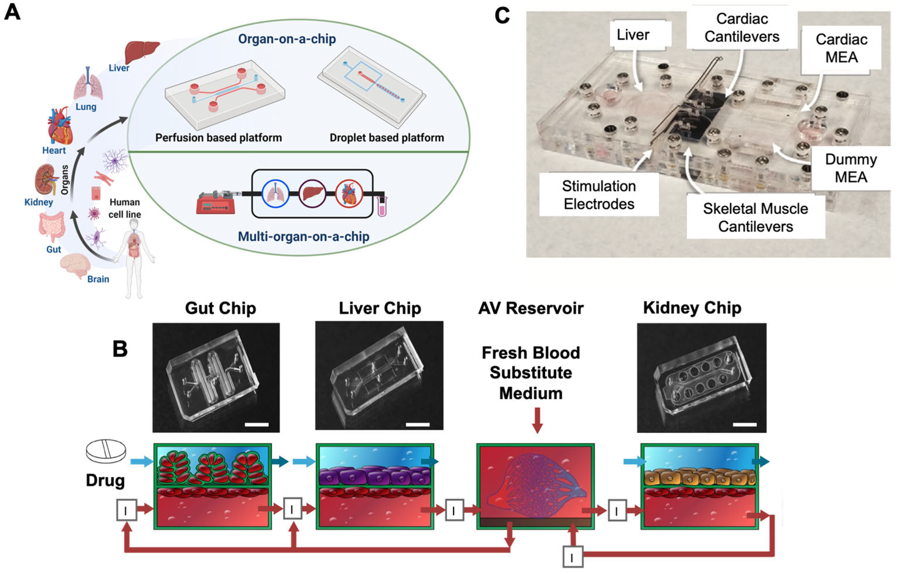
  59. Schimek, K.; Busek, M.; Brincker, S.; Groth, B.; Hoffmann, S.; Lauster, R.; Lindner, G.; Lorenz, A.; Menzel, U.; Sonntag, F.; et al. Integrating biological vasculature into a multi-organ-chip microsystem. Lab Chip 2013, 13, 3588–3598. [Google Scholar] [CrossRef] [PubMed] [Green Version]
  60. Hasenberg, T.; Mühleder, S.; Dotzler, A.; Bauer, S.; Labuda, K.; Holnthoner, W.; Redl, H.; Lauster, R.; Marx, U. Emulating human microcapillaries in a multi-organ-chip platform. J. Biotechnol. 2015, 216, 1–10. [Google Scholar] [CrossRef]
  61. Ling, Y.; Rubin, J.; Deng, Y.; Huang, C.; Demirci, U.; Karp, J.M.; Khademhosseini, A. A cell-laden microfluidic hydrogel. Lab Chip 2007, 7, 756–762. [Google Scholar] [CrossRef] [PubMed]
  62. Rahfoth, B.; Weisser, J.; Sternkopf, F.; Aigner, T.; Von Der Mark, K.; Bräuer, R. Transplantation of allograft chondrocytes embedded in agarose gel into cartilage defects of rabbits. Osteoarthr. Cartil. 1998, 6, 50–65. [Google Scholar] [CrossRef] [Green Version]
  63. Sugioka, K.; Hanada, Y.; Midorikawa, K. 3D microstructuring of glass by femtosecond laser direct writing and application to biophotonic microchips. Prog. Electromagn. Res. Lett. 2008, 1, 181–188. [Google Scholar] [CrossRef] [Green Version]
  64. Hanada, Y.; Sugioka, K.; Shihira-Ishikawa, I.; Kawano, H.; Miyawaki, A.; Midorikawa, K. 3D microfluidic chips with integrated functional microelements fabricated by a femtosecond laser for studying the gliding mechanism of cyanobacteria. Lab Chip 2011, 11, 2109–2115. [Google Scholar] [CrossRef]
  65. Sosa-Hernández, J.E.; Villalba-Rodríguez, A.M.; Romero-Castillo, K.D.; Aguilar-Aguila-Isaías, M.A.; García-Reyes, I.E. Organs-on-a-chip module: A review from the development and applications perspective. Micromachines 2018, 9, 536. [Google Scholar] [CrossRef] [PubMed] [Green Version]
  66. Chen, T.F.; Siow, K.S.; Ng, P.Y.; Majlis, B.Y. Enhancing the biocompatibility of the polyurethane methacrylate and off-stoichiometry thiol-ene polymers by argon and nitrogen plasma treatment. Mater. Sci. Eng. C 2017, 79, 613–621. [Google Scholar] [CrossRef] [PubMed]
  67. Kuo, J.S.; Ng, L.; Yen, G.S.; Lorenz, R.M.; Schiro, P.G.; Edgar, J.S.; Zhao, Y.; Lim, D.S.W.; Allen, P.B.; Jeffries, G.D.M.; et al. A new USP Class VI-compliant substrate for manufacturing disposable microfluidic devices. Lab Chip 2009, 9, 870–876. [Google Scholar] [CrossRef] [Green Version]
  68. Cherpinski, A.; Torres-Giner, S.; Vartiainen, J.; Peresin, M.S.; Lahtinen, P.; Lagaron, J.M. Improving the water resistance of nanocellulose-based films with polyhydroxyalkanoates processed by the electrospinning coating technique. Cellulose 2018, 25, 1291–1307. [Google Scholar] [CrossRef]
  69. Alfadhel, A.; Quyang, J.; Mahajan, C.G.; Forouzandeh, F.; Cormier, D.; Borkholder, D.A. Inkjet printed polyethylene glycol as a fugitive ink for the fabrication of flexible microfluidic systems. Mater. Design 2018, 150, 182–187. [Google Scholar] [CrossRef] [PubMed]
  70. Shim, K.; Kim, S.H.; Lee, N.; Kim, B.; Kim, T.H.; Jung, Y.; Choi, N.; Sung, J.H. Fabrication of micrometer-scale porous gelatin scaffolds for 3D cell culture. J. Ind. Eng. Chem. 2017, 50, 183–189. [Google Scholar] [CrossRef]
  71. Zamboni, F.; Keays, M.; Hayes, S.; Albadarin, A.; Walker, G.; Kiely, P.A.; Collins, M.N. Enhanced cell viability in hyaluronic acid coated poly(lactic-co-glycolic acid) porous scaffolds within microfluidic channels. Int. J. Pharm. 2017, 532, 595–602. [Google Scholar] [CrossRef]
  72. Miller, J.; Stevens, K.R.; Yang, M.T.; Baker, B.; Nguyen, D.-H.T.; Cohen, D.; Toro, E.; Chen, A.A.; Galie, P.; Yu, X.; et al. Rapid casting of patterned vascular networks for perfusable engineered three-dimensional tissues. Nat. Mater. 2012, 11, 768–774. [Google Scholar] [CrossRef] [PubMed]
  73. Bertassoni, L.E.; Cecconi, M.; Manoharan, V.; Nikkhah, M.; Hjortnaes, J.; Cristino, A.L.; Barabaschi, G.; Demarchi, D.; Dokmeci, M.R.; Yang, Y.; et al. Hydrogel bioprinted microchannel networks for vascularization of tissue engineering constructs. Lab Chip 2014, 14, 2202–2211. [Google Scholar] [CrossRef] [Green Version]
  74. Porter, S. Human immune response to recombinant human proteins. J. Pharm. Sci. 2011, 90, 1–11. [Google Scholar] [CrossRef]
  75. Perini, P.; Facchinetti, A.; Bulian, P.; Massaro, A.R.; De Pascalis, D.; Bertolotto, A.; Biasi, G.; Gallo, P. Interferon-beta (INF-b) antibodies in interferon-b1a-and interferon-b1b-treated multiple sclerosis patients. Prevalence, kinetics, cross-reactivity, and factors enhancing interferon-b immunogenicity in vivo. Eur. Cytokine Netw. 2001, 12, 56–61. [Google Scholar] [PubMed]
  76. Casadevall, N.; Nataf, J.; Viron, B.; Kolta, A.; Kiladjian, J.-J.; Martin-Dupont, P.; Michaud, P.; Papo, T.; Ugo, V.; Teyssandier, I.; et al. Pure Red-Cell Aplasia and Antierythropoietin Antibodies in Patients Treated with Recombinant Erythropoietin. N. Engl. J. Med. 2002, 346, 469–475. [Google Scholar] [CrossRef] [PubMed]
  77. Sanchez, S.; Barger, T.; Zhou, L.; Hale, M.; Mytych, D.; Gupta, S.; Swanson, S.J.; Civoli, F. Strategy to confirm the presence of anti-erythropoietin neutralizing antibodies in human serum. J. Pharm. Biomed. Anal. 2011, 55, 1265–1274. [Google Scholar] [CrossRef] [PubMed]
  78. Sung, J.H.; Shuler, M.L. A micro cell culture analog (µCCA) with 3-D hydrogel culture of multiple cell lines to assess metabolism-dependent cytotoxicity of anti-cancer drugs. Lab Chip 2009, 9, 1385–1394. [Google Scholar] [CrossRef]
  79. Skardal, A.; Murphy, S.V.; Devarasetty, M.; Mead, I.; Kang, H.-W.; Seol, Y.-J.; Zhang, Y.S.; Shin, S.-R.; Zhao, L.; Aleman, J.; et al. Multi-tissue interactions in an integrated three-tissue organ-on-a-chip platform. Sci. Rep. 2017, 7, 1–16. [Google Scholar] [CrossRef]
  80. Maharjan, S.; Cecen, B.; Zhang, Y.S. 3D Immunocompetent Organ-on-a-Chip Models. Small Methods 2020, 4, 2000235. [Google Scholar] [CrossRef]
  81. Qureshi, Z.P.; Seoane-Vazquez, E.; Rodriguez-Monguio, R.; Stevenson, K.B.; Szeinbach, S.L. Market withdrawal of new molecular entities approved in the United States from 1980 to 2009. Pharmacoepidemiol. Drug Saf. 2011, 20, 772–777. [Google Scholar] [CrossRef]
  82. Kang, Y.B.; Rawat, S.; Duchemin, N.; Bouchard, M.; Noh, M. Human Liver Sinusoid on a Chip for Hepatitis B Virus Replication Study. Micromachines 2017, 8, 27. [Google Scholar] [CrossRef] [Green Version]
  83. Si, L.; Bai, H.; Rodas, M.; Cao, W.; Oh, C.Y.; Jiang, A.; Nurani, A.; Zhu, D.Y.; Goyal, G.; Gilpin, S.E.; et al. Human organs-on-chips as tools for repurposing approved drugs as potential influenza and COVID19 therapeutics in viral pandemics. BioRxiv 2020. [Google Scholar] [CrossRef]
  84. Hoffmann, M.; Kleine-Weber, H.; Schroeder, S.; Krüger, N.; Herrler, T.; Erichsen, S.; Schiergens, T.S.; Herrler, G.; Wu, N.H.; Nitsche, A.; et al. SARS-CoV-2 cell entry depends on ACE2 and TMPRSS2 and is blocked by a clinically proven protease inhibitor. Cell 2020, 181, 271–280. [Google Scholar] [CrossRef]
  85. Paul, S.M.; Mytelka, D.; Dunwiddie, C.T.; Persinger, C.C.; Munos, B.H.; Lindborg, S.R.; Schacht, A.L. How to improve R&D productivity: The pharmaceutical industry’s grand challenge. Nat. Rev. Drug Discov. 2010, 9, 203–214. [Google Scholar] [CrossRef] [PubMed]
  86. Pammolli, F.; Magazzini, L.; Riccaboni, M. The productivity crisis in pharmaceutical R&D. Nat. Rev. Drug Discov. 2011, 10, 428–438. [Google Scholar] [CrossRef] [PubMed]
  87. Wu, G.; Doberstein, S.K. HTS technologies in biopharmaceutical discovery. Drug Discov. Today 2006, 11, 718–724. [Google Scholar] [CrossRef] [PubMed]
  88. Wu, M.-H.; Huang, S.-B.; Lee, G.-B. Microfluidic cell culture systems for drug research. Lab Chip 2010, 10, 939–956. [Google Scholar] [CrossRef]
  89. Bhadriraju, K.; Chen, C.S. Engineering cellular microenvironments to improve cell-based drug testing. Drug Discov. Today 2002, 7, 612–620. [Google Scholar] [CrossRef]
  90. Damiati, S.; Kompella, U.B.; Damiati, S.A.; Kodzius, R. Microfluidic Devices for Drug Delivery Systems and Drug Screening. Genes 2018, 9, 103. [Google Scholar] [CrossRef] [Green Version]
  91. Miri, A.K.; Mostafavi, E.; Khorsandi, D.; Hu, S.-K.; Malpica, M.; Khademhosseini, A. Bioprinters for organs-on-chips. Biofabrication 2019, 11, 042002. [Google Scholar] [CrossRef]
  92. Sontheimer-Phelps, A.; Hassell, B.A.; Ingber, D.E. Modelling cancer in microfluidic human organs-on-chips. Nat. Rev. Cancer 2019, 19, 65–81. [Google Scholar] [CrossRef]
  93. Samavedi, S.; Joy, N. 3D printing for the development of in vitro cancer models. Curr. Opin. Biomed. Eng. 2017, 2, 35–42. [Google Scholar] [CrossRef]
  94. Ribas, J.; Sadeghi, H.; Manbachi, A.; Leijten, J.C.H.; Brinegar, K.; Zhang, Y.S.; Ferreira, L.; Khademhosseini, A. Cardiovascular Organ-on-a-Chip Platforms for Drug Discovery and Development. Appl. Vitr. Toxicol. 2016, 2, 82–96. [Google Scholar] [CrossRef] [Green Version]
  95. Van der Meer, A.D.; Poot, A.A.; Duits, M.H.G.; Feijen, J.; Vermes, I. Microfluidic Technology in Vascular Research. J. Biomed. Biotechnol. 2009, 2009, 1–10. [Google Scholar] [CrossRef]
  96. Gold, K.; Gaharwar, A.K.; Jain, A. Emerging trends in multiscale modeling of vascular pathophysiology: Organ-on-a-chip and 3D printing. Biomaterials 2019, 196, 2–17. [Google Scholar] [CrossRef] [PubMed]
  97. Ahmed, M.; Yildirimer, L.; Khademhosseini, A.; Seifalian, A. Nanostructured Materials for Cardiovascular Tissue Engineering. J. Nanosci. Nanotechnol. 2012, 12, 4775–4785. [Google Scholar] [CrossRef] [PubMed] [Green Version]
  98. Tandon, N.; Cannizzaro, C.; Chao, P.-H.G.; Maidhof, R.; Marsano, A.; Au, H.T.H.; Radisic, M.; Vunjak-Novakovic, G. Electrical stimulation systems for cardiac tissue engineering. Nat. Protoc. 2009, 4, 155–173. [Google Scholar] [CrossRef] [Green Version]
  99. Zhang, Y.S.; Aleman, J.; Arneri, A.; Bersini, S.; Piraino, F.; Shin, S.R.; Dokmeci, M.R.; Khademhosseini, A. From cardiac tissue engineering to heart-on-a-chip: Beating challenges. Biomed. Mater. 2015, 10, 034006. [Google Scholar] [CrossRef] [PubMed]
  100. Halldorsson, S.; Lucumi, E.; Gomez-Sjoberg, R.; Fleming, R.M. Advantages and challenges of microfluidic cell culture in polydimethylsiloxane devices. Biosens. Bioelectron. 2015, 63, 218–231. [Google Scholar] [CrossRef] [Green Version]
  101. Melin, J.; Quake, S.R. Microfluidic Large-Scale Integration: The Evolution of Design Rules for Biological Automation. Annu. Rev. Biophys. Biomol. Struct. 2007, 36, 213–231. [Google Scholar] [CrossRef] [Green Version]
  102. Hong, J.; Edel, J.B.; Demello, A.J. Micro-and nanofluidic systems for high-throughput biological screening. Drug Discov. Today 2009, 14, 134–146. [Google Scholar] [CrossRef]
  103. Wang, Z.; Kim, M.-C.; Marquez, M.; Thorsen, T. High-density microfluidic arrays for cell cytotoxicity analysis. Lab Chip 2007, 7, 740–745. [Google Scholar] [CrossRef] [PubMed]
  104. Dressler, O.J.; Maceiczyk, R.M.; Chang, S.I.; Demello, A.J. Droplet-Based Microfluidics: Enabling Impact on Drug Discovery. J. Biomol. Screen. 2013, 19, 483–496. [Google Scholar] [CrossRef] [PubMed]
  105. Beebe, D.J.; Mensing, G.A.; Walker, G.M. Physics and Applications of Microfluidics in Biology. Annu. Rev. Biomed. Eng. 2002, 4, 261–286. [Google Scholar] [CrossRef] [PubMed]
  106. Shen, S.; Zhang, F.; Gao, M.; Niu, Y. Concentration Gradient Constructions Using Inertial Microfluidics for Studying Tumor Cell–Drug Interactions. Micromachines 2020, 11, 493. [Google Scholar] [CrossRef] [PubMed]
  107. Shourabi, A.Y.; Kashaninejad, N.; Saidi, M.S. An integrated microfluidic concentration gradient generator for mechanical stimulation and drug delivery. J. Sci. Adv. Mater. Devices 2021, 6, 280–290. [Google Scholar] [CrossRef]
  108. Coskun, A.F.; Su, T.-W.; Ozcan, A. Wide field-of-view lens-free fluorescent imaging on a chip. Lab Chip 2010, 10, 824–827. [Google Scholar] [CrossRef] [Green Version]
  109. Kemna, E.W.M.; Segerink, L.I.; Wolbers, F.; Vermes, I.; Berg, A.V.D. Label-free, high-throughput, electrical detection of cells in droplets. Analyst 2013, 138, 4585–4592. [Google Scholar] [CrossRef]
  110. Ferstl, W.; Klahn, T.; Schweikert, W.; Billeb, G.; Schwarzer, M.; Loebbecke, S. Inline Analysis in Microreaction Technology: A Suitable Tool for Process Screening and Optimization. Chem. Eng. Technol. 2007, 30, 370–378. [Google Scholar] [CrossRef]
  111. Sun, S.; Kennedy, R.T. Droplet Electrospray Ionization Mass Spectrometry for High Throughput Screening for Enzyme Inhibitors. Anal. Chem. 2014, 86, 9309–9314. [Google Scholar] [CrossRef] [Green Version]
  112. Zhou, Q.; Kim, T. Review of microfluidic approaches for surface-enhanced Raman scattering. Sens. Actuators B Chem. 2016, 227, 504–514. [Google Scholar] [CrossRef]
  113. Polidoro, M.A.; Ferrari, E.; Marzorati, S.; Lleo, A.; Rasponi, M. Experimental liver models: From cell culture techniques to microfluidic organs-on-chip. Liver. Int. 2021, 41, 1744–1761. [Google Scholar] [CrossRef]
  114. Jang, K.-J.; Otieno, M.A.; Ronxhi, J.; Lim, H.-K.; Ewart, L.; Kodella, K.R.; Petropolis, D.B.; Kulkarni, G.; Rubins, J.E.; Conegliano, D.; et al. Reproducing human and cross-species drug toxicities using a Liver-Chip. Sci. Transl. Med. 2019, 11, eaax5516. [Google Scholar] [CrossRef]
  115. Moya, A.; Ortega-Ribera, M.; Guimerà, X.; Sowade, E.; Zea, M.; Illa, X.; Ramon, E.; Villa, R.; Gracia-Sancho, J.; Gabriel, G. Online oxygen monitoring using integrated inkjet-printed sensors in a liver-on-a-chip system. Lab Chip 2018, 18, 2023–2035. [Google Scholar] [CrossRef] [Green Version]
  116. Ma, L.-D.; Wang, Y.-T.; Wang, J.-R.; Wu, J.-L.; Meng, X.-S.; Hu, P.; Mu, X.; Liang, Q.-L.; Luo, G.-A. Design and fabrication of a liver-on-a-chip platform for convenient, highly efficient, and safe in situ perfusion culture of 3D hepatic spheroids. Lab Chip 2018, 18, 2547–2562. [Google Scholar] [CrossRef] [PubMed]
  117. Banaeiyan, A.A.; Theobald, J.; Paukštyte, J.; Wölfl, S.; Adiels, C.B.; Goksör, M. Design and fabrication of a scalable liver-lobule-on-a-chip microphysiological platform. Biofabrication 2017, 9, 15014. [Google Scholar] [CrossRef] [PubMed]
  118. Du, G.; Fang, Q.; den Toonder, J.M. Microfluidics for cell-based high throughput screening platforms—A review. Anal. Chim. Acta 2016, 903, 36–50. [Google Scholar] [CrossRef] [PubMed] [Green Version]
  119. An, X.; Zuo, P.; Ye, B.-C. A single cell droplet microfluidic system for quantitative determination of food-borne pathogens. Talanta 2020, 209, 120571. [Google Scholar] [CrossRef] [PubMed]
  120. Occhetta, P.; Centola, M.; Tonnarelli, B.; Redaelli, A.; Martin, I.; Rasponi, M. High-throughput microfluidic platform for 3D cultures of mesenchymal stem cells. In 3D Cell Culture; Humana Press: New York, NY, USA, 2017; pp. 303–323. [Google Scholar]
  121. Fernandes, T.G.; Diogo, M.M.; Clark, D.S.; Dordick, J.S.; Cabral, J.M. High-throughput cellular microarray platforms: Applications in drug discovery, toxicology and stem cell research. Trends Biotechnol. 2009, 27, 342–349. [Google Scholar] [CrossRef] [PubMed] [Green Version]
  122. Jeon, J.S.; Bersini, S.; Gilardi, M.; Dubini, G.; Charest, J.L.; Moretti, M.; Kamm, R.D. Human 3D vascularized organotypic microfluidic assays to study breast cancer cell extravasation. Proc. Natl. Acad. Sci. USA 2015, 112, E818. [Google Scholar] [CrossRef] [PubMed] [Green Version]
  123. Robinton, D.A.; Daley, G.Q. The promise of induced pluripotent stem cells in research and therapy. Nat. Cell Biol. 2012, 481, 295–305. [Google Scholar] [CrossRef]
  124. Scott, C.W.; Peters, M.F.; Dragan, Y.P. Human induced pluripotent stem cells and their use in drug discovery for toxicity testing. Toxicol. Lett. 2013, 219, 49–58. [Google Scholar] [CrossRef]
  125. Deng, J.; Wei, W.; Chen, Z.; Lin, B.; Zhao, W.; Luo, Y.; Zhang, X. Engineered Liver-on-a-Chip Platform to Mimic Liver Functions and Its Biomedical Applications: A Review. Micromachines 2019, 10, 676. [Google Scholar] [CrossRef] [PubMed] [Green Version]
  126. Esch, M.B.; Mahler, G.J.; Stokol, T.; Shuler, M.L. Body-on-a-chip simulation with gastrointestinal tract and liver tissues suggests that ingested nanoparticles have the potential to cause liver injury. Lab Chip 2014, 14, 3081–3092. [Google Scholar] [CrossRef] [PubMed]
  127. Potkay, J.A. The promise of microfluidic artificial lungs. Lab Chip 2014, 14, 4122–4138. [Google Scholar] [CrossRef] [PubMed]
  128. Benam, K.H.; Villenave, R.; Lucchesi, C.; Varone, A.; Hubeau, C.; Lee, H.-H.; Alves, S.E.; Salmon, M.; Ferrante, T.C.; Weaver, J.C.; et al. Small airway-on-a-chip enables analysis of human lung inflammation and drug responses in vitro. Nat. Methods 2016, 13, 151–157. [Google Scholar] [CrossRef]
  129. Yang, X.; Li, K.; Zhang, X.; Liu, C.; Guo, B.; Wen, W.; Gao, X. Nanofiber membrane supported lung-on-a-chip microdevice for anti-cancer drug testing. Lab Chip 2018, 18, 486–495. [Google Scholar] [CrossRef]
  130. Li, W.; Sun, X.; Ji, B.; Yang, X.; Zhou, B.; Lu, Z.; Gao, X. PLGA Nanofiber/PDMS Microporous Composite Membrane-Sandwiched Microchip for Drug Testing. Micromachines 2020, 11, 1054. [Google Scholar] [CrossRef]
  131. Liu, Y.Y.; Sakolish, C.; Chen, Z.; Phan, D.T.; Bender, R.H.F.; Hughes, C.C.; Rusyn, I. Human in vitro vascularized micro-organ and micro-tumor models are reproducible organ-on-a-chip platforms for studies of anticancer drugs. Toxicology 2020, 445, 152601. [Google Scholar] [CrossRef]
  132. Bai, J.; Tu, T.-Y.; Kim, C.; Thiery, J.P.; Kamm, R.D. Identification of drugs as single agents or in combination to prevent carcinoma dissemination in a microfluidic 3D environment. Oncotarget 2015, 6, 36603–36614. [Google Scholar] [CrossRef] [Green Version]
  133. Lin, D.; Li, P.; Lin, J.; Shu, B.; Wang, W.; Zhang, Q.; Yang, N.; Liu, D.; Xu, B. Orthogonal Screening of Anticancer Drugs Using an Open-Access Microfluidic Tissue Array System. Anal. Chem. 2017, 89, 11976–11984. [Google Scholar] [CrossRef]
  134. Kamb, A. What’s wrong with our cancer models? Nat. Rev. Drug Discov. 2005, 4, 161–165. [Google Scholar] [CrossRef]
  135. Balkwill, F.R.; Capasso, M.; Hagemann, T. The tumor microenvironment at a glance. J. Cell Sci. 2012, 125, 5591–5596. [Google Scholar] [CrossRef] [Green Version]
  136. Gengenbacher, N.; Singhal, M.; Augustin, H.G. Preclinical mouse solid tumour models: Status quo, challenges and perspectives. Nat. Rev. Cancer 2017, 17, 751–765. [Google Scholar] [CrossRef]
  137. Joffe, A.R.; Bara, M.; Anton, N.; Nobis, N. The ethics of animal research: A survey of the public and scientists in North America. BMC Med. Ethic 2016, 17, 1–12. [Google Scholar] [CrossRef] [Green Version]
  138. Kenney, R.M.; Loeser, A.; Whitman, N.A.; Lockett, M.R. Paper-based Transwell assays: An inexpensive alternative to study cellular invasion. Analyst 2019, 144, 206–211. [Google Scholar] [CrossRef]
  139. Huang, B.-W.; Gao, J.-Q. Application of 3D cultured multicellular spheroid tumor models in tumor-targeted drug delivery system research. J. Control. Release 2018, 270, 246–259. [Google Scholar] [CrossRef]
  140. Edmondson, R.; Broglie, J.J.; Adcock, A.F.; Yang, L. Three-Dimensional Cell Culture Systems and Their Applications in Drug Discovery and Cell-Based Biosensors. ASSAY Drug Dev. Technol. 2014, 12, 207–218. [Google Scholar] [CrossRef] [PubMed] [Green Version]
  141. Del Piccolo, N.; Shirure, V.S.; Bi, Y.; Goedegebuure, S.P.; Gholami, S.; Hughes, C.C.; Fields, R.C.; George, S.C. Tumor-on-chip modeling of organ-specific cancer and metastasis. Adv. Drug Deliv. Rev. 2021, 175, 113798. [Google Scholar] [CrossRef] [PubMed]
  142. Liu, X.; Fang, J.; Huang, S.; Wu, X.; Xie, X.; Wang, J.; Liu, F.; Zhang, M.; Peng, Z.; Hu, N. Tumor-on-a-chip: From bioinspired design to biomedical application. Microsyst. Nanoeng. 2021, 7, 1–23. [Google Scholar] [CrossRef] [PubMed]
  143. Ma, C.; Witkowski, M.T.; Harris, J.; Dolgalev, I.; Sreeram, S.; Qian, W.; Tong, J.; Chen, X.; Aifantis, I.; Chen, W. Leukemia-on-a-chip: Dissecting the chemoresistance mechanisms in B cell acute lymphoblastic leukemia bone marrow niche. Sci. Adv. 2020, 6, eaba5536. [Google Scholar] [CrossRef]
  144. Nguyen, D.-H.T.; Lee, E.; Alimperti, S.; Norgard, R.J.; Wong, A.; Lee, J.J.-K.; Eyckmans, J.; Stanger, B.Z.; Chen, C.S. A biomimetic pancreatic cancer on-chip reveals endothelial ablation via ALK7 signaling. Sci. Adv. 2019, 5, eaav6789. [Google Scholar] [CrossRef] [Green Version]
  145. Marturano-Kruik, A.; Nava, M.; Yeager, K.; Chramiec, A.; Hao, L.; Robinson, S.; Guo, E.; Raimondi, M.T.; Vunjak-Novakovic, G. Human bone perivascular niche-on-a-chip for studying metastatic colonization. Proc. Natl. Acad. Sci. USA 2018, 115, 1256–1261. [Google Scholar] [CrossRef] [Green Version]
  146. Ma, C.; Peng, Y.; Li, H.; Chen, W. Organ-on-a-Chip: A New Paradigm for Drug Development. Trends Pharmacol. Sci. 2021, 42, 119–133. [Google Scholar] [CrossRef]
  147. Cui, X.; Settleman, J.; Vasudevaraja, V.; Serrano, J.; Tong, J.; Peng, Y.; DeLorenzo, M.; Shen, G.; Frenster, J.; Morales, R.-T.T.; et al. Dissecting the immunosuppressive tumor microenvironments in Glioblastoma-on-a-Chip for optimized PD-1 immunotherapy. eLife 2020, 9, 52253. [Google Scholar] [CrossRef]
  148. Liu, X.; Raju, P. 5.42–In Vitro Cancer Model for Drug Testing. In Comprehensive Biotechnology, 2nd ed.; Academic Press: Burlington, NJ, USA, 2011; pp. 543–549. [Google Scholar] [CrossRef]
  149. Nashimoto, Y.; Okada, R.; Hanada, S.; Arima, Y.; Nishiyama, K.; Miura, T.; Yokokawa, R. Vascularized cancer on a chip: The effect of perfusion on growth and drug delivery of tumor spheroid. Biomaterials 2020, 229, 119547. [Google Scholar] [CrossRef] [PubMed]
  150. Dogan, E.; Kisim, A.; Bati-Ayaz, G.; Kubicek, G.J.; Pesen-Okvur, D.; Miri, A.K. Cancer Stem Cells in Tumor Modeling: Challenges and Future Directions. Adv. NanoBiomed Res. 2021, 2100017. [Google Scholar] [CrossRef]
  151. Ching, T.; Toh, Y.-C.; Hashimoto, M.; Zhang, Y.S. Bridging the academia-to-industry gap: Organ-on-a-chip platforms for safety and toxicology assessment. Trends Pharmacol. Sci. 2021, 42, 715–728. [Google Scholar] [CrossRef] [PubMed]
  152. Low, L.A.; Mummery, C.; Berridge, B.R.; Austin, C.P.; Tagle, D.A. Organs-on-chips: Into the next decade. Nat. Rev. Drug Discov. 2021, 20, 345–361. [Google Scholar] [CrossRef] [PubMed]
Figure 1. (A) Photographs of the microfluidic platforms. Created with biorender. (B) Diagrams of the gut, liver, and kidney chips fluidically linked to each other and the reservoir; bar scale is 5 mm. Reprinted with permission from [1], Spring Nature 2020. (C) Photograph of the multi-organ housing as an immune system featuring recirculating THP-1 immune cells with cardiomyocytes microelectrode arrays (MEA), skeletal muscle, and liver in separate compartments. Reprinted with permission from [12], John Wiley and Sons, 2020.
Pharmaceutics 13 01657 g001
Figure 2. (A) (a) Characterization of the established human dermal microvascular endothelial cells (HDMECs) microvasculature at day 4, (i) live-cell viability staining (Calcein AM assay), (ii) the uniform distribution of ac-LDL uptake, (b) showed viable and evenly distributed HDMECs; bar scale is 2 mm, (c) showed HDMECs inside the microchannel. Reprinted with permission from [59], RSC 2013. (B) D rendering of cocultures in different configurations. Reprinted with permission from [60], Elsevier 2015.
Pharmaceutics 13 01657 g002
Figure 3. Fabrication of PDMS MOC for co-culture. (A) PDMS device fabrication using casting and thermal annealing. (B) Assembly of the MOC platform. (C) Bioprinting of multiple organs by applying bioink perfusion and UV crosslinking to fabricate micromodels within the culture chamber. (D) A potential design for the MOC platform. Created with biorender.
Pharmaceutics 13 01657 g003
Figure 4. (A) Timeline for the cell culture technique, the advantages and limitations of 2-D culture, 3-D culture, and liver-on-a-chip. Reprinted with permission from [113], John Wiley and Sons 2021. (B) Perfusion culture for hepatic lobule in a concave microwell designed to mimic in the in vivo liver microenvironment and a cross-sectional view of the device. Reprinted with permission from [116], RSC 2018. (C) The liver platform has a cross-sectional view with an electrochemical dissolved oxygen sensor (left) and an accurate image of a liver system with all the fluidic and electrical connections. Reprinted with permission from [115], RSC 2018. (D) Schematic of a very large-sale liver-lobule-on-a-chip device fabricated using PDMS with PDMS support pillars to support the tissue chamber and align cells, and the actual device filled with food dye. Reprinted with permission from [117], IOPscience 2017.
Pharmaceutics 13 01657 g004
Figure 5. Recapitulation of the tumor microtissue with various degrees of sophistication. (A) Schematics TME factors and (B) evolution and future directions of cancer modeling from simple 2-D cultures to tumoroid models. Reprinted with permission from [150], Adv. NanoBiomed Res 2021. (C) The evolution of different tumor microtissue research tools, (i) from static tumor spheroid culture, (ii) dynamic microfluidics culture, and (iii) vascularized organotypic ToCs. Created with biorender.
Pharmaceutics 13 01657 g005
Table 1. Biomaterials used in the fabrication of microfluidic devices.
MaterialRelevant PropertyApplicationReferences
TeflonEase of fabrication with maximum chemical resistanceVery sensitive assays, ultra-clean tools, valves, and pumps fabrication[51]
Acrylonitrile butadiene styreneHigh resolution, best topography Crafting of the master mold, study of pathogenic organisms[52]
Styrene ethylene butylene styreneLow drug absorption, optical transmittance Human lung epithelial cells, human umbilical vein endothelial HUVECs, human alveolar epithelial cells [54]
ChitosanBiocompatible, effective control of stereochemistry Biosensors, film organization [55,56]
Silkworm Biocompatible, pliable Fabrication of microfluidic platforms [57,58]
PDMSGood turnaround time, multi-material printing, long-lasting and high-temperature-resistant substance Master molding[59]
Agarose Minimal toxicity, biodegradability, tunable stability at a lower solid ratio Chondrocytes, AML-12 murine hepatocytes, sensors, and actuators[61,62]
Photocurable resin/polymerEffective resolution with small characters Study of cell growth[63,64]
Polyurethane-methacrylateEconomical to manufacture, biocompatibility, no cytotoxicity, strong electroosmotic mobilityIncreased-aspect-ratio microstructures[65,66,67]
PolyhydroxyalkanoatesBiocompatibility, tunable, biodegradabilityMicrofilm barrier for vapor and oxygen [68]
Polyethylene glycolsCheaper than many of the material, different weight categories are available, biocompatible, cytotoxicity approximately naught Microfluidic valves, microfluidic channels with an increased expiry time[69]
Gelatin methacrylatePhotopolymerizable, porous membraneMechanistic vascular and valvular biology cell support matrix[70]
Polylactic acid and polyglycolic acidMechanical biodegradationPorous scaffold for cell culture with better adhesion[71]
Synthetic hydrogelsInduration and contraction act as sensors and actuatorsSelf-regulating valves, micro-lens arrays, drug release, antigen adsorption flow sensors pH regulators[72,73]
Publisher’s Note: MDPI stays neutral with regard to jurisdictional claims in published maps and institutional affiliations.

