
Continuous Versus Intermittent Linezolid Infusion for Critically Ill Patients with Hospital-Acquired and Ventilator-Associated Pneumonia: Efficacy and Safety Challenges

 , , and
Abstract
:1. Introduction
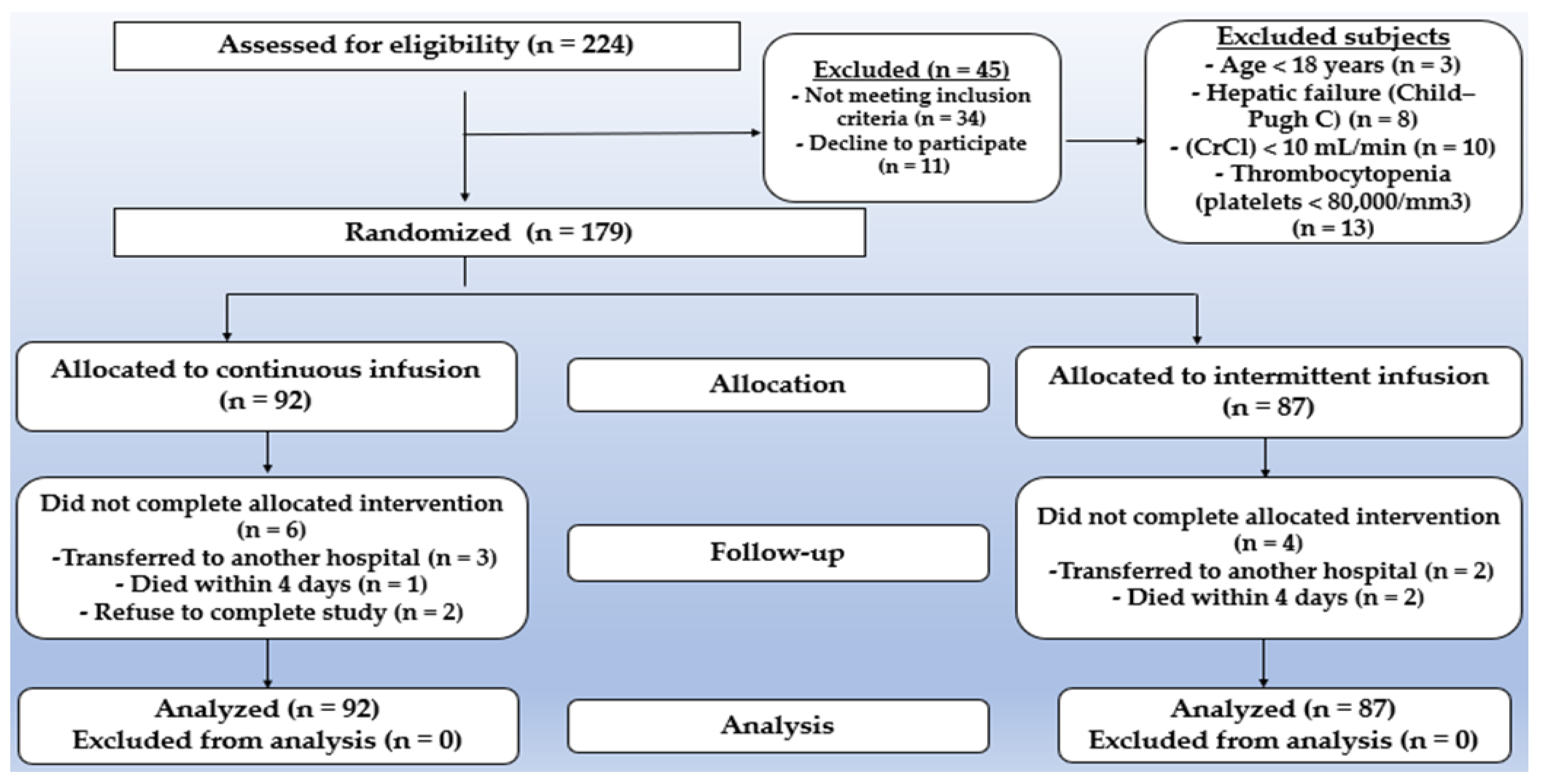
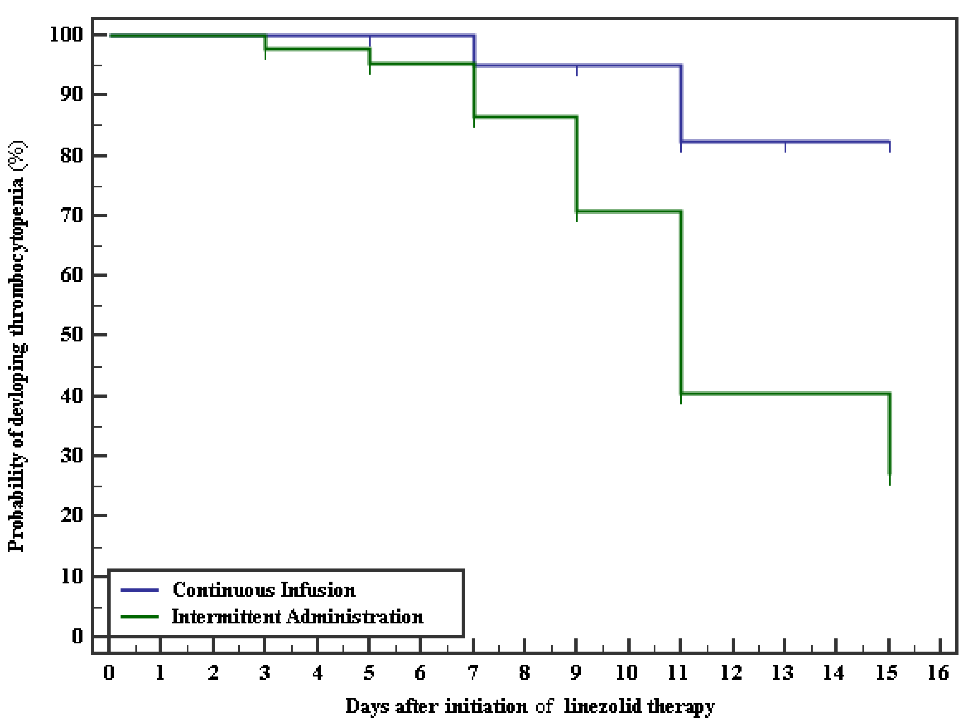
2. Results
3. Discussion
4. Materials and Methods
4.1. Study Design and Setting
4.2. Patients
4.3. Patient’s Randomization and Protocol
4.4. Patient Follow-Up and Outcome Measures
4.5. Statistical Analysis
5. Conclusions
Author Contributions
Funding
Institutional Review Board Statement
Informed Consent Statement
Data Availability Statement
Conflicts of Interest
References
- Rello, J.; Diaz, E. Pneumonia in the intensive care unit. Crit. Care Med. 2003, 31, 2544–2551. [Google Scholar] [CrossRef]
- Vincent, J.-L.; Bihari, D.J.; Suter, P.M.; Bruining, H.A.; White, J.; Nicolas-Chanoin, M.H.; Wolff, M.; Spencer, R.C.; Hemmer, M. The prevalence of nosocomial infection in intensive care units in Europe: Results of the European Prevalence of Infection in Intensive Care (EPIC) Study. JAMA 1995, 274, 639–644. [Google Scholar] [CrossRef]
- De Pascale, G.; Bello, G.; Tumbarello, M.; Antonelli, M. Severe pneumonia in intensive care: Cause, diagnosis, treatment and management a review of the literature. Curr. Opin. Pulm. Med. 2012, 18, 213–221. [Google Scholar] [CrossRef]
- Kojicic, M.; Li, G.; Hanson, A.C.; Lee, K.-M.; Thakur, L.; Vedre, J.; Ahmed, A.; Baddour, L.M.; Ryu, J.H.; Gajic, O. Risk factors for the development of acute lung injury in patients with infectious pneumonia. Crit. Care 2012, 16, R46. [Google Scholar] [CrossRef] [Green Version]
- Magill, S.S.; O’Leary, E.; Janelle, S.J.; Thompson, D.L.; Dumyati, G.; Nadle, J.; Wilson, L.E.; Kainer, M.A.; Lynfield, R.; Greissman, S.; et al. Changes in Prevalence of Health Care—Associated Infections in U.S. Hospitals. N. Engl. J. Med. 2018, 379, 1732–1744. [Google Scholar] [CrossRef]
- Sievert, D.M.; Ricks, P.; Edwards, J.R.; Schneider, A.; Patel, J.; Srinivasan, A.; Kallen, A.; Limbago, B.; Fridkin, S.; National Healthcare Safety Network (NHSN) Team; et al. Antimicrobial-resistant pathogens associated with healthcare-associated infections: Summary of data reported to the National Healthcare Safety Network at the Centers for Disease Control and Prevention, 2009–2010. Infect. Control Hosp. Epidemiol. 2013, 34, 1–14. [Google Scholar] [CrossRef]
- Ager, S.; Gould, K. Clinical update on linezolid in the treatment of Gram-positive bacterial infections. Infect. Drug Resist. 2012, 5, 87. [Google Scholar]
- Cunha, B.A. Antimicrobial Therapy of Multidrug-Resistant Streptococcus pneumoniae, Vancomycin-Resistant Enterococci, and Methicillin-Resistant Staphylococcus aureus. Med. Clin. N. Am. 2006, 90, 1165–1182. [Google Scholar] [CrossRef]
- Dryden, M.S. Linezolid pharmacokinetics and pharmacodynamics in clinical treatment. J. Antimicrob. Chemother. 2011, 66, iv7–iv15. [Google Scholar] [CrossRef] [Green Version]
- MacGowan, A.P. Pharmacokinetic and pharmacodynamic profile of linezolid in healthy volunteers and patients with Gram-positive infections. J. Antimicrob. Chemother. 2003, 51, ii17–ii25. [Google Scholar] [CrossRef]
- Stalker, D.J.; Jungbluth, G.L. Clinical Pharmacokinetics of Linezolid, a Novel Oxazolidinone Antibacterial. Clin. Pharmacokinet. 2003, 42, 1129–1140. [Google Scholar] [CrossRef]
- Rayner, C.R.; Forrest, A.; Meagher, A.K.; Birmingham, M.C.; Schentag, J.J. Clinical Pharmacodynamics of Linezolid in Seriously Ill Patients Treated in a Compassionate Use Programme. Clin. Pharmacokinet. 2003, 42, 1411–1423. [Google Scholar] [CrossRef]
- Taubert, M.; Zoller, M.; Maier, B.; Frechen, S.; Scharf, C.; Holdt, L.-M.; Frey, L.; Vogeser, M.; Fuhr, U.; Zander, J. Predictors of Inadequate Linezolid Concentrations after Standard Dosing in Critically Ill Patients. Antimicrob. Agents Chemother. 2016, 60, 5254–5261. [Google Scholar] [CrossRef] [Green Version]
- Dong, H.; Xie, J.; Wang, T.; Chen, L.; Zeng, X.; Sun, J.; Wang, X.; Dong, Y. Pharmacokinetic/pharmacodynamic evaluation of linezolid for the treatment of staphylococcal infections in critically ill patients. Int. J. Antimicrob. Agents 2016, 48, 259–264. [Google Scholar] [CrossRef]
- Yang, M.; Zhang, J.; Chen, Y.; Liang, X.; Guo, Y.; Yu, J.; Zhu, D.; Zhang, Y. Optimization of linezolid treatment regimens for Gram-positive bacterial infections based on pharmacokinetic/pharmacodynamic analysis. Future Microbiol. 2017, 12, 39–50. [Google Scholar] [CrossRef]
- Van Herendael, B.; Jeurissen, A.; Tulkens, P.M.; Vlieghe, E.; Verbrugghe, W.; Jorens, P.G.; Jorens, M. Continuous infusion of antibiotics in the critically ill: The new holy grail for beta-lactams and vancomycin? Ann. Intensive Care 2012, 2, 1–12. [Google Scholar] [CrossRef] [Green Version]
- Levison, M.E.; Levison, J.H. Pharmacokinetics and Pharmacodynamics of Antibacterial Agents. Infect. Dis. Clin. N. Am. 2009, 23, 791–815. [Google Scholar] [CrossRef] [Green Version]
- Boselli, E.; Breilh, D.; Caillault-Sergent, A.; Djabarouti, S.; Guillaume, C.; Xuereb, F.; Bouvet, L.; Rimmelé, T.; Saux, M.-C.; Allaouchiche, B. Alveolar diffusion and pharmacokinetics of linezolid administered in continuous infusion to critically ill patients with ventilator-associated pneumonia. J. Antimicrob. Chemother. 2012, 67, 1207–1210. [Google Scholar] [CrossRef] [Green Version]
- Taubert, M.; Zander, J.; Frechen, S.; Scharf, C.; Frey, L.; Vogeser, M.; Fuhr, U.; Zoller, M. Optimization of linezolid therapy in the critically ill: The effect of adjusted infusion regimens. J. Antimicrob. Chemother. 2017, 72, 2304–2310. [Google Scholar] [CrossRef]
- Adembri, C.; Fallani, S.; Cassetta, M.I.; Arrigucci, S.; Ottaviano, A.; Pecile, P.; Mazzei, T.; De Gaudio, R.; Novelli, A. Linezolid pharmacokinetic/pharmacodynamic profile in critically ill septic patients: Intermittent versus continuous infusion. Int. J. Antimicrob. Agents 2008, 31, 122–129. [Google Scholar] [CrossRef]
- Hiraki, Y.; Tsuji, Y.; Matsumoto, K.; Morita, K.; Kamimura, H.; Karube, Y. Influence of Linezolid Clearance on the Induction of Thrombocytopenia and Reduction of Hemoglobin. Am. J. Med. Sci. 2011, 342, 456–460. [Google Scholar] [CrossRef]
- Gerson, S.L.; Kaplan, S.L.; Bruss, J.B.; Le, V.; Arellano, F.M.; Hafkin, B.; Kuter, D.J. Hematologic Effects of Linezolid: Summary of Clinical Experience. Antimicrob. Agents Chemother. 2002, 46, 2723–2726. [Google Scholar] [CrossRef] [Green Version]
- Kim, H.-S.; Lee, E.; Cho, Y.-J.; Lee, Y.J.; Rhie, S.J. Linezolid-induced thrombocytopenia increases mortality risk in intensive care unit patients, a 10 year retrospective study. J. Clin. Pharm. Ther. 2018, 44, 84–90. [Google Scholar] [CrossRef] [Green Version]
- Steéphan, F.; Hollande, J.; Richard, O.; Cheffi, A.; Maier-Redelsperger, M.; Flahault, A. Thrombocytopenia in a Surgical ICU. Chest 1999, 115, 1363–1370. [Google Scholar] [CrossRef]
- Shalansky, S.J.; Verma, A.K.; Levine, M.; Spinelli, J.J.; Dodek, P.M. Risk Markers for Thrombocytopenia in Critically Ill Patients: A Prospective Analysis. Pharmacother. J. Hum. Pharmacol. Drug Ther. 2002, 22, 803–813. [Google Scholar] [CrossRef]
- Cawley, M.J.; Wittbrodt, E.T.; Boyce, E.G.; Skaar, D.J. Potential Risk Factors Associated with Thrombocytop in a Surgical Intensive Care Unit. Pharmacotherapy 1999, 19, 108–113. [Google Scholar] [CrossRef]
- Baughman, R.R.; Lower, E.E.; Flessa, H.C.; Tollerud, D.J. Thrombocytopenia in the Intensive Care Unit. Chest 1993, 104, 1243–1247. [Google Scholar] [CrossRef]
- Mimoz, O.; Montravers, P.; Paiva, J.-A. Continuous administration of linezolid in pneumonia: What is the level of proof? Int. Care Med. 2015, 41, 157–159. [Google Scholar] [CrossRef] [Green Version]
- Santimaleeworagun, W.; Changpradub, D.; Hemapanpairoa, J.; Thunyaharn, S. Optimization of Linezolid Dosing Regimens for Treatment of Vancomycin-Resistant Enterococci Infection. Infect. Chemother. 2021, 53, 503. [Google Scholar] [CrossRef]
- Beibei, L.; Yun, C.; Mengli, C.; Nan, B.; Xuhong, Y.; Rui, W. Linezolid versus vancomycin for the treatment of Gram-positive bacterial infections: Meta-analysis of randomised controlled trials. Int. J. Antimicrob. Agents 2010, 35, 3–12. [Google Scholar] [CrossRef]
- Vardakas, K.Z.; Mavros, M.N.; Roussos, N.; Falagas, M.E. Meta-Analysis of Randomized Controlled Trials of Vancomycin for the Treatment of Patients with Gram-Positive Infections: Focus on the Study Design. Mayo Clin. Proc. 2012, 87, 349–363. [Google Scholar] [CrossRef] [Green Version]
- Chen, C.-H.; Chen, Y.-M.; Chang, Y.-J.; Wang, S.-H.; Chang, C.-Y.; Yen, H.C. Continuous versus intermittent infusions of antibiotics for the treatment of infectious diseases: Meta-analysis and systematic review. Medicine 2019, 98, e14632. [Google Scholar] [CrossRef]
- De Pascale, G.; Fortuna, S.; Tumbarello, M.; Cutuli, S.L.; Vallecoccia, M.; Spanu, T.; Bello, G.; Montini, L.; Pennisi, M.A.; Navarra, P.; et al. Linezolid plasma and intrapulmonary concentrations in critically ill obese patients with ventilator-associated pneumonia: Intermittent vs continuous administration. Intensive Care Med. 2014, 41, 103–110. [Google Scholar] [CrossRef]
- Zoller, M.; Maier, B.; Hornuss, C.; Neugebauer, C.; Döbbeler, G.; Nagel, D.; Holdt, L.M.; Bruegel, M.; Weig, T.; Grabein, B.; et al. Variability of linezolid concentrations after standard dosing in critically ill patients: A prospective observational study. Crit. Care 2014, 18, R148. [Google Scholar] [CrossRef] [Green Version]
- Soraluce, A.; Barrasa, H.; Asín-Prieto, E.; Sánchez-Izquierdo, J.Á.; Maynar, J.; Isla, A.; Rodríguez-Gascón, A. Novel Population Pharmacokinetic Model for Linezolid in Critically Ill Patients and Evaluation of the Adequacy of the Current Dosing Recommendation. Pharmaceutics 2020, 12, 54. [Google Scholar] [CrossRef] [Green Version]
- Mira, J.C.; Gentile, L.F.; Mathias, B.J.; Efron, P.A.; Brakenridge, S.C.; Mohr, A.M.; Moore, F.A.; Moldawer, L.L. Sepsis pathophysiology, chronic critical illness and PICS. Crit. Care Med. 2017, 45, 253. [Google Scholar] [CrossRef]
- Mostel, Z.; Perl, A.; Marck, M.; Mehdi, S.F.; Lowell, B.; Bathija, S.; Santosh, R.; Pavlov, V.A.; Chavan, S.S.; Roth, J. Post-sepsis syndrome—An evolving entity that afflicts survivors of sepsis. Mol. Med. 2020, 26, 6. [Google Scholar] [CrossRef] [Green Version]
- Mayr, V.D.; Dünser, M.W.; Greil, V.; Jochberger, S.; Luckner, G.; Ulmer, H.; E Friesenecker, B.; Takala, J.; Hasibeder, W.R. Causes of death and determinants of outcome in critically ill patients. Crit. Care 2006, 10, R154. [Google Scholar] [CrossRef] [Green Version]
- Carcelero, E.; Soy, D.; Guerrero, L.; Poch, E.; Fernandez, J.; Castro, P.; Ribas, J. Linezolid Pharmacokinetics in Patients with Acute Renal Failure Undergoing Continuous Venovenous Hemodiafiltration. J. Clin. Pharmacol. 2012, 52, 1430–1435. [Google Scholar] [CrossRef]
- Dong, H.-Y.; Xie, J.; Chen, L.-H.; Wang, T.-T.; Zhao, Y.-R.; Dong, Y.-L. Therapeutic drug monitoring and receiver operating characteristic curve prediction may reduce the development of linezolid-associated thrombocytopenia in critically ill patients. Eur. J. Clin. Microbiol. 2014, 33, 1029–1035. [Google Scholar] [CrossRef]
- Ide, T.; Takesue, Y.; Ikawa, K.; Morikawa, N.; Ueda, T.; Takahashi, Y.; Nakajima, K.; Takeda, K.; Nishi, S. Population pharmacokinetics/pharmacodynamics of linezolid in sepsis patients with and without continuous renal replacement therapy. Int. J. Antimicrob. Agents 2018, 51, 745–751. [Google Scholar] [CrossRef]
- Dou, L.; Meng, D.; Dong, Y.; Chen, L.; Han, X.; Fan, D.; Dong, H. Dosage regimen and toxicity risk assessment of linezolid in sepsis patients. Int. J. Infect. Dis. 2020, 96, 105–111. [Google Scholar] [CrossRef]
- Pea, F.; Viale, P.; Cojutti, P.G.; Del Pin, B.; Zamparini, E.; Furlanut, M. Therapeutic drug monitoring may improve safety outcomes of long-term treatment with linezolid in adult patients. J. Antimicrob. Chemother. 2012, 67, 2034–2042. [Google Scholar] [CrossRef]
- Meyer, B.; Kornek, G.V.; Nikfardjam, M.; Karth, G.D.; Heinz, G.; Locker, G.J.; Jaeger, W.; Thalhammer, F. Multiple-dose pharmacokinetics of linezolid during continuous venovenous haemofiltration. J. Antimicrob. Chemother. 2005, 56, 172–179. [Google Scholar] [CrossRef] [Green Version]
- Takahashi, Y.; Takesue, Y.; Nakajima, K.; Ichiki, K.; Tsuchida, T.; Tatsumi, S.; Ishihara, M.; Ikeuchi, H.; Uchino, M. Risk factors associated with the development of thrombocytopenia in patients who received linezolid therapy. J. Infect. Chemother. 2011, 17, 382–387. [Google Scholar] [CrossRef]
- Sasaki, T.; Takane, H.; Ogawa, K.; Isagawa, S.; Hirota, T.; Higuchi, S.; Horii, T.; Otsubo, K.; Ieiri, I. Population Pharmacokinetic and Pharmacodynamic Analysis of Linezolid and a Hematologic Side Effect, Thrombocytopenia, in Japanese Patients. Antimicrob. Agents Chemother. 2011, 55, 1867–1873. [Google Scholar] [CrossRef] [Green Version]
- Nukui, Y.; Hatakeyama, S.; Okamoto, K.; Yamamoto, T.; Hisaka, A.; Suzuki, H.; Yata, N.; Yotsuyanagi, H.; Moriya, K. High plasma linezolid concentration and impaired renal function affect development of linezolid-induced thrombocytopenia. J. Antimicrob. Chemother. 2013, 68, 2128–2133. [Google Scholar] [CrossRef] [Green Version]
- Hirano, R.; Sakamoto, Y.; Tachibana, N.; Ohnishi, M. Retrospective analysis of the risk factors for linezolid-induced thrombocytopenia in adult Japanese patients. Int. J. Clin. Pharm. 2014, 36, 795–799. [Google Scholar] [CrossRef]
- Lin, Y.-H.; Wu, V.-C.; Tsai, I.-J.; Ho, Y.-L.; Hwang, J.-J.; Tsau, Y.-K.; Wu, C.-Y.; Wu, K.-D.; Hsueh, P.-R. High frequency of linezolid-associated thrombocytopenia among patients with renal insufficiency. Int. J. Antimicrob. Agents 2006, 28, 345–351. [Google Scholar] [CrossRef]
- Grau, S.; Morales-Molina, J.A.; Antonio, J.M.-D.; Marín-Casino, M.; Alvarez-Lerma, F. Linezolid: Low pre-treatment platelet values could increase the risk of thrombocytopenia. J. Antimicrob. Chemother. 2005, 56, 440–441. [Google Scholar] [CrossRef] [Green Version]
- Liqing, B.; Zhou, J.; Huang, M.; Zhou, S. Efficacy of linezolid on gram-positive bacterial infection in elderly patients and risk factors associated with thrombocytopenia. Chin. J. Geriatr. 2013, 29, 408–412. [Google Scholar]
- Niwa, T.; Watanabe, T.; Suzuki, A.; Ohmori, T.; Tsuchiya, M.; Suzuki, T.; Ohta, H.; Murakami, N.; Itoh, Y. Reduction of linezolid-associated thrombocytopenia by the dose adjustment based on the risk factors such as basal platelet count and body weight. Diagn. Microbiol. Infect. Dis. 2014, 79, 93–97. [Google Scholar] [CrossRef] [PubMed]
- Zhang, Z.; Liang, Z.; Li, H.; Chen, L.; She, D. Comparative evaluation of thrombocytopenia in adult patients receiving linezolid or glycopeptides in a respiratory intensive care unit. Exp. Ther. Med. 2014, 7, 501–507. [Google Scholar] [CrossRef] [PubMed] [Green Version]
- Hanai, Y.; Matsuo, K.; Ogawa, M.; Higashi, A.; Kimura, I.; Hirayama, S.; Kosugi, T.; Nishizawa, K.; Yoshio, T. A retrospective study of the risk factors for linezolid-induced thrombocytopenia and anemia. J. Infect. Chemother. 2016, 22, 536–542. [Google Scholar] [CrossRef] [PubMed]
- Calder, M.; Schonell, M. Pneumococcal Typing and the Problem of Endogenous or Exogenous Reinfection in Chronic Bronchitis. Lancet 1971, 297, 1156–1159. [Google Scholar] [CrossRef]
- Tillotson, J.R.; Finland, M. Bacterial Colonization and Clinical Superinfection of the Respiratory Tract Complicating Antibiotic Treatment of Pneumonia. J. Infect. Dis. 1969, 119, 597–624. [Google Scholar] [CrossRef] [PubMed]
- Michels, W.M.; Grootendorst, D.C.; Verduijn, M.; Elliott, E.G.; Dekker, F.W.; Krediet, R.T. Performance of the Cockcroft-Gault, MDRD, and New CKD-EPI Formulas in Relation to GFR, Age, and Body Size. Clin. J. Am. Soc. Nephrol. 2010, 5, 1003–1009. [Google Scholar] [CrossRef] [Green Version]
- Cirillo, M. Razionale, pregi e difetti della stima della filtrato glomerulare: Equazione cockcroft-gault ed equazione mdrd. G. Ital. Nefrol. 2009, 26, 310–317. [Google Scholar]
- World Medical Association. Declaration of Helsinki—Ethical Principles for Medical Research Involving Human Subjects. 2013, Adopted by the 18th WMA General Assembly, Helsinki, Finland, June 1964 (Amended by the 64th WMA General Assembly, Fortaleza, Brazil October). Available online: https://www.wma.net/policies-post/wma-declaration-of-helsinki-ethical-principles-for-medical-research-involving-human-subjects/ (accessed on 12 December 2021).
- Kumar, S.T.; Yassin, A.; Bhowmick, T.; Dixit, D. Recommendations from the 2016 Guidelines for the Management of Adults with Hospital-Acquired or Ventilator-Associated Pneumonia. P T Peer-Rev. J. Formul. Manag. 2017, 42, 767–772. [Google Scholar]
- Koenig, S.M.; Truwit, J.D. Ventilator-Associated Pneumonia: Diagnosis, Treatment, and Prevention. Clin. Microbiol. Rev. 2006, 19, 637–657. [Google Scholar] [CrossRef] [Green Version]
- Chawla, R. Epidemiology, etiology, and diagnosis of hospital-acquired pneumonia and ventilator-associated pneumonia in Asian countries. Am. J. Infect. Control. 2008, 36, S93–S100. [Google Scholar] [CrossRef] [PubMed]
- Liu, W.; Peng, L.; Hua, S. Clinical significance of dynamic monitoring of blood lactic acid, oxygenation index and C-reactive protein levels in patients with severe pneumonia. Exp. Ther. Med. 2015, 10, 1824–1828. [Google Scholar] [CrossRef] [PubMed] [Green Version]
- Chastre, J.; Fagon, J.-Y. Ventilator-associated pneumonia. Am. J. Respir. Crit. Care Med. 2002, 165, 867–903. [Google Scholar] [CrossRef] [PubMed]
- Shokouhi, S.; Darazam, I.A.; Niyati, R.; Gachkar, L.; Goharani, R.; Kahkoue, S. Resolution of Chest X-ray Opacities in Patients with Ventilator-associated Pneumonia. Infect. Disord. Drug Targets 2018, 18, 23–28. [Google Scholar] [CrossRef] [PubMed]
- Koulenti, D.; Lisboa, T.; Brun-Buisson, C.; Krueger, W.; Macor, A.; Sole-Violan, J.; Diaz, E.; Topeli, A.; DeWaele, J.; Carneiro, A.; et al. Spectrum of practice in the diagnosis of nosocomial pneumonia in patients requiring mechanical ventilation in European intensive care units. Crit. Care Med. 2009, 37, 2360–2369. [Google Scholar] [CrossRef]
- Vanderschueren, S.; De Weerdt, A.; Malbrain, M.; Vankersschaever, D.; Frans, E.; Wilmer, A.; Bobbaers, H. Thrombocytopenia and prognosis in intensive care. Crit. Care Med. 2000, 28, 1871–1876. [Google Scholar] [CrossRef] [PubMed]


| Parameters | CI Group (n = 92) | II Group (n = 87) | p-Value |
|---|---|---|---|
| Gender, n% | |||
| Male | 58 (63.0%) | 55 (63.2%) | 0.981 |
| Female | 34 (37.0%) | 32 (36.8%) | |
| Age (years) | |||
| Min.–Max. | 42.0–88.0 | 29.0–88.0 | 0.865 |
| Mean ± SD | 66.55 ± 10.56 | 66.89 ± 14.93 | |
| Body Mass Index (BMI) (kg/m2) | |||
| Min.–Max. | 20.20–53.60 | 22.0–49.10 | 0.190 |
| Mean ± SD | 30.24 ± 6.97 | 28.99 ± 5.58 | |
| (SAPS) II | |||
| Min.–Max. | 31.0–69.0 | 25.0–66.0 | 0.195 |
| Mean ± SD | 43.80 ± 8.61 | 42.10 ± 8.86 | |
| SOFA | 0.579 | ||
| Min.–Max. | 3.0–11.0 | 3.0–13.0 | |
| Median (IQR) | 5.0 (4.0–7.0) | 5.0 (3.50–7.0) | |
| Type of pneumonia, n% | |||
| HAP | 58 (63.0%) | 59 (67.8%) | 0.502 |
| VAP | 34 (37.0%) | 28 (32.2%) | |
| CPIS for VAP. | |||
| Min.–Max | 6.0–9.0 | 6.0–9.0 | 0.941 |
| Mean ± SD | 7.26 ± 0.83 | 7.25 ± 0.70 | |
| HAP patients required mechanical ventilation, n% | 13 (22.4%) | 16 (27.1%) | 0.669 |
| WBCs count (>11,000 cells/mm) | |||
| Min.–Max. | 8.30–33.90 | 5.53–42.30 | 0.959 |
| Median (IQR) | 13.15 (11.50–21.0) | 14.20 (11.10–20.20) | |
| Body temperature(>38 °C), n% | 30 (32.6%) | 30 (34.5%) | 0.791 |
| Baseline CRP (mg/dL) | |||
| Min.–Max. | 3.30–282.20 | 3.0–200.0 | 0.786 |
| Median (IQR) | 77.0 (17.30–161.0) | 75.0 (35.50–134.0) | |
| Baseline PCT (ng/mL) | |||
| Min.–Max. | 0.60–37.0 | 0.29–27.0 | 0.709 |
| Median (IQR) | 2.90 (1.90–9.80) | 3.40 (1.61–7.90) | |
| Baseline(P/F) ratio | |||
| Min.–Max. | 77.10–298.0 | 62.80–315.0 | 0.072 |
| Median (IQR) | 151.0 (110.0–190.0) | 174.0 (119.0–254.5) | |
| Baseline serum creatinine(S.Cr) (mg/dL) | |||
| Min.–Max. | 0.50–4.10 | 0.40–7.30 | <0.001 * |
| Median (IQR) | 1.21 (0.80–2.20) | 2.50 (1.02–3.96) | |
| Baseline creatinine clearance (CrCl) (mL/min) | |||
| Min.–Max. | 13.0–136.0 | 11.0–122.0 | <0.001 * |
| Median (IQR) | 53.05 (28.0–80.0) | 28.39 (18.73–64.0) | |
| Radiological findings | |||
| Multi lobar infiltrates, n% | 54 (58.7%) | 49 (56.3%) | 0.748 |
| Pleural effusion, n% | 36 (39.1%) | 44 (50.6%) | 0.124 |
| Parameters | CI Group (n = 92) | II Group (n = 87) | p-Value |
|---|---|---|---|
| Clinical cure, n% | 56 (60.9%) | 40 (46.0%) | 0.046 * |
| Development of sepsis, n% | 26 (28.3%) | 41 (47.1%) | 0.009 * |
| Mortality at the end of linezolid treatment, n% | 10 (10.9%) | 13 (14.9%) | 0.510 |
| 30-Day mortality, n% | 26 (28.3%) | 19 (21.8%) | 0.322 |
| P/F ratio at the seventh day of treatment | |||
| Min.–Max. | 79.0–429.0 | 71.0–370.0 | 0.030 * |
| Median (IQR) | 246.0 (181.0–312.0) | 198.0 (122.5–275.5) | |
| Length of ICU stay | |||
| Min.–Max. | 3.0–24.0 | 3.0–27.0 | 0.188 |
| Median (IQR) | 9.0 (7.0–11.0) | 10.0 (7.0–12.0) | |
| Length of hospital stay | |||
| Min.–Max. | 3.0–26.0 | 3.0–30.0 | 0.063 |
| Median (IQR) | 11.0 (9.50–13.0) | 12.0 (9.50–14.0) | |
| Days of treatment on Linezolid | |||
| Min.–Max. | 3.0–14.0 | 3.0–14.0 | 0.037 * |
| Median (IQR) | 7.0 (7.0–11.0) | 7.0 (7.0–9.0) | |
| Days to reach the clinical cure | |||
| Min.–Max. | 4.0–7.0 | 5.0–10.0 | <0.001 * |
| Mean ± SD | 5.82 ± 1.21 | 6.96 ± 0.97 | |
| Duration on Mechanical ventillation (MV) | |||
| Min.–Max. | 2.0–11.0 | 2.0–14.0 | 0.422 |
| Median (IQR) | 4.0 (3.0–6.0) | 5.0 (3.0–7.0) | |
| (MV) free days | |||
| Min.–Max. | 2.0–20.0 | 2.0–19.0 | 0.194 |
| Median (IQR) | 6.0 (4.0–8.0) | 5.0 (3.0–8.0) |
| Parameters | Streptococcus pneumoniae | Methicillin-Resistant Staphylococcus aureus (MRSA) | Methicillin-Susceptible Staphylococcus aureus (MSSA) |
|---|---|---|---|
| No. (%) of patients with VAP | 11 (17.7%) | 43 (69.4%) | 8 (12.9%) |
| No. (%) of patients with HAP | 15 (12.8%) | 58 (49.6%) | 44 (37.6%) |
| No. (%) of cured patients in CI group | 8 (14.3%) | 30 (53.6%) | 18 (32.1%) |
| No. (%) of cured patients in II group | 9 (22.5%) | 17 (42.5%) | 14 (35.0%) |
| Parameters | OR | 95% CI | p Value |
|---|---|---|---|
| Route of linezolid administration (continuous infusion) | 2.017 | 1.088–3.738 | 0.003 * |
| Development of sepsis | 3.037 | 1.325–6.961 | 0.008 * |
| SOFA score | 0.721 | 0.612–0.849 | 0.01 * |
| Parameters | OR | 95% CI | p-Value |
|---|---|---|---|
| Route of linezolid administration (intermittent infusion) | 4.128 | 1.681–10.139 | 0.001 * |
| Baseline platelets < 200 × 103/mm3 | 3.148 | 1.251–7.922 | 0.014 * |
| CrCl < 30 | 3.755 | 3.755–8.664 | 0.002 * |
Publisher’s Note: MDPI stays neutral with regard to jurisdictional claims in published maps and institutional affiliations. |
© 2022 by the authors. Licensee MDPI, Basel, Switzerland. This article is an open access article distributed under the terms and conditions of the Creative Commons Attribution (CC BY) license (https://creativecommons.org/licenses/by/4.0/).
Share and Cite
Abou Warda, A.E.; Sarhan, R.M.; Al-Fishawy, H.S.; Moharram, A.N.; Salem, H.F. Continuous Versus Intermittent Linezolid Infusion for Critically Ill Patients with Hospital-Acquired and Ventilator-Associated Pneumonia: Efficacy and Safety Challenges. Pharmaceuticals 2022, 15, 296. https://doi.org/10.3390/ph15030296
Abou Warda AE, Sarhan RM, Al-Fishawy HS, Moharram AN, Salem HF. Continuous Versus Intermittent Linezolid Infusion for Critically Ill Patients with Hospital-Acquired and Ventilator-Associated Pneumonia: Efficacy and Safety Challenges. Pharmaceuticals. 2022; 15(3):296. https://doi.org/10.3390/ph15030296
Chicago/Turabian StyleAbou Warda, Ahmed E., Rania M. Sarhan, Hussein Saeed Al-Fishawy, Ayman N. Moharram, and Heba F. Salem. 2022. "Continuous Versus Intermittent Linezolid Infusion for Critically Ill Patients with Hospital-Acquired and Ventilator-Associated Pneumonia: Efficacy and Safety Challenges" Pharmaceuticals 15, no. 3: 296. https://doi.org/10.3390/ph15030296