
Efficacy, Safety and Patient-Reported Outcomes with Preservative-Free (PF) Tafluprost or PF-Dorzolamide/Timolol Compared with Preserved Latanoprost: A Prospective Multicenter Study in Korean Glaucoma Patients with Ocular Surface Disease

Abstract
:1. Introduction
2. Results
2.1. Patient-Reported Symptoms and Quality of Life
2.2. Changes in IOP
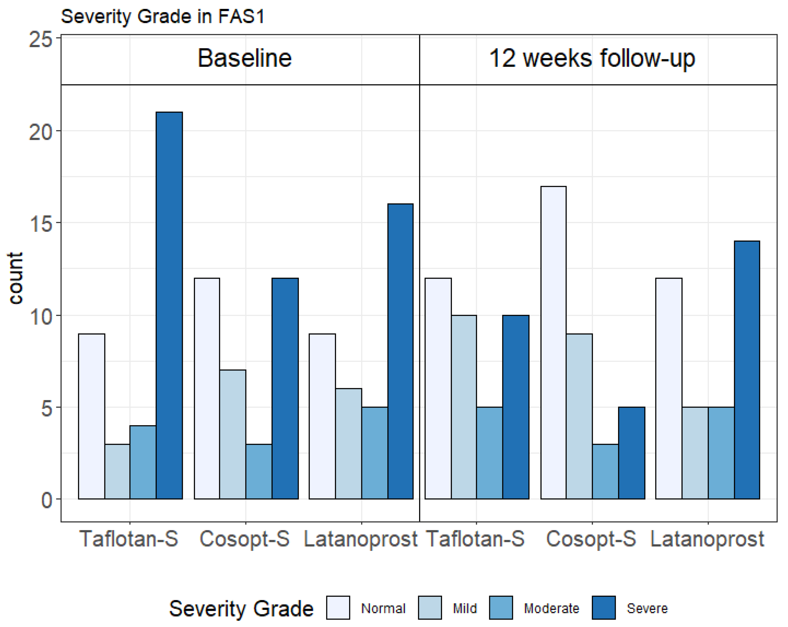
2.3. Safety
3. Discussion
4. Materials and Methods
4.1. Inclusion and Exclusion Criteria
4.2. Treatment
4.3. Efficacy Outcomes
4.4. Efficacy Assessment
4.5. Safety Assessment
4.6. Statistical Analysis
5. Conclusions
Supplementary Materials
Author Contributions
Funding
Institutional Review Board Statement
Informed Consent Statement
Data Availability Statement
Acknowledgments
Conflicts of Interest
References
- Jonas, J.B.; Aung, T.; Bourne, R.R.; Bron, A.M.; Ritch, R.; Panda-Jonas, S. Glaucoma. Lancet 2017, 390, 2183–2193. [Google Scholar] [CrossRef]
- Quigley, H.A.; Broman, A.T. The number of people with glaucoma worldwide in 2010 and 2020. Br. J. Ophthalmol. 2006, 90, 262–267. [Google Scholar] [CrossRef]
- Weinreb, R.N.; Aung, T.; Medeiros, F.A. The pathophysiology and treatment of glaucoma: A review. JAMA 2014, 311, 1901–1911. [Google Scholar] [CrossRef] [PubMed]
- Definition and Classification Subcommittee of the International Dry Eye WorkShop. The definition and classification of dry eye disease: Report of the Definition and Classification Subcommittee of the International Dry Eye WorkShop (2007). Ocul. Surf. 2007, 5, 75–92. [Google Scholar] [CrossRef]
- Labbe, A.; Terry, O.; Brasnu, E.; van Went, C.; Baudouin, C. Tear film osmolarity in patients treated for glaucoma or ocular hypertension. Cornea 2012, 31, 994–999. [Google Scholar] [CrossRef] [PubMed]
- Leung, E.W.; Medeiros, F.A.; Weinreb, R.N. Prevalence of ocular surface disease in glaucoma patients. J. Glaucoma 2008, 17, 350–355. [Google Scholar] [CrossRef] [PubMed]
- Garcia-Feijoo, J.; Sampaolesi, J.R. A multicenter evaluation of ocular surface disease prevalence in patients with glaucoma. Clin. Ophthalmol. 2012, 6, 441–446. [Google Scholar] [PubMed]
- Ruangvaravate, N.; Prabhasawat, P.; Vachirasakchai, V.; Tantimala, R. High prevalence of ocular surface disease among glaucoma patients in Thailand. J. Ocul. Pharmacol. Ther. 2018, 34, 387–394. [Google Scholar] [CrossRef] [PubMed]
- Fechtner, R.D.; Godfrey, D.G.; Budenz, D.; Stewart, J.A.; Stewart, W.C.; Jasek, M.C. Prevalence of ocular surface complaints in patients with glaucoma using topical intraocular pressure-lowering medications. Cornea 2010, 29, 618–621. [Google Scholar] [CrossRef]
- Skalicky, S.E.; Goldberg, I.; McCluskey, P. Ocular surface disease and quality of life in patients with glaucoma. Am. J. Ophthalmol. 2012, 153, 1.e2–9.e2. [Google Scholar] [CrossRef]
- Pérez-Bartolomé, F.; Martínez-de-la-Casa, J.M.; Arriola-Villalobos, P.; Fernández-Pérez, C.; Polo, V.; Julián García-Feijoó, J. Ocular surface disease in patients under topical treatment for glaucoma. Eur. J. Ophthalmol. 2017, 27, 694–704. [Google Scholar] [CrossRef]
- Boso, A.L.M.; Gasperi, E.; Fernandes, L.; Costa, V.P.; Alves, M. Impact of ocular surface disease treatment in patients with glaucoma. Clin. Ophthalmol. 2020, 14, 103–111. [Google Scholar] [CrossRef]
- Baudouin, C.; Labbé, A.; Liang, H.; Pauly, A.; Brignole-Baudouin, F. Preservatives in eyedrops: The good, the bad and the ugly. Prog. Retin. Eye Res. 2010, 29, 312–334. [Google Scholar] [CrossRef] [PubMed]
- Mantelli, F.; Tranchina, L.; Lambiase, A.; Bonini, S. Ocular surface damage by ophthalmic compounds. Curr. Opin. Allergy Clin. Immunol. 2011, 11, 464–470. [Google Scholar] [CrossRef] [PubMed]
- Actis, A.G.; Rolle, T. Ocular surface alterations and topical antiglaucomatous therapy: A review. Open Ophthalmol. J. 2014, 8, 67–72. [Google Scholar]
- Aguayo Bonniard, A.; Yeung, J.Y.; Chan, C.C.; Birt, C.M. Ocular surface toxicity from glaucoma topical medications and associated preservatives such as benzalkonium chloride (BAK). Expert Opin. Drug Metab. Toxicol. 2016, 12, 1279–1289. [Google Scholar] [CrossRef]
- Pinheiro, R.; Panfil, C.; Schrage, N.; Dutescu, R.M. The impact of glaucoma medications on corneal wound healing. J. Glaucoma 2016, 25, 122–127. [Google Scholar] [CrossRef]
- Nijm, L.M.; De Benito-Llopis, L.; Rossi, G.C.; Vajaranant, T.S.; Coroneo, M.T. Understanding the Dual Dilemma of Dry Eye and Glaucoma: An International Review. Asia Pac. J. Ophthalmol. 2020, 9, 481–490. [Google Scholar] [CrossRef] [PubMed]
- Baudouin, C.; Denoyer, A.; Desbenoit, N.; Hamm, G.; Grise, A. In vitro and in vivo experimental studies on trabecular meshwork degeneration induced by benzalkonium chloride (an American Ophthalmological Society thesis). Trans Am. Ophthalmol. Soc. 2012, 110, 40–63. [Google Scholar]
- Freeman, D.P.; Kahook, M.Y. Preservatives in topical ophthalmic medications: Historical and clinical perspectives. Expert Rev. Ophthalmol. 2009, 4, 59–64. [Google Scholar] [CrossRef]
- Rosin, L.M.; Bell, N.P. Preservative toxicity in glaucoma medication: Clinical evaluation of benzalkonium chloride-free 0.5% timolol eye drops. Clin. Ophthalmol. 2013, 7, 2131–2135. [Google Scholar] [PubMed]
- Guzmán, M.; Sabbione, F.; Gabelloni, M.L.; Vanzulli, S.; Trevani, A.S.; Giordano, M.N.; Galletti, J.G. Restoring conjunctival tolerance by topical nuclear factor-kappaB inhibitors reduces preservative-facilitated allergic conjunctivitis in mice. Investig. Ophthalmol. Vis. Sci. 2014, 55, 6116–6126. [Google Scholar] [CrossRef] [PubMed]
- Rasmussen, C.A.; Kaufman, P.L.; Kiland, J.A. Benzalkonium chloride and glaucoma. J. Ocul. Pharmacol. Ther. 2014, 30, 163–169. [Google Scholar] [CrossRef] [PubMed]
- Van Gestel, A.; Webers, C.A.B.; Beckers, H.J.M.; van Dongen, M.C.J.M.; Severens, J.L.; Hendrikse, F.; Schouten, J.S.A.G. The relationship between visual field loss in glaucoma and health-related quality-of-life. Eye 2010, 24, 1759–1769. [Google Scholar] [CrossRef]
- Gracitelli, C.P.; Abe, R.Y.; Tatham, A.J.; Rosen, P.N.; Zangwill, L.M.; Boer, E.R.; Weinreb, R.N.; Medeiros, F.A. Association between progressive retinal nerve fiber layer loss and longitudinal change in quality of life in glaucoma. JAMA Ophthalmol. 2015, 133, 384–390. [Google Scholar] [CrossRef]
- Medeiros, F.A.; Gracitelli, C.P.; Boer, E.R.; Weinreb, R.N.; Zangwill, L.M.; Rosen, P.N. Longitudinal changes in quality of life and rates of progressive visual field loss in glaucoma patients. Ophthalmology 2015, 122, 293–301. [Google Scholar] [CrossRef] [PubMed]
- Abe, R.Y.; Diniz-Filho, A.; Costa, V.P.; Gracitelli, C.P.B.; Baig, S.; Medeiros, F.A. The impact of location of progressive visual field loss on longitudinal changes in quality of life of patients with glaucoma. Ophthalmology 2016, 123, 552–557. [Google Scholar] [CrossRef]
- Sun, Y.; Lin, C.; Waisbourd, M.; Ekici, F.; Erdem, E.; Wizov, S.S.; Hark, L.A.; Spaeth, G.L. The impact of visual field clusters on performance-based measures and vision-related quality of life in patients with glaucoma. Am. J. Ophthalmol. 2016, 163, 45–52. [Google Scholar] [CrossRef]
- Takahashi, G.; Otori, Y.; Urashima, M.; Kuwayama, Y.; Quality of Life Improvement Committee. Evaluation of quality of life in Japanese glaucoma patients and its relationship with visual function. J. Glaucoma 2016, 25, e150–e156. [Google Scholar] [CrossRef]
- Guarnieri, A.; Elena Carnero, E.; Bleau, A.-M.; Alfonso-Bartolozzi, B.; Moreno-Montañés, J. Relationship between OSDI questionnaire and ocular surface changes in glaucomatous patients. Int. Ophthalmol. 2020, 40, 741–751. [Google Scholar] [CrossRef]
- Konstas, A.G.; Schmetterer, L.; Katsanos, A.; Hutnik, C.M.L.; Holló, G.; Quaranta, L.; Teus, M.A.; Uusitalo, H.; Pfeiffer, N.; Katz, L.J. Dorzolamide/timolol fixed combination: Learning from the past and looking toward the future. Adv. Ther. 2021, 38, 24–51. [Google Scholar] [CrossRef]
- Guo, Y.; Ha, J.Y.; Piao, H.L.; Sung, M.S.; Park, S.W. The protective effect of 3% diquafosol on meibomian gland morphology in glaucoma patients treated with prostaglandin analogs: A 12-month follow-up study. BMC Ophthalmol. 2020, 20, 277. [Google Scholar] [CrossRef]
- Karakus, S.; Agrawal, D.; Hindman, H.B.; Henrich, C.; Ramulu, P.Y.; Akpek, E.K. Effects of prolonged reading on dry eye. Ophthalmology 2018, 125, 1500–1505. [Google Scholar] [CrossRef] [PubMed]
- Midorikawa-Inomata, A.; Inomata, T.; Nojiri, S.; Nakamura, M.; Iwagami, M.; Fujimoto, K.; Okumura, Y.; Iwata, N.; Eguchi, A.; Hasegawa, H.; et al. Reliability and validity of the Japanese version of the Ocular Surface Disease Index for dry eye disease. BMJ Open 2019, 9, e033940. [Google Scholar] [CrossRef]
- Rossi, G.C.M.; Scudeller, L.; Lumini, C.; Mirabile, A.V.; Picasso, E.; Bettio, F.; Pasinetti, G.M.; Bianchi, P.E. An in vivo confocal, prospective, masked, 36 months study on glaucoma patients medically treated with preservative-free or preserved monotherapy. Sci. Rep. 2019, 9, 4282. [Google Scholar] [CrossRef] [PubMed]
- Mathews, P.M.; Ramulu, P.Y.; Friedman, D.S.; Utine, C.A.; Akpek, E.K. Evaluation of ocular surface disease in patients with glaucoma. Ophthalmology 2013, 120, 2241–2248. [Google Scholar] [CrossRef] [PubMed]
- Cvenkel, B.; Štunf, Š.; Srebotnik Kirbiš, I.; Strojan Fležar, M. Symptoms and signs of ocular surface disease related to topical medication in patients with glaucoma. Clin. Ophthalmol. 2015, 9, 625–631. [Google Scholar] [CrossRef]
- Saade, C.E.; Lari, H.B.; Berezina, T.L.; Fechtner, R.D.; Khouri, A.S. Topical glaucoma therapy and ocular surface disease: A prospective, controlled cohort study. Can. J. Ophthalmol. 2015, 50, 132–136. [Google Scholar] [CrossRef]
- Schiffman, R.M.; Christianson, M.D.; Jacobsen, G.; Hirsch, J.D.; Reis, B.L. Reliability and validity of the Ocular Surface Disease Index. Arch. Ophthalmol. 2000, 118, 615–621. [Google Scholar] [CrossRef]
- Guillemin, I.; Begley, C.; Chalmers, R.; Baudouin, C.; Arnould, B. Appraisal of patient-reported outcome instruments available for randomized clinical trials in dry eye: Revisiting the standards. Ocul. Surf. 2012, 10, 84–99. [Google Scholar] [CrossRef]
- Lee, W.; Lee, S.; Bae, H.W.; Kim, C.Y.; Seong, G.J. Efficacy and tolerability of preservative-free 0.0015% tafluprost in glaucoma patients: A prospective crossover study. BMC Ophthalmol. 2017, 17, 61. [Google Scholar] [CrossRef]
- Lee, N.Y.; Park, H.-Y.L.; Park, C.K. Comparison of the effects of dorzolamide/timolol fixed combination versus p-latanoprost on intraocular pressure and ocular perfusion pressure in patients with normal-tension glaucoma: A randomized, crossover clinical trial. PLoS ONE 2016, 11, e0146680. [Google Scholar] [CrossRef] [PubMed]
- Uusitalo, H.; Chen, E.; Pfeiffer, N.; Brignole-Baudouin, F.; Kaarniranta, K.; Leino, M.; Puska, P.; Palmgren, E.; Hamacher, T.; Hofmann, G.; et al. Switching from a preserved to a preservative-free prostaglandin preparation in topical glaucoma medication. Acta Ophthalmol. 2010, 88, 329–336. [Google Scholar] [CrossRef] [PubMed]
- EMC. Dorzolamide/Timolol Preservative-Free 20 mg/ml + 5 mg/ml Eye Drops, Solution in Single-Dose Container: Summary of Product Characteristics. 2020. Available online: https://www.medicines.org.uk/emc/product/5114/smpc. (accessed on 4 November 2020).
- EMC. Saflutan 15 Micrograms/ml Eye Drops, Solution, in Single-Dose Container: Summary of Product Characteristics. 2017. Available online: https://www.medicines.org.uk/emc/product/5115/smpc (accessed on 4 November 2020).
- Wong, T.T.; Aung, T.; Ho, C.L. Ocular surface status in glaucoma and ocular hypertension patients with existing corneal disorders switched from P-latanoprost 0.005% to tafluprost 0.0015%: Comparison of two prostaglandin analogues with different concentrations of benzalkonium chloride. Clin. Exp. Ophthalmol. 2018, 46, 1028–1034. [Google Scholar] [CrossRef]
- Konstas, A.G.; Quaranta, L.; Katsanos, A.; Riva, I.; Tsai, J.C.; Giannopoulos, T.; Voudouragkaki, I.C.; Paschalinou, E.; Floriani, I.; Haidich, A.B. Twenty-four hour efficacy with preservative free tafluprost compared with P-latanoprost in patients with primary open angle glaucoma or ocular hypertension. Br. J. Ophthalmol. 2013, 97, 1510–1515. [Google Scholar] [CrossRef] [PubMed]
- El Hajj Moussa, W.G.; Farhat, R.G.; Nehme, J.C.; Sahyoun, M.A.; Schakal, A.R.; Jalkh, A.E.; Abi Karam, M.P.; Azar, G.G. Comparison of efficacy and ocular surface disease index score between bimatoprost, latanoprost, travoprost, and tafluprost in glaucoma patients. J. Ophthalmol. 2018, 2018, 1319628. [Google Scholar] [CrossRef]
- Lee, S.; Kim, M.K.; Choi, H.J.; Wee, W.R.; Kim, D.M. Comparative cross-sectional analysis of the effects of topical antiglaucoma drugs on the ocular surface. Adv. Ther. 2013, 30, 420–429. [Google Scholar] [CrossRef] [PubMed]
- Kuppens, E.V.; de Jong, C.A.; Stolwijk, T.R.; de Keizer, R.J.; van Best, J.A. Effect of timolol with and without preservative on the basal tear turnover in glaucoma. Br. J. Ophthalmol. 1995, 79, 339–342. [Google Scholar] [CrossRef]
- Bartlett, J.D.; Keith, M.S.; Sudharshan, L.; Snedecor, S.J. Associations between signs and symptoms of dry eye disease: A systematic review. Clin. Ophthalmol. 2015, 9, 1719–1730. [Google Scholar] [CrossRef]
- Miller, K.L.; Walt, J.G.; Mink, D.R.; Satram-Hoang, S.; Wilson, S.E.; Perry, H.D.; Asbell, P.A.; Pflugfelder, S.C. Minimal clinically important difference for the Ocular Surface Disease Index. Arch. Ophthalmol. 2010, 128, 94–101. [Google Scholar] [CrossRef]


| Characteristic | PF-Tafluprost (n = 37) | PF-Dorzolamide/Timolol (n = 34) | P-Latanoprost (n = 36) |
|---|---|---|---|
| Age, years: mean (SD) | 63.2 (13.2) | 62.1 (14.0) | 63.0 (13.1) |
| Male: n (%) | 15 (40.5) | 17 (50) | 14 (38.9) |
| Intraocular pressure, mmHg: mean (SD) | 13.8 (2.3) | 13.1 (3.4) | 13.1 (2.7) |
| Prior medication *: n | 67 | 63 | 96 |
| Anti-anginal | 7 (10.5) | 1 (1.6) | 0 |
| Anti-asthmatic/COPD | 0 | 0 | 10 (10.4) |
| Dyslipidemic | 7 (10.5) | 7 (11.1) | 8 (8.3) |
| Oral anti-diabetic | 0 | 8 (12.7) | 10 (10.4) |
| OSDI Questionnaire | PF-Tafluprost | PF-Dorzolamide/Timolol | P-Latanoprost | |||
|---|---|---|---|---|---|---|
| Mean (SD) | p-Value † | Mean (SD) | p-Value † | Mean (SD) | p-Value † | |
| Total score | –9.5 (18.9) | 0.0042 * | –10.5 (19.8) | 0.0038 * | –1.5 (18.3) | 0.6256 |
| Dry eye symptoms | –6.8 (19.4) | 0.0412 * | –10.2 (20.3) | 0.0063 * | –3.3 (14.7) | 0.1834 |
| Visual-related function | –12.2 (23.5) | 0.0047 * | –11.1 (26.5) | 0.0291 * | –0.9 (28.8) | 0.8610 |
| Environmental triggers | –13.9 (29.6) | 0.0088 * | –14.0 (22.9) | 0.0013 * | 1.2 (32.9) | 0.8318 |
| OSDI Questionnaire | PF-Tafluprost–P-Latanoprost (a) | PF-Dorzolamide/Timolol–P-Latanoprost (b) | p-Value † | |
|---|---|---|---|---|
| Mean BGD (SD) | Mean BGD (SD) | (a) | (b) | |
| Total score | 8.0 (18.6) | 9.0 (19.0) | 0.0707 | 0.0510 |
| Dry eye symptom domain | 3.4 (17.3) | 6.8 (17.7) | 0.3998 | 0.1110 |
| Visual-related function domain | 11.3 (26.2) | 10.2 (27.7) | 0.0821 | 0.1487 |
| Environmental triggers domain | 15.1 (31.3) | 15.2 (28.5) | 0.0474 * | 0.0299 * |
| Population | Treatment Group | Highly Improved | Improved | Similar | Worse | Much Worse |
|---|---|---|---|---|---|---|
| n (%) | n (%) | n (%) | n (%) | n (%) | ||
| FAS2 (n = 105) †† | PF-tafluprost (n = 35) | 6 (16.2) | 13 (35.1) | 15 (40.5) | 1 (2.7) | 0 |
| PF-dorzolamide/timolol (n = 34) | 2 (5.9) | 10 (29.4) | 20 (58.8) | 0 | 2 (5.9) | |
| P-latanoprost (n = 36) | 1 (2.8) | 5 (13.9) | 24 (66.7) | 5 (13.9) | 1 (2.8) | |
| PPS (n = 97) ††† | PF-tafluprost (n = 32) | 6 (18.8) | 11 (34.4) | 14 (43.8) | 1 (3.1) | 0 |
| PF-dorzolamide/timolol (n = 30) | 2 (6.7) | 10 (33.3) | 17 (56.7) | 0 | 1 (3.3) | |
| P-latanoprost (n = 35) | 1 (2.9) | 5 (14.3) | 24 (68.6) | 4 (11.4) | 1 (2.9) |
| IOP | Mean (SD) IOP (mmHg) | ||
|---|---|---|---|
| PF-Tafluprost (n = 37) | PF-Dorzolamide/Timolol (n = 34) | P-Latanoprost (n = 36) | |
| Baseline | 13.76 (2.31) | 13.09 (3.41) | 13.11 (2.70) |
| 12-weeks | 13.73 (3.47) | 13.44 (2.72) | 13.08 (2.26) |
| Change from baseline | −0.03 (2.76) | 0.35 (3.66) | −0.03 (1.87) |
Publisher’s Note: MDPI stays neutral with regard to jurisdictional claims in published maps and institutional affiliations. |
© 2022 by the authors. Licensee MDPI, Basel, Switzerland. This article is an open access article distributed under the terms and conditions of the Creative Commons Attribution (CC BY) license (https://creativecommons.org/licenses/by/4.0/).
Share and Cite
Park, S.-W.; Lee, J.; Kook, M.S. Efficacy, Safety and Patient-Reported Outcomes with Preservative-Free (PF) Tafluprost or PF-Dorzolamide/Timolol Compared with Preserved Latanoprost: A Prospective Multicenter Study in Korean Glaucoma Patients with Ocular Surface Disease. Pharmaceuticals 2022, 15, 201. https://doi.org/10.3390/ph15020201
Park S-W, Lee J, Kook MS. Efficacy, Safety and Patient-Reported Outcomes with Preservative-Free (PF) Tafluprost or PF-Dorzolamide/Timolol Compared with Preserved Latanoprost: A Prospective Multicenter Study in Korean Glaucoma Patients with Ocular Surface Disease. Pharmaceuticals. 2022; 15(2):201. https://doi.org/10.3390/ph15020201
Chicago/Turabian StylePark, Sang-Woo, Jiwoong Lee, and Michael S. Kook. 2022. "Efficacy, Safety and Patient-Reported Outcomes with Preservative-Free (PF) Tafluprost or PF-Dorzolamide/Timolol Compared with Preserved Latanoprost: A Prospective Multicenter Study in Korean Glaucoma Patients with Ocular Surface Disease" Pharmaceuticals 15, no. 2: 201. https://doi.org/10.3390/ph15020201