
Role of Osteopontin as a Potential Biomarker of Pulmonary Arterial Hypertension in Patients with Systemic Sclerosis and Other Connective Tissue Diseases (CTDs)

 ,
,
Abstract
:1. Introduction
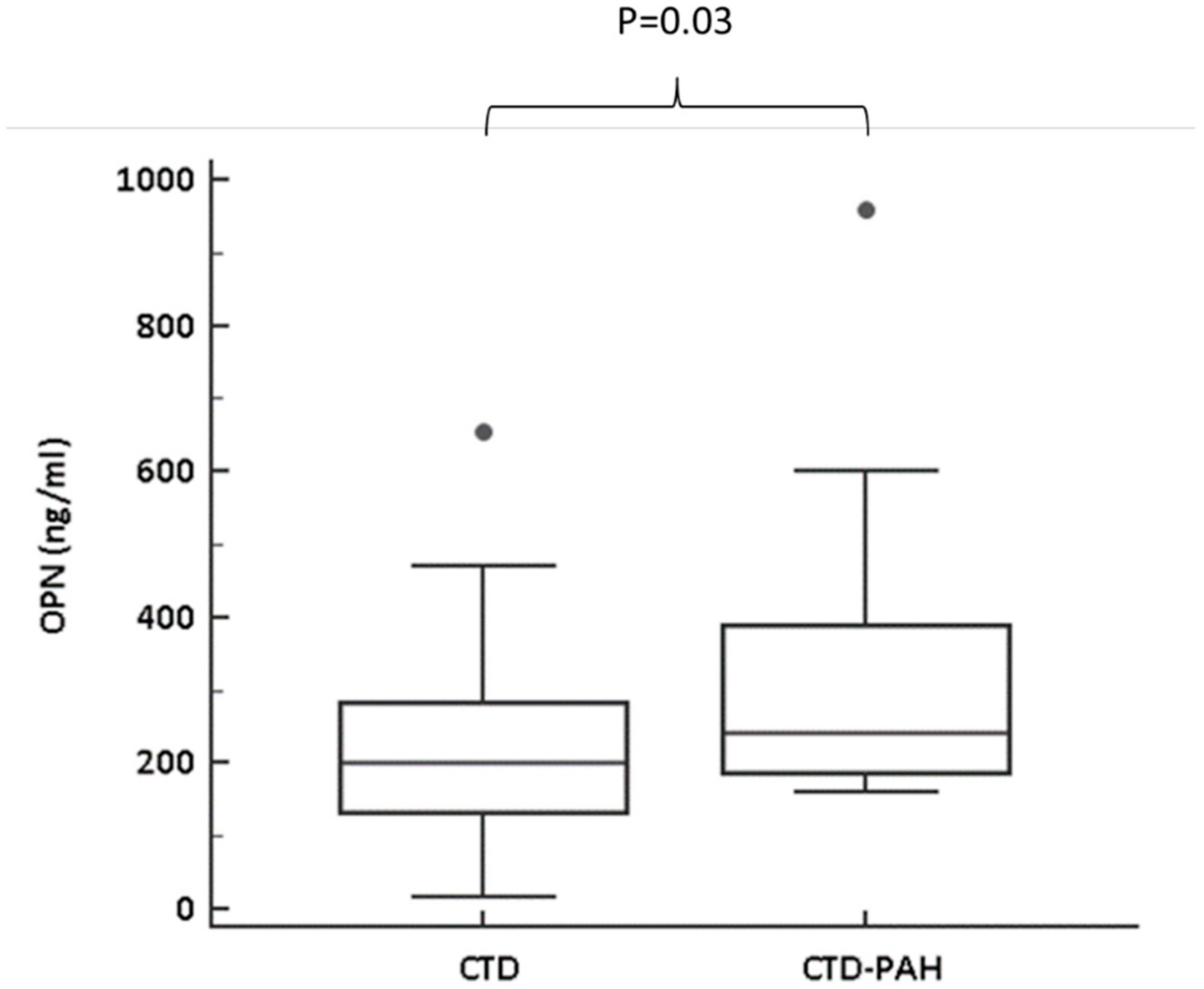
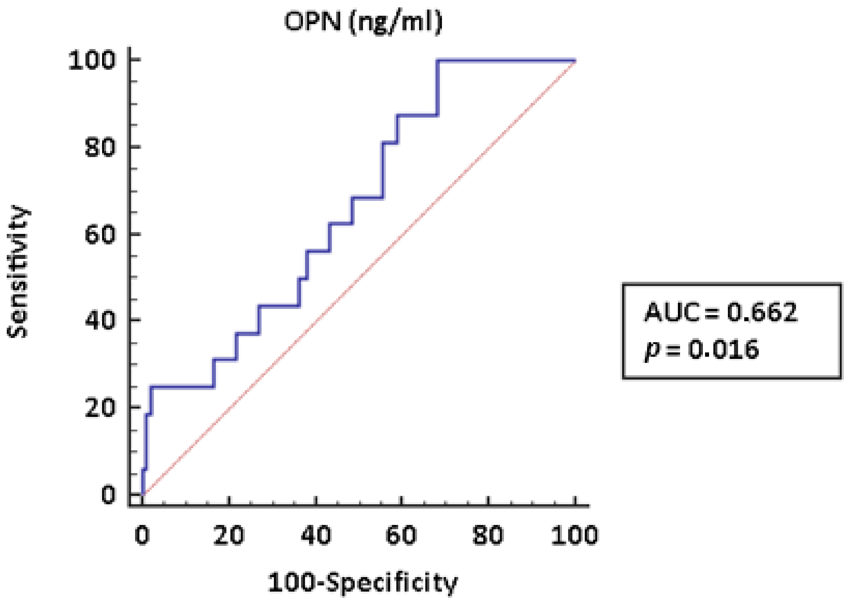
2. Results
3. Discussion
4. Materials and Methods
Statistical Analysis
5. Conclusions
Author Contributions
Funding
Institutional Review Board Statement
Informed Consent Statement
Data Availability Statement
Conflicts of Interest
References
- Galiè, N.; Humbert, M.; Vachiery, J.L.; Gibbs, S.; Lang, I.; Torbicki, A.; Simonneau, G.; Peacock, A.; Vonk Noordegraaf, A.; Beghetti, M.; et al. ESC Scientific Document Group. 2015 ESC/ERS Guidelines for the Diagnosis and Treatment of Pulmonary Hypertension. Eur. Heart J. 2016, 37, 67–119. [Google Scholar] [CrossRef] [PubMed]
- Jawad, H.; McWilliams, S.R.; Bhalla, S. Cardiopulmonary Manifestations of Collagen Vascular Diseases. Curr. Rheumatol. Rep. 2017, 19, 71. [Google Scholar] [CrossRef] [PubMed]
- Ruiz-Cano, M.J.; Escribano, P.; Alonso, R.; Delgado, J.; Carreira, P.; Velazquez, T.; Sanchez, M.A.G.; de la Calzada, C.S. Comparison of Baseline Characteristics and Survival between Patients with Idiopathic and Connective Tissue Disease–related Pulmonary Arterial Hypertension. J. Heart Lung Transplant. 2009, 28, 621–627. [Google Scholar] [CrossRef] [PubMed]
- Coghlan, J.G.; Denton, C.P.; Grünig, E.; Bonderman, D.; Distler, O.; Khanna, D.; Müller-Ladner, U.; Pope, J.E.; Vonk, M.C.; Doelberg, M.; et al. Evidence-Based Detection of Pulmonary Arterial Hypertension in Systemic Sclerosis: The DETECT Study. Ann. Rheum. Dis. 2014, 73, 1340–1349. [Google Scholar] [CrossRef] [Green Version]
- Bellan, M.; Dimagli, A.; Piccinino, C.; Giubertoni, A.; Ianniello, A.; Grimoldi, F.; Sguazzotti, M.; Nerviani, A.; Barini, M.; Carriero, A.; et al. Role of Gas6 and TAM Receptors in the Identification of Cardiopulmonary Involvement in Systemic Sclerosis and Scleroderma Spectrum Disorders. Dis. Markers 2020, 2696173. [Google Scholar] [CrossRef]
- Bellan, M.; Giubertoni, A.; Piccinino, C.; Dimagli, A.; Grimoldi, F.; Sguazzotti, M.; Burlone, M.E.; Smirne, C.; Sola, D.; Marino, P.; et al. Red Cell Distribution Width and Platelet Count as Biomarkers of Pulmonary Arterial Hypertension in Patients with Connective Tissue Disorders. Dis. Markers 2019, 4981982. [Google Scholar] [CrossRef]
- Galiè, N.; Rubin, L.J.; Hoeper, M.M.; Jansa, P.; Al-Hiti, H.; Meyer, G.; Chiossi, E.; Kusic-Pajic, A.; Simonneau, G. Treatment of Patients with Mildly Symptomatic Pulmonary Arterial Hypertension with Bosentan (EARLY study): A Dou-ble-Blind, Randomised Controlled Trial. Lancet 2008, 371, 2093–2100. [Google Scholar] [CrossRef]
- Icer, M.A.; Gezmen-Karadag, M. The Multiple Functions and Mechanisms of Osteopontin. Clin. Biochem. 2018, 59, 17–24. [Google Scholar] [CrossRef]
- Liu, L.N.; Mao, Y.M.; Zhao, C.N.; Wang, H.; Yuan, F.F.; Li, X.M.; Pan, H.F. Circulating Levels of Osteoprotegerin, Osteocalcin and Osteopontin in Patients with Rheumatoid Arthritis: A Systematic Review and Meta-Analysis. Immunol. Investig. 2019, 48, 107–120. [Google Scholar] [CrossRef]
- Castello, L.M.; Baldrighi, M.; Molinari, L.; Salmi, L.; Cantaluppi, V.; Vaschetto, R.; Zunino, G.; Quaglia, M.; Bellan, M.; Gavelli, F.; et al. The Role of Osteopontin as a Diagnostic and Prognostic Biomarker in Sepsis and Septic Shock. Cells 2019, 8, 174. [Google Scholar] [CrossRef] [Green Version]
- Ashkar, S.; Weber, G.F.; Panoutsakopoulou, V.; Sanchirico, M.E.; Jansson, M.; Zawaideh, S.; Rittling, S.R.; Denhardt, D.T.; Glimcher, M.J.; Cantor, H. Eta-1 (osteopontin): An Early Component of Type-1 (Cell-Mediated) Immunity. Science 2000, 287, 860–864. [Google Scholar] [CrossRef]
- Bellan, M.; Castello, L.M.; Pirisi, M. Candidate Biomarkers of Liver Fibrosis: A Concise, Pathophysiology-oriented Review. J Clin. Transl. Hepatol. 2018, 6, 317–325. [Google Scholar] [CrossRef] [Green Version]
- Kothari, A.N.; Arffa, M.L.; Chang, V.; Blackwell, R.H.; Syn, W.K.; Zhang, J.; Mi, Z.; Kuo, P.C. Osteopontin-A Master Regulator of Epithelial-Mesenchymal Transition. J. Clin. Med. 2016, 5, 39. [Google Scholar] [CrossRef] [Green Version]
- Wu, M.; Schneider, D.J.; Mayes, M.D.; Assassi, S.; Arnett, F.C.; Tan, F.K.; Blackburn, M.R.; Agarwal, S.K. Osteopontin in Systemic Sclerosis and its Role in Dermal Fibrosis. J. Investig. Derm. 2012, 132, 1605–1614. [Google Scholar] [CrossRef] [PubMed] [Green Version]
- Gadeau, A.P.; Campan, M.; Millet, D.; Candresse, T.; Desgranges, C. Osteopontin Overexpression is Associated with Arterial Smooth Muscle Cell Proliferation in Vitro. Arterioscler. Thromb. 1993, 13, 120–125. [Google Scholar] [CrossRef] [PubMed] [Green Version]
- Mura, M.; Cecchini, M.J.; Joseph, M.; Granton, J.T. Osteopontin Lung Gene Expression is a Marker of Disease Severity in Pulmonary Arterial Hypertension. Respirology 2019, 24, 1104–1110. [Google Scholar] [CrossRef] [Green Version]
- Hachulla, E.; Gressin, V.; Guillevin, L.; Carpentier, P.; Diot, E.; Sibilia, J.; Kahan, A.; Cabane, J.; Francès, C.; Launay, D.; et al. Early Detection of Pulmonary Arterial Hypertension in Systemic Sclerosis: A French Nationwide Prospective Multicenter Study. Arthritis Rheum. 2005, 52, 3792–3800. [Google Scholar] [CrossRef]
- Steen, V.; Medsger, T.A., Jr. Predictors of Isolated Pulmonary Hypertension in Patients with Systemic Sclerosis and Limited Cutaneous Involvement. Arthritis Rheum. 2003, 48, 516–522. [Google Scholar] [CrossRef]
- Corallo, C.; Volpi, N.; Franci, D.; Montella, A.; Biagioli, M.; Mariotti, G.; D’Onofrio, F.; Gonnelli, S.; Nuti, R.; Giordano, N. Is Osteopontin Involved in Cutaneous Fibroblast Activation? Its Hypothetical Role in Scleroderma Pathogenesis. Int. J. Immunopathol. Pharmacol. 2014, 27, 97–102. [Google Scholar] [CrossRef]
- Barizzone, N.; Marchini, M.; Cappiello, F.; Chiocchetti, A.; Orilieri, E.; Ferrante, D.; Corrado, L.; Mellone, S.; Scorza, R.; Dianzani, U.; et al. Association of Osteopontin Regulatory Polymorphisms with Systemic Sclerosis. Hum. Immunol. 2011, 72, 930–934. [Google Scholar] [CrossRef]
- Lorenzen, J.M.; Nickel, N.; Krämer, R.; Golpon, H.; Westerkamp, V.; Olsson, K.M.; Haller, H.; Hoeper, M.M. Osteopontin in Patients with Idiopathic Pulmonary Hypertension. Chest 2011, 139, 1010–1017. [Google Scholar] [CrossRef] [PubMed]
- Rosenberg, M.; Meyer, F.J.; Gruenig, E.; Schuster, T.; Lutz, M.; Lossnitzer, D.; Wipplinger, R.; Katus, H.A.; Frey, N. Osteopontin (OPN) Improves Risk Stratification in Pulmonary Hypertension (PH). Int. J. Cardiol. 2012, 155, 504–505. [Google Scholar] [CrossRef]
- Kölmel, S.; Hobohm, L.; Käberich, A.; Krieg, V.J.; Bochenek, M.L.; Wenzel, P.; Wiedenroth, C.B.; Liebetrau, C.; Hasenfuß, G.; Mayer, E.; et al. Potential Involvement of Osteopontin in Inflammatory and Fibrotic Processes in Pulmonary Embolism and Chronic Thromboembolic Pulmonary Hypertension. Thromb. Haemost. 2019, 119, 1332–1346. [Google Scholar] [CrossRef] [PubMed]
- Anwar, A.; Li, M.; Frid, M.G.; Kumar, B.; Gerasimovskaya, E.V.; Riddle, S.R.; McKeon, B.A.; Thukaram, R.; Meyrick, B.O.; Fini, M.A.; et al. Osteopontin is an Endogenous Modulator of the Constitutively Activated Phenotype of Pulmonary Adventitial Fibroblasts in Hypoxic Pulmonary Hypertension. Am. J. Physiol. Lung. Cell. Mol. Physiol. 2012, 303, L1–L11. [Google Scholar] [CrossRef]
- van den Hoogen, F.; Khanna, D.; Fransen, J.; Johnson, S.R.; Baron, M.; Tyndall, A.; Matucci-Cerinic, M.; Naden, R.P.; Medsger, T.A., Jr.; Carreira, P.E.; et al. 2013 Classification Criteria for Systemic Sclerosis: An American College of Rheumatology/European League Against Rheumatism Collaborative Initiative. Ann. Rheum. Dis. 2013, 72, 1747–1755. [Google Scholar] [CrossRef] [Green Version]
- Kasukawa, R.; Tojo, T.; Miyawaki, S. Preliminary Diagnostic Criteria for Classification of Mixed Connective Tissue Disease. In Mixed Connective Tissue Disease and Antinuclear Antibodies; Sharp, G., Ed.; Elsevier: Amsterdam, The Netherlands, 1987; p. 41. [Google Scholar]
- Balbir-Gurman, A.; Braun-Moscovici, Y. Scleroderma Overlap Syndrome. Isr. Med. Assoc. J. 2011, 13, 14–20. [Google Scholar]
- Mosca, M.; Baldini, C.; Bombardieri, S. Undifferentiated Connective Tissue Diseases in 2004. Clin. Exp. Rheumatol. 2004, 22, S14–S18. [Google Scholar]


| Clinical Features | Study Population | CTD without PAH | CTD-PAH | p |
|---|---|---|---|---|
| Female gender | 101 (89.4) | 87 (89.7) | 14 (87.5) | 0.68 |
| Median age, years | 65.0 (54.0–75.0) | 62.0 (51.0–71.0) | 74.0 (69.0–78.5) | 0.0004 |
| Hydroxychloroquine | 65 (57.7) | 57 (58.8) | 8 (50.0) | 0.59 |
| Methotrexate | 15 (13.3) | 15 (15.5) | 0 (0.0) | 0.12 |
| Steroids | 38 (33.6) | 36 (37.1) | 2 (12.5) | 0.08 |
| Phosphodiesterase 5 inhibitors | 6 (5.3) | 2 (2.1) | 6 (37.5) | <0.0001 |
| Endothelin-1 receptors antagonists | 8 (7.1) | 7 (7.2) | 8 (50.0) | <0.0001 |
| Riociguat | 1 (0.9) | 0 (0.0) | 1 (6.2) | 0.14 |
| Raynaud’s phenomenon | 102 (90.3) | 87 (89.7) | 14 (87.5) | 0.61 |
| Previous acral ulcers | 49 (43.4) | 43 (44.3) | 7 (43.7) | 1.00 |
| Digital ulcers in the past month | 5 (4.4) | 4 (4.1) | 1 (6.2) | 0.54 |
| Sclerodactyly | 66 (58.4) | 57 (58.8) | 9 (56.2) | 1.00 |
| Puffy fingers | 16 (14.2) | 16 (16.5) | 0 (0.0) | 0.12 |
| Telangiectasia | 32 (28.3) | 28 (28.9) | 5 (31.2) | 1.00 |
| Pulmonary interstitial disease | 38 (33.6) | 31 (32.0) | 7 (43.7) | 0.40 |
| Gastrointestinal involvement | 22 (19.5) | 18 (18.6) | 4 (25.0) | 0.51 |
| Renal involvement | 3 (2.7) | 2 (2.1) | 1 (6.2) | 0.37 |
| Anti-nuclear antibodies (ANA) | 104 (92.0) | 88 (90.7) | 16 (100.0) | 0.35 |
| Anti-centromere antibodies | 69 (61.1) | 56 (57.7) | 13 (81.2) | 0.10 |
| Anti-Scl-70 antibodies | 28 (24.8) | 23 (23.7) | 5 (31.2) | 0.54 |
| Anti-U1-RNP antibodies | 21 (18.6) | 16 (16.5) | 5 (31.2) | 0.17 |
| Disease duration | 5 (3–13) | 5 (4–13) | 5 (3–11) | 0.66 |
| Variable | Study Population | CTD without PAH | CTD-PAH | p |
|---|---|---|---|---|
| WBC, ×109/L | 6.49 (5.26–7.68) | 6.47 (5.26–7.63) | 6.77 (5.15–7.87) | 0.81 |
| Hb, g/dL | 12.8 (11.9–13.7) | 12.9 (12.2–13.7) | 11.3 (10.8–13.5) | 0.01 |
| PLTs, ×109/L | 228 (192–286) | 234 (202–286) | 188 (167–273) | 0.07 |
| ALT, U/L | 17 (13–22) | 18 (13–22) | 13 (12–20) | 0.33 |
| AST, U/L | 23 (20–26) | 23 (20–26) | 23 (20–27) | 0.63 |
| Creatinine, mg/dL | 0.72 (0.61–0.88) | 0.68 (0.6–0.81) | 0.95 (0.81–1.08) | <0.0001 |
| eGFR, mL/min | 90 (63.5–101.3) | 93 (72–103) | 58.5 (51.5–64.5) | <0.0001 |
| CRP, mg/dL | 0.18 (0.04–0.78) | 0.14 (0.04–0.34) | 0.77 (0.04–0.98) | 0.26 |
| ESR, mm/h | 14.5 (7–28) | 13 (7–25) | 25 (7–50) | 0.31 |
| C3, mg/dL | 104 (90–121) | 106 (91–121) | 92 (84–119) | 0.13 |
| C4, mg/dL | 24 (19–28) | 24 (20–29) | 22 (16–26) | 0.11 |
| BNP, pg/mL | 46.8 (27.1–99.6) | 39.6 (24.9–85.7) | 177.0 (82.3–305.2) | <0.0001 |
| FEV1, % | 99 (88–114) | 100.5 (88–113.5) | 94 (88.5–113) | 0.76 |
| FVC, % | 100 (90–112) | 100 (90.5–113.5) | 91 (80–104) | 0.25 |
| FEV1/FVC, % | 109 (102–113.3) | 109 (102–114) | 107.5 (101–113) | 0.74 |
| TLC, % | 98 (86.5–112) | 99 (87–112) | 82 (58–95) | 0.07 |
| DLCO-VA, % | 86 (76–99) | 87 (78–99) | 54.5 (53–76) | 0.008 |
| DLCO-Hb, % | 77 (61–91) | 80 (62–91) | 51 (43–72) | 0.03 |
| EF, % | 63 (58–67) | 63 (58–67) | 61 (58.3–66) | 0.41 |
| PAPS, mmHg | 27 (23–35) | 26 (23–30) | 44 (42–51) | <0.0001 |
| TAPSE, mm | 22 (19–24) | 22 (20–24) | 22 (18–23) | 0.42 |
| Criterion | Sens | 95% CI | Spec | 95% CI | +LR | 95% CI | −LR | 95% CI |
|---|---|---|---|---|---|---|---|---|
| >159.65 | 100.00 | 79.4–100.0 | 31.96 | 22.9–42.2 | 1.47 | 1.3–1.7 | 0.00 | |
| >159.89 | 93.75 | 69.8–99.8 | 31.96 | 22.9–42.2 | 1.38 | 1.1–1.7 | 0.20 | 0.03–1.3 |
| >174.01 | 87.50 | 61.7–98.4 | 41.24 | 31.3–51.7 | 1.49 | 1.2–1.9 | 0.30 | 0.08–1.1 |
| >185.73 | 81.25 | 54.4–96.0 | 44.33 | 34.2–54.8 | 1.46 | 1.1–2.0 | 0.42 | 0.1–1.2 |
| >188.15 | 75.00 | 47.6–92.7 | 44.33 | 34.2–54.8 | 1.35 | 1.0–1.9 | 0.56 | 0.2–1.4 |
| >201.59 | 68.75 | 41.3–89.0 | 51.55 | 41.2–61.8 | 1.42 | 1.0–2.1 | 0.61 | 0.3–1.3 |
| >225.16 | 62.50 | 35.4–84.8 | 56.70 | 46.3–66.7 | 1.44 | 0.9–2.2 | 0.66 | 0.3–1.3 |
| >236.49 | 56.25 | 29.9–80.2 | 61.86 | 51.4–71.5 | 1.47 | 0.9–2.4 | 0.71 | 0.4–1.3 |
| >243.8 | 50.00 | 24.7–75.3 | 63.92 | 53.5–73.4 | 1.39 | 0.8–2.4 | 0.78 | 0.5–1.3 |
| >271.22 | 43.75 | 19.8–70.1 | 73.20 | 63.2–81.7 | 1.63 | 0.9–3.1 | 0.77 | 0.5–1.2 |
| >293.54 | 37.50 | 15.2–64.6 | 78.35 | 68.8–86.1 | 1.73 | 0.8–3.6 | 0.80 | 0.5–1.2 |
| >316 | 31.25 | 11.0–58.7 | 83.51 | 74.6–90.3 | 1.89 | 0.8–4.4 | 0.82 | 0.6–1.2 |
| >450.44 | 25.00 | 7.3–52.4 | 97.94 | 92.7–99.7 | 12.12 | 2.4–60.8 | 0.77 | 0.6–1.0 |
| >470.97 | 18.75 | 4.0–45.6 | 98.97 | 94.4–100.0 | 18.19 | 2.0–164.2 | 0.82 | 0.6–1.0 |
| >564.24 | 12.50 | 1.6–38.3 | 98.97 | 94.4–100.0 | 12.13 | 1.2–126.1 | 0.88 | 0.7–1.1 |
| >654.78 | 6.25 | 0.2–30.2 | 100.00 | 96.3–100.0 | 0.94 | 0.8–1.1 |
| Variable | Coefficient | Standard Error | r | t | p |
|---|---|---|---|---|---|
| OPN | 0.0005 | 0.0002 | 0.19 | 2.09 | 0.04 |
| eGFR | −0.007 | 0.002 | −0.32 | −3.52 | 0.0006 |
| Age | −0.002 | 0.003 | −0.04 | −0.49 | 0.62 |
Publisher’s Note: MDPI stays neutral with regard to jurisdictional claims in published maps and institutional affiliations. |
© 2021 by the authors. Licensee MDPI, Basel, Switzerland. This article is an open access article distributed under the terms and conditions of the Creative Commons Attribution (CC BY) license (https://creativecommons.org/licenses/by/4.0/).
Share and Cite
Bellan, M.; Piccinino, C.; Tonello, S.; Minisini, R.; Giubertoni, A.; Sola, D.; Pedrazzoli, R.; Gagliardi, I.; Zecca, E.; Calzaducca, E.; et al. Role of Osteopontin as a Potential Biomarker of Pulmonary Arterial Hypertension in Patients with Systemic Sclerosis and Other Connective Tissue Diseases (CTDs). Pharmaceuticals 2021, 14, 394. https://doi.org/10.3390/ph14050394
Bellan M, Piccinino C, Tonello S, Minisini R, Giubertoni A, Sola D, Pedrazzoli R, Gagliardi I, Zecca E, Calzaducca E, et al. Role of Osteopontin as a Potential Biomarker of Pulmonary Arterial Hypertension in Patients with Systemic Sclerosis and Other Connective Tissue Diseases (CTDs). Pharmaceuticals. 2021; 14(5):394. https://doi.org/10.3390/ph14050394
Chicago/Turabian StyleBellan, Mattia, Cristina Piccinino, Stelvio Tonello, Rosalba Minisini, Ailia Giubertoni, Daniele Sola, Roberta Pedrazzoli, Ileana Gagliardi, Erika Zecca, Elisa Calzaducca, and et al. 2021. "Role of Osteopontin as a Potential Biomarker of Pulmonary Arterial Hypertension in Patients with Systemic Sclerosis and Other Connective Tissue Diseases (CTDs)" Pharmaceuticals 14, no. 5: 394. https://doi.org/10.3390/ph14050394