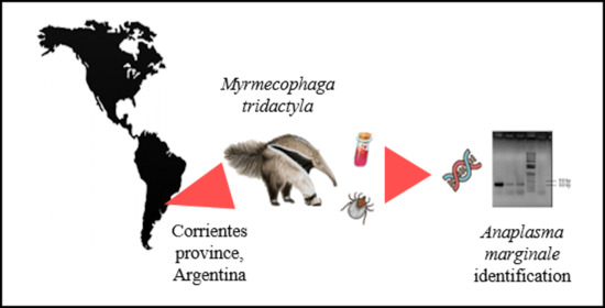
Closing the Gaps to Understand the Tick Transmission of Anaplasma marginale among Giant Anteaters (Myrmecophaga tridactyla) in Argentina

 ,
,  , ,
, ,
Abstract
:
1. Introduction
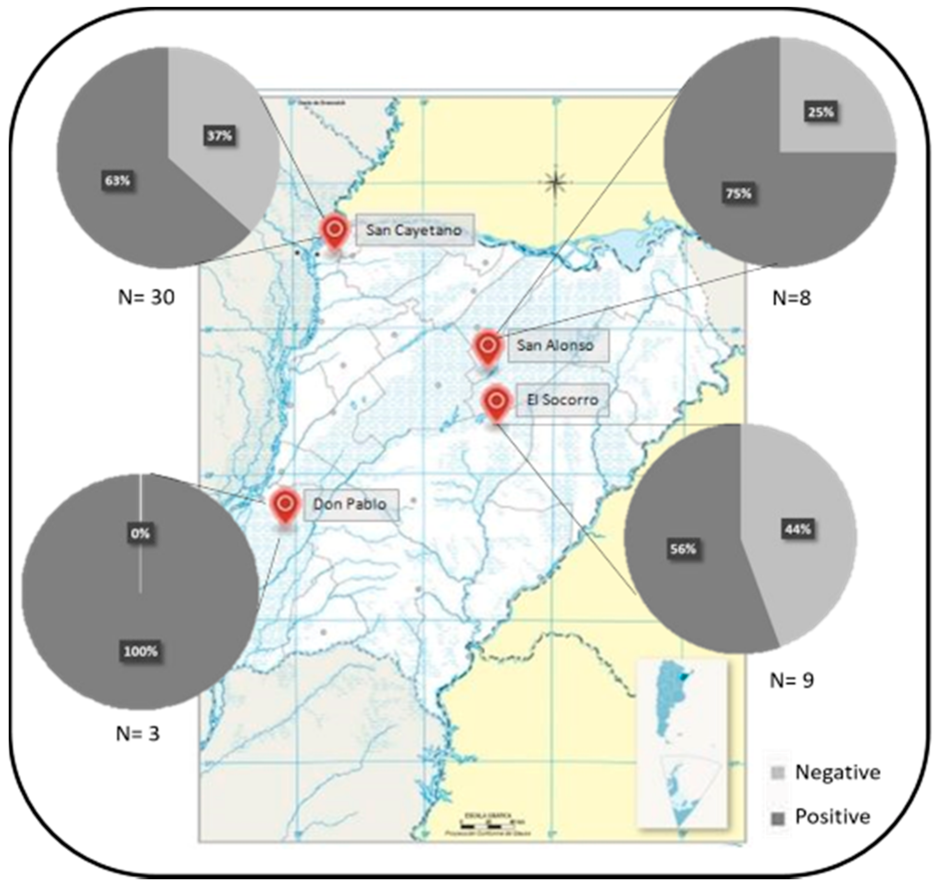
2. Results
2.1. Detection of A. marginale DNA from Blood Samples and Ticks
2.2. Molecular Characterization of A. marginale Strains
3. Discussion
4. Materials and Methods
4.1. Giant Anteaters Blood Samples and Ticks
4.2. Ticks Taxonomic Identification
4.3. DNA Extraction from Blood Samples and Ticks
4.4. Detection of A. marginale DNA from Blood Samples and Ticks
4.5. Molecular Characterization of A. marginale
5. Conclusions
Supplementary Materials
Author Contributions
Funding
Conflicts of Interest
References
- Kocan, K.M.; De Fuente, J.; Alberto, A.; Meléndez, R.D.; Fuente, D.; Guglielmone, A.A.; Mele, R.D. Antigens and Alternatives for Control of Anaplasma marginale Infection in Cattle Antigens and Alternatives for Control of Anaplasma marginale Infection in Cattle. Clin. Microbiol. Rev. 2003, 16, 698–712. [Google Scholar] [CrossRef] [PubMed] [Green Version]
- Ruybal, P.; Moretta, R.; Perez, A.; Petrigh, R.; Zimmer, P.; Alcaraz, E.; Echaide, I.; Torioni de Echaide, S.; Kocan, K.M.; de la Fuente, J.; et al. Genetic diversity of Anaplasma marginale in Argentina. Vet. Parasitol. 2009, 162, 176–180. [Google Scholar] [CrossRef] [PubMed]
- Ashraf, Q.U.A.; Khan, A.U.; Khattak, R.M.; Ali, M.; Shaikh, R.S.; Ali, M.; Iqbal, F. A report on the high prevalence of Anaplasma sp. in buffaloes from two provinces in Pakistan. Ticks Tick Borne Dis. 2013, 4, 395–398. [Google Scholar] [CrossRef] [PubMed]
- Eygelaar, D.; Jori, F.; Mokopasetso, M.; Sibeko, K.P.; Collins, N.E.; Vorster, I.; Troskie, M.; Oosthuizen, M.C. Tick-borne haemoparasites in African buffalo (Syncerus caffer) from two wildlife areas in Northern Botswana. Parasites Vectors 2015, 8, 1–11. [Google Scholar] [CrossRef] [Green Version]
- Munang’andu, H.M.; Siamudaala, V.M.; Munyeme, M.; Nalubamba, K.S. Detection of parasites and parasitic infections of free-ranging wildlife on a game ranch in Zambia: A challenge for disease control. J. Parasitol. Res. 2012, 2012, 296475. [Google Scholar] [CrossRef] [Green Version]
- Silveira, J.A.G.; Rabelo, E.M.L.; Ribeiro, M.F.B. Molecular Detection of Tick-Borne Pathogens of the Family Anaplasmataceae in Brazilian Brown Brocket Deer (Mazama gouazoubira, Fischer, 1814) and Marsh Deer (Blastocerus dichotomus, Illiger, 1815). Transbound. Emerg. Dis. 2012, 59, 353–360. [Google Scholar] [CrossRef]
- Sudan, V.; Sharma, R.L.; Borah, M.K. Subclinical anaplasmosis in camel (Camelus dromedarius) and its successful therapeutic management. J. Parasit. Dis. 2014, 38, 163–165. [Google Scholar] [CrossRef] [Green Version]
- Tonetti, N.; Berggoetz, M.; Rühle, C.; Pretorius, A.M.; Gern, L. Ticks and tick-borne pathogens from wildlife in the Free State Province, South Africa. J. Wildl. Dis. 2009, 45, 437–446. [Google Scholar] [CrossRef] [Green Version]
- Guillemi, E.C.; De La Fourniere, S.; Orozco, M.; Peña Martinez, J.; Correa, E.; Fernandez, J.; Lopez Arias, L.; Paoletta, M.; Corona, B.; Pinarello, V.; et al. Molecular identification of Anaplasma marginale in two autochthonous South American wild species revealed an identical new genotype and its phylogenetic relationship with those of bovines. Parasites Vectors 2016, 9, 305. [Google Scholar] [CrossRef] [Green Version]
- Calchi, A.C.; Vultão, J.G.; Alves, M.H.; Yogui, D.R. Ehrlichia spp and Anaplasma spp. in Xenarthra mammals from Brazil, with evidence of novel ‘Candidatus Anaplasma spp.’. Sci. Rep. 2020, 10, 12615. [Google Scholar] [CrossRef]
- Santos, P.M.; Bocchiglieri, A.; Chiarello, A.G.; Paglia, A.P.; Moreira, A.; de Souza, A.C.; Abba, A.M.; Paviolo, A.; Gatica, A.; Medeiro, A.Z.; et al. Neotropical Xenarthrans: A data set of occurrence of xenarthran species in the Neotropics. Ecology 2019, 100. [Google Scholar] [CrossRef] [PubMed] [Green Version]
- Nava, S.; Venzal, J.M.; Gonzalez-Acuña, D.; Martins, T.F.; Guglielmone, A.A. Ticks of the Southern Cone of America; Academic Press, Elsevier: London, UK, 2017; ISBN 9780128110751. [Google Scholar]
- Guillemi, E.C.; Ruybal, P.; Lia, V.; Gonzalez, S.; Lew, S.; Zimmer, P.; Lopez Arias, L.; Rodriguez, J.L.; Rodriguez, S.Y.; Frutos, R.; et al. Development of a Multilocus Sequence Typing scheme for the study of Anaplasma marginale population structure over space and time. Infect. Genet. Evol. 2015, 30, 186–194. [Google Scholar] [CrossRef] [PubMed]
- Paoletta, M.S.; López Arias, L.; de la Fournière, S.; Guillemi, E.C.; Luciani, C.; Sarmiento, N.F.; Mosqueda, J.; Farber, M.D.; Wilkowsky, S.E. Epidemiology of Babesia, Anaplasma and Trypanosoma species using a new expanded reverse line blot hybridization assay. Ticks Tick Borne Dis. 2018, 9, 155–163. [Google Scholar] [CrossRef] [PubMed]
- de la Fuente, J.; Ruybal, P.; Mtshali, M.S.; Naranjo, V.; Shuqing, L.; Mangold, A.J.; Rodríguez, S.D.; Jiménez, R.; Vicente, J.; Moretta, R.; et al. Analysis of world strains of Anaplasma marginale using major surface protein 1a repeat sequences. Vet. Microbiol. 2007, 119, 382–390. [Google Scholar] [CrossRef] [PubMed]
- Orozco, M.M.; Argibay, H.D.; Minatel, L.; Guillemi, E.C.; Berra, Y.; Schapira, A.; Di Nucci, D.; Marcos, A.; Lois, F.; Falzone, M.; et al. A participatory surveillance of marsh deer (Blastocerus dichotomus) morbidity and mortality in Argentina: First results. BMC Vet. Res. 2020, 16, 1–18. [Google Scholar] [CrossRef]
- De Sousa, K.C.M.; Calchi, A.C.; Herrera, H.M.; Dumler, J.S.; Barros-Battesti, D.M.; MacHado, R.Z.; André, M.R. Anaplasmataceae agents among wild mammals and ectoparasites in Brazil. Epidemiol. Infect. 2017, 145, 3424–3437. [Google Scholar] [CrossRef] [Green Version]
- Soares, H.S.; Marcili, A.; Barbieri, A.R.M.; Minervino, A.H.H.; Malheiros, A.F.; Gennari, S.M.; Labruna, M.B. Novel Anaplasma and Ehrlichia organisms infecting the wildlife of two regions of the Brazilian Amazon. Acta Trop. 2017, 174, 82–87. [Google Scholar] [CrossRef]
- da Silva, J.B.; da Fonseca, A.H.; Barbosa, J.D. Molecular characterization of Anaplasma marginale in ticks naturally feeding on buffaloes. Infect. Genet. Evol. 2015, 35, 38–41. [Google Scholar] [CrossRef] [Green Version]
- Pothmann, D.; Poppert, S.; Rakotozandrindrainy, R.; Hogan, B.; Mastropaolo, M.; Thiel, C.; Silaghi, C. Prevalence and genetic characterization of Anaplasma marginale in zebu cattle (Bos indicus) and their ticks (Amblyomma variegatum, Rhipicephalus microplus) from Madagascar. Ticks Tick Borne Dis. 2016, 7, 1116–1123. [Google Scholar] [CrossRef]
- Di Blanco, Y.E.; Varela, D.; Abba, A.M. Myrmecophaga tridactyla En: SAyDS–SAREM (eds.) Categorización 2019 de los Mamíferos de Argentina Según su Riesgo de Extinción Lista Roja de los Mamíferos de Argentina. Available online: http://cma.sarem.org.ar/es/especie-nativa/myrmecophaga-tridactyla (accessed on 6 December 2020).
- Miranda, F.; Bertassoni, A.; Abba, A.M. Myrmecophaga tridactyla. The IUCN Red List of Threatened Species 2014: e.T14224A47441961. 2014. Available online: https://dx.doi.org/10.2305/IUCN.UK.2014-1.RLTS.T14224A47441961.en (accessed on 6 December 2020).
- Sikes, R.S. Guidelines of the American Society of Mammalogists for the use of wild mammals in research and education. J. Mammal. 2016, 97, 663–688. [Google Scholar] [CrossRef]
- Sikes, R.S.; Gannon, W.L. Guidelines of the American Society of Mammalogists for the use of wild mammals in research. J. Mammal. 2011, 92, 235–253. [Google Scholar] [CrossRef]
- Sambrook, J.; Fritsch, E.F.; Maniatis, T. Molecular Cloning: A Laboratory Manual, 2nd ed.; Cold Spring Harbor Laboratory Press: Cold Spring Harbor, NY, USA, 1989. [Google Scholar]
- Bilgiç, H.B.; Karagenç, T.; Simuunza, M.; Shiels, B.; Tait, A.; Eren, H.; Weir, W. Experimental Parasitology Development of a multiplex PCR assay for simultaneous detection of Theileria annulata, Babesia bovis and Anaplasma marginale in cattle. Exp. Parasitol. 2013, 133, 222–229. [Google Scholar] [CrossRef] [PubMed] [Green Version]
- Torioni de Echaide, S.; Knowles, D.P.; McGuire, T.C.; Palmer, G.H.; Suarez, C.E.; McElwain, T.F. Detection of Cattle Naturally Infected with Anaplasma marginale in a Region of Endemicity by Nested PCR and a Competitive Enzyme-Linked Immunosorbent Assay Using Detection of Cattle Naturally Infected with Anaplasma marginale in a Region of Endemicity by Nested PCR and a Competitive Enzyme-linked Immunosorbent Assay Using Recombinant Major Surface Protein 5. J. Clin. Microbiol. 1998, 36, 777–782. [Google Scholar] [PubMed]
- De la Fuente, J.; Van Den Bussche, R.A.; Kocan, K.M. Molecular phylogeny and biogeography of North American isolates of Anaplasma marginale (Rickettsiaceae: Ehrlichieae). Vet. Parasitol. 2001, 97, 65–76. [Google Scholar] [CrossRef]


| Tick Species | Amblyomma dubitatum | Amblyomma sculptum | |||||
|---|---|---|---|---|---|---|---|
| Giant Anteater identification number | A. marginale in the blood sample | F | M | N | F | M | N |
| GA16 | Positive | 1 | |||||
| GA22 | Negative | 4 | 2 | ||||
| GA31 | Positive | 1 | |||||
| GA33 | Positive | 3 | |||||
| GA35 | Positive | 2 | 3 | ||||
| GA36 | Positive | 6 | |||||
| GA37 | Positive | 1 | 2 | ||||
| GA42 | No sample | 1 | |||||
| Total | 12 | 14 | |||||
| Giant Anteater Identification Number | Tick Species | Stage | Tick Organ | ||
|---|---|---|---|---|---|
| SG | OV | GUT | |||
| GA16 | A. dubitatum | F | Am | ||
| GA22 | A. dubitatum | M | / | Am | |
| A. dubitatum | M | Am | / | ||
| GA35 | A. dubitatum | M | Am | / | |
| GA36 | A. sculptum | F | Am | ||
| A. sculptum | F | Am | Am | Am | |
| A. sculptum | F | Am | |||
| A. sculptum | F | Am | |||
| A. sculptum | F | Am | Am | ||
| GA37 | A. dubitatum | M | Am | / | Am |
| GA42 | A. dubitatum | F | Am | ||
| Giant Anteater Identification Number | Genotype |
|---|---|
| GA 5 | 13 27 |
| GA 6 | 13 27 |
| GA 10 | 13 27 |
| GA 12 | 13 27 |
| GA 13 | 13 27 |
| GA 14 | 13 27 |
| GA 16 | 13 27 |
| GA 18 | 23 30 31 31 31 |
| GA 19 | 13 27 |
| GA 21 | 13 27 |
| GA 24 | 13 27 |
| Repeat | Encoded Sequence |
|---|---|
| 13 | TDSSSASGQQQESSVLSQSDQASTSSQLG |
| 23 | TDSSSASGQQQKSSVLSQSSQASTSSQLG |
| 27 | ADSSSASGQQQESSVLSQSDQASTSSQLG |
| 30 | ADSSSASGQQQKSSVLSQSSQASTSSQLG |
| 31 | ADSSSAGNQQQESSVSSQSDASTSSQLG |
Publisher’s Note: MDPI stays neutral with regard to jurisdictional claims in published maps and institutional affiliations. |
© 2020 by the authors. Licensee MDPI, Basel, Switzerland. This article is an open access article distributed under the terms and conditions of the Creative Commons Attribution (CC BY) license (http://creativecommons.org/licenses/by/4.0/).
Share and Cite
Guillemi, E.C.; Imbert, M.; de la Fournière, S.; Orozco, M.M.; Peña Martinez, J.; Rosas, A.C.; Montenegro, V.N.; Farber, M.D. Closing the Gaps to Understand the Tick Transmission of Anaplasma marginale among Giant Anteaters (Myrmecophaga tridactyla) in Argentina. Pathogens 2020, 9, 1033. https://doi.org/10.3390/pathogens9121033
Guillemi EC, Imbert M, de la Fournière S, Orozco MM, Peña Martinez J, Rosas AC, Montenegro VN, Farber MD. Closing the Gaps to Understand the Tick Transmission of Anaplasma marginale among Giant Anteaters (Myrmecophaga tridactyla) in Argentina. Pathogens. 2020; 9(12):1033. https://doi.org/10.3390/pathogens9121033
Chicago/Turabian StyleGuillemi, Eliana Carolina, Mélody Imbert, Sofia de la Fournière, María Marcela Orozco, Jorge Peña Martinez, Ana Carolina Rosas, Valeria Noely Montenegro, and Marisa Diana Farber. 2020. "Closing the Gaps to Understand the Tick Transmission of Anaplasma marginale among Giant Anteaters (Myrmecophaga tridactyla) in Argentina" Pathogens 9, no. 12: 1033. https://doi.org/10.3390/pathogens9121033