Article

An Entomological Investigation during a Recent Rift Valley Fever Epizootic/Epidemic Reveals New Aspects of the Vectorial Transmission of the Virus in Madagascar

by
Luciano Michaël Tantely
1,*,
Soa Fy Andriamandimby
2,
Maminirina Fidelis Ambinintsoa
1,
Manou Rominah Raharinirina
1,
Jean Théophile Rafisandratantsoa
2,
Jean-Pierre Ravalohery
2,
Aina Harimanana
3,
Nirina Nantenaina Ranoelison
3,
Judickaelle Irinantenaina
3,
Miamina Fidy Ankasitrahana
4,
Dany Bakoly Ranoaritiana
4,
Laurence Randrianasolo
3,
Rindra Vatosoa Randremanana
3,
Vincent Lacoste
2,
Philippe Dussart
2 and
Romain Girod
1
1
Medical Entomology Unit, Institut Pasteur de Madagascar, Antananarivo 101, Madagascar
2
Virology Unit, Institut Pasteur de Madagascar, Antananarivo 101, Madagascar
3
Epidemiology and Clinical Research Unit, Institut Pasteur de Madagascar, Antananarivo 101, Madagascar
4
Direction de la Veille Sanitaire, de la Surveillance Epidémiologique et Ripostes, Ministry of Public Health, Antananarivo 101, Madagascar
*
Author to whom correspondence should be addressed.
Pathogens 2024, 13(3), 258; https://doi.org/10.3390/pathogens13030258
Submission received: 2 February 2024 / Revised: 29 February 2024 / Accepted: 9 March 2024 / Published: 16 March 2024
(This article belongs to the Special Issue Viral Infections of Humans: Epidemiology and Control)

Abstract

:
A Rift Valley fever (RVF) outbreak occurred in at least five regions of Madagascar in 2021. The aim of this study was to provide an overview of the richness, abundance, ecology, and trophic preferences of mosquitoes in the Mananjary district and to investigate the distribution of mosquitoes that were RT-PCR-positive for RVFV. Three localities were prospected from 26 April to 4 May 2021, using light traps, BG-Sentinel traps baited with an artificial human odor, Muirhead-Thomson pit traps, and indoor pyrethroid spray catches. A total of 2806 mosquitoes belonging to at least 26 species were collected. Of 512 monospecific pools of mosquitoes tested with real-time RT-PCR, RVFV was detected in 37 pools representing 10 mosquito species. The RVFV-positive species were as follows: Aedes albopictus, Ae. argenteopunctatus, Anopheles coustani, An. gambiae s.l., An. mascarensis, An. squamosus/cydippis, Culex antennatus, Cx. decens, Cx. Tritaeniorhynchus, and Uranotaenia spp. Of the 450 tested engorged females, 78.7% had taken a blood meal on humans, 92.9% on cattle, and 71.6% had taken mixed (human–cattle) blood meals. This investigation suggests the potential role of mosquitoes in RVFV transmission within this epizootic/epidemic context and that the human populations at the three study sites were highly exposed to mosquitoes. Therefore, the use of impregnated mosquito nets as an appropriate prevention method is recommended.

1. Introduction

Rift Valley fever virus (RVFV) belongs to the Phlebovirus genus, Phenuiviridae family, within the Bunyavirales order. It was first isolated after several infections were observed in sheep during a hepatitis outbreak in Kenya almost a century ago [1]. This virus is transmitted mechanically via direct contact with the body fluids (blood, saliva, and/or nasal discharges) or infected tissues of animals or aborted ruminant fetuses. Aerosols of infected blood generated during ruminant abortion or animal slaughter can also be a source of virus transmission to humans [2,3]. This virus can also be transmitted to vertebrate hosts via the bites of infected mosquitoes [4]. For some species, the vertical transmission of RVFV from infected female mosquitoes to their offspring has been observed and is described as the principal means of RVFV maintenance in the field [5]. The epidemic/epizootic cycle involves domestic ruminants (cattle, sheep, and goat) and humans, who are considered dead-end hosts [6]. Wild ruminants are mainly involved in a sylvatic cycle [7]. Birds, reptiles, and amphibians are refractory, and livestock species, such as pigs and horses, are resistant to infection but could play a subordinate role in the RVFV transmission cycle [8,9,10].
The first evidence of the vector transmission of RVFV dates back to 1944, when the virus was isolated from wild mosquitoes of the Eretmapodites and Aedes genera [11]. Since then, several epidemics/epizootics have been observed in numerous countries on the African continent [12], the Arabian Peninsula [13], and the islands of the southwestern Indian Ocean, including Madagascar [14], the Comoros [15], and Mayotte [16]. The occurrence and spread of RVF outbreaks in these regions depend on the ecoclimatic facies (Arabian Peninsula, West Africa, and East Africa) and the composition of the vector fauna, mainly comprising mosquitoes belonging to the Aedes and Culex genera [17]. The common feature of RVFV outbreaks in West and East Africa is that they are generally observed during the rainy season, when potential vectors are abundant [18,19]. The movement of infected cattle between countries has also been associated with RVF outbreaks in West [20] and East Africa [21].
In Madagascar, the circulation of RVFV was first reported in 1979 following its isolation from mosquito pools (monospecific pools of Mansonia uniformis, as well as pools containing a mixture of Anopheles coustani/An. fuscicolor, An. pauliani/An. squamosus, Coquillettidia grandidieri/Ma. uniformis, Culex simpsoni/Cx. vansomereni/Cx. univittatus, Cx. antennatus/Cx. simpsoni/Cx. vansomereni, and Cx. simpsoni/Cx. vansomereni/Cx. annulioris) captured in the primary rainforest of Périnet in the east of the country [22]. At that time, no circulation of the virus had been demonstrated in cattle or humans. Between 1982 and 1989, serological surveys of 824 human and 882 animal sera carried out in different regions showed little maintenance of the virus [23]. Then, two epizootic foci were observed in cattle in 1990 and 1991 on the east coast and in the Central Highlands, respectively, but the virus was not isolated from any mosquitoes [24,25]. From 1996 to 1998, serological investigations also showed the low-level circulation of the virus in herds, and no attempt has been made to detect nor isolate this virus from mosquitoes [26]. Ten years later, in 2006, the World Organization for Animal Health [27] confirmed that the virus was circulating on the island, with no clinical cases of RVF observed [28]. In 2008, the Institut Pasteur de Madagascar (IPM) confirmed the infection of 59 patients with RVFV, while the Ministry of Public Health (MoPH) recorded 17 deaths from 418 suspected cases [29]. From 2008 to 2009, RVF cases were recorded in humans and cattle in the northern, central, and southeastern parts of the country [30]. The virus was detected in three species of mosquitoes (Cx. Antennatus, An. Squamosus, and An. coustani) captured in the districts of Fianarantsoa I and II [31]. Retrospective studies have shown viral circulation in cattle in almost all Malagasy districts [32]. Since this last outbreak, no circulation of RVFV has been mentioned in livestock, but the research-based literature has highlighted the presence in the country of 24 mosquito species known to be associated with RVFV transmission [17]. Seven years after the 2008–2009 epidemic, three mosquito species (Cx. antennatus, Cx. decens, and Cx. tritaeniorhynchus), collected in the west of the country during the 2016 dry season, tested positive for RVFV [33], bringing the number of species potentially associated with RVFV transmission (Cx. decens being newly included in the list) to 25. In addition, a vector competence study of locally collected Cx. antennatus and An. coustani supported the role of both species in RVFV transmission in Madagascar [34].
On 2 April 2021, an epizootic of RVF in cattle in at least five regions of Madagascar (Atsimo–Andrefana, Vatovavy–Fitovinany, DIANA, Atsimo Atsinanana, and Alaotra Mangoro) was declared by the Ministry of Agriculture, Livestock and Fisheries (Ministère de l’Agriculture, de l’Elevage et de la Pêche, MAEP). The Vatovavy–Fitovinany region was the most affected. Two weeks later, on 15 April 2021, a suspected human case was identified at the Basic Health Center (Centre de Santé de Base, CSB) of Antsenavolo in the Mananjary health district of the Vatovavy–Fitovinany region. To assess the risk of transmission to humans and to propose appropriate prevention and vector control measures, a multidisciplinary investigation team, including entomologists, virologists, and epidemiologists of the IPM and the MoPH and MAEP, performed an investigation in the district of Mananjary with the aim of documenting the epidemiological, virological, and entomological situation (Supplementary Figure S1). The aim of this paper is to (i) describe the mosquito species composition, (ii) understand their ecology and assess their trophic preferences, and (iii) investigate the distribution of mosquitoes that are RT-PCR-positive for RVFV.

2. Materials and Methods
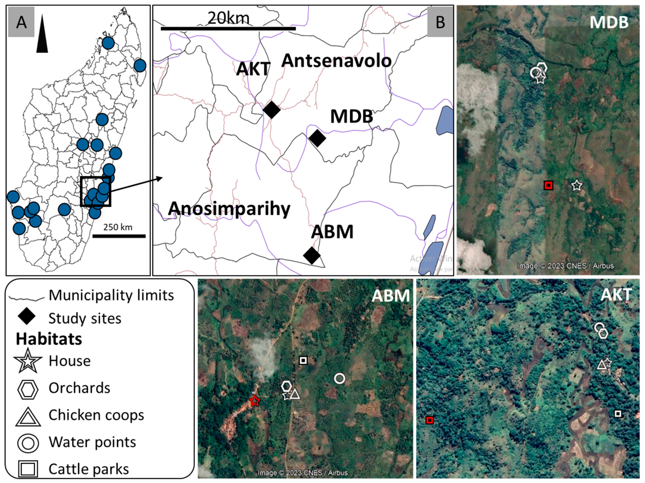
2.1. Study Sites

Entomological investigations were carried out in three villages located in the municipalities of Antsenavolo and Anosimparihy in the district of Mananjary, in the Vatovavy region (Figure 1). The villages were chosen according to the presence of (i) suspected bovine and/or human cases known before the arrival onsite of the investigation team (municipality of Antsenavolo) and/or (ii) confirmed human cases even during the investigation (municipality of Anosimparihy).
The first village, Ambodiakatra (21°23′27.94″ S, 48°1′47.64″ E), is located along National Road 25, near the village of Antsenavolo, in the municipality of Antsenavolo. The landscape is made up of secondary forests with fruit trees (coffee, orange, mango, and lychee) and banana, coconut, and ravenale trees, as well as sugar cane fields. Rice fields are also located in small valleys separating the forested areas, and clove plantations are present near human habitations. Visible domestic animals are zebu, poultry, dogs, and pigs. Most of the farmers are also beekeepers. The second village, Madiolamba (21°25′6.79″ S, 48°6′13.62″ E), is more isolated, located 10 km southeast of the village of Antsenavolo, but still in the municipality of Antsenavolo. The landscape is similar, consisting of plantations of fruit trees, fields of sugar cane, and pineapples. The village is bordered by a stream, which makes the houses inaccessible during the rainy season when the waters rise. Rice fields are present in the small valleys separating the plantations of fruit trees. Domestic animals include zebu, poultry, dogs, and pigs. The third village, Ambalamainty (21°32′46.32″ S, 48°5′44.37″ E), is located 20 km southeast of the village of Antsenavolo, in the municipality of Anosimparihy. The landscape is similar. Rice fields, slash-and-burn crops, and swamps separate the plantations of fruit trees. The domestic animals present are zebu, poultry, and dogs. Beekeeping and clove cultivation are practiced by farmers.

2.2. Mosquito Sampling

From 26 April to 4 May 2021, adult mosquitoes were collected at each of the three villages during 48-h sessions by combining six light traps (LTs), which operated from 4:00 p.m. to 7:00 a.m.; six BG-Sentinel traps baited with an artificial human odor (BGs), which operated from 5:00 a.m. to 7:00 p.m.; and six Muirhead-Thomson pit traps (MTPTs) dug outside human dwellings. Mosquito collections were carried out with a mouth aspirator between 7:30 and 8:30 a.m. Furthermore, six sets of indoor pyrethroid spray catches (IPSCs) were carried out. Knocked-down mosquitoes were collected on white sheets between 7:30 and 8:30 a.m. The four methods (Supplementary Figure S1) were implemented in six habitat types (chicken coops, forest edges, orchards, water points, cattle parks, and houses where human and cattle RVF cases were suspected or confirmed). Forest edges and orchards were considered as a single habitat for subsequent analyses and are identified as “orchards” in the rest of the paper.

2.3. Mosquito Identification and Storing

In the field, the adult mosquitoes collected were anesthetized with chloroform vapors and identified using the keys of Ravaonjanahary [35] for Aedes, Grjebine [36] for Anopheles, Doucet [37] for Coquillettidia, Edwards [38] for Culex, Brunhes and Hervy [39] for Orthopodomyia, and da Cunha Ramos and Brunhes [40] for Uranotaenia species. Immediately after morphological identification, the mosquitoes were counted; grouped into monospecific batches in cryotubes according to sex, the status of engorgement of females, the places and dates of collection, and the collection method used; and placed in liquid nitrogen. Upon return to the laboratory, they were stored at −80 °C pending further analysis.

2.4. Blood Meal Identification

The abdomens of engorged females were dissected and individualized in 2 mL microtubes. Total DNA was extracted using either a DNeasy Blood kit (Qiagen, Hilden, Germany) or QIAamp DNA kit (Qiagen). Samples were eluted in 200 µL of an AE Elution Buffer according to the manufacturer’s instructions and stored at −20 °C until use. The PCR technique developed by Pitzer et al. [41], with two forward primers designed to discriminate between blood meals taken from humans (Human-F: 5′-CTCGGCTTACTT CTCTTCC-3′) and zebu (Cattle-F: 5′-TTATCATCATAGCAATTGCC-3′), was used with a universal reverse primer (UnivRev: 5′-AGTGGGYGRAATATTATGC-3′) annealing at a conserved region for these two hosts.
The PCR mix was as follows: GoTaq Hot Start Green Master Mix (Promega, Madison, WI, USA) at a 1X final concentration, forward and reverse primers (at a 1 μM final concentration), and 2 μL of DNA diluted 1/10, in a final reaction volume of 25 μL. The PCR cycling conditions were as described by Pitzer et al. [41]. Amplicons were visualized on a 1.5% agarose gel.

2.5. RVFV Detection in Mosquitoes

Monospecific pools of 1 to 10 male mosquitoes, entire non-engorged females, or the heads and thoraxes of engorged females were created in 2 mL microtubes. The samples were then crushed in 200 µL of a cell culture medium (MEM) containing 40% fetal bovine serum, 2 mM L-glutamine, 1000 U/mL penicillin, 100 mg/mL streptomycin, and 2.5 mg/mL amphotericin B, for one minute, with 5 mm glass beads, using a TissueLyser II (Qiagen). After grinding, 200 µL of the ground material was transferred to a deep-96 well flat KF extraction plate. Viral RNA was extracted from the homogenates by using a KingFisher machine (Thermo Fisher Scientific, Waltham, MA, USA) according to the manufacturer’s instructions. Viral detection was performed via real-time RT-PCR using primers and a probe (RVF FP: 5′-TGCCAC GAGTYAGAGCCA-3′, RVF RP: 5′-GTGGGTCCGAGAGTYTGC-3′, and probe RVF P: (6FAM)-TCCTTCTCCCAGTCAGCCCCAC-(BHQ-1)) targeting the S segment of RVFV, as described by Weidmann et al. [42]. The limit of detection was determined to be 100 copies/reaction.

2.6. Statistical Analysis

All statistical analyses and most of the figures were generated using R version 4.2.2 [43]. For each mosquito genus and species, their abundance by village, type of habitat, and type of trap was compared using a Kruskal–Wallis test, followed by Dunn’s post hoc test to determine which pairs of groups were different. An Excel® heatmap was used to visualize the variations in mosquito abundance between the three villages, the five habitat types, and the collected species. For blood meal analysis, a paired differential analysis between the human and cattle blood index of each species was obtained using the ggplot2 and ggpubr packages. Wilcoxon matched-pair signed-rank tests were used to compare paired human and blood indices. By using the ggalluvial packages in ggplot2, alluvial plots were generated to visualize the change in the number of engorged females collected across mosquito species, types of blood meals, habitat types, and sites.

3. Results

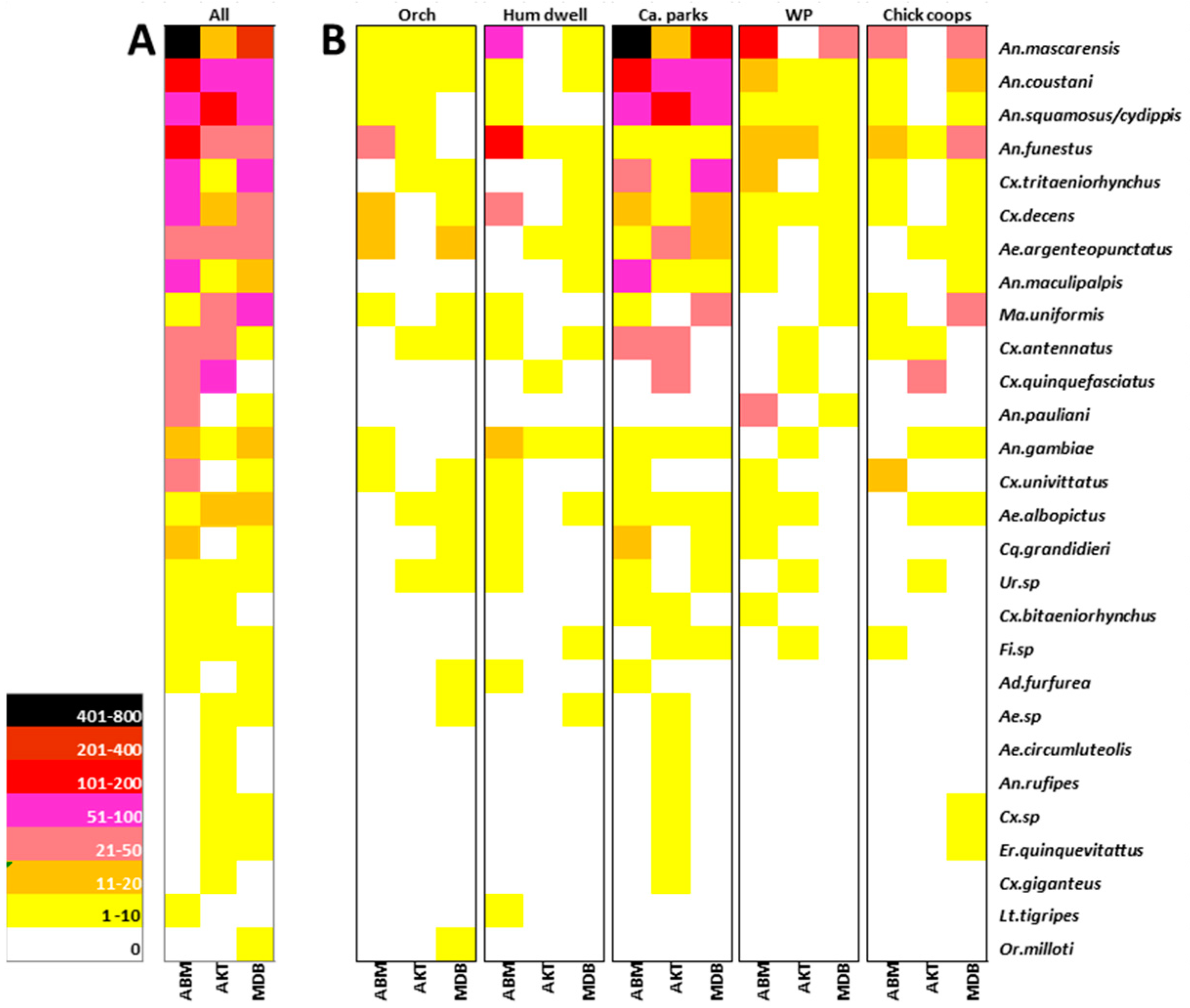
3.1. Mosquito Species Distribution and Abundance

Over six days, a total of 2806 mosquitoes were collected. Eleven genera and at least twenty-six species were identified, which were distributed as follows: Aedeomyia (one species), Aedes (at least three species), Anopheles (eight species), Culex (at least seven species), Coquillettidia (one species), Eretmapodites (one species), Lutzia (one species), Mansonia (one species), Ficalbia (at least one species), Orthopodomyia (one species), and Uranotaenia (at least one species). Four specimens could not be assigned to a genus (Table 1). Four species endemic to Madagascar (Or. milloti, An. pauliani, Cq. grandidieri, and Cx. giganteus) and one species endemic to Madagascar and the Comoros archipelago (An. mascarensis) were collected.
The number of mosquitoes collected varied significantly according to the method used (Kruskal–Wallis X2 = 59.35, df = 3, p < 0.001). Mosquitoes were mainly collected using LTs (2638 mosquitoes, 26 species), with the exception of Cx. Quinquefasciatus, which was mainly collected with MTPs, and Ae. albopictus, with BGs. In terms of abundance, light trap collections were followed by IPSCs (76 mosquitoes, 5 species), BGs (53 mosquitoes, 10 species), and finally MTPTs (39 mosquitoes, 9 species) (Figure 2).
The genus Anopheles accounted for 74.3% (2806) of all collections, followed by the genus Culex (16.6%, 467). The other nine genera accounted for 9.0% (253) of the total. The number of mosquitoes collected varied significantly between species (Kruskal–Wallis X2 = 191.52, df = 25, p < 0.001).
Anopheles mascarensis was the most abundant species, accounting for 37.4% of all mosquitoes caught and 50.3% of all Anopheles collected. Anopheles squamosus/cydippis and An. coustani each accounted for 10.6% of all mosquitoes caught, An. funestus for 9.6%, Culex tritaeniorhynchus 6.0%, and Cx. decens 3.7%.
The number of mosquitoes collected varied significantly between the three villages (Kruskal–Wallis X2 = 44.481, df = 2, p < 0.001) and between the five habitats (Kruskal–Wallis X2 = 76.075, df = 4, p < 0.001) (Figure 2). The highest number of mosquitoes, with a total number of 1624 (57.9%), was collected in Ambalamainty, followed by the villages of Madiolamba (742, 26.4%) and Ambodiakatra (440, 15.7%). The predominance of An. mascarensis and An. coustani was particularly notable in Ambalamainty, while An. squamosus/cydippis was predominant in Ambodiakatra (Figure 2A). For the most abundant species, the majority of mosquitoes were caught near cattle parks (62.9%, 1764). Mosquitoes were more rarely collected at forest edges, regardless of the site (Figure 2B). The predominance of An. gambiae, An. funestus, and Cx. decens near human habitation was observed in Ambalamainty. Fourteen species were common to all three sites and a further ten to two of the three sites (Figure 2B).
Of the 26 species collected, 14 have already been reported to be involved in RVFV transmission [17]. They belonged to the Anopheles, Aedes, Coquillettidia, Culex, and Mansonia genera. Of these, Anopheles coustani (n = 299), Anopheles squamosus/cydippis (n = 297), Culex tritaeniorhynchus (n = 168), and Culex decens (n = 104) were the most abundant known vector species.

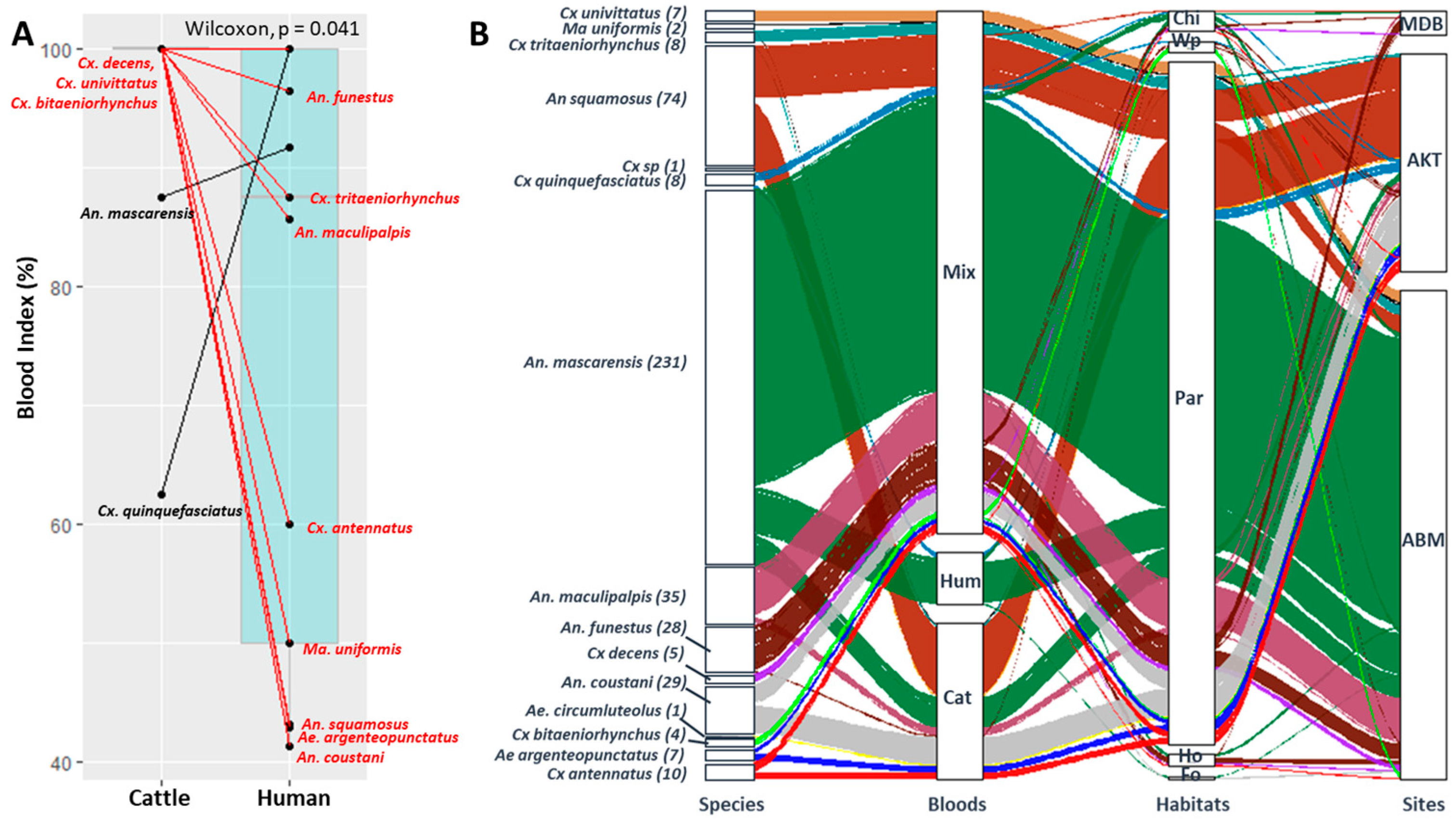
3.2. Blood Meal Analysis

A total of 450 engorged females belonging to 14 species (plus one unidentified Culex sp.) were individually tested via PCR to determine the origins of their blood meals. Mosquitoes that had taken blood from cattle were significantly more numerous than those that had taken blood from humans (Wilcoxon test p < 0.05). Only An. mascarensis and Cx. quinquefasciatus had a greater percentage of blood meals taken from humans (Figure 3A).
Of the 450 engorged females, 301 were collected from ABM, 134 from AKT, and 15 from MDB (Figure 3B). Cattle parks provided 420 engorged females, followed by chicken coops (n = 13), human dwellings (n = 8), water points (n = 7), and forested habitats (n = 2). They were mostly collected via LTs (91.56%, n = 412), while 4.7% (n = 21) were from MTPTs, 3.11% (n = 14) from ISPCs, and only three from BGs.
Blood meals originating solely from humans or cattle were identified from 32 (7.1%) and 96 (21.3%) engorged females, respectively. Mixed blood meals, taken from humans and cattle, were identified from 322 (71.6%) engorged females. Hence, 92.9% of engorged females had taken a blood meal from cattle and 78.7% from humans. The three types of blood meals (human only, cattle only, and mixed) were only observed for An. mascarensis (Figure 3B).

3.3. Detection of RVFV in Mosquitoes

A total of 512 monospecific pools consisting of 2806 mosquitoes, including 249, 120, and 143 pools from ABM, AKT, and MDB, respectively, were tested via real-time RT-PCR. The viral genome was not detected in any of the 53 pools of male mosquitoes (for a total of 130 mosquitoes) (Table 2). Viral RNA was detected in 37 pools of female mosquitoes (n = 213), including 25 pools of non-engorged females (n = 152) and 12 pools of engorged females (n = 61), belonging to ten species (38.5% of the species). Thirteen positive pools were from ABM, 12 were from AKT, and the last 12 were from MDB (Table 2). The species that were found to be RVFV-positive were as follows: Aedes albopictus (1 out of 18 non-engorged female pools), Ae. argenteopunctatus (1 out of 18 non-engorged female pools and 2 out of 2 engorged female pools), Anopheles coustani (1 out of 35 non-engorged females pools and 2 out of 5 engorged females pools), An. gambiae s.l. (1 out of 12 non-engorged female pools), An. mascarensis (6 out of 92 non-engorged female pools and 3 out of 29 engorged female pools), An. squamosus/cydippis (6 out of 30 non-engorged female pools and 2 out of 11 engorged female pools), Culex antennatus (2 out of 15 non-engorged female pools and 2 out of 5 engorged female pools), Cx. decens (1 out of 17 non-engorged female pools and 1 out of 5 engorged female pools), Cx. tritaeniorhynchus (5 out of 24 non-engorged female pools), and Uranotaenia sp (one out of seven non-engorged female pools).

3.4. RVFV Detection and Blood Meal Sources

Twelve monospecific pools of engorged females from six species were found to be positive for RVFV. The two positive pools of Ae. argenteopunctatus, as well as two of Cx. antennatus, contained females that had taken a mixed meal or a meal on cattle exclusively (Table 3). Three pools of An. mascarensis contained females that had taken either a mixed meal or a meal on humans exclusively. The pools of An. coustani, An. squamosus/cydippis, and Cx. decens contained only females that had taken a mixed blood meal. In all, 74.6% (44/59) of engorged females from the RVFV-positive pools had taken a mixed blood meal (Table 3).

4. Discussion

The reappearance of RVF in Madagascar in 2021, twelve years after the second major outbreak reported on the island (2008–2009), showed the persistence of a recurrent substantial risk for the human population and livestock locally. It should be remembered that RVF epidemics/epizootics occur when the circulation of RVFV is amplified by mosquito populations in an environment favorable to their proliferation [44]. In this context, the entomological component was inventoried during a multidisciplinary investigation conducted in the Mananjary district of the Vatovavy–Fitovinany region from 26 April to 4 May 2021.
A total of 2806 mosquitoes belonging to at least 26 species were caught. Among them, 14 species were already known to be associated with RVFV transmission in Madagascar [17]. Anopheles mascarensis was the most abundant species, accounting for 37.4% of all mosquitoes caught. This species has never been reported to be infected with RVFV in the literature [17]. In addition, of the 2806 mosquitoes, 450 blood-fed females were caught. A mixed blood meal was taken by 71.6% of engorged females and three quarters of those from RVFV-positive pools. These results led us to identify the mosquito species likely to act as RVFV vectors at the three study sites during this epidemic.

4.1. Species Diversity and Abundance

Most mosquitoes (94.0% of total individuals) were caught with light traps. The use of LTs was based not only on their higher productivity as compared to other types of traps [23,45,46,47] but also on their suitability for the collection of nocturnal mosquito species involved in RVFV transmission [31]. This method is extensively used during RVFV outbreak investigation [31] and inter-epizootic periods [33].
The variation in mosquito abundance according to the type of trap, sampling site, and habitat type recorded in this study has already been reported in other studies [47,48,49]. The highest abundance of Cx. quinquefasciatus in Ambodiakatra, the most anthropized area, was expected due to the urban nature of the species [47]. With the exception of species belonging to the Orthopodomyia, Uranotaenia, and Ficalbia genera, the majority of the species collected in this study have been commonly collected in previous entomological studies on RVFV [31]. Notably, this is the first study to report the predominance of Anopheles mascarensis, raising questions about the role of this species in RVFV transmission and in the 2021 RVF epidemic.
Of the twenty-six-mosquito species collected, six (An. squamosus/cydippis, An. coustani, Cx. antennatus, Cx. decens, Cx. tritaeniorhynchus, and Ma. uniformis) were already found to be naturally infected with RVFV in Madagascar [23,31,33], six others (Ae. circumluteolus, An. gambiae sl, Cx. bitaeniorhynchus, Cx. quinquefasciatus, Cx. univittatus, and Er. quinquevittatus) were found in other countries, and two species (Ae. albopictus and Ae. argenteopunctatus) had known vector competence [17].

4.2. Trophic Behavior

A second condition necessary for viral circulation and epidemic emergence is the meeting between a virus and its vertebrate host [50], probably during mosquito feeding. In the case of RVFV, the trophic preferences of the different mosquito species towards domestic ruminants and humans are a determining element. In the present study, PCR primers allowing for discrimination between human and cattle blood meals were used based on the involvement of these two hosts in the RVFV epidemiological cycle in Madagascar [12]. With the exception of Ae. circumluteolus, all of the mosquito species shown to feed on cattle in the present study were shown to feed on humans too, supporting previous studies that these species are opportunistic feeders [47,51]; however, although An. mascarensis and Cx. quinquefasciatus were shown to feed on cattle, they were shown to be mostly anthropophilic, as already reported [47,52]. For An. mascarensis, two sibling species (one mostly anthropophilic and other mostly zoophilic species) have been reported in Madagascar [52]. It is possible that this anthropophilic An. mascarensis population is one sibling species of this species, as already mentioned by Fontenille [52].
The high number of mixed blood meals (more than 70%) observed here was in line with that reported in host-feeding pattern studies of culicine mosquitoes, allowing for the explanation of the 2006–2007 Kenyan RVFV outbreak [53] and regular dengue, as well as malaria outbreaks in Thailand [54].

4.3. Virus Detection

Another necessary condition for viral circulation and the emergence of an epidemic is the “compatibility” of a vector to a virus [50]. Tantely et al. [17] proposed the classification of mosquito vectors according to three criteria: natural infection, vector competence, and vector–host contact in the field. Based on these criteria, three species were validated as being major vectors of RVFV: two as candidate vectors and one as a potential vector in Madagascar (Table 2). In the present study, ten species tested positive for RVFV. The repeated detection of RVFV in An. squamosus, An. coustani, Cx antennatus, Cx. decens, and Cx. tritaeniorhynchus in Madagascar supports the full incrimination of these species as vectors according to Hamon et al. [55].
An. squamosus/cydippis: An. squamosus and An. cydippis are morphologically identical and indistinguishable from each other. While Anopheles cydippis has never been implicated in pathogen transmission, Anopheles squamosus was found to be positive for RVFV during the 2008–2009 epidemic [31]. This species is known for its zoo-anthropophilic behavior [36,45]. In this study, 43.2% of the blood meals identified from this species were mixed blood meals taken from cattle and humans. The detection of RVFV in this species and its abundance, particularly near cattle parks, suggest its role as a candidate vector in the RVF epizootic/epidemic in the Mananjary district. Vectorial competence is the only missing information for its consideration as a major vector.
An. coustani: Ratovonjato et al. [31] were the first to identify An. coustani as being naturally infected with RVFV in Madagascar, while this species was already found to be naturally infected with RVFV in mainland Africa [56]. This species is mostly zoophilic, and no information is available on its ability to transmit RVFV. Based only on the experimental infection rates reported in the literature [34], not enough information is available to evaluate this species as a major RVFV vector.
Cx. antennatus: This species is zoo-anthropophilic [51]. RVFV was isolated from it during the 2009 epidemic/epizootic in the districts of Fianarantsoa I and Ambalavao [31], as well as in 2016, during the inter-epizootic period, in the Ambatoboeny district [33]. During this study, Cx. antennatus collections were significant at the Ambalamainty and Ambodiakatra sites, particularly near the zebu parks. In the literature, this species has been reported to be susceptible to RVFV infection and capable of transmitting it [4,34,57]. All these observations confirm its role as a major vector of RVFV [17].
Cx. decens: This species can exhibit anthropophilic behavior [23] and may be attracted to livestock and poultry [45]. The RVFV genome was detected from Cx. decens in 2016 in the Ambatoboeny and Anivorano districts in the north of the country [33]. Significant densities of this species were observed during the present survey, but, given that no information is available on its potential to transmit RVFV, Cx. decens remains a candidate vector for RVFV.
Cx. tritaeniorhynchus: The species has already been found to be PCR-positive for RVFV in the Ambatoboeny district in 2016 [33] and was reported as experimentally susceptible to RVFV infection [58]. The zoo-anthropophilic nature of this species is confirmed by the high proportion of females (87.5%) with mixed blood meals obtained during this study. Cx. tritaeniorhynchus can therefore be considered an RVFV candidate vector pending information on its potential to transmit RVFV.
This study is the first to report the RT-PCR positivity of An. gambiae sl., Aedes albopictus, Ae. argenteopunctuatus, and an unidentified Uranotaenia sp. for RVFV in Madagascar.
An. gambiae sl: Three species of this complex are present in Madagascar: Anopheles gambiae, An. arabiensis, and An. merus [59]. RVFV was detected via RT-PCR from wild-caught populations of An. arabiensis in Sudan [60]. In addition, RVFV infection and dissemination were also reported from laboratory colonies of An. gambiae [61,62,63,64].
Ae. albopictus: To our knowledge, this is the first report of Ae. albopictus testing positive via RT-PCR for RVFV, although RVFV infection and transmission have been reported from laboratory colonies [62]. This species is diurnal and highly anthropophilic [23]. The three criteria that make this species a major vector are met; however, the highly anthropophilic behavior of this species [23] should not lead to new outbreaks due to the fact that humans are considered dead-end hosts [65]. Nonetheless, RVFV maintenance by Ae. albopictus via vertical transmission, as already suggested in Madagascar [17], should not be neglected.
Ae. argenteopunctatus: This study is also the first to mention the detection of RVFV via RT-PCR in Ae. argenteopunctatus. It is an anthropophilic species [23] reported to exhibit zoophilic behavior by feeding on domestic ruminants in Africa [66]. RVFV transmission was also reported from laboratory colonies [63]. This species belongs to the Aedimorphus subgenus, for which RVFV vertical transmission has already been suggested [67]. The three criteria that make this species a major vector are met, but this species should exploit this major vector role locally (where this species occurs in abundance).
Uranotaenia: This is the first study to report a mosquito belonging to the genus Uranotaenia as being RT-PCR-positive for RVFV, but this genus was already found to harbor a Phlebovirus in the Ivory Coast [64]. With the information obtained in this study, the Uranotaenia genus should be considered as a potential vector for RVFV.
An. mascarensis: Among the ten species found to be RT-PCR-positive for RVFV, An. mascarensis deserves special attention. This species, endemic to Madagascar and the Comoros archipelago, was already found to be infected with Plasmodium [52], Ngary virus [23], and Wuchereria bancrofti [68], but has never been associated with RVFV transmission. In the present study, RVFV was detected in six out of nine pools of non-engorged females, suggesting that RVFV had already disseminated [49]. The detection of RVFV via RT-PCR in wild-caught populations of An. mascarensis suggests that this species could be susceptible to the virus [57]. Unfortunately, no information is available on its ability to transmit it. Although this species is abundant and displays highly zoo-anthropophilic behavior, its role as an RVFV candidate vector remains to be further explored through viral isolation and experimental infection attempts.
This species is reputed to be a vector of Plasmodium parasites in the country [69], and the Mananjary district is located in an area of high and stable malaria transmission [70]. The co-infection of An. mascarensis with Plasmodium parasites could facilitate RVFV transmission due to the disruption of the salivary glands [71]. Furthermore, although An. mascarensis is assumed to be an incompetent vector of RVFV, the high abundance and zoo-anthropophilic behavior of this species could favor RVFV outbreaks. Indeed, according to yellow fever’s epidemiology [72], high population densities, such as those observed here, of an incompetent mosquito vector could initiate and maintain viral transmission.

4.4. Implications for RVFV Transmission to Humans and Animals and Recommendations

This multidisciplinary investigation allowed for the identification of the occurrence of mosquitoes, vertebrate hosts, and RVFV in the same place and at the same time, thus meeting the first necessary condition for the circulation of the virus and the emergence of RVF in livestock and humans. Entomologically, there are now eleven species that have been found to be RT-PCR-positive for RVFV from wild-caught populations in Madagascar, with An. gambiae sl., An. mascarensis, Ae. albopictus, Ae. argenteopunctuatus, and an unidentified species of Uranotaenia newly included in the list.
By combining the 24 mosquito species potentially associated with RVFV transmission listed by Tantely et al. [17], plus the two species (Cx. decens and Cx. tritaeniorhynchus) found to be RT-PCR-positive for RVFV in 2016 in the western region of Madagascar [33] and An. mascarensis, there are now 27 species associated with RVFV transmission in Madagascar [17].
Compared with previous RVFV epidemics/epizootics [14,24,30,32], the 2021 outbreak was the largest epizootic ever recorded in the country [27]. Cattle movements within the island, as observed in Mananjary, are a possible cause of this widespread RVFV outbreak [14,24,73]. In addition, we observed that cattle were an important blood meal source for all mosquito species, suggesting that they could have served as virus-amplifying hosts for mosquito species able to transmit RVFV [7]. Indeed, susceptible cattle rapidly develop high-titer viremia (1.0 × 10² PFUs/mL of blood) two days post-infection [74], and viremia higher than 101.3 PFUs/mL is enough to infect mosquito vectors [62].
The larval stages of mosquitoes associated with RVFV can develop in streams, lakes, ponds, marshes, banks, backwaters, irrigation drains, nursery waters, brackish waters, lagoons, wet ponds, and rice fields [23,51]. The configuration of the larval breeding habitats of these species makes it difficult to envisage the application of larval control measures. As the mosquito species involved in RVFV transmission are nocturnal [17], the use of impregnated mosquito nets appears relevant to reduce exposure to endophagic and endophilic mosquitoes; however, the application of indoor vector control measures (the use of impregnated mosquito nets and the indoor spraying of residual insecticides) is of limited effectiveness, due to the fact that all species found to be positive for RVFV also exhibit exophagic and exophilic behavior [48], except Uranotaenia, with unknown behavior.
Fortunately, the distribution of mosquito nets impregnated with long-acting insecticides is already planned as part of the national malaria control program in the Mananjary district. Indeed, some of the species in question (An. mascarensis and An. gambiae sl) are also known to transmit malaria parasites. Moreover, the implementation of indoor spraying operations might not be accepted by villagers, in an attempt to avoid an undesirable effect on their beekeeping activity.

5. Conclusions

The abundance, diversity of species, origin of blood meals, and identification of RVFV in some monospecific pools of mosquitoes enable the incrimination of mosquito vectors in the transmission of RVFV during this epidemic/epizootic. Anopheles mascarensis could initiate an RVFV outbreak, and this species, with the remaining nine RVFV-positive species, may have acted as bridge vectors for the virus, contributing to the high numbers of infected animals and humans [75,76]. In addition, with the high number of mixed blood meals observed, high numbers of human cases were expected; however, few careful clinical observations are generally reported in African countries. In any case, with the close association of humans with domestic animals being an important risk factor for infection, important epidemics are expected after epizootic activity [77]. Hitherto, the long inter-epizootic periods, of 12 to 17 years, stated for the country [45] are still highlighted, and the detection of RVFV in mosquitoes (Cx. decens, Cx. tritaeniorhynchus, and Cx. antennatus) from the northwest region in 2016 [33] supports the existence of low-noise viral circulation in livestock during inter-epizootic periods [22,23,25,26,30]. The high percentage of blood meals taken on humans shows that the human populations at the three investigated study sites in the Mananjary district were highly exposed to potentially infectious mosquito bites. Nevertheless, vector competence experiments should be performed to confirm that these mosquito species can transmit the virus. Promoting the use of impregnated mosquito nets appears to be the most appropriate prevention method in order to limit the transmission of RVFV in the human population of the Mananjary district.

Supplementary Materials

The following supporting information can be downloaded at: https://www.mdpi.com/article/10.3390/pathogens13030258/s1, Supplementary Figure S1: Illustration of the multidisciplinary team performing an investigation in the district of Mananjary with a mobile laboratory in a four-wheel-drive vehicle used to perform the laboratory procedures in the field conditions. B: Field workers crossing a river to reach Madiolamba village. C: Field workers preparing and deploying mosquito traps in the village of Madiolamba. LT: light trap, IPSC: indoor pyrethroid spray catch; BG: BG-Sentinel trap, and MTPT: Muirhead-Thomson pit traps.

Author Contributions

Conceptualization, L.M.T., S.F.A., P.D. and R.G.; methodology, L.M.T., S.F.A., P.D., V.L. and R.G.; validation, L.M.T., S.F.A., P.D., V.L. and R.G.; formal analysis, L.M.T.; investigation, L.M.T., M.F.A. (Maminirina Fidelis Ambinintsoa), M.F.A. (Miamina Fidy Ankasitrahana), D.B.R., J.T.R., J.-P.R., A.H., N.N.R., J.I. and L.R.; data curation, L.M.T., S.F.A., M.F.A. (Maminirina Fidelis Ambinintsoa) and M.R.R.; writing—original draft preparation, L.M.T.; writing—review and editing, L.M.T., S.F.A., P.D., V.L. and R.G.; visualization, L.M.T. and R.G.; supervision, R.G., S.F.A., P.D. and V.L.; project administration, R.G., P.D., V.L. and R.V.R.; funding acquisition, R.G., P.D., V.L. and R.V.R. All authors have read and agreed to the published version of the manuscript.

Funding

The publication process as well as the field and laboratory work have been funded by the U.S. Agency for International Development (USAID) under the Research, Innovation, Surveillance and Evaluation (RISE) program (Cooperative Agreement #72068719CA0001). The findings and conclusions in this report are those of the author(s) and do not necessarily represent the official position of USAID.

Institutional Review Board Statement

Not applicable.

Informed Consent Statement

Not applicable.

Data Availability Statement

All data provided and analysed in this study are publicly available. Please contact Luciano Michaël Tantely.

Acknowledgments

The authors thank the medical and veterinary staff in the municipalities of Anosimparihy and Antsenavolo, within the Manjary district. The authors greatly thank the MDB, AKT, and ABM villagers for their warm welcome and for allowing us to trap mosquitoes in their houses.

Conflicts of Interest

The authors declare no conflicts of interest. The funders had no role in the design of the study; in the collection, analyses, or interpretation of the data; in the writing of the manuscript; or in the decision to publish the results.

References

  1. Daubney, R.; Hudson, J.R.; Garnham, P.C. Enzootic hepatitis or Rift Valley fever, an undescribed virus disease of sheep, cattle and man from East Africa. J. Pathol. Bacteriol. 1931, 34, 545–579. [Google Scholar] [CrossRef]
  2. Flick, R.; Bouloy, M. Rift Valley Fever Virus. Curr. Mol. Med. 2005, 5, 827–834. [Google Scholar] [CrossRef] [PubMed]
  3. Swanepoel, R.; Coetzer, J.A.W. Rift Valley fever. Synonyms, Enzootic hepatitis, Slenkdalkoors (Afrik). In Infectious Diseases of Livestock with Special Reference to Southern Africa; Coetzer, J.A.W., Thomson, G.R., Tustin, R.C., Eds.; Oxford University Press: Cape Town, South Africa, 1994. [Google Scholar]
  4. Turell, M.J.; Presley, S.M.; Gad, A.M.; Cope, S.E.; Dohm, D.J.; Morrill, J.C.; Arthur, R.R. Vector competence of egyptian mosquitoes for Rift Valley fever virus. Am. J. Trop. Med. Hyg. 1996, 54, 136–139. [Google Scholar] [CrossRef] [PubMed]
  5. Linthicum, K.J.; Davies, F.G.; Kairo, A. Rift Valley fever virus (family Bunyviridae, genus Phlebovirus). Isolations from diptera collected during an inter-epizootic period in Kenya. J. Hyg. 1985, 95, 197–209. [Google Scholar] [CrossRef] [PubMed]
  6. Lefèvre, P.C.; Blancou, J.; Chermette, R. Principales maladies infectieuses et parasitaires du bétail, Europe et régions chaudes, Tec and Doc, Paris. Bull. De L’académie Vétérinaire De Fr. 2003, 1824, 643–657. [Google Scholar]
  7. Evans, A.; Gakuya, F.; Paweska, J.T.; Rostal, M.; Akoolo, L.; Van Vuren, P.J.; Manyibe, T.; Macharia, J.M.; Ksiazek, T.G.; Feikin, D.R.; et al. Prevalence of antibodies against Rift Valley fever virus in Kenyan wildlife. Epidemiol. Infect. 2008, 136, 261–269. [Google Scholar] [CrossRef] [PubMed]
  8. Easterday, B.C.; Murphy, L.C.; Bennett, D.G. Experimental Rift Valley fever in calves, goats, and pigs. Am. J. Vet. Res. 1962, 23, 1224–1230. [Google Scholar]
  9. Swanepoel, R.; Coetzer, J.A.W. Rift Valley fever. In Infectious Diseases of Livestock, 2nd ed.; Coetzer, J.A., Tustin, R.C., Eds.; Oxford university Press: Cape Town, Southern Africa, 2004; pp. 1037–1070. [Google Scholar]
  10. Rissmann, M.; Kley, N.; Ulrich, R.; Stoek, F.; Balkema-Buschmann, A.; Eiden, M.; Groschup, M.H. Competency of Amphibians and Reptiles and Their Potential Role as Reservoir Hosts for Rift Valley Fever Virus. Viruses 2020, 12, 1206. [Google Scholar] [CrossRef]
  11. Smithburn, K.C.; Haddow, A.J.; Gillett, J.D. Rift Valley fever, isolation of the virus from wild mosquitoes. Br. J. Exp. Pathol. 1948, 29, 107–121. [Google Scholar]
  12. AFSSA. Risque de Propagation de la Fièvre de la Vallée du Rift FVR dans l’Océan Indien La Réunion et Mayotte; AFSSA: Maisons-Alfort, France, 2008. [Google Scholar]
  13. CDC. 2000 Update, Outbreak of Rift Valley Fever. Saudi Arabia, August–November 2000. MMWR Morb. Mortal. Wkly Rep. 2000, 49, 982–985. [Google Scholar]
  14. Morvan, J.; Fontenille, D.; Sulazzo, J.-F.; Coulanges, P. Possible Rift Valley Fever outbreak in man and cattle in Madagascar. Trans. R. Soc. Trop. Med. Hyg. 1991, 85, 108. [Google Scholar] [CrossRef]
  15. Roger, M.; Girard, S.; Faharoudine, A.; Halifa, M.; Bouloy, M.; Cetre-Sossah, C.; Cardinale, E. Rift Valley fever in ruminants, republic of Comoros, 2009. Emerg. Infect. Dis. 2011, 17, 1319–1320. [Google Scholar] [CrossRef] [PubMed]
  16. Sissoko, D.; Giry, C.; Gabrie, P.; Tarantola, A.; Pettinelli, F.; Collet, L.; d’Ortenzio, E.; Renault, P.; Pierre, V. Rift Valley Fever, Mayotte, 2007–2008. Emerg. Infect. Dis. 2009, 15, 568–570. [Google Scholar] [CrossRef] [PubMed]
  17. Tantely, M.L.; Boyer, S.; Fontenille, D. A review of mosquitoes associated with Rift Valley fever virus in Madagascar. Am. J. Trop. Med. Hyg. 2015, 92, 722–729. [Google Scholar] [CrossRef] [PubMed]
  18. Davies, F.G.; Linthicum, K.J.; James, A.D. Rainfall and epizootic Rift valley Fever. Bull. World Health Organ. 1985, 63, 941–943. [Google Scholar] [PubMed]
  19. Zeller, H.G.; Fontenille, D.; Traore-Lamizana, M.; Thiongane, Y.; Digoutte, J.P. Enzootic activity of Rift Valley fever virus in Senegal. Am. J. Trop. Med. Hyg. 1997, 56, 265–272. [Google Scholar] [CrossRef]
  20. Chevalier, V.; De La Rocque, S.; Baldet, T.; Vial, L.; Roger, F. Epidemiological processes involved in the emergence of vector borne diseases West Nile Fever, Rift Valley Fever, Japanese Encephalitis and Crimean-Congo Fever. Rev. Sci. Tech. Off. Int. Epiz. 2004, 23, 535–555. [Google Scholar] [CrossRef] [PubMed]
  21. Davies, F.G. Rift Valley Fever and Livestock Trade from the Horn of Africa (Ethiopia, Somalia, Kenya); FAO: Rome, Italy, 1998; pp. 1–28. [Google Scholar]
  22. Clerc, Y.; Coulanges, P.; Rodhain, F.; Ricklin, B.; Ranaivosata, J.; Albignac, R. Le programme arbovirus de l’Institut Pasteur de Madagascar, Bilan actuel. Arch. Inst. Pasteur Mad. 1981, 48, 247–257. [Google Scholar]
  23. Fontenille, D. Arbovirus transmission cycles in Madagascar. Arch. Inst. Pasteur Madag. 1989, 55, 1–317. [Google Scholar]
  24. Morvan, J.; Saluzzo, J.F.; Fontenille, D.; Rollin, P.E.; Coulanges, P. Rift Valley fever on the east coast of Madagascar. Res. Virol. 1991, 142, 475–482. [Google Scholar] [CrossRef]
  25. Morvan, J.; Rollin, P.E.; Laventure, S.; Rakotoarivony, I.; Roux, J. Rift Valley fever epizootic in Central Highlands of Madagascar. Res. Virol. 1992, 143, 407–415. [Google Scholar] [CrossRef]
  26. Zeller, H.G.; Roux, J. Surveillance de la Fièvre de la Vallée du Rift à Madagascar; Rapport Final du Projet Janvier 1996 mars 1998; Institut Pasteur de Madagascar: Antananarivo, Madagascar, 1998; p. 18. [Google Scholar]
  27. WOAH. 25th Conference of WOAH. Analysis of the Animal Health Situation in Members on the Region during 2021 and 2022; Regional Commission for Africa: Gaborone, Botswana, 2023. [Google Scholar]
  28. Corso, B.; Pinto, J.; Beltràn-Alcrudo, D.; De Simone, L.; Lubroth, J. Rift Valley Fever Outbreaks in Madagascar and Potential Risks to Neighbouring Countries; FAO Empress Watch: Rome, Italy, 2008; p. 8. [Google Scholar]
  29. WHO. Rift Valley fever, Madagascar. Wkly. Epidemiol. Rec. 2008, 83, 157–168. [Google Scholar]
  30. Andriamandimby, S.F.; Randrianarivo-Solofoniaina, A.E.; Jeanmaire, E.M.; Ravololomanana, L.; Razafimanantsoa, L.T.; Rakotojoelinandrasana, T.; Razainirina, J.; Hoffman, J.; Ravalohery, J.P.; Razafisandratantsoa, J.T.; et al. Rift Valley Fever during rainy seasons, Madagascar, 2008 and 2009. Em. Inf. Dis. 2010, 16, 963–970. [Google Scholar] [CrossRef]
  31. Ratovonjato, J.; Olive, M.M.; Tantely, M.L.; Andrianaivolambo, L.; Tata, E.; Razainirina, J.; Jeanmaire, E.M.; Reynes, J.M.; Elissa, N. Detection, isolation, and genetic characterisation of Rift Valley Fever virus from Anopheles (Anopheles) coustani, Anopheles (Anopheles) squamosus, Culex (Culex) antennatus of the Haute Matsiatra region, Madagascar. Vector-Borne Zoo. Dis. 2010, 11, 753–759. [Google Scholar] [CrossRef]
  32. Jeanmaire, E.; Rabenarivahiny, R.; Biarmann, M.; Rabibisoa, L.; Ravaomanana, F.; Randriamparany, T.; Andriamandimby, S.F.; Squarzoni, D.C.; Fenozara, P.; de La Rocque, S.; et al. Prevalence of Rift Valley Fever infection in ruminants in Madagascar after the 2008 outbreak. Vector-Borne Zoo. Dis. 2011, 11, 395–402. [Google Scholar] [CrossRef] [PubMed]
  33. Jeffries, C.; Tantely, M.L.; Raharimalala, F.; Hurn, E.; Boyer, S.; Walker, T. Diverse novel resident Wolbachia strains in Culicine mosquitoes from Madagascar. Sci. Rep. 2018, 8, 17456. [Google Scholar] [CrossRef] [PubMed]
  34. Nepomichene, T.N.J.J.; Raharimalala, F.N.; Andriamandimby, S.F.; Ravalohery, J.-P.; Failloux, A.-B.; Heraud, J.-M.; Boyer, S. Vector competence of Culex antennatus and Anopheles coustani mosquitoes for Rift Valley fever virus in Madagascar. Med. Vet. Entomol. 2018, 32, 259–262. [Google Scholar] [CrossRef] [PubMed]
  35. Ravaonjanahary, C. Les Aedes de Madagascar (Diptera-Culicidae). Étude monographique du genre. 2. Biologie d’Aedes (Diceromyia) tiptoni. Trav. Doc. L’orstom 1978, 87, 1–210. [Google Scholar]
  36. Grjébine, A. Insectes Diptères Culicidae Anophelinae; Faune de Madagascar; ORSTOM: Paris, France, 1966; pp. 1–500. [Google Scholar]
  37. Doucet, J. Étude des Culicidae de la région de Vangaindrano (Diptera). Mém. Inst. Sci. Mad. 1951, 6, 83–113. [Google Scholar]
  38. Edwards, F.W. Mosquitoes of the Ethiopian Region. III. Culicine Adults and Pupae; British Museum (Natural History): London, UK, 1941. [Google Scholar]
  39. Brunhes, J.; Hervy, J. Insectes Diptères Culicidae Culicinae Genre Orthopodomyia de la sous-région malgache et de la région afrotropicale. Mus. Natl. Hist. Nat. 1995, 85, 1–120. [Google Scholar]
  40. da Cunha Ramos, H.; Brunhes, J. Insecta, Diptera, Culicidae, Uranotaenia. Faune de Madagascar 91, 3rd ed.; CIRAD: Montpellier, Paris, 2004; p. 461. [Google Scholar]
  41. Pitzer, J.B.; Kaufman, P.E.; Tenbroeck, S.H.; Maruniak, J.E. Host blood meal identification by multiplex polymerase chain reaction for dispersal evidence of stable flies (Diptera, Muscidae) between livestock facilities. J. Med. Entomol. 2011, 48, 53–60. [Google Scholar] [CrossRef] [PubMed]
  42. Weidmann, M.; Sanchez-Seco, M.P.; Sall, A.A.; Ly, P.O.; Thiongan, Y.; Lô, M.M.; Schley, H.; Hufert, F.T. Rapid detection of important human pathogenic Phleboviruses. J. Clin. Virol. 2008, 41, 138–142. [Google Scholar] [CrossRef] [PubMed]
  43. R Core Team. A Language and Environment for Statistical Computing. Foundation for Statistical Computing; R Core Team: Vienna, Austria, 2021; Available online: https://www.R–project.org/ (accessed on 1 February 2024).
  44. de La Rocque, S.; Formenty, P. Rift Valley fever, Disease ecology and early warning. Sustainable Management of Animal Production and Health; Odongo, N., Garcia, M., Viljoen, G., Eds.; FAO: Rome, Italy, 2010; pp. 327–333. [Google Scholar]
  45. Tantely, M.L.; Rakotoniaina, J.C.; Andrianaivolamb, O.L.; Tata, E.; Razafindrasata, F.; Fontenille, D.; Elissa, N. Biology of mosquitoes that are potential vectors of Rift Valley fever virus in different biotopes of the Central Highlands of Madagascar. J. Med. Entomol. 2013, 50, 603–610. [Google Scholar] [CrossRef] [PubMed]
  46. Nepomichene, T.; Tata, E.; Boyer, S. Malaria case in Madagascar.; probable implication of a new vector, Anopheles coustani. Malaria J. 2015, 14, 475. [Google Scholar] [CrossRef]
  47. Tantely, M.L.; Guis, H.; Randriananjantenaina, I.; Raharinirina, M.R.; Velonirina, H.J.; Cardinale, E.; Raveloarijaona, N.; Cêtre-Sossah, C.; Garros, C.; Girod, R. Mosquito species associated with horses in Madagascar, A review of their vector status with regard to the epidemiology of West Nile fever. Med. Vet. Entomol. 2021, 36, 13. [Google Scholar] [CrossRef] [PubMed]
  48. Andrianaivolambo, L.; Domarle, O.; Randrianarivelojosia, M.; Ratovonjato, J.; Le Goff, G.; Talman, A.; Robert, V. Anthropophilic mosquitoes and malaria transmission in the eastern foothills of the central highlands of Madagascar. Acta. Trop. 2010, 116, 240–245. [Google Scholar] [CrossRef] [PubMed]
  49. Tantely, M.L.; Cêtre–Sossah, C.; Rakotondranaivo, T.; Cardinale, E.; Boyer, S. Population dynamics of mosquito species in a West Nile Virus endemic area in Madagascar. Parasite 2017, 24, 3. [Google Scholar] [CrossRef] [PubMed]
  50. Fontenille, D.; Powell, J.R. From anonymous to public enemy, how does a mosquito become a feared Arbovirus vector? Pathogens 2020, 9, 265. [Google Scholar] [CrossRef]
  51. Tantely, M.L.; Le Goff, G.; Boyer, S.; Fontenille, D. An updated checklist of mosquito species (Diptera, Culicidae) from Madagascar. Parasite 2016, 23, 42. [Google Scholar] [CrossRef]
  52. Fontenille, D.; Campbell, G.H. Is Anopheles mascarensis a new malaria vector in Madagascar? Am. J. Trop. Med. Hyg. 1992, 46, 28–30. [Google Scholar] [CrossRef]
  53. Muturi, E.J.; Muriu, S.; Shililu, J.; Mwangangi, J.M.; Jacob, B.G.; Mbogo, C.; Githure, J.; Novak, R.J. Blood-feeding patterns of Culex quinquefasciatus and other culicines and implications for disease transmission in Mwea rice scheme, Kenya. Parasitol. Res. 2008, 102, 1329–1335. [Google Scholar] [CrossRef]
  54. Khaklang, S.; Kittayapong, P. Species composition and blood meal analysis of mosquitoes collected from a tourist island, Koh Chang, Thailand. J. Vector Ecol. 2014, 39, 448–452. [Google Scholar] [CrossRef]
  55. Hammon, W.M.; Reeves, W.C. Laboratory Transmission of St. Louis Encephalitis Virus by Three Genera of Mosquitoes. J. Exp. Med. 1943, 78, 241–253. [Google Scholar] [CrossRef]
  56. McIntosh, B.M. Rift Valley fever. 1. Vector studies in the field. J. S. Afr. Vet. Med. Assoc. 1972, 43, 391–395. [Google Scholar] [PubMed]
  57. Turell, M.; Dohm, D.J.; Mores, C.N.; Terracina, L.; Wallette, D.L.; Hribar, L.J.; Pecor, J.E.; Blow, J.A. Potential for North American mosquitoes to transmit Rift Valley fever virus. J. S. Afr. Vet. Med. Assoc. 2008, 24, 502–507. [Google Scholar] [CrossRef]
  58. Jupp, P.G.; Kemp, A.; Grobbelaar, A.; Leman, P.; Burt, F.J.; Alahmed, A.M.; Al Mujalli, D.; Khamees, A.; Swanepoel, R. The 2000 epidemic of Rift Valley fever in Saudi Arabia, mosquito vector studies. Med. Vet. Entomol. 2002, 16, 245–252. [Google Scholar] [CrossRef] [PubMed]
  59. Léong Pock Tsy, J.M.; Duchemin, J.B.; Marrama, L.; Rabarison, P.; Le Goff, G.; Rajaonarivelo, V.; Robert, V. Distribution of the species of the Anopheles gambiae complex and first evidence of Anopheles merus as a malaria vector in Madagascar. Malaria J. 2003, 2, 33. [Google Scholar] [CrossRef] [PubMed]
  60. Seufi, A.M.; Galal, F.H. Role of Culex and Anopheles mosquito species as potential vectors of Rift Valley fever virus in Sudan outbreak, 2007. Infect. Dis. 2010, 10, 65. [Google Scholar] [CrossRef] [PubMed]
  61. Moutailler, S.; Krida, G.; Schaffner, F.; Vazeille, M.; Failloux, A.B. Potential vectors of Rift Valley fever virus in the Mediterranean Region. Vector Borne Zoonotic Dis. 2008, 8, 749–754. [Google Scholar] [CrossRef] [PubMed]
  62. Turell, M.; Bailey, C.; Beaman, J. Vector competence of a Houston, Texas strain of Aedes albopictus for Rift Valley fever virus. J. Am. Mosq. Control. Assoc. 1988, 4, 94–96. [Google Scholar]
  63. Jupp, P.; Cornel, A.; Turell, M.; Bailey, C.; Beaman, J. Vector competence tests with Rift Valley fever virus and five south African species of mosquito. Am. Mosq. Control. Assoc. 1988, 4, 4–8. [Google Scholar]
  64. Junglen, S.; Kurth, A.; Kuehl, H.; Quan, P.L.; Ellerbrok, H.; Pauli, G.; Nitsche, A.; Nunn, C.; Rich, S.M.; Lipkin, W.I.; et al. Examining landscape factors influencing relative distribution of mosquito genera and frequency of virus infection. EcoHealth 2009, 6, 239–249. [Google Scholar] [CrossRef] [PubMed]
  65. Chevalier, V.; Pépin, M.; Plée, L.; Lancelot, R. Rift Valley fever—A threat for Europe? Eurosurveillance 2010, 15, 19506. [Google Scholar] [CrossRef] [PubMed]
  66. Hamon, J.; Sales, S.; Coz, J.; Ouedraogo, C.S.; Dyemkouma, A.; Diallo, B. Observations sur les préférences alimentaires des moustiques de la République de Haute-Volta. Bull. Soc. Pathol. Exot. 1964, 57, 1133–1150. [Google Scholar]
  67. Mondet, B.; Diaite, A.; Ndione, J.A.; Fall, A.G.; Chevalier, V.; Lancelot, R.; Ndiaye, M.; Ponçon, N. Rainfall patterns and population dynamics of Aedes (Aedimorphus) vexans arabiensis, Patton 1905 (Diptera, Culicidae).; a potential vector of Rift Valley fever virus in Senegal. J. Vector. Ecol. 2005, 30, 102–106. [Google Scholar] [PubMed]
  68. Brunhes, J. Épidémiologie de la filariose de Bancroft à Madagascar. II. Recherches sur Wuchereria voceli Galliard.; 1959 et étude morphologique comparée des souches malgache et comorienne de Wuchereria bancrofti Cobbold, 1877. Sér. Ent. Méd. Parasitol. 1972, 10, 193–205. [Google Scholar]
  69. Fontenille, D.; Lepers, J.P.; Campbell, G.H.; Coluzzi, M.; Rakotoarivony, I.; Coulanges, P. Malaria transmission and vector biology in Manarintsoa, High Plateaux of Madagascar. Am. J. Trop. Med. Hyg. 1990, 43, 107–115. [Google Scholar] [CrossRef]
  70. Rice, B.L.; Golden, C.D.; Randriamady, H.J.; Rakotomalala, A.A.N.A.; Vonona, M.A.; Anjaranirina, E.J.G.; Hazen, J.; Castro, M.C.; Metcalf, J.E.; Hartl, D.L. Fine-scale variation in malaria prevalence across ecological regions in Madagascar, a cross-sectional study. BMC Public Health 2021, 21, 1018. [Google Scholar] [CrossRef]
  71. Vaughan, J.A.; Turell, M.J. Facilitation of Rift Valley Fever virus transmission by Plasmodium berghei sporozoites in Anopheles stephensi mosquitoes. Am. J. Trop. Med. Hyg. 1996, 55, 407–409. [Google Scholar] [CrossRef]
  72. Miller, B.R.; Monath, T.P.; Tabachnick, W.J.; Ezike, V.I. Epidemic yellow fever caused by an incompetent mosquito vector. Trop. Med. Parasitol. 1989, 40, 396–399. [Google Scholar]
  73. Lancelot, R.; Béral, M.; Rakotoharinome, V.M.; Andriamandimby, S.F.; Heraud, J.M.; Costea, C.; Apollonia, A.; Squarzoni-Diawg, C.; de La Rocquea, S.; Formenty, P.B.H.; et al. Drivers of Rift Valley fever epidemics in Madagascar. Proc. Natl. Acad. Sci. USA 2017, 114, 938–943. [Google Scholar] [CrossRef]
  74. Bird, B.H.; Ksiazek, T.G.; Nichol, S.T.; McLachlan, J. Rift Valley fever virus. J. Am. Vet. Med. Assoc. 2009, 234, 883–893. [Google Scholar] [CrossRef] [PubMed]
  75. Linthicum, K.J.; Britch, S.C.; Anyamba, A. Rift Valley fever, an emerging mosquito-borne disease. Annu. Rev. Entomol. 2016, 61, 395–415. [Google Scholar] [CrossRef] [PubMed]
  76. Lumley, S.; Hernández-Triana, L.M.; Horton, D.L.; de Marco, M.D.M.F.; Medlock, J.M.; Hewson, R.; Fooks, A.R.; Johnson, N. Competence of mosquitoes native to the United Kingdom to support replication and transmission of Rift Valley fever virus. Par. Vect. 2018, 11, 308. [Google Scholar] [CrossRef] [PubMed]
  77. Peters, C.J.; Linthicum, K.J. Rift Valley fever. In Handbook of Zoonoses, 2nd ed.; Beran, G.W., Ed.; CRC Press: Boca Raton, FL, USA, 1994; pp. 125–138. [Google Scholar]
Figure 1. (A): The distribution of outbreaks of RVF in Madagascar, between 1 January 2021 and 25 November 2022, as reported to the WOAH through the early warning system [27]. (B): The locations of the three villages in the two municipalities of Antsenavolo and Anosimparihy and the five mosquito collection sites in each village. MDB: Madiolamba, ABM: Ambalamainty, and AKT: Ambodiakatra. A red star indicates a “house” habitat with a confirmed human case, and a red square indicates a “cattle park” habitat with a confirmed bovine case. The remaining habitats without confirmed human and bovine cases are white-colored.
Pathogens 13 00258 g001
Figure 2. The abundance of mosquito species (in rows) collected by (A) site (ABM: Ambalamainty, AKT: Ambodiakatra, and MDB: Madiolamba) and (B) biotope (Orch: orchards and forest edges, Hum dwell: human dwellings, Ca. parks: cattle parks, WP: water points, and Chick coops: chicken coops).
Pathogens 13 00258 g002
Figure 3. The analysis of blood meals. (A) The paired analysis of human and cattle blood indices (dots indicate the mosquito species; red lines represent the species that are predominantly zoophagic; and black lines indicate those that are predominantly anthropophagic). (B) Alluvial plots showing the changes in the number of engorged females for each species as a function of the type of blood meal (Cat: cattle, Hum: human, and Mix: mixed blood meals), habitat type (Fo: orchard, Ho: house, Par: cattle park, Wp: water point, and Chi: chicken coop), and site (ABM: Ambalamainty, AKT: Ambodiakatra, and MDB: Madiolamba). On the left-hand side of each block, the number of engorged females tested by species is indicated.
Pathogens 13 00258 g003
Table 1. The distribution of adult mosquitoes collected by species, collection site, and collection method.
SpeciesAmbalamaintyAmbodiakatraMadiolamba Total
BGsLTsMTPTsIPSCsBGsLTsMTPTsIPSCsBGsLTsMTPTsIPSCs
Ad. furfurea0200000001003
Ae. albopictus *70008012650029
Ae. argenteopunctatus *02300031100360091
Ae. circumluteolus £, V0000020000002
Ae. sp.0000010002003
An. coustani **, V0143000690008700299
An. funestus0190641172603319269
An. gambiae sl  £, V1130008010110034
An. maculipalpis0640007002120085
An. mascarensis5765180018000241201049
An. pauliani050000000010051
An. rufipes0000020000002
An. squamosus/cydippis **0910001230008300297
Cq. grandidieri014000000050019
Cx. antennatus ***0440002641120078
Cx. bitaeniorhynchus  £0500001000006
Cx. decens **064000120002710104
Cx. giganteus0000010000001
Cx. quinquefasciatus **, £0000174153000075
Cx. tritaeniorhynchus ***263000100009300168
Cx. univittatus **, £, V032000000010033
Cx. sp.0000010001002
Er. quinquevitattus **, £0000100001002
Ficalbia sp.0100030002006
Ma. uniformis **090000000730082
Or. milloti0000000001001
Ur. sp.04001300011010
Lt tigripes0100000000001
Unidentified genus1200010000004
Total1615802442833910639719592806
In bold are species already found naturally infected with RVFV in Madagascar in monospecific batches. £: species found infected with RVFV in other regions. V: species capable of vertical transmission. BGs: BG-Sentinel traps baited with an artificial human odor. LTs: light traps. MTPTs: Muirhead-Thomson pit traps. IPSCs: indoor pyrethroid spray catches. Tantely et al. [17] proposed the classification of mosquito vectors according to three criteria, these being natural infection, vector competence, and field vector–host contact. These three criteria are validated in RVFV major vectors (***), two of them in candidate vectors (**), and one of them in potential vectors (*).
Table 2. The number of monospecific pools of mosquitoes tested and found positive via RT-PCR for RVFV by species, sex, the status of engorgement for females, and site.
AmbalamaintyAmbodiakatraMadiolambaTotal
FemalesMalesFemalesMalesFemalesMales
SpeciesGoNon-Go GoNon-Go GoNon-Go
Ad. furfurea-2-----1-3
Ae. albopictus *** , µ-51-6 (1)1-7121 (1)
Ae. argenteopunctatus  *** , µ-422 (2)6 (1)--8-22 (3)
Ae. circumluteolus  £, V---11----2
Ae. sp.----1--2-3
An. coustani **, V1 (1)17 (1)34 (1)61-12145 (3)
An. funestus423236237353
An. gambiae sl **, £, V, µ-42-42-4 (1)117 (1)
An. maculipalpis36-11-14-16
An. mascarensis **, µ26 (3)60423 (1)-129 (5)2127 (9)
An. pauliani-5-----1-6
An. rufipes----2----2
An. squamosus/cydippis **212 (3)18 (2)9 (2)-19(1)-42 (8)
Coquillettidia grandidieri-4-----217
Cx. antennatus ***1 (1)7 (1)-4 (1)6 (1)--2-20 (4)
Cx. bitaeniorhynchus  £11-1-----3
Cx. decens **2 (1)9 (1)4-3-15226 (2)
Cx. giganteus----1----1
Cx. quinquefasciatus **, £---3610---19
Cx. tritaeniorhynchus ***19-23--12 (5)128 (5)
Cx. univittatus **, £, V162----1-10
Cx. sp.---1---1-2
Er. quinquevitattus **, £-----1-1-2
Ficalbia sp.-1--21-2-6
Ma. uniformis **15-----10117
Or. milloti--------11
Ur. sp. *, µ-2 (1)--3--2-7 (1)
Lt. tigripes-1-------1
Genre sp.-2--1----3
Total43 (6)185 (7)2132 (6)70 (6)187122 (12)14512 (37)
Go: number of monospecific pools of engorged females tested; Non-Go: number of monospecific pools of non-engorged females tested; and Males: number of monospecific pools of males tested. Between brackets: number of monospecific pools found positive via RT-PCR. In bold: species naturally infected with RVFV or RT-PCR-positive for RVFV in Madagascar. µ: species newly found to be RT-PCR-positive for RVFV in Madagascar. £: species found to be infected with RVFV in other regions. *** main vector, ** candidate vector, and * potential vector species. V: species capable of vertical transmission.
Table 3. The origins of the blood meals taken by engorged female mosquitoes constituting the monospecific pools found to be positive for RVFV.
SpeciesEngorged PositivesBlood Meals (Mixed and Single)
RVFV + PoolsInd Go/Pool+MixedHumanCattle
Ae. argenteopunctatus24 * + 5304
An. coustani21 + 5600
An. mascarensis310 + 10 + 1020100
An. squamosus/cydippis22 + 101200
Cx. antennatus21 + 2201
Cx. decens11100
RVFV + pools: number of monospecific pools found to be positive for RVFV, Ind go/pool: number of engorged females per RVFV-positive pool, and *: two engorged females without an abdomen (origin of blood meal unknown).
Disclaimer/Publisher’s Note: The statements, opinions and data contained in all publications are solely those of the individual author(s) and contributor(s) and not of MDPI and/or the editor(s). MDPI and/or the editor(s) disclaim responsibility for any injury to people or property resulting from any ideas, methods, instructions or products referred to in the content.

