Article

Comparison of Analytical Sensitivity (Limit of Detection) of Xpert MTB/RIF and Xpert MTB/RIF Ultra for Non-Sputum Specimens

1
Department of Pathology, University of Texas Medical Branch, Galveston, TX 77555, USA
2
Department of Immunology and Microbiology, University of Texas Medical Branch, Galveston, TX 77555, USA
3
Department of Pathology, Infectious Disease Testing, Montefiore Medical Center, Bronx, NY 10467, USA
*
Authors to whom correspondence should be addressed.
Pathogens 2023, 12(2), 157; https://doi.org/10.3390/pathogens12020157
Submission received: 12 December 2022 / Revised: 15 January 2023 / Accepted: 17 January 2023 / Published: 18 January 2023
(This article belongs to the Special Issue Molecular Diagnostics of Emerging Pathogens)

Abstract

:
Tuberculosis (TB) is a significant public health threat and has remained a leading cause of death in many parts of the world. Rapid and accurate testing and timely diagnosis can improve treatment efficacy and reduce new exposures. The Cepheid Xpert® MTB/RIF tests have two marketed products (US-IVD and Ultra) that are widely accepted for diagnosis of TB but have not yet been approved for non-sputum specimens. Despite numerous studies in the literature, no data for the analytical sensitivity of these two products on the non-sputum samples are available to date. This is the first study that systematically determined the analytical sensitivities of both US-IVD and Ultra tests on cerebrospinal fluid (CSF), tissue, and bronchoalveolar lavage (BAL). The limits of detection (LoDs) on the US-IVD test for both Mycobacterium tuberculosis and rifampin resistance in CFU/mL, respectively, were as follows: CSF (3.3 and 4.6), tissue (15 and 23), and bronchoalveolar lavage (BAL) (45 and 60), and on the Ultra test: CSF (0.16 and 2.7), tissue (0.11 and 12), and BAL (0.65, and 7.5). Overall, the analytical sensitivities of the Ultra test were substantially better than US-IVD for all sample types tested. This study provided a foundation for using either the US-IVD or Ultra test for the early detection of both pulmonary and extrapulmonary (EP) TB. Furthermore, using Ultra could result in higher TB case detection rates in subjects with paucibacillary TB and EP TB, positively impacting WHO goals to eradicate TB.

1. Background

Mycobacterium tuberculosis (MTB) remains a significant cause of morbidity and mortality worldwide. Even though MTB is ranked as one of the leading causes of death by a single infectious agent, over the past decade, there had been a slowly declining trajectory in cases globally [1]. However, that was substantially reversed by the COVID-19 pandemic due to a reduction in healthcare access, close household contact during lockdowns, and testing, even in the setting of increased mask use and other public health initiatives to mitigate the spread of SARS-CoV-2 [1].
Early and rapid detection of MTB and rifampin (RIF) resistance is crucial to help prevent transmission and overcome significant treatment challenges associated with drug-resistant MTB. The gold standard for MTB diagnosis is currently culture-based. Since MTB grows slowly and identification capabilities may be limited in many laboratories, the Cepheid Xpert® MTB/RIF test performed on the GeneXpert® system (Cepheid, Sunnyvale, CA, USA) is widely accepted for the initial diagnosis of suspected pulmonary tuberculosis (TB) [2,3]. Even though major public health strategies have been designed to mitigate the spread of MTB, an estimated 1/3 of all cases remain undiagnosed [4]. MTB in extrapulmonary (EP) sources accounted for most undiagnosed or delayed diagnosed cases. Although EP TB is not as prevalent as pulmonary TB in the United States, some forms of EP TB, such as meningeal or spinal TB, can be devastating and life-threatening [5,6,7].
Xpert® MTB/RIF (US-IVD) is an in vitro diagnostic (IVD) test in the US that can simultaneously detect MTB DNA and RIF-resistance-associated mutations of the rpoB gene only from sputum samples. It is a qualitative, nested real-time polymerase chain reaction (PCR) test in which the primers are designed to amplify the 81 base pair core region of the rpoB gene, also called the RIF resistance-determining region (RRDR). In addition, five probes are designed to differentiate the conserved wild-type sequence and mutations in the RRDR [8,9]. They are Probe A for codons 507–511, Probe B for codons 511–518, Probe C for codons 518–523, Probe D for codons 523–529, and Probe E for codons 529–533 [10].
Xpert® MTB/RIF Ultra (Ultra) is approved by the European Medicines Agency (EMA) for IVD use on unprocessed sputum samples or concentrated sediments prepared from induced or expectorated sputum. However, it has neither been approved by US Food and Drug Administration (FDA), nor is it available in the US. Unlike the US-IVD test, Ultra is a semi-quantitative, nested real-time PCR test with melt peak detection. The Ultra test primers amplify the same core region of the rpoB gene as the US-IVD test, but with four probes. Mutations in the RRDR are captured by a shift in the melting temperature (Tm) of at least one of the four probes away from the Tm expected to occur in the presence of wild-type [11]. In addition, the primers in the Ultra test amplify portions of the multi-copy insertion elements IS1081 and IS6110, which enhance the detection of MTB [12]. Ultra has been documented to be more sensitive than US-IVD, but may be less specific based on the disease prevalence and underlying comorbidities [13,14].
Currently, US-IVD is FDA approved for use on sputum only and has a documented LoD of 131 CFU/mL [15]. Ultra has demonstrated high sensitivity/specificity for MTB and RIF-resistance detection in patients with pulmonary TB [3]. Chakravorty et al. reported the improved detection of MTB and RIF resistance by Ultra with an LoD of 15.6 CFU/mL of sputum [8]. However, EP TB can be notoriously challenging to diagnose, partly due to low concentrations of the organism. This study aimed to compare the analytical sensitivities of both US-IVD and Ultra on non-sputum samples.

2. Methods

2.1. Preparation of MTB Stock

An RIF-resistant MTB clinical isolate 10330068 was subcultured on a Lowenstein–Jensen Medium (LJ) agar slant (Thermo Fisher Scientific, Waltham, MA, USA) for 12–15 days at 36 °C. The organism was suspended in sterile saline and adjusted to 1 McFarland (0.39 OD600) to obtain an estimated stock concentration of 1.97 × 10⁶ CFU/mL [16].

2.2. Sample Preparation

MTB-negative bronchoalveolar lavage (BAL), cerebrospinal fluid (CSF), and lymph node tissue specimens from TB culture-negative patients were pooled based on sources. After pooling, pooled samples from each source were tested by US-IVD and Ultra to ensure MTB was not detectable. These pooled MTB-negative specimens were used as matrices for each source analyzed. Lymph node tissue was mixed with sterile phosphate-buffered saline (PBS) (1:1) and ground using a sterile tissue grinder. The MTB–saline stock was spiked into one portion of the appropriate pooled specimens. Then, 1:2 serial dilutions were performed using the other portions to obtain representative concentrations ranging from 3940 colony forming units per mL (CFU/mL) to 0.004 CFU/mL and 18–22 replicates of each concentration (636 total tests) were frozen at −80 °C until tested. About ten each were used for US-IVD and Ultra tests, respectively. To further determine the CFU/mL for each specimen type, 100 μL of the suspensions with expected concentrations between 25–150 CFU/mL were plated on Middlebrook 7H11 agar plates (Thermo ScientificTM RemelTM, San Diego, CA, USA) in triplicate. After 3 weeks of incubation at 36 °C, colonies were counted. MTB concentrations were calculated based on colony counts for each specimen type.

2.3. Xpert MTB/RIF US-IVD and Xpert MTB/RIF Ultra Tests

Aliquots were treated with a 1:1 Sample Reagent provided in the testing kits to sample volume for CSF [17] and 2:1 for BAL [18] and tissue [19], per the manufacturer’s guidelines for non-sputum specimens. After vigorous mixing and room temperature incubation for 15 min, 2 mL of the treated sample was loaded into either a US-IVD or Ultra cartridge and tested per the manufacturer’s instructions.

2.4. Statistical Analysis

Linear regressions (dose–response analysis) were performed for each specimen type tested by US-IVD and Ultra, respectively, using MedCalc® statistical software (MedCalc Software Ltd., Ostend, Belgium)

3. Results

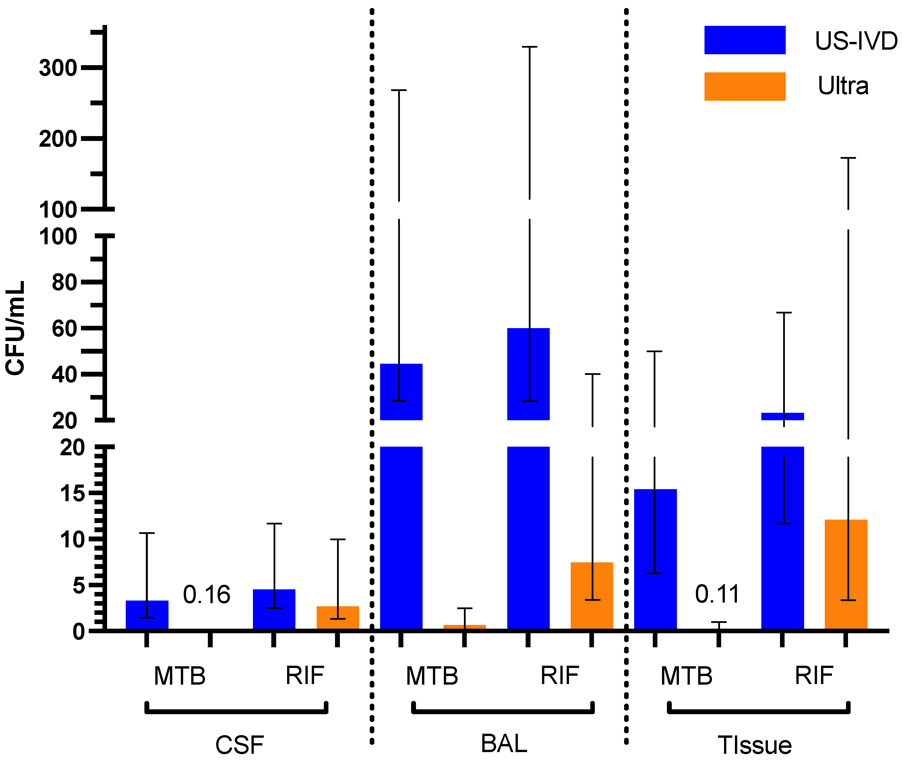
3.1. Limit of Detection (LoD)

The LoDs for MTB and RIF were determined for US-IVD and Ultra on all three sample types. The US-IVD test had the lowest LoDs for both MTB and RIF on CSF (3.3 CFU/mL and 4.6 CFU/mL, respectively) and the highest LoDs for both MTB and RIF on BAL (45 CFU/mL and 60 CFU/mL, respectively) among these three sample types. In comparison, the Ultra test had the lowest LoD for MTB (0.11 CFU/mL), but the highest LoD for RIF (12 CFU/mL) on tissue samples among the three sample types (Table 1). The LoD of MTB on BAL was highest (0.65 CFU/mL) and the LoD of RIF on CSF (2.7 CFU/mL) was lowest for the Ultra test (Table 1). Overall, the analytical sensitivity of the Ultra test is significantly higher than that of US-IVD, regardless of the sample types (p < 0.05) (Table 1 and Figure 1).

3.2. Linearity for US-IVD and Ultra

The rpoB gene was amplified and detected by five probes (A–E) in US-IVD, with the cycle threshold (Ct) set at 39 for A, B, and C, and 36 for D and E [9]. As expected, Ct values for each probe increased as the concentration decreased in all sample types (Figure 2A–D). Mutations associated with the Probe E (codons 529–533) region were identified as the most common rpoB gene mutations [10]. Based on the Ct values, the RIF-resistant MTB isolate used in this study had mutations in the probe E region of the rpoB gene. The software reported probe E target Ct values as 0 for all tests run on the US-IVD test in our study.
MTB detection with Ultra is defined as one or both of the probes that detect the multi-copy insertion elements IS6110 and IS1081 being positive, with Ct less than 37 and at least two rpoB probes with Ct less than 40 [20]. Ct values corresponding to the CFU/mL were plotted for IS (Figure 2E) and rpoB with four probes (Figure 2F–I) to visualize the relationship between Ct values and CFU/mL, with both the US-IVD and Ultra tests for each probe. With Ultra, the IS targets (Figure 2E) were more sensitive than the rpoB gene probes (Figure 2F–I). These regressions could be used to help predict the concentration (CFU/mL) of MTB in a sample based on the Ct values.

3.3. Semi-Quantitative Results by Ultra

The Ultra test is designed to report semi-quantitative results (high, medium, low, very low, and trace) corresponding to a Ct value range in one or more targets detected. For example, if one or both probes for the IS targets are positive with Ct less than 37 and no more than one rpoB probe has a Ct less than 40, Ultra calls it “MTB detected, trace” [20]. Here, we plotted the lowest Ct value among the four probes (B1, B2, B3, and B4) used for the rpoB gene for each sample versus the semi-quantitative categorical results (Figure 3A). All the samples tested in this study that had “trace” semi-quantitative results only had Ct values for the IS target, but not for any of the probes on the rpoB target. Therefore, the trace category was not included in Figure 3A. Figure 3B shows the CFU/mL in log scale versus the semi-quantitative categorical results. Figure 3 demonstrates the relationships between the amount of MTB in the specimens and the semi-quantitative categories.

4. Discussion

Overall, the analytical sensitivities of the Ultra test were substantially better than US-IVD for all sample types tested. Among the three sample types, BAL had the poorest analytical sensitivity on both the US-IVD and Ultra tests. BAL is not as homogenous as CSF and is more difficult to dilute than CSF, which may have accounted for some difference in the reported sensitivity. Since BAL and bronchoalveolar brush samples are typically considered superior non-sputum specimen types for MTB detection [6], we believe this is due to the higher organism burden in these specimen types clinically and not due to the sensitivity of the test. In contrast, pleural fluid is notoriously paucibacillary and therefore was not used to establish the analytical sensitivities of either test. In addition, Carnevale et al. reported that high cellular components and red blood cells could interfere with MTB detection in BAL and pleural fluid when certain extraction and amplification products were utilized [21]. While the pooled BAL matrix may have had moderate levels of interfering cellular contents, the breakdown of cellular components during storage may also have impacted the test performance and analytical sensitivity. Nonetheless, studies have shown that both Ultra and US-IVD can be used with BAL as an alternative to sputum for accurate detection of MTB in patients with low-yield sputum, smear-negative sputum, or tracheobronchial MTB [5,6]. Our results further demonstrated that both US-IVD and Ultra had even lower LoDs with BAL than sputum specimens in the experimental setting [9,12]. However, since these data were based on MTB prevalent regions, studies on the performance of Ultra on BAL samples compared to US-IVD in low prevalence regions are needed.
Lymphatic TB has consistently been the leading cause of EP TB in the US, based on the CDC data from 2010 to 2020 (https://www.cdc.gov/tb/statistics/surv/surv2020/default.htm, accessed on 7 August 2022). In our study, Ultra demonstrated the lowest LoD for MTB in lymph node tissues among all three sample types tested. Since we pooled lymph nodes from various anatomic locations, our data represent test performance on tissues from any lymphatic origin. However, since the pooled lymph node tissue was spiked post-homogenization, it does not truly represent patient lymph node samples containing organisms within the tissue. Therefore, the LoD for MTB in lymph node tissues reported in this study could be overestimated.
In the World Health Organization (WHO)’s report, US-IVD and Ultra are strongly recommended for establishing the diagnosis of meningeal TB from CSF samples [22]. There were discussions about the performance of US-IVD vs. Ultra for TB meningitis and whether one was more sensitive than the other. This could be explained by co-infection status with HIV and MTB prevalence in the population. A higher diagnostic accuracy from CSF samples was observed with Ultra in the HIV-negative group, whereas US-IVD performed better in HIV-positive group [23]. These differences were also likely influenced by whether or not the CSF was concentrated prior to testing [24].
In line with previous studies, the LoDs of MTB and RIF were lower (i.e., more sensitive) with Ultra than US-IVD in our study. This could also indicate that HIV-negative individuals with a lower bacterial burden than their counterparts would be more likely diagnosed by Ultra than US-IVD [17,24]. While WHO recommended using US-IVD for the initial diagnosis of TB meningitis, caution must be exercised in interpreting results depending on the disease prevalence in the region and underlying comorbidities, such as HIV status. Most studies describing the high accuracy of Ultra were performed in TB-prevalent areas. Although meningeal TB prevalence in the US was only 4% of the total EP TB per CDC’s report in 2020 (https://www.cdc.gov/tb/statistics/surv/surv2020/default.htm, accessed on 7 August 2022), the actual number of cases might be slightly higher due to possible misdiagnoses. Follow-up studies in the US entailing clinical correlations of TB meningitis and the use of Ultra in establishing the diagnosis are needed.
Because Ultra can detect MTB at “trace” levels, this feature can be helpful in establishing TB diagnoses in paucibacillary conditions. Studies have shown that the “trace” calls result in the early diagnosis of TB in smear-negative, culture-positive paucibacillary situations, leading to prompt treatments [25,26]. This highlights the significant clinical impact of Ultra’s “trace” feature in critical situations where the diagnosis could have been missed or delayed otherwise. The WHO report in 2019 also indicated that the TB diagnosis made from “trace” calls by Ultra should be based on defined criteria, such as EP TB, pediatric population, or underlying HIV infection, where the bacterial burden can be typically low [4].
Although only one MTB strain was used in this study, our findings confirmed that the analytical sensitivities of both US-IVD and Ultra were higher on non-sputum than sputum specimens [9,12]. In addition, Ultra had even lower LoDs than US-IVD for all sample types. These results suggest that using Ultra could result in more TB case detection rates in subjects with paucibacillary TB and EP TB, positively impacting WHO goals to eradicate TB.

Author Contributions

Conceptualization, P.R.; methodology, P.M.T.; formal analysis, M.C.N., P.M.T. and P.R.; investigation, M.C.N., P.M.T. and P.R.; resources, M.C.N. and P.R.; data curation, M.C.N., P.C. and R.P., supervision, S.L. and P.R.; writing—original draft, M.C.N., P.M.T. and P.R.; writing—review and editing, M.C.N., P.M.T. and P.R., funding acquisition, P.R. All authors have read and agreed to the published version of the manuscript.

Funding

Cepheid provided the Xpert MTB/RIF and Xpert MTB/RIF Ultra testing kits.

Institutional Review Board Statement

Not applicable.

Informed Consent Statement

Not applicable.

Data Availability Statement

All raw data are available upon request.

Acknowledgments

We thank Cepheid for providing the Xpert MTB/RIF and Xpert MTB/RIF Ultra testing kits.

Conflicts of Interest

The authors declare no conflict of interest.

References

  1. Dheda, K.; Perumal, T.; Moultrie, H.; Perumal, R.; Esmail, A.; Scott, A.J.; Udwadia, Z.; Chang, K.C.; Peter, J.; Pooran, A.; et al. The intersecting pandemics of tuberculosis and COVID-19: Population-level and patient-level impact, clinical presentation, and corrective interventions. Lancet Respir. Med. 2022, 10, 603–622. [Google Scholar] [CrossRef] [PubMed]
  2. Steingart, K.R.; Schiller, I.; Horne, D.J.; Pai, M.; Boehme, C.C.; Dendukuri, N. Xpert® MTB/RIF assay for pulmonary tuberculosis and rifampicin resistance in adults. Cochrane Database Syst. Rev. 2014, CD009593. [Google Scholar] [CrossRef] [PubMed]
  3. Horne, D.J.; Kohli, M.; Zifodya, J.S.; Schiller, I.; Dendukuri, N.; Tollefson, D.; Schumacher, S.G.; Ochodo, E.A.; Pai, M.; Steingart, K.R. Xpert MTB/RIF and Xpert MTB/RIF Ultra for pulmonary tuberculosis and rifampicin resistance in adults. Cochrane Database Syst. Rev. 2019, 6, CD009593. [Google Scholar] [CrossRef] [PubMed]
  4. World Health Organization. Global Tuberculosis Report 2019; World Health Organization: Geneva, Switzerland, 2019; ISBN 9789241565714. [Google Scholar]
  5. Aisenberg, G.M.; Jacobson, K.; Chemaly, R.F.; Rolston, K.V.; Raad, I.I.; Safdar, A. Extrapulmonary tuberculosis active infection misdiagnosed as cancer: Mycobacterium tuberculosis disease in patients at a Comprehensive Cancer Center (2001–2005). Cancer 2005, 104, 2882–2887. [Google Scholar] [CrossRef] [PubMed]
  6. Pedrazzoli, D.; Abubakar, I.; Potts, H.; Hunter, P.R.; Kruijshaar, M.E.; Kon, O.M.; Southern, J. Risk factors for the misdiagnosis of tuberculosis in the UK, 2001–2011: TABLE 1. Eur. Respir. J. 2015, 46, 564–567. [Google Scholar] [CrossRef] [Green Version]
  7. Gomez, C.A.; Pinsky, B.A.; Liu, A.; Banaei, N. Delayed Diagnosis of Tuberculous Meningitis Misdiagnosed as Herpes Simplex Virus-1 Encephalitis with the FilmArray Syndromic Polymerase Chain Reaction Panel. Open Forum Infect. Dis. 2017, 4, ofw245. [Google Scholar] [CrossRef]
  8. Chakravorty, S.; Simmons, A.M.; Rowneki, M.; Parmar, H.; Cao, Y.; Ryan, J.; Banada, P.P.; Deshpande, S.; Shenai, S.; Gall, A.; et al. The New Xpert MTB/RIF Ultra: Improving Detection of Mycobacterium tuberculosis and Resistance to Rifampin in an Assay Suitable for Point-of-Care Testing. MBio 2017, 8, e00812-17. [Google Scholar] [CrossRef] [Green Version]
  9. Cepheid|Package Inserts. Available online: https://www.cepheid.com/en_US/package-inserts/1608 (accessed on 15 October 2022).
  10. Alemu, A.; Tadesse, M.; Seid, G.; Mollalign, H.; Eshetu, K.; Sinshaw, W.; Abebaw, Y.; Amare, M.; Dagne, B.; Diriba, G.; et al. Does Xpert® MTB/RIF assay give rifampicin resistance results without identified mutation? Review of cases from Addis Ababa, Ethiopia. BMC Infect. Dis. 2020, 20, 87. [Google Scholar] [CrossRef] [Green Version]
  11. Cao, Y.; Parmar, H.; Simmons, A.M.; Kale, D.; Tong, K.; Lieu, D.; Persing, D.; Kwiatkowski, R.; Alland, D.; Chakravorty, S. Automatic Identification of Individual rpoB Gene Mutations Responsible for Rifampin Resistance in Mycobacterium tuberculosis by Use of Melting Temperature Signatures Generated by the Xpert MTB/RIF Ultra Assay. J. Clin. Microbiol. 2019, 58, e00907-19. [Google Scholar] [CrossRef] [Green Version]
  12. Cepheid|MTB/RIF Molecular Test—Xpert MTB/RIF Ultra. Available online: https://www.cepheid.com/en/tests/Critical-Infectious-Diseases/Xpert-MTB-RIF-Ultra (accessed on 15 October 2022).
  13. Dorman, S.E.; Schumacher, S.G.; Alland, D.; Nabeta, P.; Armstrong, D.T.; King, B.; Hall, S.L.; Chakravorty, S.; Cirillo, D.M.; Tukvadze, N.; et al. Study team Xpert MTB/RIF Ultra for detection of Mycobacterium tuberculosis and rifampicin resistance: A prospective multicentre diagnostic accuracy study. Lancet Infect. Dis. 2018, 18, 76–84. [Google Scholar] [CrossRef]
  14. Zifodya, J.S.; Kreniske, J.S.; Schiller, I.; Kohli, M.; Dendukuri, N.; Schumacher, S.G.; Ochodo, E.A.; Haraka, F.; Zwerling, A.A.; Pai, M.; et al. Xpert Ultra versus Xpert MTB/RIF for pulmonary tuberculosis and rifampicin resistance in adults with presumptive pulmonary tuberculosis. Cochrane Database Syst. Rev. 2021, 2, CD009593. [Google Scholar] [CrossRef]
  15. Marlowe, E.M.; Novak-Weekley, S.M.; Cumpio, J.; Sharp, S.E.; Momeny, M.A.; Babst, A.; Carlson, J.S.; Kawamura, M.; Pandori, M. Evaluation of the Cepheid Xpert MTB/RIF assay for direct detection of Mycobacterium tuberculosis complex in respiratory specimens. J. Clin. Microbiol. 2011, 49, 1621–1623. [Google Scholar] [CrossRef] [Green Version]
  16. Peñuelas-Urquides, K.; Villarreal-Treviño, L.; Silva-Ramírez, B.; Rivadeneyra-Espinoza, L.; Said-Fernández, S.; de León, M.B. Measuring of Mycobacterium tuberculosis growth. A correlation of the optical measurements with colony forming units. Braz. J. Microbiol. 2013, 44, 287–289. [Google Scholar] [CrossRef] [Green Version]
  17. Bahr, N.C.; Nuwagira, E.; Evans, E.E.; Cresswell, F.V.; Bystrom, P.V.; Byamukama, A.; Bridge, S.C.; Bangdiwala, A.S.; Meya, D.B.; Denkinger, C.M.; et al. ASTRO-CM Trial Team Diagnostic accuracy of Xpert MTB/RIF Ultra for tuberculous meningitis in HIV-infected adults: A prospective cohort study. Lancet Infect. Dis. 2018, 18, 68–75. [Google Scholar] [CrossRef] [Green Version]
  18. Le Palud, P.; Cattoir, V.; Malbruny, B.; Magnier, R.; Campbell, K.; Oulkhouir, Y.; Zalcman, G.; Bergot, E. Retrospective observational study of diagnostic accuracy of the Xpert® MTB/RIF assay on fiberoptic bronchoscopy sampling for early diagnosis of smear-negative or sputum-scarce patients with suspected tuberculosis. BMC Pulm. Med. 2014, 14, 137. [Google Scholar] [CrossRef] [Green Version]
  19. Denkinger, C.M.; Schumacher, S.G.; Boehme, C.C.; Dendukuri, N.; Pai, M.; Steingart, K.R. Xpert MTB/RIF assay for the diagnosis of extrapulmonary tuberculosis: A systematic review and meta-analysis. Eur. Respir. J. 2014, 44, 435–446. [Google Scholar] [CrossRef] [Green Version]
  20. World Health Organization. WHO Meeting Report of a Technical Expert Consultation: Non-Inferiority Analysis of Xpert MTB/RIF Ultra Compared to Xpert MTB/RIF; World Health Organization: Geneva, Switzerland, 2017. [Google Scholar]
  21. Carnevale, G.G.; Vargas, F.S.; Caiaffa-Filho, H.H.; Acencio, M.M.P.; Marçal, L.J.; Sales, R.K.B.; Teixeira, L.R.; Antonangelo, L. Preanalytical conditions can interfere with M. tuberculosis detection by PCR in respiratory samples. Clinics 2018, 73, e410. [Google Scholar] [CrossRef]
  22. WHO Consolidated Guidelines on Tuberculosis: Module 3: Diagnosis: Rapid Diagnostics for Tuberculosis Detection. 2021 Update. Available online: https://www.who.int/publications/i/item/9789240029415 (accessed on 23 October 2022).
  23. Donovan, J.; Thu, D.D.A.; Phu, N.H.; Dung, V.T.M.; Quang, T.P.; Nghia, H.D.T.; Oanh, P.K.N.; Nhu, T.B.; Chau, N.V.V.; Ha, V.T.N.; et al. Xpert MTB/RIF Ultra versus Xpert MTB/RIF for the diagnosis of tuberculous meningitis: A prospective, randomised, diagnostic accuracy study. Lancet Infect. Dis. 2020, 20, 299–307. [Google Scholar] [CrossRef] [Green Version]
  24. Patel, V.B.; Theron, G.; Lenders, L.; Matinyena, B.; Connolly, C.; Singh, R.; Coovadia, Y.; Ndung’u, T.; Dheda, K. Diagnostic accuracy of quantitative PCR (Xpert MTB/RIF) for tuberculous meningitis in a high burden setting: A prospective study. PLoS Med. 2013, 10, e1001536. [Google Scholar] [CrossRef] [Green Version]
  25. Amedeo, A.; Beci, G.; Giglia, M.; Lombardi, G.; Bisognin, F.; Chiarucci, F.; Corsini, I.; Dal Monte, P.; Tadolini, M. Evaluation of trace calls by Xpert MTB/RIF ultra for clinical management in low TB burden settings. PLoS ONE 2022, 17, e0272997. [Google Scholar] [CrossRef]
  26. Biswas, S.; Uddin, M.K.M.; Paul, K.K.; Ather, M.F.; Ahmed, S.; Nasrin, R.; Kabir, S.; Heysell, S.K.; Banu, S. Xpert MTB/RIF Ultra assay for the detection of Mycobacterium tuberculosis in people with negative conventional Xpert MTB/RIF but chest imaging suggestive of tuberculosis in Dhaka, Bangladesh. Int. J. Infect. Dis. 2022, 114, 244–251. [Google Scholar] [CrossRef] [PubMed]
Figure 1. Limit of Detections between US-IVD and Ultra on different sample types for both MTB and RIF. The bars reflect the 95% confidence intervals.
Pathogens 12 00157 g001
Figure 2. (AD) Linear regression for the rpoB gene with detection probes A, B, C, and D (US-IVD), respectively. (EI) Linear regression for the IS and the rpoB gene with detection probes B1, B2, B3, and B4 (Ultra), respectively. The orange area indicates the 95% confidence interval.
Pathogens 12 00157 g002
Figure 3. Semi-quantitative category reported on the Ultra test plotted against the lowest Ct values among the 4 rpoB gene probes for each sample (A) and the corresponding CFU/mL (log10) (B). The box represents the interquartile ranges from the lower quartiles (Q1) through the upper quartiles (Q3). The median and the mean are displayed as a solid horizontal line and × inside the box, respectively. The outliers are shown as data points outside the standard deviation bar.
Pathogens 12 00157 g003
Table 1. LoD95 in CFU/mL for US-IVD and Ultra for each sample type.
Sample Type US-IVD (95% CI)Ultra (95% CI)
CSFMTB3.3 (2.2–11)0.16 (0.095–0.52)
RIF4.6 (2.9–14)2.7 (1.4–10)
BALMTB45 (22–260)0.65 (0.39–2.3)
RIF60 (28– 353)7.5 (3.6–46)
TissueMTB15 (8.0–49)0.11 (0.077–0.29)
RIF23 (12–70)12 (3.7–151)
Disclaimer/Publisher’s Note: The statements, opinions and data contained in all publications are solely those of the individual author(s) and contributor(s) and not of MDPI and/or the editor(s). MDPI and/or the editor(s) disclaim responsibility for any injury to people or property resulting from any ideas, methods, instructions or products referred to in the content.

