
Application of Fluorescence In Situ Hybridization (FISH) in Oral Microbial Detection
by
 and
and
Junjie Gu
1,†, 
Huayu Wang
1,†,
Mengye Zhang
1,
Yichen Xiong
1,
Lei Yang
1,
Biao Ren
1,* and
Ruijie Huang
1,2,* 1
State Key Laboratory of Oral Diseases, National Clinical Research Center for Oral Diseases, West China School of Stomatology, Sichuan University, Chengdu 610041, China
2
Department of Pediatric Dentistry, West China Hospital of Stomatology, Sichuan University, Chengdu 610041, China
*
Authors to whom correspondence should be addressed.
†
These authors contributed equally to this work.
Pathogens 2022, 11(12), 1450; https://doi.org/10.3390/pathogens11121450
Submission received: 10 October 2022
/
Revised: 26 November 2022
/
Accepted: 29 November 2022
/
Published: 1 December 2022
(This article belongs to the Special Issue Insights in Oral Microbiota)
Abstract
:Varieties of microorganisms reside in the oral cavity contributing to the occurrence and development of microbes associated with oral diseases; however, the distribution and in situ abundance in the biofilm are still unclear. In order to promote the understanding of the ecosystem of oral microbiota and the diagnosis of oral diseases, it is necessary to monitor and compare the oral microorganisms from different niches of the oral cavity in situ. The fluorescence in situ hybridization (FISH) has proven to be a powerful tool for representing the status of oral microorganisms in the oral cavity. FISH is one of the most routinely used cytochemical techniques for genetic detection, identification, and localization by a fluorescently labeled nucleic acid probe, which can hybridize with targeted nucleic acid sequences. It has the advantages of rapidity, safety, high sensitivity, and specificity. FISH allows the identification and quantification of different oral microorganisms simultaneously. It can also visualize microorganisms by combining with other molecular biology technologies to represent the distribution of each microbial community in the oral biofilm. In this review, we summarized and discussed the development of FISH technology and the application of FISH in oral disease diagnosis and oral ecosystem research, highlighted its advantages in oral microbiology, listed the existing problems, and provided suggestions for future development..
1. Introduction
The human oral microbiota consists of more than 700 species, and the microbial communities are diverse at different niches in the oral cavity [1]. Human oral microbiota mainly consists of bacteria, fungi, viruses, protists, and oral archaea [2,3]. According to the human oral microbiome database (HOMD), 70% of the oral bacteria are culturable, 30% are unculturable, and only 57% of culturable species have been named [4].
Human oral microbiota has distinct characteristics [5,6]. The human oral cavity provides an unexplored, structurally complex environment and a differentiated set of habitats [7], and saliva promotes the interactions of various local environments [8]. The spatial organization of microorganisms in the oral cavity is formed by dynamic equilibrium flow and adhesion, abscission, and colonization, as well as the interaction between microorganisms and host [7,8], which makes oral microbiota an important biological model of the human microbiome [9]. The synergies and interactions of variable oral microorganisms help the human body to maintain oral health, but the imbalance of oral microbiota leads to the development of oral diseases, even systemic diseases [10], indicating that monitoring of oral microbiota is important for the diagnosis or analysis of oral diseases and some systemic diseases [11].
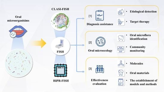
Various oral microbiological detection methods have been developed in labs and clinics [12]. Fluorescence in situ hybridization (FISH), a non-radioactive technique combining cytogenetics and molecular biology, was developed based on radiogenic in situ hybridization in the late 1980s to replace isotope markers with fluorescent markers [13]. The application of FISH in microbial detection usually targets microbial ribosomal RNA (rRNA) genes [14]. FISH can also be used to detect the effects of different external factors on colonies and the interactions between colonies [15]. In present review, we have summarized and discussed the applications of FISH in oral microbiology to highlight its advantages in oral microbial detection and oral diseases diagnosis (Figure 1).
2. Research Progress of FISH
FISH is one of the most routinely used cytochemical techniques for genetic detection, identification, and localization by a fluorescently labeled nucleic acid probe. It is more specifically used in hybridizing processes with nucleic acid sequences of interest [16]. The vitality of this technology is continuously proved by the evolution of probe design, signal amplification, and multiplexing, broadening the application of experimental research and clinical diagnoses in this field of study [17]. Specifically, FISH and its variants have been applied to nucleic acid investigation, cell metabolic research, oncology diagnostics, and microbiological research [18].
2.1. Development of FISH
In the 1960s before the advent of FISH, cytochemical methods of detecting and localizing specific intracellular molecules were mainly immunocytochemistry based on in situ hybridization with radiolabeled probes and antigen-antibody interaction with fluorescein-labelled immunoglobulins [19,20,21]. However, radioactive RNA or DNA binding to DNA sequences in situ used to be limited to detecting, characterizing, and localizing specific DNA segments. Due to the low resolving power and long exposure time in autoradiography, this methodology is inaccurate in quantification [17,22]. To compensate for this deficiency, indirect immunofluorescence, a type of immunocytochemistry technique developed in 1965, has been successfully applied in quantitatively analyze biological markers via fluorophore-labeled immunoglobulins that target marker-binding antibodies [21].
As a combination of in situ hybridization and indirect immunofluorescence, a method described by Rudkin et al. in 1977 replaced autoradiography with fluorescence microscopy to detect in situ signals from antibodies against DNA-RNA hybrids [23]. Before long, based on the methods above of nucleic acid hybridization and indirect immunofluorescence without intermediated antibodies, Bauman et al. first applied fluorophore-covalent-labeled RNA probes in specific DNA sequence detection in 1980, representing the birth of FISH [22]. Nonetheless, the application of this original FISH still needs further improvement in probe affinity and signal amplification due to the low signal intensity caused by target inaccessibility and low copy numbers [24].
The optimization of probe design and synthesis is crucial to the development of FISH. The establishment of genetic databases made it possible to design probe sequences targeting complementary gene sequences of interest [25]. Probe preparation underwent a development from manual multiple sequence alignment for conserved target regions to probe sequence auto-selecting programs and even web-based platforms for theoretical evaluation of the probe performances in FISH [25,26,27,28,29]. The new technology makes it easier to control the length, complementary sequence, thermodynamic property, potential secondary structure, and specificity of the probe, which are directly related to the successful application of FISH [30].
With accurate probe design methods, FISH properties became even more modern, with changes that include increasing signal intensity and stability, range of targets, and sensitivity. Commonly used variants are: catalyzed reporter deposition FISH (CARD-FISH), gene-FISH, recognition of individual genes FISH (RING-FISH), nucleic acid mimics FISH (NAM-FISH), combinatorial labeling, and spectral imaging FISH (CLASI-FISH), double labeling of oligonucleotide probes for FISH (DOPE-FISH), as described below.
The polynucleotide probe also has an advantage in signal intensity with multiple labels and secondary structures mediating probes-connected networks [31]. RING-FISH achieves high detection efficiency to single genes with multiple labeled transcript polynucleotide probes generating halo-like signals [32]. As an alternative to RING-FISH for prokaryotic cellular microorganisms, two-pass TSA-FISH provides higher efficiency and a better signal-to-noise ratio, making it a useful protocol based on the functional gene for single microbial cell detection [33]. By combining rRNA CARD-FISH and polynucleotide probe gene detection, gene-FISH can be an innovative technique to provide a stable signal and high sensitivity [34]. Furthermore, direct gene-FISH for gene signal quantification can be formed by replacing CARD (and its disabilities) with fluorochrome-labeled probes and super-resolution microscopy [35].
The nucleic acid mimics probe has become the most promisingly high-efficient probe with higher affinity, specificity, and better stability based on resistance to enzymatic degradation [36], used to overcome the issues with weak signal caused by low affinity and vulnerability of DNA or RNA probes. The NAM probes currently being utilized are peptide nucleic acid (PNA), 2’-O-Methyl-RNAs, and locked nucleic acid (LNA) [37].
The variants mentioned above show corresponding advantages in detecting microorganisms, allowing direct identification without cultivation and further detection of microbial community structure and individual function [36,38]. As microbiological research kept expanding from individuals to communities, multiplexing identification was developed to simultaneously analyze multi-microbes and microbiota efficiently. A typical example is CLASI-FISH, which introduces combinatorial labeling and spectral imaging (CLASI) into FISH and can be applied to distinguish several microbes at once by linear unmixing the spectra of fluorophores from overlapping spectra [39]. However, CLASI-FISH application may be limited due to internal sensitivity loss and potential probe binding bias caused by binary combinations, limiting the application of this technique for quantification analyses [40]. Compared to CLASI-FISH, DOPE-FISH provides a double signal intensity as well as stable specificity and higher affinity to targets, but the detectable number of microbes is much lower [41].
The variants of FISH are improving, as are their applications based on the detection of nucleic acids. Apart from microbiological applications, FISH has been a gold standard technique in absolute gene copy number quantification in cancer, allowing implementation of precise treatment strategies [42]. Based on imaging spatial transcriptomics, FISH performs cell segmentation for cell interactions and the state of complex tissues for analysis of, for example cell organization in the cerebral cortex and cell fate decisions in organogenesis [43,44,45]. Expansion-Assisted Iterative (EASI)-FISH was developed for the 3D organization of cell types in thick tissue, contributing to characterizing the architecture underpinning brain function [46]. It is reported that the combination of FISH and small and ultrabright fluorescent polymeric nanoparticles functionalized with DNA allows a simpler, faster and sensitive single-cell RNA imaging method for transcriptomic analysis [47]. The characteristic chromosomal abnormalities in cancer cells can be obtained by high-resolution karyotyping by FISH, which can help in differential diagnosis [48]. Other uses of FISH karyotype analysis include genetic diagnosis, prenatal screening, and plant and animal genetic studies [48,49,50,51,52]. But karyotyping is highly dependent on frequency of cytogenetically abnormal cells, in need of enrichment methods such as fluorescence-activated cell sorter (FACS) to improve the sensitivity [53]. Thus, the combination of FISH with other suitable techniques can compensate for some inherent drawbacks and break the application limitations of FISH. The coupling of Flow Cytometry and FISH enables high-throughput quantification of complex whole-cell populations, and the association with FACS (FLOW-FISH-FACS) enables sorting of target microorganisms [54]. In drug discovery, FISH was introduced into high-content screening for intracellular imaging of mRNA to screen mRNA-associated drugs and assess their pharmacological activity [55]. Resolution After Single-strand Exonuclease Resection (RASER)-FISH provides a robust generation of single-stranded DNA with excellent preservation of chromatin structure, nuclear integrity and improved hybridization efficiency, which is achieved by exonuclease digestion rather than physical denaturation by heat and exposure to formamide [56].
2.2. Procedures and Principles of FISH and Its Variants Used for Oral Microbial Detection
The general protocols of FISH include (a) specimen treatment, (b) probe denaturation, (c) hybridization, (d) elution, (e) hybridization signal amplification (applicable to biotin-labeled probes), (f) re-staining, (g) encapsulation, and (h) fluorescence microscope observation of FISH results [40,57,58]. Probe labeling and specimen processing require different processing methods according to different detection needs, in order to obtain clearer detection results in the subsequent hybridization process [59,60]. For example, to substantially enlarge the number of distinct taxa in one FISH experiment, CLASI-FISH was created, and the established FISH protocols are ordinarily suitable for the hybridization, but still some changes for the protocol were proposed, such as fixing samples with PFA followed by ethylalcohol [39]. Take CARD-FISH as another instance, to enhance sensitivity and whittle background interference, the protocol resembled the typical FISH, but tyramide-fluorophore and HRP was introduced to replace fluorophore on the probe [61]. All the protocols are detailedly elaborated by the inventors of the FISH variants, so that researchers can consult relevant papers according to application needs. Different physical and chemical environments during hybridization will affect the effect of hybridization, and redyeing will significantly change the clarity of the observed objects, which will lead to significantly different results under the optical microscope [62,63].
FISH can evaluate various types of test samples, such as tumor tissue, pathological sections, local animal samples, human tissue samples, etc. For tissue samples: 4% paraformaldehyde fixation (paraffin section), or put into liquid nitrogen, −80 °C storage, sample not less than 100 mg; For cell samples: cell slides prepared with 6-well plates, with no less than 2 × 10⁶ cells per well; For environmental bacteria: sludge samples or sludge particles and other sample forms that can be used for smear or sectioning [6,39,64,65].
Though FISH is widely used in microbiological detection and biofilm analysis, some techniques belonging to nucleic acid, immunity, and single-cell techniques also have been applied in different emphases of microbial research due to their different advantages. Ten common techniques (including FISH) in microbial research were selected and compared based on several performance factors, including detectable resolution, applications in oral microbiology, culture reliance, quantification appliance, capability for microbiota analysis, and unsearched species (Table 1). In summary, some features of FISH make it an advantageous technique, e.g., precision, culture-independent, unsearched species detectability for microbiota analysis for extra semi-quantification and intuitive spectral imaging.
Furthermore, most oral microorganisms are unculturable, and the community structure and components are complex and changeable, and these properties make FISH very suitable for oral microbial research. Among many variants of FISH, classical FISH and CLASI-FISH are the most popular in terms of their application in oral microbiology. However, it is noteworthy that a new variant called HiPR-FISH is considered to have a promising future. The procedure and principles of these three currently used FISH techniques in oral microbiology will be elaborated throughout this study.
2.2.1. FISH
The most common procedure followed by researchers who apply FISH involves the following four steps: (1) fixation for dehydration to inhibit the action of enzymes without cell and nucleic acid structures destruction (some special cells need extra permeabilization treatment that degrade the cell walls to increase membrane permeability); (2) hybridization between target nucleic acid sequences and specific complementary probes labeled with fluorescent dyes or reporter molecules to be detected by fluorescent antibodies; (3) washing to remove the unbound or loosely bound (usually mismatched) probes; (4) detection and visualization of the probe-bounded cells by typically fluorescence microscopy and some advanced microscopy systems, then access to information for species detection or microbiota analysis [16,36,87]. Hybridization is one of the most decisive steps, strongly influenced by probe properties on specificity and sensitivity [87]. These four steps are also the basic standpoints of modified FISH variants.
2.2.2. CLASI-FISH
Regardless of some minor adaptions to classical FISH protocols for CLASI-FISH application, the main improvement of CLASI-FISH are probe design and image acquisition/analysis. CLASI-FISH probes are particularly effective in synthesizing two probes for one targeted sequence and two different sequence-particular fluorophores; thus, the cells of different species are labeled by specific combinations of two highest-intensity fluorophores [88]. For detection, CLASI-FISH applies confocal laser scanning microscopy (CLSM) in image acquisition, allowing the linear spectral unmixing computational analysis to identify the fluorophores with overlapping spectra and analyze the fluorophore composition in each cell [39].
2.2.3. HiPR-FISH
Regarding the expansion of the number of distinguishable species in a single image as CLASI-FISH, it has been previously verified that High Phylogenetic Resolution FISH (HiPR-FISH) achieves significantly higher taxonomic resolution and multiplexity due to a two-step hybrid approach and a routine for automated image segmentation [89]. Two kinds of probes were used for hybridization: the encoding probe in the first step and the readout probe in the second step. The encoding probes are taxon-specific probes that include targeting sequences modified with different flanking readout sequences. The fluorescently labeled readout probes target readout sequences and stochastically bind to the bound encoding probes, representing equal proportions of fluorophores.
Ten distinct fluorophores encode up to 1023 fluorophore combinations, and one species corresponds to a 10-bit binary barcode derived from designed one or more encoding probes binding with relevant readout probes and presenting different spectral components. After image acquisition, automated image segmentation classifies the spectra of images and assigned cells the corresponding barcodes. The reference spectrum for each barcode is established by the Förster resonance energy transfer (FRET) model, and the k-means clustering-denoised and straightened images of cultured single-cell and biofilm samples are segmented by the watershed algorithm that the seed is defined by developed Local Neighborhood Enhancement (LNE). For spatial analysis, the adjacency segmentation is also generated by the watershed algorithm and then calculates the intuitive spatial association matrix. The super-resolution images and 3D datasets are generated the same way, and the 4D data cube needs rendering in ipyvolume.
3. Research and Application of FISH in Oral Microbial Detection
FISH has been widely used in various fields of oral microbial research. From the basic research at the practical level to the potential clinical application and outcome transformation, the important role of this methodology has been witnessed by scientists in different scenarios inside the research community. In this section, we summarize the three major applications of FISH and explain its relevant details (Table 2).
3.1. Diagnosis Assistance
This function of FISH has been used to identify the species of pathogenic microorganisms in the simplest way, but it is still in the basic research stage in terms of predicting the risk of disease and prognosis, which needs to be further tested and verified in clinical practice. In the context of modern medicine, the requirements of patients and clinicians for diagnosis have upgraded from merely knowing the type of diseases to detecting specific causes and predicting disease progression. For patients with diseases related to oral microorganisms (caries, pulpitis, periodontal disease, etc.), most of them exerted evident symptoms at the time of its’ initial diagnosis, such as pain, inflammation, and changes in biological characteristics. And increasing studies have shown that cancer, atherosclerosis, diabetes and other diseases can be reflected in oral flora. Oral microorganisms play an important role in the occurrence and development of these diseases, and the changes in community structure can somehow indicate how it has been progressing [110]. Therefore, we can understand the types of pathogens based on determining the types of diseases and improve the accuracy of diagnosis to outline potential target therapies. At present, most of the detection methods of pathogenic microorganisms in hospitals are still traditional identification methods, including Gram staining, microbial culture and biochemical tests. However, for the complexity of oral microenvironment, traditional methods are not well qualified for multi-species identification, community analysis and other aspects, let alone judging the risk of disease and prognosis. As a highly-stable, high-resolution, simple, and intuitive nucleic acid probe technology, FISH can effectively facilitate the application of such functions. However, more detailed databases are still needed to support accurate diagnosis due to inadequate personalized research and global data sets.
Clinically, FISH can be used to detect and analyze various disease-causing factors, such as HIV and Epstein-Barr virus, providing a faster way to diagnose such conditions. However, due to the spreading of the new COVID-19 pandemic, nucleic acid testing has become a part of people’s lives, so we urgently need a more rapid and more convenient way to detect the virus. Hepp et al. developed a rapid FISH protocol capable of quantitatively detecting influenza virus, avian infectious bronchitis virus, and SARS-CoV-2 in approximately 20 minutes by combining nasal and throat swabs with the added virus in virus cultures. This rapid and simple method can be used both as a commonly used detection technique in the laboratory and to aid the future diagnosis of enveloped viruses with accessible genomes [62].
Modern research shows considerable differences in the type, proportion, and structure of oral microbiota between healthy and pathological states, which can be used as breakthrough points to diagnose the actual causes of the disease. FISH was initially used to detect relatively single and known pathogenic microorganisms, but with the development of new technology, a variety of microorganisms can now be detected simultaneously. Clinicians and researchers use FISH to detect microbes’ information, such as species, and community structure, after extracting samples from patients. This kind of data is included in the databases and also used as feedback to clinicians to develop more accurate treatment plans. Taking oral lichen planus (OLP) as an example, there are significant differences in microflora between OLP patients and healthy controls. By applying FISH, Zheng et al. [91] and Wang et al. [92] revealed species, location, and average optical density (AOD) variations of oral microorganisms between OLP patients and healthy individuals. These significant differences might all become targets for diagnosis and treatment in the future.
In addition to being used in the diagnosis of diseases, the results of FISH can be used in the formulation of treatment options. Take the most common and inconvenient form of bad breath as an example. Bernardi et al. visualized and quantified the dorsal biofilm through the combination of FISH and CLSM and stained eubacteria, Streptococcus spp., and F. nucleatum using specific fluorescent probes. In their experiment, they found a significantly higher proportion of F. nucleatum and Streptococcus spp. in the biofilm of the halitosis group. It was concluded that the relative ratio of total microorganisms to these two bacteria could be considered a relevant factor in causing bad breath; hence it can be included in the treatment goals [93].
Apart from oral diseases, FISH is also used for microbial detection of lesions of other diseases. For example, it has been found that there are many common oral bacteria in oral squamous cell carcinoma [111]. Although oral microorganisms are also found in atherosclerotic plaques [112] and Alzheimer’s disease patients’ brains [113], the detection method used is PCR. Therefore, FISH is almost equal to PCR in determining whether there is a certain bacterium or not. Nevertheless, considering that FISH technology does not depend on the isolation and culture of bacteria and that not all laboratories have the experimental conditions for FISH, the specific method to choose eventually depends on the experimenters. However, if we want to expand the detection scope to more species, community distribution, and even predicting risk and prognosis, FISH will be more advantageous.
3.2. Oral Microecology
Currently, research focus on oral microecology occupies an important position in stomatology [114], and many past research results showed that oral microbes remain a major cause of a variety of oral diseases [63]. Many contemporary studies indicate that FISH is a powerful tool for studying oral microecology [115]. In recent years, due to the rapid development of this technique, it has become a highly automated process with two major functions, microbial species identification and community monitoring [116]. Furthermore, with the continuous combination with other molecular biology techniques, it has been shown that FISH can even be used to detect the effects of different external factors on colonies and interactions between them [15].
According to the Expanded Human Oral Microbiome Database (eHOMD), only 57% of oral microorganisms can be cultivated and formally named, 13% of oral bacteria can be cultivated but not named, and 30% of the oral microorganisms fail to be separated from the biofilm, hence hindering the study of oral micro-ecological research [117]. FISH, which does not require bacteria culture, can identify microbial populations into genera and species by using fluorescently labeled specific oligonucleotide fragments as probes to hybridize with DNA molecules in the environmental genome. This feature helps to avoid the limitations of traditional culture methods for identification and counting. It has high application value in identifying oral microflora, bringing a new detection method for some diseases caused by bacterial infection.
Annett et al. detected the presence of Treponema pallidum in the tissue samples of syphilis patients by a specific 16S rRNA FISH probe against T. pallidum. Their effective methodology avoided possible false results of serological identification in HIV-positive patients and provided a faster and more effective technique for disease monitoring [90]. Using similar methods, Fernandes et al. identified bacteria in pulp infections by FISH and confirmed the involvement of bacteria such as Pyramidobacter piscolens and Fretibacterium fastidiosum. These two bacteria are also challenging to detect in endodontic infections because they are difficult to separate and culture [96]. In addition, FISH can also visualize oral colonies with a confocal laser scanning microscope (CLSM), thus locating microorganisms’ data in clinical samples and assisting in speculating the process of invasion, colonization, and transmission. Bertl et al. successfully monitored the position of the oxygenic pathogens in the biofilm of the vocal cord prosthesis by the combination of FISH and CLSM, supporting that they were not the cause of the breakdown of vocal cord prosthesis equipment [97]. In contrast, in Zangeneh et al.’s experiment, they used PCR-DGGE to detect only the type and number of bacteria in the oral cavity of multiple sclerosis patients [79]. This shows that compared to traditional microbial detection techniques such as PCR, FISH not only has the advantage of being a simple and fast process, but by combining it with other detection methods, more information about the colonies can be obtained.
Due to its ability to display location information apart from identifying species, researchers have increasingly applied FISH to monitor microbial communities in recent years. The technique can be used to study colonies that are difficult to cultivate and clearly present the overall microbial environment without requiring additional bias-prone steps such as extraction and amplification. FISH can accurately and quickly reflect the in-situ distribution of microbial communities in various systems, avoiding the complex process of other community analysis methods [118]. Through FISH, we can quickly and efficiently analyze the microbial communities in pathological conditions and then compare them with the normal ones to assist doctors with disease diagnosis.
Bring et al. spotted the distribution of TM7 bacteria, which is difficult to culture in the oral microecology through FISH and further reveals the mechanism of their metastatic infection [59]. Shi et al. created a micron-scale map of the location and identity of hundreds of microbial species in a complex microbial community by high-phylogenetic-resolution microbiome mapping by fluorescence in situ hybridization (HiPR-FISH), based on spectral imaging and decoding. Their findings revealed the disruption of the spatial network in the mouse gut microbiota by antibiotic treatment and the longitudinal stability of the spatial structure in the oral plaque microbiota, providing a framework for the spatial analysis of microbial communities at single-cell resolution [89]. By testing the oral microbial community at different times, FISH plays an essential role in static detection; it dynamically observes the changes of the microbial community in the mouth and monitors the occurrence and progression of the disease.
Using the combination of FISH and CLSM, AI-Ahmad et al. measured microflora changes in oral plaque within eight weeks with a particular focus on individuals with poor oral hygiene [57]. Combining with other different molecular biological technologies, the quantity, location, and morphological changes of different microbial communities in the biofilm could be dynamically observed, which further yields the influence of various factors on specific bacteria and the interaction between microbial communities. Similarly, Esteves et al. used FISH to detect the interactions of different microbial communities in dental plaque, revealing the impact of these interactions on the generation and development of paradentosis [98]. Moreover, FISH combined with flow cytometry for detecting microbes is a promising technology to diagnose and evaluate microbial community structure and its dynamics. Its highly automated operation makes it more suitable than other methods for frequent and rapid monitoring of specific colonies [119].
Because of its two effective features for species identification and community detection, FISH is often used to study dental plaque biofilm. Mature dental plaque consists of multiple species of biofilms and contains more than 500 different bacteria, causing common oral diseases such as periodontitis, dental caries, and gingivitis [120]. Combined with CLSM, this technique allows the visualization of biofilm systems, and the dynamic monitoring of plaque biofilms can assist doctors in the judgment of disease progression while avoiding the traditional post-culture detection, identification, and quantification. For instance, Gmür et al. identified and analyzed single microbial cells in the dental plaque through immunofluorescence and FISH [94]. Karygianni et al. found that combining CLSM and FISH could create high-resolution 3D images of individual bacteria in their natural environment to better visualize bacterial communities in dental plaque, allowing professionals to effectively monitor the 3D spatial distribution of many different bacteria in oral biofilms. After obtaining high-resolution images, image processing and data analysis can quantify the biomass of different targets in oral biofilms [95]. Dige et al. successfully applied FISH to analyze spatial relationships in dental plaque and the changes of specific microflora over time [64]. Welch et al. used CLASI-FISH to discover a distinctive, multigenic consortium in the microbiome of supragingival dental plaque, which consisted of a radially arranged, nine-taxon structure organized around cells of filamentous corynebacteria. The size of this consortium varies from ten to hundreds of microns and follows a trend that promotes the spatial distribution of its flora. It was found that anaerobic taxa tend to be located in the interior, while parthenogenic or exclusively aerobic bacteria tend to be distributed in the periphery. Based on this finding, they propose a new morphological model of the microbial community in dental plaque biofilms [121]. In summary, FISH can accomplish both the identification of individual strains and achieve dynamic monitoring of the colony system. By combining with CLSM, it is even possible to visualize information such as the location of a particular strain in the biofilm, which cannot be achieved by traditional detection methods.
3.3. Effectiveness Evaluation
The purpose of studying oral microorganisms is to maintain a healthy oral microecology, so scientists make efforts to establish a fairly complete effectiveness evaluation system. At present, the research topics related to using FISH as one of the detection methods mainly include molecules, oral materials, and the establishment of models and methods.
3.3.1. Evaluation of the Effectiveness of Molecules
In the long history of people dealing with oral pathogens, researchers have found that some molecules can specifically inhibit some of these pathogens and promote the reproduction of probiotics. The first kind of molecules are part of our daily lives, and it is commonly known that some have medicinal properties, such as salicylic acid, podophyllotoxin, and vinblastine. Hannig et al. [99] suggested that gargling with some polyphenol beverages and ingesting related foods could reduce the adhesion of initial bacteria to dental enamel and might help prevent diseases caused by oral biofilm. Similarly, Hertel et al. [100] used FISH to observe the anti-acid effect of Inula viscosa and its effect on the initial formation of oral biofilm and found that flushing with Inula viscosa for more than 8h had an effect on bacterial colonization on the enamel surface, but no effect on the anti-acid performance of the biofilm.
The second kind of molecule is related to the artificial synthesis and improvement of natural compounds. Lyu et al. [101] studied the effect of LCG-N25 (a new small molecule exploited from known inartificial lead compounds) on the constituent parts of multi-species biofilm by using species-specific FISH. Their results showed that with good anti-bacterial activity and low cytotoxicity, this molecule cut down the proportion of Streptococcus mutans, Streptococcus sanguinis and Streptococcus gordonii but did not induce drug resistance to cariogenic S. mutans, manifesting that LCG-N25 could be a hopeful adjuvant for the treatment of caries.
After exploring the role of individual molecules, our team identified that it was equally important to understand the effects of the interaction of different molecules on our results, so a new series of experiments was carried out. Cheng et al. [102] investigated the combined impact of arginine and fluoride on oral bacteria and spotted that arginine might increase the ecological benefit of fluoride by enhancing the alkali-producing bacteria in plaque biofilm, playing a synergistic role with fluoride in the control of caries. In addition to the interaction of medicinal molecules, the gain effects of medicinal molecules on functional but non-medicinal molecules were also previously studied by other scholars. Liu et al. [103] added dimethylaminododecyl methacrylate (DMADDM) to a root canal sealant called EndoREZ and detected the composition of multi-strain biofilm by FISH and RT-qPCR. It was unveiled that when the mass fraction of DMADDM increased to 5%, the cytotoxicity, apical sealing ability, antibacterial, and solubility of the sealant were considerably distinguished from the control group. Therefore, it was concluded that the EndoREZ sealant containing DMADDM could be used for clinical prevention and treatment of persistent periapical periodontitis.
In addition to FISH, other technologies are also widely utilized, such as using PCR to explore the effects of terminalia chebula extracted from ethylalcohol on S. mutans [122], using DNA microarray to study the effect of secondary carbon dioxide laser irradiation on S. mutans [123], and so on. However, because PCR can only provide data analysis and can not show the results more intuitively, and DNA microarray can only be used to study the species that have been identified, FISH is more advantageous in evaluating the effectiveness of molecules.
3.3.2. Evaluation of the Bacteriostatic Effect of Oral Materials
The oral cavity is an essential human structure closely related to food nutrition absorption, gas exchange, and speech expression. Consequently, dental materials are required to have biosafety, biocompatibility, and biofunction. However, the biofilm formed by microbes attached to the material surface can lead to drug-resistant infection and the contamination of medical devices [124]. Accordingly, the scientific community has explored different means to make bacteriostatic dental materials. In the field of dental implant materials, for instance, the development of bacteria-driven mucosal inflammation and peri-implant inflammation may lead to the failure of treatment. Al-Ahmad et al. [104] used FISH to detect the thickness, surface coverage, and oral Streptococcus spp. content in the biofilm, revealing that the surface roughness of the zirconia surface of the oral implant with low roughness was similar to that of titanium surface in terms of initial bacterial adhesion or oral Streptococcus spp. content in the biofilm. Similarly, PCR was used to unveal how TiO2 in the form of nanotube inserted into GIC affect S. mutans [125], but it was limited to gene expression analysis. If FISH can be used in this study, researchers can further analyze the morphological changes and distribution of bacteria.
3.3.3. Verification of the Establishment of Models and Methods
The model method is a bridge between scientific theory, an effective means of successful scientific research, and a carrier for the sublimation of creative thinking. The research community has been exploring this method and expanding the knowledge about it each day, shedding light on the crucial role it can play in modern medicine. Many important experimental results have been achieved via teaching models in the classroom, experimental animal models and cell models, bioreactors, computer-aided 3D modeling, and mathematical modeling. At present, the model research on oral diseases caused by oral microbes can be divided into in vivo and in vitro models. For in vivo models, more clinically significant indicators such as plaque index and alveolar bone resorption are often used to judge whether the model is successful or not. As for the in vitro models, especially when the results are verified by FISH, the bioreactor is widely used to simulate the physiological or pathological environment. Different models, including an oral biofilm model of dental pulp disease established on hydroxyapatite and dentin disc [105], a repeatable and easy-to-use model for cultivating oral multi-species biofilms in a flow chamber system [106], a model combining in vivo and in vitro oral biofilm growth [107], and an in vitro “submucosal” biofilm model for peri-implantitis [108] have been established by in situ identification using FISH. It is believed that more different ones will be developed in the future, bringing upgrades into the scientific community and strengthening clinic knowledge.
If the model method is an up-to-date summary of the major findings of systematic research, then the establishment of the new methodology represents the improvement or subversive creation of the existing methods. However, the improvement or innovation of oral microbiological research methods requires more convincing proof methods, e.g., conventional slide detection, immunological methods, and molecular biology techniques [126], which makes FISH stand out as a technology with reasonable specificity, fast detection, and strong visibility. For instance, a microscopic method for macroscopic non-invasive monitoring of oral biofilms was created by Karygianni to depict the spatial distribution of biofilms on the bovine enamel surface (BES), and used FISH to verify the effectiveness of this new method. In this specific study, microbes were stained using specific-FISH probes, and signals were captured by CLSM and monitored by Scan∧R [109]. Analogously, Jackson et al. [127] used RT-qPCR to assist validation and establish a three-dimensional oral tissue model to study HPV. This model is of vital essence for the prevention and treatment of HPV-related diseases. If FISH can be introduced in the subsequent use to analyze the distribution and dynamic changes of HPV, the research results will be more meaningful.
4. Discussion and Perspectives
In recent years, FISH has significantly impacted clinical and microbiological detection methods due to its high specificity and efficiency, making it one of the most promising methods for clinical microbiological detection. In addition, FISH is commonly used in daily life experiments and may become a standardized experimental method in future research. Multiple optimizing standpoints of FISH have spawned numerous variants. Based on four fundamental procedures, innovations in probe design have been successfully put into practice, greatly expanding the applications of this methodology. Regarding potential applications, the specific fields are featured with diagnosis assistance, oral microecology research, and effectiveness evaluation, manifesting the great applicability of FISH in oral microbiology.
Although the multimolecular affinity and good biocompatibility of FISH make it capable of providing a new and effective method for detecting oral microorganisms, the method still has its shortcomings, especially when it comes to potential interference with false-negative results due to signal loss. Albeit multiple variants of FISH have been improved from several perspectives, the ensuing more complex procedures and higher demands on experimental manipulation and equipment limit their practical applications. For basic FISH, the probes present the problem of the inability to achieve 100% hybridization rate, especially with cDNA probes which are complementary to mRNA. The detection of mRNA is mainly used for transcriptomic analysis but the level of mRNA does not fully reflect the level of expressed proteins. In addition, a lack of standardized processes for the analysis of FISH results exists, which leads to the need for experienced analysts to ensure relatively correct judgment, which undoubtedly brings extra training costs. At present, some disease sites only use detection technology to confirm the existence of oral microorganisms, and most of the research on FISH detection of oral microorganisms focuses on the impact on oral diseases, lacking research on other diseases that have been proved to be related to oral microorganisms. Therefore, FISH and its variants can be used in the future to analyze the community structure, dynamic evolution process, interaction with other cells and the relationship with disease occurrence and development of oral microorganisms in these lesions, so as to help doctors and researchers better develop and use drugs and other interventions.
With the advent of the era of precision medicine, microbiome and its precise regulation are exceedingly important in the research of microbial-related diseases. Thereby, the common application with other technologies and the innovation of FISH itself has become a general trend within the scholarly community, and the emergence of CLASI-FISH and HiPR-FISH are relevant examples. The emergence of these new technologies enables us to understand the interaction among genes, metabolites, and signal pathways in the oral microbial community from an ecological point of view rather than the previous relatively single interpretation [128].
Altogether, the future direction of FISH can be divided into two categories: (1) The improvement of FISH itself. For example, the further improvement of the accuracy of probes, the way to expand the variety and total number of probes while reducing the interference effect, and cost reduction. (2) The cooperative application of FISH and other technologies. For instance, to develop a full-process automated FISH experimental device by combining the image recognition technology of machine learning and deep learning and using FISH to analyze the three-dimensional models of oral microorganisms with real-time and dynamic monitoring. Hypothetically, if the ideas get actualized and widely accepted in the future, problems that need to be discussed from the perspective of “microorganism-host-system” can be studied more conveniently, quickly, and accurately. For instance, the establishment of a dynamic competitive ecological model of dominant bacteria in oral cavity, study on the evolution of pathogenic bacteria in hotbeds such as dental plaque, and the potential ways of oral pathogens to invade other parts of the body. Based on the dynamic three-dimensional database of oral microecology obtained by FISH, we can further explore the fundamental relationship between oral microorganisms and tumors, atherosclerosis, Alzheimer’s disease and other diseases, so as to develop monitoring equipment for oral microbial detection based on FISH probes to cooperate with other auxiliary tests to predict the risk of disease, determine the degree of disease development and judge the prognosis. This flourishing research field will scale to new heights based on these achievements.
Author Contributions
Conceptualization, J.G., H.W. and M.Z.; funding acquisition, R.H. and B.R.; project administration, J.G.; supervision, J.G., L.Y., R.H. and B.R.; visualization, J.G., H.W. and M.Z.; writing – original draft, J.G., H.W., M.Z. and Y.X.; writing – review and editing, J.G., L.Y., R.H. and B.R. All authors have read and agreed to the published version of the manuscript.
Funding
This work was supported by grants from the National Natural Science Foundation of China grants (82170947, 81600858, 81870778, 81991500, 81991501), the Key Research and Development Projects of Science and Technology Department of Sichuan Province (2021YFQ0064), the Applied Basic Research Programs of Sichuan Province (2020YJ0227), and the Technology Innovation R&D Project of Chengdu (2022-YF05-01401-SN).
Institutional Review Board Statement
Not applicable.
Informed Consent Statement
Not applicable.
Data Availability Statement
Not applicable.
Acknowledgments
The figures were created with islides. Thanks to Hong Feng from the College of Life Sciences at Sichuan University for his strong support for this review. Thanks to Fengshuo Liu from the West China School of Stomatology at Sichuan University for his contribution to the beautification of the graphical abstracts.
Conflicts of Interest
The authors declare that there are no competing financial interest.
References
- Arweiler, N.B.; Netuschil, L. The Oral Microbiota. Adv. Exp. Med. Biol. 2016, 902, 45–60. [Google Scholar] [PubMed]
- He, J.; Li, Y.; Cao, Y.; Xue, J.; Zhou, X. The oral microbiome diversity and its relation to human diseases. Folia Microbiol. 2014, 60, 69–80. [Google Scholar] [CrossRef] [PubMed]
- Wade, W.G. The oral microbiome in health and disease. Pharmacol. Res. 2013, 69, 137–143. [Google Scholar] [CrossRef] [PubMed]
- Verma, D.; Garg, P.K.; Dubey, A.K. Insights into the human oral microbiome. Arch. Microbiol. 2018, 200, 525–540. [Google Scholar] [CrossRef] [PubMed]
- Baker, J.L.; Bor, B.; Agnello, M.; Shi, W.; He, X. Ecology of the Oral Microbiome: Beyond Bacteria. Trends Microbiol. 2017, 25, 362–374. [Google Scholar] [CrossRef] [Green Version]
- Xian, P.; Xuedong, Z.; Xin, X.; Yuqing, L.; Yan, L.; Jiyao, L.; Xiaoquan, S.; Shi, H.; Jian, X.; Ga, L. The Oral Microbiome Bank of China. Int. J. Oral Sci. 2018, 10, 6. [Google Scholar] [CrossRef]
- Welch, J.L.M.; Ramírez-Puebla, S.T.; Borisy, G.G. Oral Microbiome Geography: Micron-Scale Habitat and Niche. Cell Host Microbe 2020, 28, 160–168. [Google Scholar] [CrossRef]
- Scannapieco, F.A. Saliva-Bacterium Interactions in Oral Microbial Ecology. Crit. Rev. Oral Biol. Med. 1994, 5, 203–248. [Google Scholar] [CrossRef] [Green Version]
- Segata, N.; Haake, S.K.; Mannon, P.; Lemon, K.P.; Waldron, L.; Gevers, D.; Huttenhower, C.; Izard, J. Composition of the adult digestive tract bacterial microbiome based on seven mouth surfaces, tonsils, throat and stool samples. Genome Biol. 2012, 13, R42. [Google Scholar] [CrossRef] [Green Version]
- Gao, L.; Xu, T.; Huang, G.; Jiang, S.; Gu, Y.; Chen, F. Oral microbiomes: More and more importance in oral cavity and whole body. Protein Cell 2018, 9, 488–500. [Google Scholar] [CrossRef]
- Pascale, A.; Marchesi, N.; Marelli, C.; Coppola, A.; Luzi, L.; Govoni, S.; Giustina, A.; Gazzaruso, C. Microbiota and metabolic diseases. Endocrine 2018, 61, 357–371. [Google Scholar] [CrossRef]
- Mira, A. Oral Microbiome Studies: Potential Diagnostic and Therapeutic Implications. Adv. Dent. Res. 2018, 29, 71–77. [Google Scholar] [CrossRef]
- Prudent, E.; Raoult, D. Fluorescence in situ hybridization, a complementary molecular tool for the clinical diagnosis of infectious diseases by intracellular and fastidious bacteria. FEMS Microbiol. Rev. 2018, 43, 88–107. [Google Scholar] [CrossRef]
- Amann, R.; Fuchs, B.M. Single-cell identification in microbial communities by improved fluorescence in situ hybridization techniques. Nat. Rev. Microbiol. 2008, 6, 339–348. [Google Scholar] [CrossRef]
- Fröjd, V.; Linderbäck, P.; Wennerberg, A.; de Paz, L.C.; Svensäter, G.; Davies, J.R. Effect of nanoporous TiO2 coating and anodized Ca2+ modification of titanium surfaces on early microbial biofilm formation. BMC Oral Heal. 2011, 11, 8. [Google Scholar] [CrossRef] [Green Version]
- Guimaraes, N.M.; Azevedo, N.F.; Almeida, C. FISH Variants. Methods Mol. Biol. 2021, 2246, 17–33. [Google Scholar]
- Levsky, J.M.; Singer, R.H. Fluorescence in situ hybridization: Past, present and future. J. Cell Sci. 2003, 116, 2833–2838. [Google Scholar] [CrossRef] [Green Version]
- Veselinyová, D.; Mašlanková, J.; Kalinová, K.; Mičková, H.; Mareková, M.; Rabajdová, M. Selected In Situ Hybridization Methods: Principles and Application. Molecules 2021, 26, 3874. [Google Scholar] [CrossRef]
- Gall, J.G.; Pardue, M.L. ormation and detection of RNA-DNA hybrid molecules in cytological preparations. Proc. Natl. Acad. Sci. USA 1969, 63, 378–383. [Google Scholar] [CrossRef] [Green Version]
- John, H.A.; Birnstiel, M.L.; Jones, K.W. RNA-DNA Hybrids at the Cytological Level. Nature 1969, 223, 582–587. [Google Scholar] [CrossRef]
- Beutner, E.H.; Holborow, E.J.; Johnson, G.D. A New Fluorescent Antibody Method: Mixed Antiglobulin Immunofluores-cence or Labelled Antigen Indirect Immunofluorescence Staining. Nature 1965, 208, 353–355. [Google Scholar] [CrossRef] [PubMed]
- Bauman, J.G.; Wiegant, J.; Borst, P.; Van Duijn, P. A new method for fluorescence microscopical localization of specific DNA sequences by in situ hybridi-zation of fluorochromelabelled RNA. Exp. Cell Res. 1980, 128, 485–490. [Google Scholar] [CrossRef] [PubMed]
- Rudkin, G.T.; Stollar, B.D. High resolution detection of DNA–RNA hybrids in situ by indirect immunofluorescence. Nature 1977, 265, 472–473. [Google Scholar] [CrossRef] [PubMed]
- Zwirglmaier, K. Fluorescence in situ hybridisation (FISH)—The next generation. FEMS Microbiol. Lett. 2005, 246, 151–158. [Google Scholar] [CrossRef] [PubMed] [Green Version]
- Hu, M.; Yang, B.; Cheng, Y.; Radda, J.S.D.; Chen, Y.; Liu, M.; Wang, S. ProbeDealer is a convenient tool for designing probes for highly multiplexed fluorescence in situ hybridization. Sci. Rep. 2020, 10, 22031. [Google Scholar] [CrossRef]
- Teixeira, H.; Sousa, A.L.; Azevedo, A.S. Bioinformatic Tools and Guidelines for the Design of Fluorescence In Situ Hy-bridization Probes. Methods Mol. Biol. 2021, 2246, 35–50. [Google Scholar]
- Yilmaz, L.S.; Parnerkar, S.; Noguera, D.R. mathFISH, a Web Tool That Uses Thermodynamics-Based Mathematical Models for In Silico Evaluation of Oligonucleotide Probes for Fluorescence In Situ Hybridization. Appl. Environ. Microbiol. 2011, 77, 1118–1122. [Google Scholar] [CrossRef] [Green Version]
- Hershberg, E.A.; Camplisson, C.K.; Close, J.L.; Attar, S.; Chern, R.; Liu, Y.; Akilesh, S.; Nicovich, P.R.; Beliveau, B.J. PaintSHOP enables the interactive design of transcriptome- and genome-scale oligonucleotide FISH experiments. Nat. Methods 2021, 18, 937–944. [Google Scholar] [CrossRef]
- Liu, G.; Zhang, T. Single Copy Oligonucleotide Fluorescence In Situ Hybridization Probe Design Platforms: Development, Application and Evaluation. Int. J. Mol. Sci. 2021, 22, 7124. [Google Scholar] [CrossRef]
- Zwirglmaier, K.; Ludwig, W.; Schleifer, K.H. Recognition of individual genes in a single bacterial cell by fluorescence in situ hybridization—RING-FISH. Mol. Microbiol. 2003, 51, 89–96. [Google Scholar] [CrossRef] [Green Version]
- Dugan, L.C.; Pattee, M.S.; Williams, J.; Sorensen, K.; Bedford, J.S.; Christian, A.T. Polymerase chain reaction-based suppression of repetitive sequences in whole chromosome painting probes for FISH. Chromosom. Res. 2005, 13, 27–32. [Google Scholar] [CrossRef] [Green Version]
- Pratscher, J.; Stichternoth, C.; Fichtl, K.; Schleifer, K.-H.; Braker, G. Application of Recognition of Individual Genes-Fluorescence In Situ Hybridization (RING-FISH) To Detect Nitrite Reductase Genes ( nirK ) of Denitrifiers in Pure Cultures and Environmental Samples. Appl. Environ. Microbiol. 2009, 75, 802–810. [Google Scholar] [CrossRef]
- Kawakami, S.; Hasegawa, T.; Imachi, H.; Yamaguchi, T.; Harada, H.; Ohashi, A.; Kubota, K. Detection of single-copy functional genes in prokaryotic cells by two-pass TSA-FISH with polynucleotide probes. J. Microbiol. Methods 2012, 88, 218–223. [Google Scholar] [CrossRef] [Green Version]
- Moraru, C.; Lam, P.; Fuchs, B.M.; Kuypers, M.M.M.; Amann, R. GeneFISH—An in situ technique for linking gene presence and cell identity in environmental microorganisms. Environ. Microbiol. 2010, 12, 3057–3073. [Google Scholar] [CrossRef]
- Barrero-Canosa, J.; Moraru, C.; Zeugner, L.; Fuchs, B.M.; Amann, R. Direct-geneFISH: A simplified protocol for the simultaneous detection and quantification of genes and rRNA in microorganisms. Environ. Microbiol. 2017, 19, 70–82. [Google Scholar] [CrossRef]
- Frickmann, H.; Zautner, A.E.; Moter, A.; Kikhney, J.; Hagen, R.M.; Stender, H.; Poppert, S. Fluorescence in situ hybridization (FISH) in the microbiological diagnostic routine laboratory: A review. Crit. Rev. Microbiol. 2017, 43, 263–293. [Google Scholar] [CrossRef]
- Fontenete, S.; Carvalho, D.; Guimarães, N.; Madureira, P.; Figueiredo, C.; Wengel, J.; Azevedo, N.F. Application of locked nucleic acid-based probes in fluorescence in situ hybridization. Appl. Microbiol. Biotechnol. 2016, 100, 5897–5906. [Google Scholar] [CrossRef]
- Geier, B.; Sogin, E.; Michellod, D.; Janda, M.; Kompauer, M.; Spengler, B.; Dubilier, N.; Liebeke, M. Spatial metabolomics of in situ host–microbe interactions at the micrometre scale. Nat. Microbiol. 2020, 5, 498–510. [Google Scholar] [CrossRef]
- Valm, A.M.; Welch, J.L.M.; Borisy, G.G. CLASI-FISH: Principles of combinatorial labeling and spectral imaging. Syst. Appl. Microbiol. 2012, 35, 496–502. [Google Scholar] [CrossRef] [Green Version]
- Behnam, F.; Vilcinskas, A.; Wagner, M.; Stoecker, K. A Straightforward DOPE (Double Labeling of Oligonucleotide Probes)-FISH (Fluorescence In Situ Hybridization) Method for Simultaneous Multicolor Detection of Six Microbial Populations. Appl. Environ. Microbiol. 2012, 78, 5138–5142. [Google Scholar] [CrossRef] [Green Version]
- Stoecker, K.; Dorninger, C.; Daims, H.; Wagner, M. Double Labeling of Oligonucleotide Probes for Fluorescence In Situ Hybridization (DOPE-FISH) Improves Signal Intensity and Increases rRNA Accessibility. Appl. Environ. Microbiol. 2010, 76, 922–926. [Google Scholar] [CrossRef] [PubMed] [Green Version]
- Onozato, M.L.; Yapp, C.; Richardson, D.; Sundaresan, T.; Chahal, V.; Lee, J.; Sullivan, J.P.; Madden, M.W.; Shim, H.S.; Liebers, M.; et al. Highly Multiplexed Fluorescence in Situ Hybridization for in Situ Genomics. J. Mol. Diagn. 2019, 21, 390–407. [Google Scholar] [CrossRef] [PubMed]
- Petukhov, V.; Xu, R.J.; Soldatov, R.A.; Cadinu, P.; Khodosevich, K.; Moffitt, J.R.; Kharchenko, P.V. Cell segmentation in imaging-based spatial transcriptomics. Nat. Biotechnol. 2021, 40, 345–354. [Google Scholar] [CrossRef] [PubMed]
- Fang, R.; Xia, C.; Close, J.L.; Zhang, M.; He, J.; Huang, Z.; Halpern, A.R.; Long, B.; Miller, J.A.; Lein, E.S. Conservation and divergence of cortical cell organization in human and mouse revealed by MERFISH. Science 2022, 377, 56–62. [Google Scholar] [CrossRef] [PubMed]
- Lohoff, T.; Ghazanfar, S.; Missarova, A.; Koulena, N.; Pierson, N.; Griffiths, J.A.; Bardot, E.S.; Eng, C.-H.L.; Tyser, R.C.V.; Argelaguet, R.; et al. Integration of spatial and single-cell transcriptomic data elucidates mouse organogenesis. Nat. Biotechnol. 2021, 40, 74–85. [Google Scholar] [CrossRef]
- Wang, Y.; Eddison, M.; Fleishman, G.; Weigert, M.; Xu, S.; Wang, T.; Rokicki, K.; Goina, C.; Henry, F.E.; Lemire, A.L.; et al. EASI-FISH for thick tissue defines lateral hypothalamus spatio-molecular organization. Cell 2021, 184, 6361–6377.e24. [Google Scholar] [CrossRef]
- Egloff, S.; Melnychuk, N.; Da Silva, E.C.; Reisch, A.; Martin, S.; Klymchenko, A.S. Amplified Fluorescence in Situ Hybridization by Small and Bright Dye-Loaded Polymeric Nanoparticles. ACS Nano 2021, 16, 1381–1394. [Google Scholar] [CrossRef]
- Cui, C.; Shu, W.; Li, P. Fluorescence In situ Hybridization: Cell-Based Genetic Diagnostic and Research Applications. Front. Cell Dev. Biol. 2016, 4, 89. [Google Scholar] [CrossRef] [Green Version]
- Upendram, P.; Sahni, S.; Mohiuddin, K.; Poornima, S.; Gourishankar, B.; Vattam, K.K.; Boddala, P.; Jayashankar, E.; Mohiuddin, S.; Kamineni, V.; et al. Amplification of specific chromosomal regions assessed by fluorescent in situ hybridization on Pap smears to be added as screening tool for identifying women at risk of progressing to cervical cancer. Tumor Biol. 2017, 39. [Google Scholar] [CrossRef] [Green Version]
- Pauciullo, A.; Versace, C.; Perucatti, A.; Gaspa, G.; Li, L.-Y.; Yang, C.-Y.; Zheng, H.-Y.; Liu, Q.; Shang, J.-H. Oocyte aneuploidy rates in river and swamp buffalo types (Bubalus bubalis) determined by Multi-color Fluorescence In Situ Hybridization (M-FISH). Sci. Rep. 2022, 12, 8440. [Google Scholar] [CrossRef]
- Waminal, N.E.; Yang, T.-J.; In, J.-G.; Kim, H.H. Five-color fluorescence in situ hybridization system for karyotyping of Panax ginseng. Hortic. Environ. Biotechnol. 2020, 61, 869–877. [Google Scholar] [CrossRef]
- Ju, D.; Li, X.; Shi, Y.; Ma, Y.; Guo, L.; Wang, Y.; Ma, R.; Zhong, Y.; Zhang, Y.; Xue, F. Evaluation of the practical applications of fluorescence in situ hybridization in the prenatal diagnosis of positive noninvasive prenatal screenings. J. Matern. Neonatal Med. 2021, 35, 7422–7429. [Google Scholar] [CrossRef]
- Ha, J.; Cho, H.; Lee, T.G.; Shin, S.; Chung, H.; Jang, J.E.; Kim, S.-J.; Cheong, J.-W.; Lee, S.-T.; Kim, J.S.; et al. Cytogenetic testing by fluorescence in situ hybridization is improved by plasma cell sorting in multiple myeloma. Sci. Rep. 2022, 12, 8287. [Google Scholar] [CrossRef]
- Pereira, A.C.; Tenreiro, A.; Cunha, M.V. When FLOW-FISH met FACS: Combining multiparametric, dynamic approaches for microbial single-cell research in the total environment. Sci. Total Environ. 2021, 806, 150682. [Google Scholar] [CrossRef]
- Querido, E.; Dekakra-Bellili, L.; Chartrand, P. RNA fluorescence in situ hybridization for high-content screening. Methods 2017, 126, 149–155. [Google Scholar] [CrossRef]
- Brown, J.M.; De Ornellas, S.; Parisi, E.; Schermelleh, L.; Buckle, V.J. RASER-FISH: Non-denaturing fluorescence in situ hybridization for preservation of three-dimensional interphase chromatin structure. Nat. Protoc. 2022, 17, 1306–1331. [Google Scholar] [CrossRef]
- Al-Ahmad, A.; Roth, D.; Wolkewitz, M.; Wiedmann-Al-Ahmad, M.; Follo, M.; Ratka-Krüger, P.; Deimling, D.; Hellwig, E.; Hannig, C. Change in diet and oral hygiene over an 8-week period: Effects on oral health and oral biofilm. Clin. Oral Investig. 2009, 14, 391–396. [Google Scholar] [CrossRef]
- Alraies, A.; Canetta, E.; Waddington, R.J.; Moseley, R.; Sloan, A.J. Discrimination of Dental Pulp Stem Cell Regenerative Heterogeneity by Single-Cell Raman Spectroscopy. Tissue Eng. Part C Methods 2019, 25, 489–499. [Google Scholar] [CrossRef]
- Brinig, M.M.; Lepp, P.W.; Ouverney, C.; Armitage, G.C.; Relman, D.A. Prevalence of Bacteria of Division TM7 in Human Subgingival Plaque and Their Association with Disease. Appl. Environ. Microbiol. 2003, 69, 1687–1694. [Google Scholar] [CrossRef] [Green Version]
- Chen, Y.; Wong, W.K.; Seneviratne, J.C.; Huang, S.; McGrath, C.; Hagg, U. Associations between salivary cytokines and periodontal and microbiological parameters in orthodontic pa-tients. Medicine 2021, 100, e24924. [Google Scholar] [CrossRef]
- Kubota, K. CARD-FISH for environmental microorganisms: Technical advancement and future applications. Microbes Environ. 2013, 28, 3–12. [Google Scholar] [CrossRef] [Green Version]
- Hepp, C.; Shiaelis, N.; Robb, N.C.; Vaughan, A.; Matthews, P.C.; Stoesser, N.; Crook, D.; Kapanidis, A.N. Viral detection and identification in 20 min by rapid single-particle fluorescence in-situ hybridization of viral RNA. Sci. Rep. 2021, 11, 19579. [Google Scholar] [CrossRef] [PubMed]
- Mosaddad, S.A.; Tahmasebi, E.; Yazdanian, A.; Rezvani, M.B.; Seifalian, A.; Yazdanian, M.; Tebyanian, H. Oral microbial biofilms: An update. Eur. J. Clin. Microbiol. Infect. Dis. 2019, 38, 2005–2019. [Google Scholar] [CrossRef] [PubMed]
- Dige, I.; Nilsson, H.; Kilian, M.; Nyvad, B. In situ identification of streptococci and other bacteria in initial dental biofilm by confocal laser scanning mi-croscopy and fluorescence in situ hybridization. Eur. J. Oral Sci. 2007, 115, 459–467. [Google Scholar] [CrossRef] [PubMed]
- Petruzzi, M.; Lucchese, A.; Contaldo, M.; Tampoia, M.; Frassanito, M.A.; Lauritano, D.; della Vella, F. ELISA detection of anti-desmoglein 1 and anti-desmoglein 3 and indirect immunofluorescence in oral pemphigus: A retrospective study. Oral Dis. 2021, 28, 1149–1156. [Google Scholar] [CrossRef]
- Wade, W.; Prosdocimi, E. Profiling of Oral Bacterial Communities. J. Dent. Res. 2020, 99, 621–629. [Google Scholar] [CrossRef]
- Salipante, S.J.; Jerome, K.R. Digital PCR—An Emerging Technology with Broad Applications in Microbiology. Clin. Chem. 2019, 66, 117–123. [Google Scholar] [CrossRef]
- Lochman, J.; Zapletalova, M.; Poskerova, H.; Holla, L.I.; Linhartova, P.B. Rapid Multiplex Real-Time PCR Method for the Detection and Quantification of Selected Cariogenic and Periodontal Bacteria. Diagnostics 2019, 10, 8. [Google Scholar] [CrossRef] [Green Version]
- Kuypers, J.; Jerome, K.R. Applications of Digital PCR for Clinical Microbiology. J. Clin. Microbiol. 2017, 55, 1621–1628. [Google Scholar] [CrossRef] [Green Version]
- Ahn, J.; Yang, L.; Paster, B.J.; Ganly, I.; Morris, L.; Pei, Z.; Hayes, R.B. Oral Microbiome Profiles: 16S rRNA Pyrosequencing and Microarray Assay Comparison. PLoS ONE 2011, 6, e22788. [Google Scholar] [CrossRef] [Green Version]
- Mougeot, J.L.; Stevens, C.B.; Cotton, S.L.; Morton, D.S.; Krishnan, K.; Brennan, M.T.; Lockhart, P.B.; Paster, B.J.; Bahrani Mougeot, F.K. Concordance of HOMIM and HOMINGS technologies in the microbiome analysis of clinical samples. J. Oral Microbiol. 2016, 8, 30379. [Google Scholar] [CrossRef] [Green Version]
- Caselli, E.; Fabbri, C.; D’Accolti, M.; Soffritti, I.; Bassi, C.; Mazzacane, S.; Franchi, M. Defining the oral microbiome by whole-genome sequencing and resistome analysis: The complexity of the healthy picture. BMC Microbiol. 2020, 20, 120. [Google Scholar] [CrossRef]
- Fuks, G.; Elgart, M.; Amir, A.; Zeisel, A.; Turnbaugh, P.J.; Soen, Y.; Shental, N. Combining 16S rRNA gene variable regions enables high-resolution microbial community profiling. Microbiome 2018, 6, 17. [Google Scholar] [CrossRef] [Green Version]
- Belstrøm, D.; Grande, M.A.; Sembler-Møller, M.L.; Kirkby, N.; Cotton, S.L.; Paster, B.J.; Holmstrup, P. Influence of periodontal treatment on subgingival and salivary microbiotas. J. Periodontol. 2018, 89, 531–539. [Google Scholar] [CrossRef] [Green Version]
- Belstrøm, D.; Paster, B.J.; Fiehn, N.-E.; Bardow, A.; Holmstrup, P. Salivary bacterial fingerprints of established oral disease revealed by the Human Oral Microbe Identification using Next Generation Sequencing (HOMINGS) technique. J. Oral Microbiol. 2016, 8, 30170. [Google Scholar] [CrossRef] [Green Version]
- The Human Microbiome Project Consortium. A framework for human microbiome research. Nature 2012, 486, 215–221. [Google Scholar] [CrossRef] [Green Version]
- Sano, H.; Wakui, A.; Kawachi, M.; Washio, J.; Abiko, Y.; Mayanagi, G.; Yamaki, K.; Tanaka, K.; Takahashi, N.; Sato, T. Profiling system of oral microbiota utilizing polymerase chain reaction-restriction fragment length polymor-phism analysis. J. Oral Biosci. 2021, 63, 292–297. [Google Scholar] [CrossRef]
- Takeshita, T.; Nakano, Y.; Yamashita, Y. Improved accuracy in terminal restriction fragment length polymorphism phy-logenetic analysis using a novel internal size standard definition. Oral Microbiol. Immunol. 2007, 22, 419–428. [Google Scholar] [CrossRef]
- Zangeneh, Z.; Abdi-Ali, A.; Khamooshian, K.; Alvandi, A.; Abiri, R. Bacterial variation in the oral microbiota in multiple sclerosis patients. PLoS ONE 2021, 16, e0260384. [Google Scholar] [CrossRef]
- Sun, F.; Ahmed, A.; Wang, L.; Dong, M.; Niu, W. Comparison of oral microbiota in orthodontic patients and healthy individuals. Microb. Pathog. 2018, 123, 473–477. [Google Scholar] [CrossRef]
- Wei, Y.-S.; Chang, Y.-R.; Tsai, Y.-T.; Yang, Y.-T.; Weng, S.-H.; Tseng, L.-F.; Chou, H.-C.; Hu, A.T.; Liao, E.-C.; Chen, H.-Y.; et al. The distribution of cultivable oral anaerobic microbiota identified by MALDI-TOF MS in healthy subjects and in patients with periodontal disease. J. Pharm. Biomed. Anal. 2020, 192, 113647. [Google Scholar] [CrossRef] [PubMed]
- Chen, X.-F.; Hou, X.; Xiao, M.; Zhang, L.; Cheng, J.-W.; Zhou, M.-L.; Huang, J.-J.; Zhang, J.-J.; Xu, Y.-C.; Hsueh, P.-R. Matrix-Assisted Laser Desorption/Ionization Time of Flight Mass Spectrometry (MALDI-TOF MS) Analysis for the Identification of Pathogenic Microorganisms: A Review. Microorganisms 2021, 9, 1536. [Google Scholar] [CrossRef] [PubMed]
- Calderaro, A.; Martinelli, M.; Motta, F.; Larini, S.; Arcangeletti, M.C.; Medici, M.C.; Chezzi, C.; De Conto, F. Comparison of peptide nucleic acid fluorescence in situ hybridization assays with culture-based matrix-assisted laser desorption/ionization-time of flight mass spectrometry for the identification of bacteria and yeasts from blood cultures and cerebrospinal fluid cultures. Clin. Microbiol. Infect. 2014, 20, O468–O475. [Google Scholar] [PubMed] [Green Version]
- Garg, K.; Meriläinen, L.; Franz, O.; Pirttinen, H.; Quevedo-Diaz, M.; Croucher, S.; Gilbert, L. RETRACTED ARTICLE: Evaluating polymicrobial immune responses in patients suffering from tick-borne diseases. Sci. Rep. 2018, 8, 15932. [Google Scholar] [CrossRef] [PubMed] [Green Version]
- Yan, S.; Qiu, J.; Guo, L.; Li, D.; Xu, D.; Liu, Q. Development overview of Raman-activated cell sorting devoted to bacterial detection at single-cell level. Appl. Microbiol. Biotechnol. 2021, 105, 1315–1331. [Google Scholar] [CrossRef]
- Lee, K.S.; Pereira, F.C.; Palatinszky, M.; Behrendt, L.; Alcolombri, U.; Berry, D.; Wagner, M.; Stocker, R. Optofluidic Raman-activated cell sorting for targeted genome retrieval or cultivation of microbial cells with specific functions. Nat. Protoc. 2020, 16, 634–676. [Google Scholar] [CrossRef]
- Moter, A.; Göbel, U.B. Fluorescence in situ hybridization (FISH) for direct visualization of microorganisms. J. Microbiol. Methods 2000, 41, 85–112. [Google Scholar] [CrossRef]
- Valm, A.M.; Welch, J.L.M.; Rieken, C.W.; Hasegawa, Y.; Sogin, M.L.; Oldenbourg, R.; Dewhirst, F.E.; Borisy, G.G. Systems-level analysis of microbial community organization through combinatorial labeling and spectral imaging. Proc. Natl. Acad. Sci. USA 2011, 108, 4152–4157. [Google Scholar] [CrossRef] [Green Version]
- Shi, H.; Shi, Q.; Grodner, B.; Lenz, J.S.; Zipfel, W.R.; Brito, I.L.; De Vlaminck, I. Highly multiplexed spatial mapping of microbial communities. Nature 2020, 588, 676–681. [Google Scholar] [CrossRef]
- Petrich, A.; Rojas, P.; Schulze, J.; Loddenkemper, C.; Giacani, L.; Schneider, T.; Hertel, M.; Kikhney, J.; Moter, A. Fluorescence in situ hybridization for the identification of Treponema pallidum in tissue sections. Int. J. Med Microbiol. 2015, 305, 709–718. [Google Scholar] [CrossRef]
- Zheng, S.W.; Xu, P.; Cai, L.T.; Tan, Z.W.; Guo, Y.T.; Zhu, R.X.; He, Y. The presence of Prevotella melaninogenica within tissue and preliminary study on its role in the pathogenesis of oral lichen planus. Oral Dis. 2021, 28, 1580–1590. [Google Scholar] [CrossRef]
- Wang, X.; Zhao, Z.; Tang, N.; Zhao, Y.; Xu, J.; Li, L.; Qian, L.; Zhang, J.; Fan, Y. Microbial Community Analysis of Saliva and Biopsies in Patients with Oral Lichen Planus. Front. Microbiol. 2020, 11, 629. [Google Scholar] [CrossRef]
- Bernardi, S.; Continenza, M.A.; Al-Ahmad, A.; Karygianni, L.; Follo, M.; Filippi, A.; Macchiarelli, G. Streptococcus spp. and Fusobacterium nucleatum in tongue dorsum biofilm from halitosis patients: A fluo-rescence in situ hybridization (FISH) and confocal laser scanning microscopy (CLSM) study. New Microbiol. 2019, 42, 108–113. [Google Scholar] [PubMed]
- Gmür, R.; Lüthi-Schaller, H. A combined immunofluorescence and fluorescent in situ hybridization assay for single cell analyses of dental plaque microorganisms. J. Microbiol. Methods 2007, 69, 402–405. [Google Scholar] [CrossRef]
- Karygianni, L.; Hellwig, E.; Al-Ahmad, A. Multiplex fluorescence in situ hybridization (M-FISH) and confocal laser scan-ning microscopy (CLSM) to analyze multispecies oral biofilms. Methods Mol. Biol. 2014, 1147, 65–72. [Google Scholar]
- Do Cabo Fernandes, C.; Rechenberg, D.K.; Zehnder, M.; Belibasakis, G.N. Identification of Synergistetes in endodontic infections. Microb. Pathog. 2014, 73, 1–6. [Google Scholar] [CrossRef] [Green Version]
- Bertl, K.; Zijnge, V.; Zatorska, B.; Leonhard, M.; Schneider-Stickler, B.; Harmsen, H.J.M. Oral cavity anaerobic pathogens in biofilm formation on voice prostheses. Head Neck 2014, 37, 524–529. [Google Scholar] [CrossRef] [Green Version]
- Esteves, G.; Pereira, J.; Azevedo, N.; Azevedo, A.; Mendes, L. Friends with Benefits: An Inside Look of Periodontal Microbes’ Interactions Using Fluorescence In Situ Hybridization—Scoping Review. Microorganisms 2021, 9, 1504. [Google Scholar] [CrossRef]
- Hannig, C.; Sorg, J.; Spitzmüller, B.; Hannig, M.; Al-Ahmad, A. Polyphenolic beverages reduce initial bacterial adherence to enamel in situ. J. Dent. 2009, 37, 560–566. [Google Scholar] [CrossRef]
- Hertel, S.; Graffy, L.; Pötschke, S.; Basche, S.; Al-Ahmad, A.; Hoth-Hannig, W.; Hannig, M.; Hannig, C. Effect of Inula viscosa on the pellicle’s protective properties and initial bioadhesion in-situ. Arch. Oral Biol. 2016, 71, 87–96. [Google Scholar] [CrossRef]
- Lyu, X.; Li, C.; Zhang, J.; Wang, L.; Jiang, Q.; Shui, Y.; Chen, L.; Luo, Y.; Xu, X. A Novel Small Molecule, LCG-N25, Inhibits Oral Streptococcal Biofilm. Front. Microbiol. 2021, 12, 654692. [Google Scholar] [CrossRef] [PubMed]
- Zheng, X.; Cheng, X.; Wang, L.; Qiu, W.; Wang, S.; Zhou, Y.; Li, M.; Li, Y.; Cheng, L.; Li, J.; et al. Combinatorial Effects of Arginine and Fluoride on Oral Bacteria. J. Dent. Res. 2014, 94, 344–353. [Google Scholar] [CrossRef] [PubMed] [Green Version]
- Liu, D.; Peng, X.; Wang, S.; Han, Q.; Li, B.; Zhou, X.; Ren, B.; Xu, H.H.K.; Weir, M.D.; Li, M.; et al. A novel antibacterial resin-based root canal sealer modified by Dimethylaminododecyl Methacrylate. Sci. Rep. 2019, 9, 10632. [Google Scholar] [CrossRef] [PubMed]
- Al-Ahmad, A.; Karygianni, L.; Wartenhorst, M.S.; Bächle, M.; Hellwig, E.; Follo, M.; Vach, K.; Han, J.S. Bacterial adhesion and biofilm formation on yttria-stabilized, tetragonal zirconia and titanium oral im-plant materials with low surface roughness—An in situ study. J. Med. Microbiol. 2016, 65, 596–604. [Google Scholar] [CrossRef] [PubMed] [Green Version]
- Lukic, D.; Karygianni, L.; Flury, M.; Attin, T.; Thurnheer, T. Endodontic-Like Oral Biofilms as Models for Multispecies Interactions in Endodontic Diseases. Microorganisms 2020, 8, 674. [Google Scholar] [CrossRef]
- Kommerein, N.; Doll, K.; Stumpp, N.S.; Stiesch, M. Development and characterization of an oral multispecies biofilm implant flow chamber model. PLoS ONE 2018, 13, e0196967. [Google Scholar] [CrossRef] [Green Version]
- Klug, B.; Santigli, E.; Westendorf, C.; Tangl, S.; Wimmer, G.; Grube, M. From Mouth to Model: Combining in vivo and in vitro Oral Biofilm Growth. Front. Microbiol. 2016, 7, 1448. [Google Scholar] [CrossRef]
- Thurnheer, T.; Belibasakis, G.N. Incorporation of staphylococci into titanium-grown biofilms: An in vitro "submucosal" biofilm model for peri-implantitis. Clin. Oral Implants Res. 2016, 27, 890–895. [Google Scholar] [CrossRef] [Green Version]
- Karygianni, L.; Follo, M.; Hellwig, E.; Burghardt, D.; Wolkewitz, M.; Anderson, A.; Al-Ahmad, A. Microscope-Based Imaging Platform for Large-Scale Analysis of Oral Biofilms. Appl. Environ. Microbiol. 2012, 78, 8703–8711. [Google Scholar] [CrossRef] [Green Version]
- Le Bars, P.; Matamoros, S.; Montassier, E.; Le Vacon, F.; Potel, G.; Soueidan, A.; Jordana, F.; De La Cochetière, M.-F. The oral cavity microbiota: Between health, oral disease, and cancers of the aerodigestive tract. Can. J. Microbiol. 2017, 63, 475–492. [Google Scholar] [CrossRef] [Green Version]
- Hooper, S.J.; Crean, S.J.; Fardy, M.J.; Lewis, M.A.; Spratt, D.A.; Wade, W.G.; Wilson, M.J. A molecular analysis of the bacteria present within oral squamous cell carcinoma. J. Med. Microbiol. 2007, 56, 1651–1659. [Google Scholar] [CrossRef] [Green Version]
- Koren, O.; Spor, A.; Felin, J.; Fåk, F.; Stombaugh, J.; Tremaroli, V.; Behre, C.J.; Knight, R.; Fagerberg, B.; Ley, R.E.; et al. Human oral, gut, and plaque microbiota in patients with atherosclerosis. Proc. Natl. Acad. Sci. USA 2011, 108, 4592–4598. [Google Scholar] [CrossRef] [Green Version]
- Riviere, G.R.; Riviere, K.H.; Smith, K.S. Molecular and immunological evidence of oral Treponema in the human brain and their association with Alzheimer’s disease. Oral Microbiol. Immunol. 2002, 17, 113–118. [Google Scholar] [CrossRef]
- Shao, D.T.; Li, M.J.; Chen, R.; Wei, W.W. Progress in research of influencing factors of oral microbiome and association between oral microbiome and upper gastrointestinal cancer. Chin J Epidemiol 2020, 41, 1160–1164. [Google Scholar]
- Yakun, J.; Junyi, L. Fluorescence in situ hybridization and its application in the detection of oral microorganisms. Int. J. Stomatol. 2006, 41, 12–14. [Google Scholar]
- Sunde, P.T.; Olsen, I.; Göbel, U.B.; Theegarten, D.; Winter, S.; Debelian, G.J.; Tronstad, L.; Moter, A. Fluorescence in situ hybridization (FISH) for direct visualization of bacteria in periapical lesions of asymp-tomatic root-filled teeth. Microbiology 2003, 149, 1095–1102. [Google Scholar] [CrossRef] [Green Version]
- Escapa, I.F.; Chen, T.; Huang, Y.; Gajare, P.; Dewhirst, F.E.; Lemon, K.P. New Insights into Human Nostril Microbiome from the Expanded Human Oral Microbiome Database (eHOMD): A Resource for the Microbiome of the Human Aerodigestive Tract. mSystems 2018, 3, e00187-18. [Google Scholar] [CrossRef] [Green Version]
- Yujiao, S.; Yong, W.; Xia, H. Application of fluorescence in situ hybridization in analysis of environmental microbial ecology. Tech. Equip. Environ. Pollut. Control. 2004, 5(11), 14–20. [Google Scholar]
- Weerkamp, F.; Dekking, E.; Ng, Y.Y.; Van Der Velden, V.H.J.; Wai, H.; Böttcher, S.; Brüggemann, M.; Van Der Sluijs, A.J.; Koning, A.; Boeckx, N.; et al. Flow cytometric immunobead assay for the detection of BCR–ABL fusion proteins in leukemia patients. Leukemia 2009, 23, 1106–1117. [Google Scholar] [CrossRef]
- Foster, J.S.; Kolenbrander, P.E. Development of a Multispecies Oral Bacterial Community in a Saliva-Conditioned Flow Cell. Appl. Environ. Microbiol. 2004, 70, 4340–4348. [Google Scholar] [CrossRef] [Green Version]
- Mark Welch, J.; Rossetti, B.; Rieken, C.; Dewhirst, F.; Borisky, G. Biogeography of a human oral microbiome at the micron scale. Proc. Natl. Acad. Sci. USA 2016, 113, E791–E800. [Google Scholar] [CrossRef] [PubMed] [Green Version]
- Nam, Y.J.; Hwang, Y.S. Antibacterial and antioxidant effect of ethanol extracts of Terminalia chebula on Streptococcus mutans. Clin. Exp. Dent. Res. 2021, 7, 987–994. [Google Scholar] [CrossRef] [PubMed]
- Sol, A.; Feuerstein, O.; Featherstone, J.D.B.; Steinberg, D. Effect of sublethal CO2 laser irradiation on gene expression of streptococcus mutans immobilized in a biofilm. Caries Res. 2011, 45, 361–369. [Google Scholar] [CrossRef] [PubMed]
- Song, F.; Koo, H.; Ren, D. Effects of Material Properties on Bacterial Adhesion and Biofilm Formation. J. Dent. Res. 2015, 94, 1027–1034. [Google Scholar] [CrossRef] [PubMed]
- Araujo, I.J.D.S.; Ricardo, M.G.; Gomes, O.P.; Giovani, P.A.; Puppin-Rontani, J.; Pecorari, V.A.; Martinez, E.F.; Napimoga, M.H.; Nociti Junior, F.H. Titanium dioxide nanotubes added to glass ionomer cements affect S. mutans viability and mechanisms of viru-lence. Braz. Oral Res. 2021, 35, e062. [Google Scholar] [CrossRef]
- Rajapaksha, P.; Elbourne, A.J.; Gangadoo, S.; Brown, R.; Cozzolino, D.; Chapman, J. A review of methods for the detection of pathogenic microorganisms. Analyst 2018, 144, 396–411. [Google Scholar] [CrossRef]
- Jackson, R.; Maarsingh, J.D.; Herbst-Kralovetz, M.M.; Van Doorslaer, K. 3D Oral and Cervical Tissue Models for Studying Papillomavirus Host-Pathogen Interactions. Curr. Protoc. Microbiol. 2020, 59, e129. [Google Scholar] [CrossRef]
- Marx, V. Engineers embrace microbiome messiness. Nat. Chem. Biol. 2019, 16, 581–584. [Google Scholar] [CrossRef]
Figure 1.
Applications of FISH in oral microbial detection. This figure summarizes the applications of FISH in oral microbiology into three categories: diagnosis assistance, oral microecology, and effectiveness evaluation. The specific applications consist of target therapy, etiological detection, community monitoring, microflora identification, molecular interaction, oral materials, and idea verification.


Table 1.
Comparison between FISH and common microbial detection techniques.
| Method | Resolution | Culture Reliance | Unsearched Species | Quantification | Microbiota Analysis | Oral Microbiology Applications | Specialty | Reference |
|---|---|---|---|---|---|---|---|---|
| Fluorescence in situ hybridization (FISH) | Genus to Species | No | Detectable | Semi-quantification (affected by hybridizing rate) | Digital image analysis provides spatial resolution | Experimental study Rapid Clinical detection and diagnosis | Intuitive imaging and spectral quantitative positioning analysis | [13,33,34,36] |
| Polymerase chain reaction (PCR) | Subspecies to strain | Isolate nucleic acids from pure cultured bacterial cells | Detectable | Absolute quantification without calibrated standard or highly efficient amplification | Detect and quantify the community compositions | Experimental study Clinical detection and diagnosis | Some subtypes can identify microbes by themselves or be as a sample preparing process for other identification methods | [66,67,68,69] |
| DNA Microarray | Species | No | Only detectable to previously identified species | Quantitative detection of multiple bacteria | Monitor the changes of multiple species, capture the major species | Experimental study Epidemiologic investigation | Based on the pre-constructed microarray chip | [70,71] |
| Next-generation sequencing of 16s rRNA | Subspecies | No | Detectable | Unable | Assess taxonomic diversity of microbiota | Experimental study Clinical treatment evaluation Develop the databases of bacterial genomes | Mainly applied for community composition, evolutionary relationships, and diversity | [71,72,73,74] |
| Next-generation sequencing of whole-genome sequencing | Strain | No | Detectable | Absolute quantification | Unable | Develop the databases of bacterial genomes | Massive DNA sequencing with a high throughput but high cost | [73,75,76] |
| Restriction fragment length polymorphism (RFLP) | Species of several genera | Same as PCR | Detectable | Unable | Assess the diversity of complex microbiota and rapidly compare the structure from different environments | Oral microbiota analysis in smaller laboratories | An inexpensive but complex identification via obtained RFLP patterns | [77,78] |
| Denaturing gradient gel electrophoresis (DGGE) | Species | No, based on 16S rDNA amplified by PCR | Detectable | Semi-quantitative accompanied by real-time PCR | Generate 16S rDNA band patterns as species fingerprints | Experimental study Clinical treatment evaluation | isolate at least 10 different bacteria in each sample | [79,80] |
| Matrix-assisted laser desorption/ionization-Time-of-flight mass spectrometry (MALDI-TOF MS) | Genus to Species | Directly identified by protein or nucleic acid from samples, but the accuracy is higher after separation and purification | detectable | Relative quantification of targeted biomarkers | Mass spectral patterns represent bacterial distribution and relative abundance | Experimental study Rapid clinical detection and diagnosis | Significantly decreases the processing time, but requires expensive instrumentation fail in the identification of mixed infections | [81,82,83] |
| Enzyme-linked immunosorbent assay (ELISA) | Species | No | Only detectable to targeted species corresponding to infection | The level of inflammatory cytokines and immunoglobulins correlate to bacterial load | Unable | Clinical infection diagnostic examinations | Commonly adjunctive tool in clinical practice | [60,65,84] |
| Single-cell Raman spectra (SCRS) | Species | No | Detectable | Quantitative detection of individual droplets | Explore the mechanism of individual microorganisms, but unable to discern bacteria in complex environments | Single-cell investigation Rapid identification and classification | Label-free and non-destructive, but low throughput | [58,85,86] |
Table 2.
Research and application of FISH in oral microbial detection.
| Areas | Aspects | Ref | ||
|---|---|---|---|---|
| Diagnosis assistance | Etiological detection | Human immunodeficiency virus | [90] | |
| Epstein-Barr virus | ||||
| Influenza virus | ||||
| Avian infectious bronchitis virus | ||||
| SARS-CoV-2 | ||||
| Treponema pallidum | ||||
| Target therapy | Oral lichen planus | Prevotella melaninogenica | [91,92] | |
| Capnocytophaga | ||||
| Gemella | ||||
| Escherichia-Shigella | ||||
| Megasphaera | ||||
| Carnobacteriaceae | ||||
| Flavobacteriaceae | ||||
| Halitosis | Eubacteria | [93] | ||
| Fusobacterium nucleatum | ||||
| Streptococcus spp. | ||||
| Oral microecology | Oral microflora identification | In situ identification | [93,94] | |
| Biomass quantification | [95] | |||
| 3D spatial distribution | [89,94] | |||
| Cobacteria in infection | [96] | |||
| Community monitoring | Process of invasion, colonization and transmission | [59,97] | ||
| Interactions between colonies | [98] | |||
| Microbial identity & location map | [89] | |||
| Effects of external factors | [15] | |||
| Occurrence and progression of the disease | [98] | |||
| Effectiveness evaluation | Molecules | Natural molecules | Polyphenol beverages: reduce the adhesion of initial bacteria | [99] |
| Inula viscosa: anti-acid and initial biofilm formation effect | [100] | |||
| Artificial compounds | LCG-N25: adjuvant for the treatment of caries | [101] | ||
| Combined molecules | Arginine + fluoride: synergistic control of dental caries | [102] | ||
| DMADDM + EndoREZ: clinical treatment of periapical periodontitis | [103] | |||
| Oral materials | Dental implant materials | Bacteriostatic effect: oral streptococcus content in the biofilm | [104] | |
| The establishment of models and methods | Oral biofilm model of dental pulp disease established on hydroxyapatite and dentin disc | [105] | ||
| Repeatable and easy-to-use model for cultivating oral multi-species biofilms in a flow chamber system | [106] | |||
| In vivo and in vitro oral biofilm growth model | [107] | |||
| In vitro “submucosal” biofilm model for peri-implantitis | [108] | |||
| FISH: a microscopic method for macroscopic non-invasive monitoring of oral biofilms | [109] | |||
Publisher’s Note: MDPI stays neutral with regard to jurisdictional claims in published maps and institutional affiliations. |
© 2022 by the authors. Licensee MDPI, Basel, Switzerland. This article is an open access article distributed under the terms and conditions of the Creative Commons Attribution (CC BY) license (https://creativecommons.org/licenses/by/4.0/).
Share and Cite
MDPI and ACS Style
Gu, J.; Wang, H.; Zhang, M.; Xiong, Y.; Yang, L.; Ren, B.; Huang, R. Application of Fluorescence In Situ Hybridization (FISH) in Oral Microbial Detection. Pathogens 2022, 11, 1450. https://doi.org/10.3390/pathogens11121450
AMA Style
Gu J, Wang H, Zhang M, Xiong Y, Yang L, Ren B, Huang R. Application of Fluorescence In Situ Hybridization (FISH) in Oral Microbial Detection. Pathogens. 2022; 11(12):1450. https://doi.org/10.3390/pathogens11121450
Chicago/Turabian StyleGu, Junjie, Huayu Wang, Mengye Zhang, Yichen Xiong, Lei Yang, Biao Ren, and Ruijie Huang. 2022. "Application of Fluorescence In Situ Hybridization (FISH) in Oral Microbial Detection" Pathogens 11, no. 12: 1450. https://doi.org/10.3390/pathogens11121450
Note that from the first issue of 2016, this journal uses article numbers instead of page numbers. See further details here.