Article

Mycobacterium abscessus Mutants with a Compromised Functional Link between the Type VII ESX-3 System and an Iron Uptake Mechanism Reliant on an Unusual Mycobactin Siderophore

by
Glennon V. Bythrow
1,2,
Manal F. Farhat
1,2,
Keith Levendosky
1,2,
Poornima Mohandas
1,2,
Gabrielle A. Germain
1,2,
Barney Yoo
3 and
Luis E. N. Quadri
1,2,4,*
1
Department of Biology, Brooklyn College, City University of New York, 2900 Bedford Avenue, Brooklyn, NY 11210, USA
2
Biology Program, Graduate Center, City University of New York, 365 Fifth Avenue, New York, NY 10016, USA
3
Department of Chemistry, Hunter College, City University of New York, 695 Park Avenue, New York, NY 10065, USA
4
Biochemistry Program, Graduate Center, City University of New York, 365 Fifth Avenue, New York, NY 10016, USA
*
Author to whom correspondence should be addressed.
Pathogens 2022, 11(9), 953; https://doi.org/10.3390/pathogens11090953
Submission received: 15 July 2022 / Revised: 12 August 2022 / Accepted: 18 August 2022 / Published: 23 August 2022

Abstract

:
The opportunistic pathogen Mycobacterium abscessus subsp. abscessus (Mab) has become an emerging public health threat due to the increasing number of Mab-associated chronic pulmonary disease cases. Treatment requires multiple drug courses and is often combined with surgical resection. Cure rates are only ~50% due to treatment failure and comorbidities. Deeper understanding of the biology of Mab is required to illuminate potential avenues for the development of better therapeutics against Mab infections. The ESX-3 type VII protein secretion system of Mab has an important role in host inflammatory and pathological responses during infection. In this work, we demonstrate a functional link between ESX-3 and an iron uptake system based on an unusual mycobactin-type siderophore (designated MBT Ab) and exploit this link to implement a large screen for transposon mutants with an impaired ESX-3. Most mutants we identified carry insertions in genes encoding predicted ESX-3 secretion machinery components or potential ESX-3 substrates. The mutants overproduce MBT Ab, a trait consistent with an iron uptake defect. Our characterization of MBT Ab revealed structural features reminiscent of nocardial mycobactin-like compounds with cytotoxicity. This finding raises the possibility that MBT Ab may play roles in pathogenesis unlinked to iron homeostasis. The mutants generated herein will facilitate research to better understand the role of ESX-3 and its interplay with the siderophore system.

1. Introduction

Mycobacterium abscessus subsp. abscessus (Mab), the best-known member of the M. abscessus complex, is a ubiquitous opportunistic nontuberculous mycobacterial pathogen responsible for community-acquired and healthcare-associated infections [1,2,3,4,5,6,7,8]. Mab has become an emerging public health menace due to the rising number of Mab-associated tuberculosis-like chronic pulmonary disease cases [1,3,4,5,6,8]. Mab is considered the most pathogenic of the rapidly growing Mycobacterium species and accounts for 65–80% of the chronic pulmonary disease cases associated with this group [1,3,4,5,6]. Patients with Mab-associated chronic pulmonary disease often have underlying risk factors such as cystic fibrosis, chronic obstructive pulmonary disease, previous mycobacterial lung infections, lipoid pneumonia, lung transplantation, or cancer [9,10,11,12]. The pulmonary disease caused by Mab is a daunting therapeutic challenge. Despite aggressive drug regimens, patients with Mab-associated chronic pulmonary disease are seldom cured (30–50% cure rate) due to drug treatment failure [13,14]. A deeper knowledge of the biology of Mab is required to illuminate potential avenues for the development of better therapeutics against Mab infections.
Recently, considerable attention has been given to conserved mycobacterial type VII protein secretion systems as potential targets for the development of tuberculosis drugs with novel mechanisms of action [15,16,17,18]. These systems are encoded by six paralogous chromosomal loci, known as esx-1, -2, -3, -4, -5, and -4-bis/-4EV. The number of esx loci present in each species varies. For example, there are five loci in Mycobacterium tuberculosis (Mtb) (esx-1 through -5), four loci in Mycobacterium avium (esx-2 through -5), three loci in Mycobacterium leprae (esx-1, -3, and -5), and two loci in Mab (esx-3 and -4) [17,19,20,21]. The heterogeneous distribution of ESX paralogues in mycobacteria raises the possibility of species-specific functional redundancies, specializations, and crosstalk between ESX systems. This scenario underscores the need for species-focused studies of these complex mycobacterial protein secretion systems.
Notably, esx-3 appears to be the only esx locus ubiquitous in mycobacteria of clinical significance [17]. A study aimed at discovering novel tuberculosis drug targets validated the EccB3 component of Mtb ESX-3 as a target candidate not exploited by existing tuberculosis drugs [16]. Experimental data accumulated by various studies indicate that ESX-3 is essential for Mtb growth under standard culturing conditions in iron-rich Middlebrook (MB) media and involved in mycobactin (MBT)/carboxymycobactin (cMBT) siderophore-mediated iron uptake, heme utilization, zinc homeostasis, immune response modulation, and virulence via both iron acquisition-dependent and iron acquisition-independent modalities [22,23,24,25,26,27,28,29,30,31,32,33,34]. The essentiality of Mtb ESX-3 can be bypassed by supplementing the MB media with a suitable alternative source of iron (i.e., an excess of hemin or MBT J–Fe3+ siderophore complex) [33]. The peculiar conditional essentiality of Mtb ESX-3 places the system in the group of conditionally essential target candidates for antimicrobial drug development [35].
The ESX-3 deficient Mtb mutant cultured in the growth-permissive medium experiences a deficiency of iron attributed to a defect in the utilization of MBT/cMBT-bound iron. Under this culture condition, the mutant displays a distinct orange pigmentation (OP) phenotype due to an excessive accumulation of MBT/cMBT–Fe3+ complexes [33]. The Mtb proteins Pe5 and Ppe4 encoded in the esx-3 locus are secreted by ESX-3 and thought to be implicated in the utilization of MBT/cMBT-bound iron and virulence in an iron acquisition-dependent manner [33]. On the other hand, the Mtb ESX-3 substrates Pe15 and Ppe20, which are encoded outside the esx-3 locus, are believed to be involved in virulence in an iron-uptake independent manner [33]. Overall, the emerging picture links the essentiality of Mtb ESX-3 to its critical function in the utilization of MBT/cMBT-bound iron and indicates that the secretion system plays multiple roles in Mtb biology and host–pathogen interaction that are not well understood.
Recent studies describing a Mab strain (Δesx-3) with a 14-kb chromosomal deletion including the esx-3 locus (MAB_2224c through MAB_2234c) and a genome-wide analysis of gene essentiality revealed that Mab ESX-3 is not essential for growth under routine laboratory culturing conditions (standard, iron-rich MB media) [36,37]. The dispensability of Mab ESX-3 under standard culturing conditions contrasts with the essential nature documented for its counterpart in Mtb. However, paralleling the observations made in Mtb, Mab ESX-3 plays an important role in pathogenesis [36]. The Mab Δesx-3 mutant has impaired survival in human macrophages and causes less pathology in mice than the wild-type (WT) strain [36]. However, Mab ESX-3’s involvement in the utilization of siderophore-bound iron has not yet been determined. Moreover, the production of MBT/cMBT-type siderophores by Mab has not been validated thus far. Isolation of presumptive MBT-type siderophores from Mycobacterium chelonae subsp. abscessus strains was reported almost thirty years ago [38,39]. However, the strains used in these early studies have unverifiable correspondence to contemporary Mab strains, including the type strain ATCC 19977T [40] used in most laboratory studies and herein. We have recently highlighted the presence of orthologues of the genes encoding the nonribosomal peptide synthetases, polyketide synthases, and other proteins involved in the synthesis of MBT/cMBT siderophores of Mtb and Mycobacterium smegmatis (Msm) [41,42] in Mab ATCC 19977T and other species [43]. The presence of these orthologues in Mab suggests that the bacterium has the capacity to produce an MBT-type siderophore. Overall, it is clear that additional studies on the ESX-3 and siderophore systems of Mab are warranted.
In this work, we demonstrate a functional link between the ESX-3 and siderophore systems of Mab and exploit this link to identify mutants with an impaired ESX-3. We also show that Mab produces an unusual MBT-type variant with features reminiscent of nocardial MBT-like compounds with cytotoxicity or antiproliferative activity. Our findings provide further insight into the ESX-3 and siderophore systems of Mab and underline the differences between the iron-acquisition capabilities of the opportunistic pathogen and Mtb. The collection of novel Mab mutants generated in this study will facilitate future research to better understand the functional dimensions of ESX-3 and its interplay with the siderophore system in Mab.

2. Results

2.1. Principle and Validation of the Screen for ESX-3-Impaired Mutants and Siderophore Production Probe

As noted above, Mtb ESX-3 mutants cultured under growth-permissive conditions that bypass the essentiality of ESX-3 display a distinct orange pigmentation (i.e., OP) phenotype caused by excessive accumulation of siderophore–Fe3+ complexes. This observation led us to speculate whether ESX-3 mutants of Mab, in which the secretion system is not essential [36], would display OP on s7H11 plates, and whether, if so, such a phenotype could be used to screen libraries of Tn mutants for strains with a dysfunctional ESX-3 or other defects leading to OP. Before exploring these ideas, we probed the capacity of Mab to produce a presumptive MBT-type siderophore. We previously reported an orthology analysis of MBT/cMBT biosynthesis genes that highlighted the potential of Mab to produce an MBT-like siderophore [43]. These genes include mbtA, which encodes the predicted salicylic acid-specific adenylation enzyme (MbtA) that catalyzes the first committed step in MBT/cMBT backbone biosynthesis [42,43]. We have also shown that exogenous [14C]salicylic acid can be utilized for biosynthesis of radiolabeled MBTs/cMBTs in Mtb and Msm cultures [42,43,44]. We used this labeling approach herein to probe the ability of Mab cultures to produce presumptive salicylic acid-derived siderophores by radio-TLC analysis. The results of this experiment revealed the production of a salicylic acid-derived compound (a presumptive MBT-type siderophore) with a retention factor (Rf) different from the Rf of the MBT from Msm (Figure 1a). When the results of the TLC analysis are interpreted in the context of the published Mtb and Msm work, they suggest that Mab may indeed produce an MBT variant, an inference confirmed by the MS analysis described below.
We have previously shown that the rationally designed Mtb MbtA inhibitor salicyl-AMS suppresses MBT/cMBT biosynthesis and has potent antimicrobial activity against Mtb and Msm (strain producing only MBT/cMBT siderophores) conditional to culturing in iron-limiting medium, where the siderophores are needed for growth [44,45]. We assessed the antimicrobial activity of the inhibitor against Mab. The analysis revealed robust antimicrobial activity conditional to culturing in iron-limiting medium (iron-rich IC50 to iron-limiting IC50 ratio ~2000; iron-rich MIC to iron-limiting MIC ratio ~100) (Figure 1b). The activity of salicyl-AMS against Mab paralleled the effect of the inhibitor against Mtb and Msm [44,45]. The findings of the antimicrobial testing against Mab suggest that a siderophore is likely to be particularly critical for the bacterium’s growth in iron-limiting medium.
Having validated the production of a salicylic acid-derived compound (a presumptive MBT-type siderophore) in Mab, we proceeded to explore the screen concept. To this end, we generated a small pilot library of Tn mutants (~2500) and screened the library for colonies with OP on s7H11 plates. The pilot screen rendered one colony with OP (isolate M1). The orange coloration of M1 was clearly distinguishable from the off-white color of the rest of the colonies on the plates and resembled the color of an MBT J–Fe3+ standard [46] (Figure 2). Tn insertion site analysis indicated that the mutant had a single insertion (Figure S1), and it mapped to eccC3 (MAB_2232c), the gene encoding the predicted core component EccC3 of the ESX-3 secretion machinery (Figure 3). By analogy with the Mtb ESX-3 mutant, we hypothesized that the OP of M1 (hereafter referred to as M1eccC3) is caused by accumulation of a siderophore–Fe3+ complex secondary to an ESX-3 malfunction.
Unexpectedly, during our initial experiments, we noticed a sporadic weakening or loss of the OP mutant phenotype on s7H11 plates. We eventually linked this puzzling phenomenon to changes in the lots of the commercial bovine serum albumin (BSA) used to prepare the standard ADN supplement added to 7H11. We found that BSA was needed in the agar to observe the OP phenotype (Figure S2a), and that some BSA lots supported the development of OP poorly or not at all. We also found that increasing the concentration of the BSA from such lots in the medium supplement from the standard 0.5% to 1.25–2.5% afforded reproducible OP development (Figure S2b). In view of these early findings, we subsequently prepared the ADN supplement with BSA at higher concentrations (typically 1.5%) depending on each BSA lot’s pre-assessed performance in supporting OP development by previously collected mutant strains. In all, the findings of our pilot experiments provided the first indication of a functional link between the ESX-3 and siderophore systems of Mab, validated the screening approach, and set the methodological stage for a large-scale screen.

2.2. Large-Scale Screen and Genetic Characterization of Isolates with OP Phenotype

The results described above encouraged us to scale up the screen. We screened a total of ~196,000 mutants, a library reaching an average of one Tn insertion per 26 bp of genomic DNA. This frequency corresponds to a theoretical probability of the Tn missing the reference ~1000-bp average size gene of Mab by chance of ~10−16 (Figure S3). Moreover, the library could be considered to be near-saturation (i.e., having mutants of nearly all nonessential genes represented) based on the observation that reaching near-saturation in bacterial Tn insertion libraries requires 6000–7000 mutants per megabase of genome [50,51,52]. This threshold was surpassed by ~6-fold in our library.
The screen led to the identification of 54 isolates with OP phenotype (including M1eccC3). The results of the Southern blot analysis confirmed a single insertion in 52 of the isolates (Figure S1). The remaining two isolates (M7 and M63) showed two hybridization bands that could have originated from two insertions or incomplete DNA digestion (not shown). However, both M7 and M63 had at least one insertion in an esx-3 locus gene. The results of the insertion site determinations for all isolates are summarized in Figure 3a and Table 1. Of the 52 isolates with a single insertion, 47 carried an insertion in one of the eleven genes in the esx-3 locus, and two had an insertion in a putative promoter region at the 5′-end of the locus (Figure 3). The remaining five isolates had insertions outside either the esx-3 locus or its predicted promoter region (Table 1). Four of these insertions mapped to four different genes, i.e., MAB_1912c, MAB_2276c, MAB_4275c, and MAB_4783. The fifth insertion mapped to the putative promoter region of MAB_4537c. These five genes encode proteins of unknown function and different degrees of conservation across Mycobacterium species (Table S4). To our knowledge, there are no reports of functional links between any of these genes and ESX or siderophore systems, or iron or zinc homeostasis. Notably, however, MAB_4783 encodes a paralogue of the predicted ESX-3 substrate Ppe4 (MAB_2230c; 37% amino acid identity) with close orthologues only present in the M. abscessus complex, M. chelonae, and four species closely related to the M. chelonaeM. abscessus group (Table S4).

2.3. Colony and Macrocolony Phenotypes of Mutant Isolates

Figure 4 shows the OP phenotype of single colonies and macrocolonies (arising from spot-inoculation of liquid cultures onto agar [54,55]) of nine representative esx-3 locus mutants (M65eccA3, M59eccB3, M23eccC3, M75eccD3, M5eccE3, M57mycP3, M72esxH, M45ppe4, and M64pe5; highlighted in Figure 3), two mutants with an insertion outside the esx-3 locus (M83MAB_4783 and M50MAB_2276c), and the respective genetic complementation control strain for each of the eleven mutants. Robust complementation was achieved for M65eccA3, M59eccB3, M75eccD3, M5eccE3, M57mycP3, and M45ppe4, as demonstrated by a drastic reduction in pigmentation intensity in single colonies (Figure 4a) and macrocolonies (Figure 4b) of their respective complementation control strains. Weak partial complementation was observed for the remaining five mutants (i.e., M23eccC3, M72esxH, M64pe5, M50MAB_2276c, and M83MAB_4783). The complementation controls of these mutants showed only a slight reduction in pigmentation intensity, which was more evident in single colonies (Figure 4a) than in macrocolonies (Figure 4b). Interestingly, the esxH mutant could be fully complemented by an esxG-esxH fragment (Figure S4). The remaining three mutants with insertions outside the esx-3 locus (M56MAB_1912c, P5MAB_4275c, and M55MAB_4537c) did not show signs of complementation (not shown) and were not further investigated.
Figure 4. Phenotype of representative single colonies (a) and macrocolonies (b) of M. abscessus strains. Top and bottom rows in (a,b) depict the Tn mutants and their corresponding genetic complementation control strains, respectively. The wild-type strain (WT) and the mutants in the top rows carried pML1335 (empty), the vector used in the genetic complementation experiments, so that all strains could be grown in the same antibiotic-containing medium. Images of single colonies (7 days old) and spot inoculation-derived macrocolonies (5 days old; 1.5 µL inoculum, culture OD600 = 1.0) were digitally captured using an Olympus SZX7 stereo microscope (Olympus Corp., Center Valley, PA, USA) and a T2i DSLR camera (Canon Inc., Melville, NY, USA), respectively. Scale bars are shown.
Pathogens 11 00953 g004

2.4. Sequence Bioinformatics Suggests the esx-3 Locus of M. abscessus Is Regulated in Response to Iron by IdeR

As noted above, M74peccA3 and M68peccA3 have an insertion in a putative promoter region upstream of eccA3 (Figure 3, Table 1). A search for promoter elements in this region revealed potential -10 and -35 sequence motifs upstream of the Tn insertions (Figure 5). Notably, these sequence motifs differed by only two mismatches (-10 motif) from those in the promoters upstream of the Mtb and Msm eccA3 orthologues. These findings are in line with the notion that disruption of the transcription of esx-3 locus genes downstream of the Tn insertion in M74peccA3 and M68peccA3 causes the OP phenotype of the mutants. Future studies to probe the hypothesized basis of the phenotype observed in M74peccA3 and M68peccA3 are warranted.
Figure 5. Diagram of the proposed promoter region of M. abscessus (Mab) eccA3, the orthologous promoter regions in M. smegmatis (Msm) and M. tuberculosis (Mtb), and the proposed promoter region of MAB_4783. Predicted -35 and -10 sequence elements (yellow boxes), binding sites for IdeR (IB, red boxes) and Zur (ZB, blue box), and transcription start sites (TSS, green boxes) are depicted. Genes are shown as arrows (light blue), with arrowheads indicating gene orientation. Numbers displayed between elements indicate the number of nucleotides between them. The information presented for the Msm and Mtb eccA3 promoter regions is a compilation from reported studies [22,27,56,57,58]. The -35 and -10 elements and iron boxes in the promoters of Mab eccA3 and MAB_4783 were predicted as described in the Materials and Methods section. The iron boxes of Mab eccA3 and MAB_4783 correspond to putative iron boxes 24C and 52 in Figure 6, respectively. The TSS of Mab genes (denoted by a boxed question mark) is unknown. The positions of the Tn insertions in mutant M68 (257 nucleotides away from the annotated start codon) and mutant M74 (6 nucleotides away from the annotated start codon) are depicted.
Pathogens 11 00953 g005
The promoter of the Mab eccA3 orthologue in Msm (MSMEG_0615) is controlled by the iron-dependent regulator IdeR [22,56,58]. In contrast, the promoter of the eccA3 counterpart in Mtb (Rv0282) is controlled by both IdeR and the zinc-dependent regulator Zur [22,27,56,58]. We searched the putative promoter region of Mab eccA3 for potential binding sites for IdeR (iron box) and for Zur (Zur box). The searches revealed a potential iron box with a location analogous to that of the iron boxes present in the esx-3 loci of Msm and Mtb (Figure 5). The analysis, however, did not reveal a putative Zur box. We extended the iron box search to the rest of the Mab genome to identify other potential IdeR-regulated genes. The search revealed several iron boxes in the chromosome, many of which have counterparts in orthologous loci of other mycobacteria (Figure 6). Notably, a putative iron box was found in the predicted promoter regions of MAB_4783 (the ppe4 paralogue disrupted in M83MAB_4783) and the MBT biosynthesis genes mbtT (MAB_2121c), mbtE (MAB_2122), mbtA (MAB_2247c), mbtE′ (MAB_2248), and mbtI (MAB_2245) [42,43] (Figure 6). These results support the idea that transcription of the Mab esx-3 locus genes, MAB_4783, and the MBT biosynthesis genes might be co-regulated in response to intracellular iron levels by Mab IdeR (MAB_3029; 86% identity with Mtb IdeR). Overall, these findings strengthen the hypothesized physiological interplay between the ESX-3 and MBT siderophore systems of Mab noted above and underscore a role of Mab ESX-3 in iron homeostasis. Our findings also highlight directions for future studies on the expression levels of esx-3 and MBT biosynthesis genes and their hypothesized iron/IdeR-dependent regulation.
Figure 6. Putative IdeR binding sites in M. abscessus. All binding sites in the alignment are shown in the 5′ to 3′ direction. Bases on a black background match the iron box sequence consensus. The locus tags of annotated M. abscessus (Mab) genes located at up to 350 nucleotides downstream of the 3′ end of each iron box are shown to the left of the alignment. The number of bases between the 5′ end of each of these genes and the 3′ end of the iron box is shown in parentheses to the right of the alignment. Adjacent genes with divergent transcription orientation and potentially regulated by IdeR binding to the same site are boxed. The superscripted letter “c” adjacent to the iron box number displayed for one member in each of these boxed gene pairs indicates that the iron box is the reverse-complement of the iron box with the same number (e.g., 20c is the reverse-complement of 20). Locus tags of predicted orthologues of Mab genes in M. tuberculosis (Mtb), M. smegmatis (Msm), and Mycobacterium avium subsp. paratuberculosis (Mavp) are shown. The nm notation in the orthology column indicates no orthologue match found. Orthologues marked by an asterisk (*) are reported to have an iron box (predicted or experimentally validated) in their respective promoter regions [57,58,59,60,61].
Pathogens 11 00953 g006

2.5. The Orange Pigmentation Phenotype of M. abscessus Cultures Is Influenced by Iron Availability

Based on our analysis of putative promoter regions, we hypothesize that growth in iron-limiting conditions will cause de-repression of IdeR-regulated esx-3 and MBT biosynthesis genes. In addition, we predict that the upregulation of MBT biosynthesis genes will lead to an increase in MBT accumulation in both WT and mutant cultures. To test this view, we probed for OP in cultures of selected esx-3 mutants (M45ppe4, M57mycP3, M65eccA3, and M75eccD3) grown in GAST (iron-limiting, BSA free) and GAST+Fe (GAST supplemented with 100 µM FeCl3), two media where the mutants and the WT strain display comparable growth (see below). In line with our expectation, an OP analogous to that seen in the colonies and macrocolonies of the mutants was observed in the GAST cultures. While these cultures had no visible OP initially (presumably because restricted iron in the medium does not support substantial formation of the orange MBT–Fe3+ complex), post-culturing addition of excess FeCl3 (5 mM) to the cultures for maximal conversion of colorless MBT into MBT–Fe3+ led to an instantaneous development of OP in the WT and the four mutants tested; i.e., M45ppe4 and M57mycP3 (Figure 7a and Figure S5a), and M65eccA3 and M75eccD3 (not shown). The pigmentation was chiefly associated with the cells, as determined by visual inspection of both the cell pellets and spent culture supernatants (Figure 7a and Figure S5b). Nonetheless, the supernatants retained some pigmentation detectable by the naked eye and quantifiable by spectrophotometric analysis (Figure 7b and Figure S5b). The pigmentation of the esx-3 mutants was markedly more intense than that observed in the WT strain (M45ppe4 and M57mycP3, Figure 7a and Figure S5a) or essentially indistinguishable from it (M65eccA3 and M75eccD3, not shown). In the former case, genetic complementation reduced the pigmentation intensity to WT levels. Growth of the WT and mutant strains in GAST+Fe, however, did not lead to visible pigmentation in any of the cultures, with or without post-culturing addition of excess FeCl3 (Figure 7a and Figure S5a). The findings with the GAST+Fe are in agreement with the hypothesized IdeR-dependent downregulation of MBT biosynthesis in both WT and mutant strains in the iron-rich medium.

2.6. Growth Characterization of esx-3 Locus Mutants, M83MAB_4783, and M50MAB_2276c

To gain further insight into the effect of the insertions in the mutants, we compared the growth of the WT strain and the representative mutants displayed in Figure 4 in standard s7H9 (iron rich), GAST (iron limiting), and GAST+Fe (iron rich) media. Figure 8 summarizes the results of growth curves shown in Figure S6. In s7H9, only the eccC3 mutant had an appreciable growth delay compared to the WT strain. The defect was modest, and it was not present in the mutant’s complementation control strain (Figure S6a). In GAST, however, all esx-3 locus mutants with insertions in genes of ESX-3 components predicted to be involved in secretion (ecc genes and mycP3) showed some degree of growth delay relative to the WT strain (Figure S6b). The delay was small for the eccA3, eccB3, eccD3, eccE3, and mycP3 mutants, but drastic for the eccC3 mutant. The growth of the complementation control strains of these six mutants was no different from that of the WT strain (Figure S6b). The growth defect of the eccA3, eccB3, eccD3, eccE3, and mycP3 mutants was also absent in GAST+Fe (Figure S6c). In contrast, the growth defect of the eccC3 mutant was not fully suppressed in GAST+Fe, where the mutant had an appreciable growth delay analogous to that seen in the iron-rich s7H9 medium. The defect was, however, eliminated by genetic complementation (Figure S6c).
Unlike the secretion machinery mutants noted above, the mutants with insertions in predicted ESX-3 substrate genes (i.e., pe5, ppe4, and esxH mutants) did not show a growth abnormality in GAST (Figure S6b). These mutants also had normal growth in GAST+Fe (Figure S6c). The medium-specific growth pattern of the ppe4 mutant was not recapitulated in the MAB_4783 mutant, which carried the insertion in a ppe4 paralogue (Figure S6a–c). Both the MAB_4783 mutant and its complementation control strain showed a slight growth delay in GAST (Figure S6b), a defect absent in GAST+Fe (Figure S6c). Lastly, the mutant with the insertion in MAB_2276c (encoding a putative regulatory protein, Table S4) had no growth delay in any media (Figure S6b,c). Overall, the growth characterization data indicate that insertions disrupting the ESX-3 secretion apparatus lead to a fitness cost conditional to (or in the case of eccC3 exacerbated by) culturing in the iron-limiting medium. In contrast, insertions disrupting the predicted canonical ESX-3 substrate genes do not impact fitness in iron-limiting or iron-rich media.

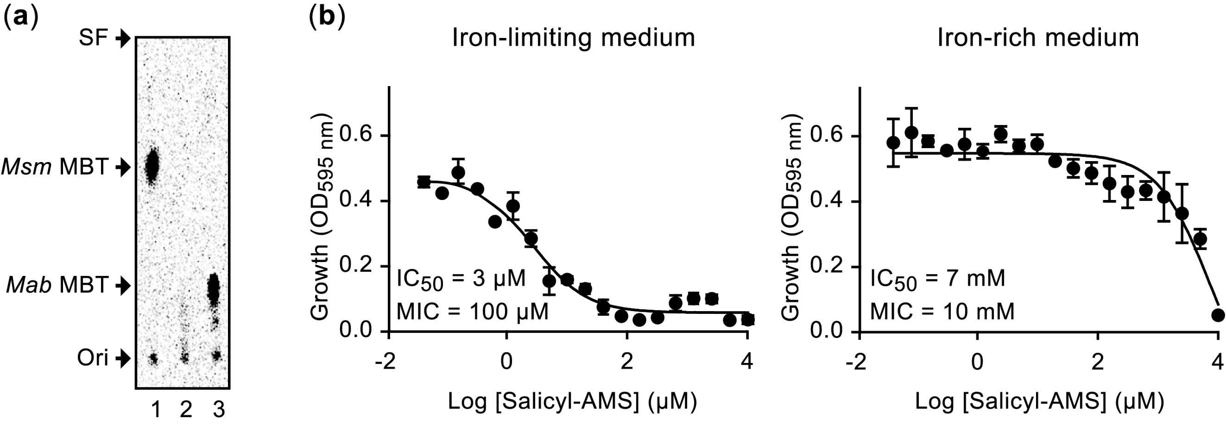
2.7. Mass Spectrometry Analysis of M. abscessus Mycobactin

To gain insight into the structural features of the presumptive MBT-type siderophore of Mab (hereafter called MBT Ab), we undertook a siderophore-targeted, LC-MS/MS-based metabolomics approach. To this end, we explored the structure of the siderophore produced by the WT strain and the mycP3 Tn mutant, which has a disruption of the gene encoding the predicted MycP (mycosin protease) component conserved in type VII secretion systems [18]. As noted above, the mutant displayed siderophore overproduction, and a robust OP phenotype on iron-rich s7H11 plates and iron-limiting broth readily complemented by a WT copy of the gene (Figure 4 and Figure S5). We grew both strains in iron-limiting broth to upregulate siderophore production, and processed cell pellets and spent culture supernatants for isolation of MBT-type siderophores. The samples were then subjected to high-resolution LC-MS/MS analysis using an iron isotope-assisted screen for iron-containing metabolites in the mass range expected for MBT/cMBT siderophores. LC-MS conditions were initially guided by protocols we previously developed for the siderophore of Msm [43], and then iteratively optimized for presumptive siderophores in Mab samples. Commercially available MBT J was used as a structural reference in the LC-MS/MS studies. Our explorative pilot experiments (not shown) revealed similar presumptive siderophore ions and MS/MS signatures in the WT and mycP3::Tn strains, and across samples from cell pellets and culture supernatants. The analysis showed that MBT J and the presumptive Mab siderophore exhibit similar MS/MS fragmentation patterns, while also sharing a few of the same daughter ions. The pilot experiments also demonstrated a higher total (summed) siderophore abundance in the mutant than in the WT strain, a finding consistent with the more intense OP of the esx-3 mutant (Figure 7). In view of these results, we carried out our analysis to obtain a detailed structural characterization of the Mab siderophore with the more readily available siderophore obtained from the cells of the mutant.
Based on the findings of LC-MS analysis (Figure S8), we propose the structure of the scaffold of MBT Ab shown in Figure 9. The figure also shows structural information for other mycobacterial siderophores and related compounds from Nocardia species reported in the literature [43,46,62,63]. Surprisingly, two unusual structural features differentiate MBT Ab from most previously characterized mycobacterial siderophore variants (e.g., Mtb and Msm MBTs, and MBT J) (Figure 9). First, MBT Ab does not have an acyl substituent on the Nε of the internal hydroxylysine residue. In other mycobacteria, the biosynthesis and addition of this substituent have been shown to require four genes (mbtK, mbtL, mbtM, mbtN) located in the so-called mbt-2 gene cluster [64,65,66]. Our orthology analysis (not shown) and analysis by others [66] did not reveal an mbt-2 locus orthologue in Mab. This finding is consistent with the lack of an acyl substituent on the internal hydroxylysine residue of MBT Ab. Second, MBT Ab has a long alkyl chain of variable length in the moiety predicted to be assembled by the MbtC (MAB_2120c)-MbtD (MAB_2119c) polyketide synthase system of the pathway [42,43]. MBT Ab shares structural features with a recently proposed structure of the MBT of M. marinum (MBT M [62]; Figure 9), which also lacks an acyl substituent on the internal hydroxylysine and has an alkyl chain with or without a terminal carboxylic acid functionality at the equivalent position of the variable alkyl chain of MBT Ab (Figure 9). Notably, the two salient structural features of MBT Ab noted above are common in several nocardial compounds with core scaffolds remarkably similar to those of MBTs [43] (Figure 9).
Following the structure elucidation of MBT Ab, we used the LC-MS platform to investigate siderophore abundance in cultures of the WT and mycP3::Tn strains in iron-limiting and iron-rich growth media. To this end, we analyzed aggregated (summed) data of siderophore abundance determined by ion peak integrations from extracted ion chromatograms collected for seven different MBT Ab structural variants in samples of cell pellet-associated and supernatant-associated siderophore extracts (Table S5). The results of this analysis, which provides a more reliable picture than an analysis based on any single MBT Ab variant could afford, are shown in Figure 10. In the iron-rich condition, the ESX-3 mutant displayed a 17-fold increase in total MBT abundance relative to the WT strain reference. In contrast, the ratio of culture supernatant-associated MBT to cell pellet-associated MBT did not diverge drastically between the strains (1.6-fold change). In the iron-limiting condition, both strains had increased MBT abundance relative to their respective iron-rich condition reference. The increase was more drastic for the WT strain (32-fold) than for the mutant (5-fold), which had already higher MBT abundance in the iron-rich condition than the WT reference (17-fold). Notably, while there was a relatively modest 3-fold difference in total MBT abundance between the strains in the iron-limiting medium, there was a drastic 11-fold change in the ratio of culture supernatant-associated MBT to cell pellet-associated MBT between the strains, with the mutant having a significantly larger proportion of cell-associated MBT. Overall, the increased abundance of MBT in the mutant is consistent with its OP phenotype. All together, these findings support the hypothesis that the mutant overproduces MBT due to an inability to secure a suitable iron supply in the presence of a malfunctioning ESX-3 with an impaired capacity for utilization of MBT-bound iron in both iron-limiting and iron-rich media.

3. Discussion

ESX-3 is essential for Mtb and M. bovis BCG growth in standard MB growth media, a property of ESX-3 thought to be due to its requirement for utilization of MBT/cMBT-bound iron by a yet unknown mechanism. Notably, the ESX-3 essentiality seen in these slow-growing mycobacteria is not recapitulated in Mab, despite the involvement of Mab ESX-3 in iron homeostasis, as determined by this study. Our findings demonstrate a functional link between the ESX-3 and siderophore systems of Mab. We found that Mab Tn mutants with insertions in the esx-3 locus have an OP phenotype reminiscent of the pigmentation observed in the Mtb ESX-3 mutant. Based on our results and by analogy to the observation made for Mtb, we attribute the pigmentation of the Mab mutants to an abnormally high accumulation of MBT–Fe3+ complex. We hypothesize that a dysfunctional ESX-3 in the Mab mutants impairs the utilization of MBT-bound iron, a defect that, in turn, leads to a constant intracellular iron-deficiency state accompanied by de-repression of IdeR-regulated MBT biosynthesis genes and excessive accumulation of the MBT–Fe3+ complex. Our laboratory hopes to explore the experimental validation of this hypothesized model in the future.
Our results show that the impact of a compromised ESX-3 on the iron uptake capability of the Mab mutants is not severe enough to prevent growth under iron-limiting or iron-rich conditions. This finding underlines a difference between the iron-acquisition capabilities of Mab and the slow-growing mycobacteria noted above. Paralleling results seen with Mtb [45], however, we found that the MBT/cMBT biosynthesis inhibitor salicyl-AMS (which targets MbtA [44,45]) produces a drastic Mab growth inhibition conditional to culturing in iron-limiting medium. This finding suggests a critical need of MBT-based iron scavenging and acquisition for Mab growth under iron-limiting conditions. Our findings also suggest that the ability of the Mab ESX-3 mutants to utilize MBT-bound iron is diminished rather than abolished. There are two possible non-mutually exclusive explanations for the iron acquisition ability of the mutants. They could acquire iron using a partially functional ESX-3 or via a secondary mechanism that allows for utilization of MBT-bound iron in an ESX-3 independent manner. Future Mab studies are warranted to probe the hypothesized essentiality of mbtA and siderophore production for growth in iron-limiting media, explore the salicyl-AMS’ mechanism of antimicrobial activity, and further investigate bacterial iron acquisition.
Our serendipitous finding that the OP phenotype of the colonies of Mab ESX-3 mutants is conditional to the presence of BSA on s7H11 plates and is BSA lot-dependent was unexpected. The mechanism underlying this phenomenon remains unknown. Nevertheless, the observation that the properties of the BSA added to the growth medium can influence the behavior of mycobacterial cultures is not unprecedented [67]. Moreover, a study reporting that addition of BSA to cultures of Pseudomonas putida leads to an enhancement of siderophore accumulation in the medium provides another example of a puzzling link between BSA in the medium and siderophore production [68]. The finding that our Mab mutants do not display the OP phenotype and have WT-like growth on 7H11 plates lacking BSA suggests that the mutants are able to secure a suitable (non-limiting) iron supply (conceivably via MBT-dependent and/or MBT-independent mechanisms) in the absence of BSA. The addition of BSA to the 7H11 medium appears to compromise the ability of Mab to procure a suitable iron supply in the absence of a WT ESX-3. A tantalizing mechanistic possibility behind this phenomenon is that binding of MBT and/or the MBT–Fe3+ complex to BSA may exacerbate the malfunctioning of an already weakened iron acquisition system in the mutants. The documented binding of the enterobactin siderophore to BSA and the earlier proposal that serum albumin may act in conjunction with other proteins in serum to restrict the iron supply needed for pathogen growth provide a conceptual framework for this possibility [69].
We expected our screen of a near-saturation library with ~196,000 Tn mutants to provide the first collection of Mab mutants with gene knockouts of individual genes in the 14-kb esx-3 locus and, possibly, illuminate any genetic determinants outside the locus that are involved in ESX-3 function or transport of ESX substrates through the mycolate layer [21]. The screen rendered a collection of 54 mutants. Of the 54 mutants, 47 had an insertion in one of nine (out of eleven) genes in the esx-3 locus, two had an insertion in a hypothesized promoter region at the 5′-end of the locus, and five had an insertion distant from the locus. Interestingly, the screen did not render mutants of esxG or espG3, two genes for which, given their size, the theoretical probability of not having their Tn mutants in our library by chance is in the order of 1 × 10⁻⁵ and 1 × 10⁻¹⁵, respectively (Figure S3). Thus, our results suggest that disruption of esxG or espG3 does not lead to the OP phenotype. Functional redundancy might be a reason for the lack of esxG and espG3 mutant isolates. Mab has two esxG paralogues encoding proteins with 98% (MAB_0048) and 99% (MAB_0665) sequence identity to EsxG (Figure S7a,b). One or both of these paralogues might complement an esxG knockout. Mab appears to have only one espG3 paralogue (MAB_0147c) encoding an EspG family protein with ~22% amino acid identity to EspG3 (not shown). The low sequence identity between the two proteins is not surprising, as EspG paralogues tend to have low sequence identity (<25%) [70,71]. Perhaps MAB_0147c can complement an espG knockout to a degree sufficient to prevent the pigmentation phenotype. Interestingly, MAB_0147c is preceded by, and possibly in the same operon with, MAB_0149c and MAB_0148c, which encode a PE protein (17% identity with Pe5, Figure S7a) and a PPE protein (21% identity with Ppe4, Figure S7b), respectively. The MAB_0149c−MAB_0147c cluster is not in proximity to either the esx-3 locus or the esx-4 locus (MAB_3759c−MAB_3753c), the only two esx loci in Mab [17,72]. Of note, the MAB_0149c−MAB_0147c array is homologous with the pe-ppe-espG array of the so-called esx-4EVOL locus present in Mab subsp. bolletii and Nocardia brasiliensis [17]. The function of the genes in the MAB_0149c−MAB_0147c cluster remains unknown.
Our mutant complementation analysis showed weak partial complementation for some mutants with insertions in esx-3 locus genes. The poor complementation seen for some of these genes could be due to polar effect and/or loss of translational coupling. The latter possibility is likely in the case of the esxH mutant, which can be fully complemented by an esxG-esxH fragment. Interestingly, complementation of the Mtb esxH mutant has been reported only with the cognate esxG-esxH pair [33]. Moreover, folding of the co-expressed Mtb EsxG and EsxH proteins is coupled to formation of a stable 1:1 heterodimeric complex [28,73,74], a process likely facilitated by translational coupling. Of the five mutants with insertions outside the esx-3 locus, only MAB_2276c and MAB_4783 could be complemented, albeit partially. Prediction of operonic arrangements suggests that the weak and lack of complementation of the MAB_4783 and MAB_1912c mutants, respectively, might be caused by polar effects resulting from the Tn insertions (Table S4). Conversely, polar effect is unlikely to be responsible for the weak complementation of the MAB_2276c mutant or the lack of complementation of the MAB_4275c and MAB_4537c mutants (Table S4). Since only one Tn mutant was isolated for each of the five loci outside the esx-3 gene cluster, it is also possible that spontaneous mutations in the genome (e.g., possibly in the esx-3 locus) are ultimately responsible for the pigmentation seen in at least some of these mutants. Future experiments to further investigate genotype–phenotype associations in the mutants with weak or no complementation will be needed.
The number of Tn mutant isolates with insertions in the 14-kb esx-3 locus is lower than expected given the size of the library screened, which corresponds to an average of one Tn insertion per 26 bp. Assuming that no insertion compromises viability and that the probability of insertion is uniform across the genome, one could have predicted approximately 540 insertions in the esx-3 locus. Thus, it is somewhat surprising that the screen rendered 49 isolates with insertions in the locus (one Tn/286 bp observed average), even considering that not all insertions in the locus could be expected to produce colonies with OP phenotype. For example, some gene knockouts might not lead to the phenotype due to functional redundancies (e.g., esxG), some intragenic insertions near a gene’s 3′ end might not effectively disrupt function, and some insertions might be selected against due to generation of toxic protein truncations. A trans-complementation mechanism preventing the OP phenotype on the screen plates might have also contributed to lower the number of isolates. Mutants with an impaired ESX-3 could perhaps utilize ESX-3 substrates involved in acquisition of MBT-bound iron release into the medium by nearby colonies with a functional secretion system. The observation that the growth defect of the Mtb ESX-3 mutant can be eliminated by filtered supernatant from a culture of the WT strain or by co-culturing with the WT strain [24] provides some conceptual support for the possibility of a trans-complementation phenomenon in our screen plates.
The Tn insertion distribution pattern in the pool of isolates with insertions in the esx-3 locus was also unexpected. For example, we found four insertions in esxH (291 bp; one Tn/73 bp average) and fifteen insertions in ppe4 (1572 bp; one Tn/105 bp average), but only six insertions in eccC3 (4032 bp; one Tn/672 bp average). Interestingly, an irregular insertion pattern across the length of some genes was also evident. Most notably, the seven insertions in eccA3 (1863 bp) were exclusively distributed across the first half of the gene (~1000-bp segment; one Tn/143 bp local average), and the only three insertions in eccB3 (1566 bp) mapped to the first third of the gene (545-bp segment; one Tn/182 bp local average). The unexpected distribution of insertions in the locus is unlikely to be due to pure chance. As noted above, selection against insertions leading to production of toxic protein truncations might have contributed to biases in insertion patterns. On the other hand, the insertion pattern in some of the genes might signify the presence of functionally dispensable and functionally indispensable (sub)domains in their protein products. This is a tempting possibility for eccA3. EccA3 proteins have an N-terminal domain containing tetratricopeptide repeats (postulated to mediate interactions with ESX-3 substrates) and a C-terminal ATPase domain [75]. All the eccA3 mutants identified had insertions distributed across the N-terminal domain, perhaps suggesting that disruption of the ATPase domain does not lead to an OP phenotype. Intragenic Tn insertion patterns displaying segments with and segments without insertions that correspond to dispensable and essential protein domains, respectively, have in fact been reported [76,77].
Our structural analysis of the Mab siderophore indicates that MBT Ab is an unusual MBT variant. The siderophore has considerable similarity to the recently proposed structure MBT M from M. marinum, a slow-growing mycobacterium that causes tuberculosis-like disease in fishes and opportunistic infections in humans, most commonly leading to skin and soft tissue disease. Both MBTs have structural features uncommon in mycobacterial siderophores, but evocative of nocardial MBT-like compounds with cytotoxicity or antiproliferative activity. This finding raises the possibility of roles for MBTs Ab and M in pathogenesis unlinked to iron uptake. MBTs Ab and M lack a characteristic long-chain acyl substituent found on the internal hydroxylysine residue of other MBTs characterized thus far (e.g., Mtb and Msm MBTs). The lipid tail installed on the hydroxylysine is thought to be critical for the ability of the siderophores to interact with biological membranes and capture iron within macrophages through lipid trafficking [65,78]. The long alkyl chains in the polyketide synthase-derived hydroxy acid moieties of MBT Ab and MBT M (and related nocardial compounds) are likely the functional equivalent of the lipid tail moieties in the MBTs of Mtb, Msm, and other mycobacteria. It appears that evolution might have led to two different strategies to provide the hydrophobic tail needed for the interactions of the siderophores with lipidic environments of both bacterial and host cells.

4. Materials and Methods

4.1. Routine Culturing Conditions, Molecular Biology Techniques, and Reagents

Unless otherwise stated, Mab (type strain ATCC 19977T) and its derivatives were cultured under standard conditions in MB 7H9 broth (Difco, Becton-Dickinson and Co., Franklin Lakes, NJ, USA) supplemented with 10% ADN (5% BSA, 2% dextrose, 0.85% NaCl) and 0.05% Tween 80 (s7H9 broth), or ADN-supplemented MB 7H11 agar (s7H11) (Difco), as reported [43]. Growth curve experiments were carried out using a 96-well plate-based platform as described previously [79]. Where appropriate, hygromycin (Hyg, 1000 μg/mL) and/or zeocin (Zeo, 50 μg/mL) were added to the mycobacterial growth media. Escherichia coli strains were cultured under standard conditions in Luria–Bertani media [80]. When required, ampicillin (Amp, 100 μg/mL), kanamycin (Km, 30 μg/mL), Hyg (200 μg/mL), Zeo (50 μg/mL), and/or 5-bromo-4-chloro-3-indolyl-β-d-galactopyranoside (20–30 μg/mL) were added to the Luria–Bertani media. Unless indicated otherwise, DNA manipulations were carried out using established protocols and E. coli as the primary cloning host [80]. PCR-generated DNA fragments used in plasmid constructions were sequenced to verify fidelity (Genewiz, Azenta Life Sciences, Chelmsford, MA, USA). Mab genomic DNA isolation, plasmid electroporation into Mab, and selection of Mab transformants were carried out following standard protocols [43,44,81]. Unless otherwise stated, reagents were purchased from Sigma-Aldrich Inc. (St. Louis, MO, USA), Thermo Fisher Scientific Inc. (Branchburg, NJ, USA), New England Biolabs Inc. (Ipswich, MA, USA), Qiagen LLC. (Germantown, MD, USA), or VWR International, LLC (Radnor, PA, USA). Information on oligonucleotides, mycobacterial gene expression plasmids, and mycobacterial strains used in this study is provided in Tables S1–S3, respectively.

4.2. Construction of Plasmid pMOD3Zeo and Preparation of EZ-Tn5-Derived Transposome

The EZ-Tn5-carrying plasmid pMOD-3 from the EZ-Tn5 Custom Transposome Construction Kit (Epicentre Biotechnologies Corp., Madison, WI, USA) was modified by introducing a Zeo resistance (ZeoR) selectable marker for Mab [81]. To this end, the promoter-ZeoR gene (sh ble) fragment from plasmid pMSG360Zeo [82] was amplified with primers PM57 and PM56. The latter primer introduced the rrnB-T1 transcription terminator [83] at the 3′ end of the promotersh ble cassette. The resulting amplicon was cloned into vector pCR-2.1-TOPO (TOPO TA Cloning Kit, Thermo Fisher Scientific Inc.). The promotersh ble─terminator cassette insert in the pCR-2.1-TOPO construct was subsequently recovered as a 727-bp KpnI-PstI fragment and subcloned into pMOD-3 digested with KpnI and PstI. The resulting plasmid, named pMOD-3Zeo, carried a Tn5 transposon derivative (hereafter referred to as Tn5Zeo) with a ZeoR marker. Plasmid pMOD-3Zeo was used as a source of Tn5Zeo for the generation of the transposome complex in vitro, following recommended protocols (EZ-Tn5 Transposome Kit, Epicentre Biotechnologies Technologies Corp.).

4.3. Generation and Screening of Mutant Libraries

Transposome electroporations into Mab were carried out as noted above for plasmids, except for the addition of a glycine treatment during the preparation of electrocompetent cells [84]. Prior to library screening, the number of Tn mutants per milliliter resulting from the electroporations was determined via titration on standard s7H11 plates containing Zeo. The titers obtained guided subsequent bacterial plating for library screening on the same medium or s7H11 with an optimized BSA concentration (s7H11*) as noted in the results section and at a frequency of 100–125 colonies per plate (standard Petri dish size). Plates were incubated at 37 °C and visually screened for colonies with OP phenotype on days 4, 5, and 6. Colonies with OP were recovered from the plates and subjected to colony purification (two rounds of streak plating for colony isolation) and phenotype confirmation. Glycerol stocks (s7H9 containing 25% glycerol) were prepared for each confirmed isolate and stored at −80 °C until needed.

4.4. Insertion Site Determination and Southern Blot Hybridization Analysis

Cloning of Tn-containing genomic fragments by plasmid rescue in E. coli EC100D-pir+ (Epicentre Bioechnologies Corp.) and Tn-genome junction sequencing were carried out as reported [54], except for the use of Tn5Zeo-specific sequencing primers (primers GB27 and GB28). The genomic sequences identified were mapped onto the Mab ATCC 19977T genome (chromosome: GenBank CU458896.1; plasmid pMAB23: GenBank CU458745.1). Southern blot analysis was performed using standard methods described elsewhere [54], except for the use of a Tn5Zeo-specific DNA hybridization probe. The probe was a DIG-labeled DNA fragment (751-bp) generated by PCR amplification (primers GG3 and ME-Plus9-FWD) from pMOD-3Zeo using a PCR digoxigenin-labeled probe synthesis kit (F. Hoffmann-La Roche, Ltd., Nutley, NJ, USA) according to the manufacturer’s instructions.

4.5. Construction of Mycobacterial Gene Expression Plasmids

To construct pML1335-Pmyc1tetO-eccB3, a PCR-generated fragment containing eccB3 (primers MF88 and MF78) and a PCR-generated fragment containing the TetR-responsive promoter Pmyc1tetO (primers MF86 and MF87) from plasmid pSE100 [85] were independently cloned into pCR-2.1-TOPO using E. coli TetR (strain DH5α carrying the TetR-expressing plasmid pACBB-TetR-LVA [86]) as the cloning host. The eccB3 and Pmyc1tetO inserts in the pCR-2.1-TOPO constructs were subsequently recovered as AflII-NotI and BspHI-AflII excerpts, respectively, and the two excerpts were subcloned into the BspHI/NotI-linearized pML1335 vector backbone [87] using E. coli TetR as the cloning host. The construction placed eccB3 under the control of the Pmyc1tetO promoter [85]. To create pML1335-Pmyc1tetO-eccA3, a PCR-generated fragment containing eccA3 (primers MF79 and MF80) was digested with AflII and SbfI, and then cloned into the pML1335-Pmyc1tetO vector backbone (obtained by AflII-SbfI digestion of pML1335-Pmyc1tetO-eccB3) using E. coli TetR as the cloning host. The construction placed eccA3 under the control of the Pmyc1tetO promoter. To generate plasmids pML1335-Pmyc1tetO-eccC3, -esxH, -mycP3 and -MAB_2276c+, four PCR-generated fragments each containing a specific gene (eccC3: primers MF75 and MF76; esxH: primers MF73 and MF74; mycP3: primers KL45 and KL46; and MAB_2276c+: primers MF81 and MF82) were independently cloned into pCR-2.1-TOPO. Then, the eccC3, esxH, mycP3, and MAB_2276c+ inserts of the respective pCR-2.1-TOPO constructs were recovered as AflII-NotI fragments and independently cloned into the pML1335-Pmyc1tetO vector backbone (obtained by AflII-NotI digestion of pML1335-Pmyc1tetO-eccB3) using E. coli TetR as the cloning host. The constructions placed each Mab gene under the control of the Pmyc1tetO promoter. To obtain plasmids pML1335-WCB2-eccD3, -eccE3, -pe5, -ppe4, -esxGH, -MAB_1912c, -MAB_4275c, -MAB_4537c and -MAB_4783, nine PCR-generated fragments each containing a specific gene (eccD3: primers MF7 and MF60; eccE3: primers MF3b and MF56; pe5: primers MF17 and MF62; ppe4: primers MF15 and MF51; esxGH: primers MF13 and MF50; MAB_1912c: primers MF44b and MF54; MAB_4275c: primers MF64 and MF65; MAB_4537c: primers MF46 and MF55; and MAB_4783: primers MF66 and MF67) were independently cloned into pCR-2.1-TOPO. Then, the insert of each pCR-2.1-TOPO construct was recovered as a PsiI-MfeI fragment and independently cloned into the pML1335-WCB2 vector backbone (obtained by PsiI-MfeI digestion of pML1335-WCB2 [54]). The constructions placed each Mab gene under the control of the strong constitutive synthetic mycobacterial promoter MOP [88].

4.6. Sequence Bioinformatics

Potential IdeR binding sites were identified by querying the Mab genome with the IdeR binding site consensus (a.k.a. iron box, TWAGGTWAGSCTWACCTWA; where W = A/T and S = G/C) [57,58]. Potential Zur binding sites were identified by querying selected promoter regions in the chromosome of Mab or Msm (strain MC2 155; Genbank: NC_008596.1) with the Zur binding site consensus (SNTRWYGAWAAYMRTKKYCRWYADNV; where S = G/C, R = A/G, W = A/T, Y = C/T, M = A/C, K = T/G, D = A/G/T, V = A/C/G, and N = any base) [27,56]. The computational queries were done using the Virtual Footprint tool of the Prokaryotic Database of Gene Regulation (http://www.prodoric.de, accessed on 17 August 2022) [89]. The searches were set to identify sequences with up to five mismatches relative to the consensus, a binding site search criterion used with other mycobacteria [57,60]. Predicted -10 and -35 promoter sequences were identified using the BPROM promoter prediction tool of the Softberry application package (www.softberry.com, accessed on 17 August 2022; Softberry, Inc., Mount Kisco, NY, USA). Potential orthologues of Mab genes in Mtb (strain H37Rv, Genbank: NC_000962.3), Msm, and M. avium subsp. paratuberculosis (strain K-10, Genbank NC_002944.2) were identified using the orthologue database OrtholugeDB (http://www.pathogenomics.sfu.ca/ortholugedb, accessed on 17 August 2022) [90] or the reciprocal BLAST feature embedded in the GVIEW Server (https://server.gview.ca/, accessed on 17 August 2022) with expected cut-off, alignment length cut-off, and percent identity cut-off of 1 × 10⁻¹⁰, 100, and 60, respectively. Routine sequence alignments were performed with Clustal W embedded in the MegAlign module of the DNASTAR Lasergene software package (DNASTAR, Inc., Madison, WI, USA).

4.7. Growth Inhibition Assay

Dose−response experiments were carried out using a microdilution assay comparable to those we have reported [44,45,91]. Briefly, mid-log phase cultures in s7H9 medium were spun down and resuspended in the iron-limiting GAST medium or in GAST supplemented with 200 µM FeCl3 (GAST+Fe) [44,45,91]. The resulting cell suspensions were used to start multi-well plate microcultures (200 μL/well) at an initial optical density at 595 nm (OD595) of 0.001. The MBT/cMBT biosynthesis inhibitor salicyl-AMS (obtained as reported [92]) was evaluated using a 2-fold dilution series covering a 0.004−10,000 μM range. The inhibitor was added from a 10% DMSO stock solution. The final DMSO concentration in inhibitor-treated cultures and DMSO controls (no inhibitor) was 0.5%. Growth was assessed as OD595 after four days of incubation (37 °C, 170 rpm) using a DTX 880 multimode detector microplate reader (Beckman Coulter, Inc., Brea, CA, USA). Dose–response data were analyzed with Prism v6.01 (GraphPad Software, Inc., San Diego, CA, USA).

4.8. Determination of Mycobactin Production by Spectrophotometric and Radiometirc Assays

Radio-thin layer chromatography (TLC) analysis of radiolabeled MBT was carried out as reported [43,44,45]. Briefly, 14C-labeled MBT was obtained by feeding the MBT-specific radiotracer [14C]salicylic acid (sp. Act. = 55 µCi/µmol; American Radiolabeled Chemicals, Inc., St. Louis, MO, USA) to cultures in iron-limiting GAST medium, the labeled MBT was extracted from the cultures with organic solvents, and the extracts were subjected to radio-TLC analysis on silica gel plates. Developed TLC plates were exposed to phosphor screens, which were subsequently scanned using a Cyclone Plus Storage Phosphor system (Perkin-Elmer Life and Analytical Sciences, Inc., Boston, MA, USA). Presence of the MBT–Fe3+ complex in culture supernatants was evaluated using a spectrophotometric assay measuring the characteristic absorbance of the complex at 450 nm [93]. Absorbance determinations were done using the plate reader noted above.

4.9. Mass Spectrometry Analysis of Mycobactins

Cultures of Mab WT and the mutant M22mycP3 (5 mL, 220 rpm, 50 mL culture tubes) in iron-limiting GASTD medium or in GASTD supplemented with 100 µM FeCl3 (GASTD+Fe) [44,45,91] were grown to saturation, normalized to an OD600 of 1.4, and centrifuged to obtain cell pellets and spent culture supernatants. MBT siderophores associated with pellets and supernatants were extracted into organic solvents using established protocols reported for isolation of MBT and cMBT siderophores [43,45]. After extraction, the solvent was evaporated and the remaining residue was lyophilized to dryness. The dried siderophore extracts were analyzed by high-resolution liquid chromatography-mass spectrometry (LC-MS). LC-MS was performed on an Agilent 6550 iFunnel Q-TOF mass spectrometer coupled to an Agilent 1290 Infinity LC system (including a binary pump, diode array detector, and autosampler). Data were analyzed using Agilent’s MassHunter Qualitative Software (version B.06.00; Agilent Technologies, Lexington, MA, USA). Chromatography was performed using an Agilent Poroshell 120 SB-C18 column (2.7 μm, 2.1 × 50 mm) at 45 °C and a gradient of solvents A (water, 0.1% formic acid) and B (acetonitrile, 0.1% formic acid) from 5–95% solvent B or 50–100% solvent B in 10 min at a flow rate of 0.4 mL/min. Stock solutions of the siderophore samples were prepared by dissolving the extracts in acetonitrile and routinely stored at −80 °C until needed. Small aliquots of these stocks were diluted 100× and 5 μL of the diluted samples were injected for analysis. The following settings were applied to the electrospray ionization source: gas temperature, 250 °C; nebulizer, 30 psig; sheath gas temperature, 250 °C; vcap, 3500 V; and nozzle voltage, 2000 V. For MS analysis, full scan mass spectra (m/z = 100–1500) were acquired in positive-ion mode. For targeted MS/MS analyses, collision energies (CE) of 10–70 were evaluated. Compounds of interest were identified by both assessing UV chromatograms at 450 nm, and mass spectra exhibiting characteristic Fe isotopic distribution. The commercially available MBT J from Mycobacterium avium subsp. paratuberculosis [46] (Allied Monitor, Inc., Fayette, MO, USA) was analyzed by MS/MS, and its fragmentation pattern was used as a template to help determine potential structures of the siderophore analogues found in the extracts.

5. Conclusions

Overall, our studies provide further insight into the ESX-3 and siderophore systems of Mab and expand our knowledge of the biology of this recalcitrant pulmonary pathogen. The findings of this work highlight the need for further research to better understand the functional dimensions of ESX-3 and its interplay with the MBT Ab-mediated iron acquisition system. The collection of novel mutants generated in this study will facilitate progress on these fronts. Mutants generated herein will also be useful to probe the relevance of individual ESX-3 components in cellular and animal infection models. The structural features of the MBT Ab revealed by our study suggest that the siderophore might have cytotoxic properties that contribute to pathogenesis. This possibility warrants exploration and may open new lines of experimental inquiry into the pathogenesis of Mab.

Supplementary Materials

The following supporting information can be downloaded at: https://www.mdpi.com/article/10.3390/pathogens11090953/s1, Figure S1: Southern blot analysis of M. abscessus mutants; Figure S2: Effect of BSA on the development of the orange pigmentation phenotype in M. abscessus mutants; Figure S3: Probability (p) of missing a gene with “x” base pairs (bp) in a library of “n” transposon (Tn) mutants of M. abscessus; Figure S4: Complementation of the esxH mutant; Figure S5: The orange pigmentation phenotype of M. abscessus cultures is influenced by iron availability; Figure S6: Growth of M. abscessus strains in iron-rich and iron-limiting media; Figure S7: Substrate candidates for the secretome of the ESX systems of M. abscessus; Figure S8: Representative structures and fragmentation patterns of mycobactin Ab (a) and mycobactin J (b); Table S1: Primers used in the study; Table S2: Mycobacterial gene expression plasmids used in this study; Table S3: M. abscessus strains included in phenotypic characterizations; Table S4: Genetic loci outside the esx-3 cluster with transposon insertions; Table S5: Ion peak integration data collected from extracted ion chromatograms for seven different MBT Ab structural variants in samples of supernatant-associated (SA) and cell pellet-associated (CPA) siderophore extracts from cultures in the iron-limiting or iron-rich growth media.

Author Contributions

Conceptualization, L.E.N.Q.; validation, G.V.B., M.F.F., K.L., P.M., G.A.G., B.Y. and L.E.N.Q.; formal analysis, G.V.B., B.Y. and L.E.N.Q.; investigation, G.V.B., M.F.F., K.L., P.M., G.A.G., B.Y. and L.E.N.Q.; writing—original draft preparation, L.E.N.Q. and G.V.B.; writing—review and editing, G.V.B., M.F.F., K.L., P.M., G.A.G., B.Y. and L.E.N.Q.; visualization, L.E.N.Q., G.V.B. and B.Y.; supervision, L.E.N.Q.; project administration, L.E.N.Q.; funding acquisition, L.E.N.Q. All authors have read and agreed to the published version of the manuscript.

Funding

This research was supported in part by funding from the Potts Memorial Foundation Grant, Professional Staff Congress-City University of New York Award # 65408-00 53, the Weill Cornell Medical College Howard and Abby Milstein Program in Chemical Biology, and the endowment support from Carol and Larry Zicklin to L.E.N.Q., G.V.B. and K.L. were supported in part by City University of New York Doctoral Student Research Grant Program Awards. G.V.B and K.L. were also supported in part by an Alfred Alberts Award in Biochemistry and a Lila Lustig Science Scholarship, respectively. G.A.G. and M.F.F. were supported in part by Estelle and James Ramey Scholarships. G.A.G. was also supported in part by a Sandi Sulam Memorial Award.

Institutional Review Board Statement

Not applicable.

Informed Consent Statement

Not applicable.

Data Availability Statement

Not applicable.

Acknowledgments

We thank Shelley Jain and Claudia Zmijewski (undergraduate research assistants, L.E.N.Q. laboratory) for helping with recombinant DNA cloning. We are grateful to Michael Niederweis (Department of Microbiology, University of Alabama at Birmingham, Birmingham, AL, United States) for providing the plasmid pML1335.

Conflicts of Interest

The authors declare no conflict of interest. The funders had no role in the design of the study; in the collection, analyses, or interpretation of data; in the writing of the manuscript; or in the decision to publish the results.

References

  1. Medjahed, H.; Gaillard, J.L.; Reyrat, J.M. Mycobacterium abscessus: A new player in the mycobacterial field. Trends Microbiol. 2010, 18, 117–123. [Google Scholar] [CrossRef] [PubMed]
  2. Mueller, P.S.; Edson, R.S. Disseminated Mycobacterium abscessus infection manifesting as fever of unknown origin and intra-abdominal lymphadenitis: Case report and literature review. Diagn. Microbiol. Infect. Dis. 2001, 39, 33–37. [Google Scholar] [CrossRef]
  3. Varghese, B.; Shajan, S.E.; Al, M.O.; Al-Hajoj, S.A. First case report of chronic pulmonary lung disease caused by Mycobacterium abscessus in two immunocompetent patients in Saudi Arabia. Ann. Saudi Med. 2012, 32, 312–314. [Google Scholar] [CrossRef] [PubMed]
  4. Petrini, B. Mycobacterium abscessus: An emerging rapid-growing potential pathogen. APMIS 2006, 114, 319–328. [Google Scholar] [CrossRef] [PubMed]
  5. Koh, W.J.; Jeon, K.; Lee, N.Y.; Kim, B.J.; Kook, Y.H.; Lee, S.H.; Park, Y.K.; Kim, C.K.; Shin, S.J.; Huitt, G.A.; et al. Clinical significance of differentiation of Mycobacterium massiliense from Mycobacterium abscessus. Am. J. Respir. Crit. Care Med. 2011, 183, 405–410. [Google Scholar] [CrossRef]
  6. Nessar, R.; Cambau, E.; Reyrat, J.M.; Murray, A.; Gicquel, B. Mycobacterium abscessus: A new antibiotic nightmare. J. Antimicrob. Chemother. 2012, 67, 810–818. [Google Scholar] [CrossRef] [PubMed]
  7. Tortoli, E.; Kohl, T.A.; Brown-Elliott, B.A.; Trovato, A.; Leao, S.C.; Garcia, M.J.; Vasireddy, S.; Turenne, C.Y.; Griffith, D.E.; Philley, J.V.; et al. Emended description of Mycobacterium abscessus, Mycobacterium abscessus subsp. abscessus and Mycobacterium abscessus subsp. bolletii and designation of Mycobacterium abscessus subsp. massiliense comb. nov. Int. J. Syst. Evol. Microbiol. 2016, 66, 4471–4479. [Google Scholar] [CrossRef] [PubMed]
  8. Ratnatunga, C.N.; Lutzky, V.P.; Kupz, A.; Doolan, D.L.; Reid, D.W.; Field, M.; Bell, S.C.; Thomson, R.M.; Miles, J.J. The rise of non-tuberculosis mycobacterial lung disease. Front. Immunol. 2020, 11, 303. [Google Scholar] [CrossRef]
  9. Daley, C.L.; Griffith, D.E. Pulmonary disease caused by rapidly growing mycobacteria. Clin. Chest Med. 2002, 23, 623–632. [Google Scholar] [CrossRef]
  10. Jeong, Y.J.; Lee, K.S.; Koh, W.J.; Han, J.; Kim, T.S.; Kwon, O.J. Nontuberculous mycobacterial pulmonary infection in immunocompetent patients: Comparison of thin-section CT and histopathologic findings. Radiology 2004, 231, 880–886. [Google Scholar] [CrossRef]
  11. Wang, C.C.; Lin, M.C.; Liu, J.W.; Wang, Y.H. Nontuberculous mycobacterial lung disease in southern Taiwan. Chang Gung Med. J. 2009, 32, 499–508. [Google Scholar] [PubMed]
  12. Redelman-Sidi, G.; Sepkowitz, K.A. Rapidly growing mycobacteria infection in patients with cancer. Clin. Infect. Dis. 2010, 51, 422–434. [Google Scholar] [CrossRef] [PubMed]
  13. Griffith, D.E.; Aksamit, T.; Brown-Elliott, B.A.; Catanzaro, A.; Daley, C.; Gordin, F.; Holland, S.M.; Horsburgh, R.; Huitt, G.; Iademarco, M.F.; et al. An official ATS/IDSA statement: Diagnosis, treatment, and prevention of nontuberculous mycobacterial diseases. Am. J. Respir. Crit. Care Med. 2007, 175, 367–416. [Google Scholar] [CrossRef] [PubMed]
  14. van Ingen, J.; Boeree, M.J.; van Soolingen, D.; Mouton, J.W. Resistance mechanisms and drug susceptibility testing of nontuberculous mycobacteria. Drug Resist. Updates 2012, 15, 149–161. [Google Scholar] [CrossRef]
  15. Bottai, D.; Serafini, A.; Cascioferro, A.; Brosch, R.; Manganelli, R. Targeting type VII/ESX secretion systems for development of novel antimycobacterial drugs. Curr. Pharm. Des. 2014, 20, 4346–4356. [Google Scholar] [CrossRef] [PubMed]
  16. Feltcher, M.E.; Sullivan, J.T.; Braunstein, M. Protein export systems of Mycobacterium tuberculosis: Novel targets for drug development? Future Microbiol. 2010, 5, 1581–1597. [Google Scholar] [CrossRef] [PubMed]
  17. Newton-Foot, M.; Warren, R.M.; Sampson, S.L.; van Helden, P.D.; Gey van Pittius, N.C. The plasmid-mediated evolution of the mycobacterial ESX (Type VII) secretion systems. BMC Evol. Biol. 2016, 16, 62. [Google Scholar] [CrossRef] [PubMed]
  18. Rivera-Calzada, A.; Famelis, N.; Llorca, O.; Geibel, S. Type VII secretion systems: Structure, functions and transport models. Nat. Rev. Microbiol. 2021, 19, 567–584. [Google Scholar] [CrossRef] [PubMed]
  19. Ates, L.S.; Houben, E.N.G.; Bitter, W. Type VII secretion: A highly versatile secretion system. Microbiol. Spectr. 2016, 4, 357–384. [Google Scholar] [CrossRef]
  20. Groschel, M.I.; Sayes, F.; Simeone, R.; Majlessi, L.; Brosch, R. ESX secretion systems: Mycobacterial evolution to counter host immunity. Nat. Rev. Microbiol. 2016, 14, 677–691. [Google Scholar] [CrossRef]
  21. Bitter, W.; Houben, E.N.; Bottai, D.; Brodin, P.; Brown, E.J.; Cox, J.S.; Derbyshire, K.; Fortune, S.M.; Gao, L.Y.; Liu, J.; et al. Systematic genetic nomenclature for type VII secretion systems. PLoS Pathog. 2009, 5, e1000507. [Google Scholar] [CrossRef] [PubMed]
  22. Serafini, A.; Pisu, D.; Palu, G.; Rodriguez, G.M.; Manganelli, R. The ESX-3 secretion system is necessary for iron and zinc homeostasis in Mycobacterium tuberculosis. PLoS ONE 2013, 8, e78351. [Google Scholar]
  23. Sassetti, C.M.; Boyd, D.H.; Rubin, E.J. Genes required for mycobacterial growth defined by high density mutagenesis. Mol. Microbiol. 2003, 48, 77–84. [Google Scholar] [CrossRef] [PubMed]
  24. Serafini, A.; Boldrin, F.; Palu, G.; Manganelli, R. Characterization of a Mycobacterium tuberculosis ESX-3 conditional mutant: Essentiality and rescue by iron and zinc. J. Bacteriol. 2009, 191, 6340–6344. [Google Scholar] [CrossRef] [PubMed]
  25. Siegrist, M.S.; Unnikrishnan, M.; McConnell, M.J.; Borowsky, M.; Cheng, T.Y.; Siddiqi, N.; Fortune, S.M.; Moody, D.B.; Rubin, E.J. Mycobacterial Esx-3 is required for mycobactin-mediated iron acquisition. Proc. Natl. Acad. Sci. USA 2009, 106, 18792–18797. [Google Scholar] [CrossRef]
  26. Siegrist, M.S.; Steigedal, M.; Ahmad, R.; Mehra, A.; Dragset, M.S.; Schuster, B.M.; Philips, J.A.; Carr, S.A.; Rubin, E.J. Mycobacterial Esx-3 requires multiple components for iron acquisition. mBio 2014, 5, e01073-14. [Google Scholar] [CrossRef]
  27. Maciag, A.; Dainese, E.; Rodriguez, G.M.; Milano, A.; Provvedi, R.; Pasca, M.R.; Smith, I.; Palu, G.; Riccardi, G.; Manganelli, R. Global analysis of the Mycobacterium tuberculosis Zur (FurB) regulon. J. Bacteriol. 2007, 189, 730–740. [Google Scholar] [CrossRef]
  28. Ilghari, D.; Lightbody, K.L.; Veverka, V.; Waters, L.C.; Muskett, F.W.; Renshaw, P.S.; Carr, M.D. Solution structure of the Mycobacterium tuberculosis EsxG.EsxH complex: Functional implications and comparisons with other M. tuberculosis Esx family complexes. J. Biol. Chem. 2011, 286, 29993–30002. [Google Scholar] [CrossRef]
  29. Sweeney, K.A.; Dao, D.N.; Goldberg, M.F.; Hsu, T.; Venkataswamy, M.M.; Henao-Tamayo, M.; Ordway, D.; Sellers, R.S.; Jain, P.; Chen, B.; et al. A recombinant Mycobacterium smegmatis induces potent bactericidal immunity against Mycobacterium tuberculosis. Nat. Med. 2011, 17, 1261–1268. [Google Scholar] [CrossRef]
  30. Billeskov, R.; Vingsbo-Lundberg, C.; Andersen, P.; Dietrich, J. Induction of CD8 T cells against a novel epitope in TB10.4: Correlation with mycobacterial virulence and the presence of a functional region of difference-1. J. Immunol. 2007, 179, 3973–3981. [Google Scholar] [CrossRef]
  31. Hervas-Stubbs, S.; Majlessi, L.; Simsova, M.; Morova, J.; Rojas, M.J.; Nouze, C.; Brodin, P.; Sebo, P.; Leclerc, C. High frequency of CD4+ T cells specific for the TB10.4 protein correlates with protection against Mycobacterium tuberculosis infection. Infect. Immun. 2006, 74, 3396–3407. [Google Scholar] [CrossRef] [PubMed]
  32. Skjot, R.L.; Brock, I.; Arend, S.M.; Munk, M.E.; Theisen, M.; Ottenhoff, T.H.; Andersen, P. Epitope mapping of the immunodominant antigen TB10.4 and the two homologous proteins TB10.3 and TB12.9, which constitute a subfamily of the esat-6 gene family. Infect. Immun. 2002, 70, 5446–5453. [Google Scholar] [CrossRef] [PubMed]
  33. Tufariello, J.M.; Chapman, J.R.; Kerantzas, C.A.; Wong, K.W.; Vilcheze, C.; Jones, C.M.; Cole, L.E.; Tinaztepe, E.; Thompson, V.; Fenyo, D.; et al. Separable roles for Mycobacterium tuberculosis ESX-3 effectors in iron acquisition and virulence. Proc. Natl. Acad. Sci. USA 2016, 113, E348–E357. [Google Scholar] [CrossRef] [PubMed]
  34. Zhang, L.; Hendrickson, R.C.; Meikle, V.; Lefkowitz, E.J.; Ioerger, T.R.; Niederweis, M. Comprehensive analysis of iron utilization by Mycobacterium tuberculosis. PLoS Pathog. 2020, 16, e1008337. [Google Scholar] [CrossRef] [PubMed]
  35. Quadri, L.E. Strategic paradigm shifts in the antimicrobial drug discovery process of the 21st century. Infect. Disord. Drug Targets 2007, 7, 230–237. [Google Scholar] [CrossRef] [PubMed]
  36. Kim, Y.S.; Yang, C.S.; Nguyen, L.T.; Kim, J.K.; Jin, H.S.; Choe, J.H.; Kim, S.Y.; Lee, H.M.; Jung, M.; Kim, J.M.; et al. Mycobacterium abscessus ESX-3 plays an important role in host inflammatory and pathological responses during infection. Microbes Infect. 2017, 19, 5–17. [Google Scholar] [CrossRef]
  37. Rifat, D.; Chen, L.; Kreiswirth, B.N.; Nuermberger, E.L. Genome-wide essentiality analysis of Mycobacterium abscessus by saturated transposon mutagenesis and deep sequencing. mBio 2021, 12, e0104921. [Google Scholar] [CrossRef]
  38. Bosne, S.; Levy-Frebault, V.V. Mycobactin analysis as an aid for the identification of Mycobacterium fortuitum and Mycobacterium chelonae subspecies. J. Clin. Microbiol. 1992, 30, 1225–1231. [Google Scholar] [CrossRef]
  39. Hall, R.M.; Ratledge, C. Mycobactins as chemotaxonomic characters for some rapidly growing mycobacteria. J. Gen. Microbiol. 1984, 130, 1883–1892. [Google Scholar] [CrossRef]
  40. Ripoll, F.; Pasek, S.; Schenowitz, C.; Dossat, C.; Barbe, V.; Rottman, M.; Macheras, E.; Heym, B.; Herrmann, J.L.; Daffe, M.; et al. Non mycobacterial virulence genes in the genome of the emerging pathogen Mycobacterium abscessus. PLoS ONE 2009, 4, e5660. [Google Scholar] [CrossRef]
  41. Quadri, L.E. Biosynthesis of mycobacterial lipids by polyketide synthases and beyond. Crit. Rev. Biochem. Mol. Biol. 2014, 49, 179–211. [Google Scholar] [CrossRef] [PubMed]
  42. Quadri, L.E.; Sello, J.; Keating, T.A.; Weinreb, P.H.; Walsh, C.T. Identification of a Mycobacterium tuberculosis gene cluster encoding the biosynthetic enzymes for assembly of the virulence-conferring siderophore mycobactin. Chem. Biol. 1998, 5, 631–645. [Google Scholar] [CrossRef]
  43. Chavadi, S.S.; Stirrett, K.L.; Edupuganti, U.R.; Sadhanandan, G.; Vergnolle, O.; Schumacher, E.; Martin, C.; Qiu, W.G.; Soll, C.E.; Quadri, L.E.N. Mutational and phylogenetic analyses of the mycobacterial mbt gene cluster. J. Bacteriol. 2011, 193, 5905–5913. [Google Scholar] [CrossRef] [PubMed]
  44. Bythrow, G.V.; Mohandas, P.; Guney, T.; Standke, L.C.; Germain, G.A.; Lu, X.; Ji, C.; Levendosky, K.; Chavadi, S.S.; Tan, D.S.; et al. Kinetic analyses of the siderophore biosynthesis inhibitor salicyl-AMS and analogues as MbtA inhibitors and antimycobacterial agents. Biochemistry 2019, 58, 833–847. [Google Scholar] [CrossRef] [PubMed]
  45. Ferreras, J.A.; Ryu, J.S.; Di Lello, F.; Tan, D.S.; Quadri, L.E. Small-molecule inhibition of siderophore biosynthesis in Mycobacterium tuberculosis and Yersinia pestis. Nat. Chem. Biol. 2005, 1, 29–32. [Google Scholar] [CrossRef] [PubMed]
  46. Ghosh, C.; Pal, S.; Patel, A.; Kapur, M. Total synthesis of the proposed structure of mycobactin J. Org. Lett. 2018, 20, 6511–6515. [Google Scholar] [CrossRef]
  47. DeJesus, M.A.; Gerrick, E.R.; Xu, W.; Park, S.W.; Long, J.E.; Boutte, C.C.; Rubin, E.J.; Schnappinger, D.; Ehrt, S.; Fortune, S.M.; et al. Comprehensive essentiality analysis of the Mycobacterium tuberculosis genome via saturating transposon mutagenesis. mBio 2017, 8, e02133-16. [Google Scholar] [CrossRef]
  48. Famelis, N.; Rivera-Calzada, A.; Degliesposti, G.; Wingender, M.; Mietrach, N.; Skehel, J.M.; Fernandez-Leiro, R.; Bottcher, B.; Schlosser, A.; Llorca, O.; et al. Architecture of the mycobacterial type VII secretion system. Nature 2019, 576, 321–325. [Google Scholar] [CrossRef]
  49. Poweleit, N.; Czudnochowski, N.; Nakagawa, R.; Trinidad, D.D.; Murphy, K.C.; Sassetti, C.M.; Rosenberg, O.S. The structure of the endogenous ESX-3 secretion system. eLife 2019, 8, e52983. [Google Scholar] [CrossRef]
  50. Jacobs, M.A.; Alwood, A.; Thaipisuttikul, I.; Spencer, D.; Haugen, E.; Ernst, S.; Will, O.; Kaul, R.; Raymond, C.; Levy, R.; et al. Comprehensive transposon mutant library of Pseudomonas aeruginosa. Proc. Natl. Acad. Sci. USA 2003, 100, 14339–14344. [Google Scholar] [CrossRef]
  51. Cameron, D.E.; Urbach, J.M.; Mekalanos, J.J. A defined transposon mutant library and its use in identifying motility genes in Vibrio cholerae. Proc. Natl. Acad. Sci. USA 2008, 105, 8736–8741. [Google Scholar] [CrossRef] [PubMed]
  52. Gallagher, L.A.; Ramage, E.; Patrapuvich, R.; Weiss, E.; Brittnacher, M.; Manoil, C. Sequence-defined transposon mutant library of Burkholderia thailandensis. mBio 2013, 4, e00604–e00613. [Google Scholar] [CrossRef] [PubMed]
  53. Gallagher, L.A.; Ramage, E.; Jacobs, M.A.; Kaul, R.; Brittnacher, M.; Manoil, C. A comprehensive transposon mutant library of Francisella novicida, a bioweapon surrogate. Proc. Natl. Acad. Sci. USA 2007, 104, 1009–1014. [Google Scholar] [CrossRef] [PubMed]
  54. Budell, W.C.; Germain, G.A.; Janisch, N.; McKie-Krisberg, Z.; Jayaprakash, A.D.; Resnick, A.E.; Quadri, L.E.N. Transposon mutagenesis in Mycobacterium kansasii links a small RNA gene to colony morphology and biofilm formation and identifies 9,885 intragenic insertions that do not compromise colony outgrowth. Microbiologyopen 2020, 9, e988. [Google Scholar] [CrossRef]
  55. Mohandas, P.; Budell, W.C.; Mueller, E.; Au, A.; Bythrow, G.V.; Quadri, L.E. Pleiotropic consequences of gene knockouts in the phthiocerol dimycocerosate and phenolic glycolipid biosynthetic gene cluster of the opportunistic human pathogen Mycobacterium marinum. FEMS Microbiol. Lett. 2016, 363, fnw016. [Google Scholar] [CrossRef]
  56. Maciag, A.; Piazza, A.; Riccardi, G.; Milano, A. Transcriptional analysis of ESAT-6 cluster 3 in Mycobacterium smegmatis. BMC Microbiol. 2009, 9, 48. [Google Scholar] [CrossRef]
  57. Gold, B.; Rodriguez, G.M.; Marras, S.A.; Pentecost, M.; Smith, I. The Mycobacterium tuberculosis IdeR is a dual functional regulator that controls transcription of genes involved in iron acquisition, iron storage and survival in macrophages. Mol. Microbiol. 2001, 42, 851–865. [Google Scholar] [CrossRef]
  58. Rodriguez, G.M.; Voskuil, M.I.; Gold, B.; Schoolnik, G.K.; Smith, I. ideR, an essential gene in Mycobacterium tuberculosis: Role of IdeR in iron-dependent gene expression, iron metabolism, and oxidative stress response. Infect. Immun. 2002, 70, 3371–3381. [Google Scholar] [CrossRef]
  59. Yellaboina, S.; Ranjan, S.; Vindal, V.; Ranjan, A. Comparative analysis of iron regulated genes in mycobacteria. FEBS Lett. 2006, 580, 2567–2576. [Google Scholar] [CrossRef]
  60. Janagama, H.K.; Senthilkumar, T.M.A.; Bannantine, J.P.; Rodriguez, G.M.; Smith, I.; Paustian, M.L.; McGarvey, J.A.; Sreevatsan, S. Identification and functional characterization of the iron-dependent regulator (IdeR) of Mycobacterium avium subsp. paratuberculosis. Microbiology 2009, 155, 3683–3690. [Google Scholar] [CrossRef]
  61. Prakash, P.; Yellaboina, S.; Ranjan, A.; Hasnain, S.E. Computational prediction and experimental verification of novel IdeR binding sites in the upstream sequences of Mycobacterium tuberculosis open reading frames. Bioinformatics 2005, 21, 2161–2166. [Google Scholar] [CrossRef] [PubMed]
  62. Knobloch, P.; Koliwer-Brandl, H.; Arnold, F.M.; Hanna, N.; Gonda, I.; Adenau, S.; Personnic, N.; Barisch, C.; Seeger, M.A.; Soldati, T.; et al. Mycobacterium marinum produces distinct mycobactin and carboxymycobactin siderophores to promote growth in broth and phagocytes. Cell. Microbiol. 2020, 22, e13163. [Google Scholar] [CrossRef] [PubMed]
  63. De Voss, J.J.; Rutter, K.; Schroeder, B.G.; Barry, C.E. Iron acquisition and metabolism by mycobacteria. J. Bacteriol. 1999, 181, 4443–4451. [Google Scholar] [CrossRef] [PubMed]
  64. Krithika, R.; Marathe, U.; Saxena, P.; Ansari, M.Z.; Mohanty, D.; Gokhale, R.S. A genetic locus required for iron acquisition in Mycobacterium tuberculosis. Proc. Natl. Acad. Sci. USA 2006, 103, 2069–2074. [Google Scholar] [CrossRef] [PubMed]
  65. Madigan, C.A.; Martinot, A.J.; Wei, J.R.; Madduri, A.; Cheng, T.Y.; Young, D.C.; Layre, E.; Murry, J.P.; Rubin, E.J.; Moody, D.B. Lipidomic analysis links mycobactin synthase K to iron uptake and virulence in M. tuberculosis. PLoS Pathog. 2015, 11, e1004792. [Google Scholar] [CrossRef]
  66. Li, W.; He, J.; Xie, L.; Chen, T.; Xie, J. Comparative genomic insights into the biosynthesis and regulation of mycobacterial siderophores. Cell. Physiol. Biochem. 2013, 31, 1–13. [Google Scholar] [CrossRef] [PubMed]
  67. Gold, B.; Warrier, T.; Nathan, C. A multi-stress model for high throughput screening against non-replicating Mycobacterium tuberculosis. Methods Mol. Biol. 2015, 1285, 293–315. [Google Scholar]
  68. Joshi, H.; Dave, R.; Venugopalan, V.P. Protein as chemical cue: Non-nutritional growth enhancement by exogenous protein in Pseudomonas putida KT2440. PLoS ONE 2014, 9, e103730. [Google Scholar]
  69. Konopka, K.; Neilands, J.B. Effect of serum albumin on siderophore-mediated utilization of transferrin iron. Biochemistry 1984, 23, 2122–2127. [Google Scholar] [CrossRef]
  70. Ekiert, D.C.; Cox, J.S. Structure of a PE-PPE-EspG complex from Mycobacterium tuberculosis reveals molecular specificity of ESX protein secretion. Proc. Natl. Acad. Sci. USA 2014, 111, 14758–14763. [Google Scholar] [CrossRef]
  71. Tuukkanen, A.T.; Freire, D.; Chan, S.; Arbing, M.A.; Reed, R.W.; Evans, T.J.; Zenkeviciute, G.; Kim, J.; Kahng, S.; Sawaya, M.R.; et al. Structural variability of EspG chaperones from mycobacterial ESX-1, ESX-3, and ESX-5 type VII secretion systems. J. Mol. Biol. 2019, 431, 289–307. [Google Scholar] [CrossRef] [PubMed]
  72. Dumas, E.; Christina Boritsch, E.; Vandenbogaert, M.; Rodriguez de la Vega, R.C.; Thiberge, J.M.; Caro, V.; Gaillard, J.L.; Heym, B.; Girard-Misguich, F.; Brosch, R.; et al. Mycobacterial pan-genome analysis suggests important role of plasmids in the radiation of type VII secretion systems. Genome Biol. Evol. 2016, 8, 387–402. [Google Scholar] [CrossRef] [PubMed]
  73. Lightbody, K.L.; Ilghari, D.; Waters, L.C.; Carey, G.; Bailey, M.A.; Williamson, R.A.; Renshaw, P.S.; Carr, M.D. Molecular features governing the stability and specificity of functional complex formation by Mycobacterium tuberculosis CFP-10/ESAT-6 family proteins. J. Biol. Chem. 2008, 283, 17681–17690. [Google Scholar] [CrossRef]
  74. Renshaw, P.S.; Lightbody, K.L.; Veverka, V.; Muskett, F.W.; Kelly, G.; Frenkiel, T.A.; Gordon, S.V.; Hewinson, R.G.; Burke, B.; Norman, J.; et al. Structure and function of the complex formed by the tuberculosis virulence factors CFP-10 and ESAT-6. EMBO J. 2005, 24, 2491–2498. [Google Scholar] [CrossRef] [PubMed]
  75. Gaur, A.; Sharma, V.K.; Shree, S.; Rai, N.; Ramachandran, R. Characterization of EccA3, a CbbX family ATPase from the ESX-3 secretion pathway of M. tuberculosis. Biochim. Biophys. Acta Proteins Proteom. 2017, 1865, 715–724. [Google Scholar] [CrossRef]
  76. DeJesus, M.A.; Zhang, Y.J.; Sassetti, C.M.; Rubin, E.J.; Sacchettini, J.C.; Ioerger, T.R. Bayesian analysis of gene essentiality based on sequencing of transposon insertion libraries. Bioinformatics 2013, 29, 695–703. [Google Scholar] [CrossRef]
  77. Chao, M.C.; Pritchard, J.R.; Zhang, Y.J.; Rubin, E.J.; Livny, J.; Davis, B.M.; Waldor, M.K. High-resolution definition of the Vibrio cholerae essential gene set with Hidden Markov Model-based analyses of transposon-insertion sequencing data. Nucleic Acids Res. 2013, 41, 9033–9048. [Google Scholar] [CrossRef]
  78. Luo, M.; Fadeev, E.A.; Groves, J.T. Mycobactin-mediated iron acquisition within macrophages. Nat. Chem. Biol. 2005, 1, 149–153. [Google Scholar] [CrossRef]
  79. Chavadi, S.S.; Edupuganti, U.R.; Vergnolle, O.; Fatima, I.; Singh, S.M.; Soll, C.E.; Quadri, L.E. Inactivation of tesA reduces cell-wall lipid production and increases drug susceptibility in mycobacteria. J. Biol. Chem. 2011, 286, 24616–24625. [Google Scholar] [CrossRef]
  80. Sambrook, J.; Russell, D.W. Molecular Cloning: A Laboratory Manual, 3rd ed.; Cold Spring Harbor Pres: Cold Spring Harbor, NY, USA, 2001. [Google Scholar]
  81. Medjahed, H.; Singh, A.K. Genetic manipulation of Mycobacterium abscessus. In Current Protocols in Microbiology; John Wiley & Sons, Inc.: New York, NY, USA, 2010. [Google Scholar]
  82. Barkan, D.; Stallings, C.L.; Glickman, M.S. An improved counterselectable marker system for mycobacterial recombination using galK and 2-deoxy-galactose. Gene 2011, 470, 31–36. [Google Scholar] [CrossRef]
  83. Orosz, A.; Boros, I.; Venetianer, P. Analysis of the complex transcription termination region of the Escherichia coli rrnB gene. Eur. J. Biochem. 1991, 201, 653–659. [Google Scholar] [CrossRef] [PubMed]
  84. Goude, R.; Roberts, D.M.; Parish, T. Electroporation of mycobacteria. Methods. Mol. Biol. 2015, 1285, 117–130. [Google Scholar] [PubMed]
  85. Guo, X.V.; Monteleone, M.; Klotzsche, M.; Kamionka, A.; Hillen, W.; Braunstein, M.; Ehrt, S.; Schnappinger, D. Silencing Mycobacterium smegmatis by using tetracycline repressors. J. Bacteriol. 2007, 189, 4614–4623. [Google Scholar] [CrossRef]
  86. Vick, J.E.; Johnson, E.T.; Choudhary, S.; Bloch, S.E.; Lopez-Gallego, F.; Srivastava, P.; Tikh, I.B.; Wawrzyn, G.T.; Schmidt-Dannert, C. Optimized compatible set of BioBrick vectors for metabolic pathway engineering. Appl. Microbiol. Biotechnol. 2011, 92, 1275–1286. [Google Scholar] [CrossRef] [PubMed]
  87. Huff, J.; Czyz, A.; Landick, R.; Niederweis, M. Taking phage integration to the next level as a genetic tool for mycobacteria. Gene 2010, 468, 8–19. [Google Scholar] [CrossRef] [PubMed]
  88. George, K.M.; Yuan, Y.; Sherman, D.R.; Barry, C.E. The biosynthesis of cyclopropanated mycolic acids in Mycobacterium tuberculosis. Identification and functional analysis of CMAS-2. J. Biol. Chem. 1995, 270, 27292–27298. [Google Scholar] [CrossRef] [PubMed]
  89. Munch, R.; Hiller, K.; Grote, A.; Scheer, M.; Klein, J.; Schobert, M.; Jahn, D. Virtual Footprint and PRODORIC: An integrative framework for regulon prediction in prokaryotes. Bioinformatics 2005, 21, 4187–4189. [Google Scholar] [CrossRef]
  90. Whiteside, M.D.; Winsor, G.L.; Laird, M.R.; Brinkman, F.S. OrtholugeDB: A bacterial and archaeal orthology resource for improved comparative genomic analysis. Nucleic Acids Res. 2013, 41, D366–D376. [Google Scholar] [CrossRef]
  91. Ferreras, J.A.; Gupta, A.; Amin, N.D.; Basu, A.; Sinha, B.N.; Worgall, S.; Jayaprakash, V.; Quadri, L.E. Chemical scaffolds with structural similarities to siderophores of nonribosomal peptide-polyketide origin as novel antimicrobials against Mycobacterium tuberculosis and Yersinia pestis. Bioorgan. Med. Chem. Lett. 2011, 21, 6533–6537. [Google Scholar] [CrossRef]
  92. Lun, S.; Guo, H.; Adamson, J.; Cisar, J.S.; Davis, T.D.; Chavadi, S.S.; Warren, J.D.; Quadri, L.E.; Tan, D.S.; Bishai, W.R. Pharmacokinetic and in vivo efficacy studies of the mycobactin biosynthesis inhibitor salicyl-AMS in mice. Antimicrob. Agents Chemother. 2013, 57, 5138–5140. [Google Scholar] [CrossRef]
  93. Gobin, J.; Moore, C.H.; Reeve, J.R.; Wong, D.K.; Gibson, B.W.; Horwitz, M.A. Iron acquisition by Mycobacterium tuberculosis: Isolation and characterization of a family of iron-binding exochelins. Proc. Natl. Acad. Sci. USA 1995, 92, 5189–5193. [Google Scholar] [CrossRef] [PubMed]
Figure 1. Radio-thin layer chromatography probe for presumptive mycobactin (MBT)−type siderophore in M. abscessus and antimicrobial activity of salicyl-AMS. (a) Thin layer chromatography (TLC) analysis. Lanes 1, 2, and 3 are samples from M. smegmatis (Msm), Msm ΔmbtA (MBT deficient mutant), and M. abscessus (Mab), respectively. The image shows the entire TLC plate. Solvent system: petroleum ether–n–butanol–ethyl acetate (2:3:3). Ori, origin; SF, solvent front. (b) Dose–response curves showing susceptibility of Mab to salicyl-AMS in GAST (iron-limiting) and GAST+Fe (iron-rich) media. The data represent means and standard deviations of triplicate cultures.
Pathogens 11 00953 g001
Figure 2. Plate of the pilot screen with the colony of isolate M1. The inset (labeled M1) shows the enlarged image of the plate section containing the colony of M1 (5.4× magnification). An image (labeled J) of an aliquot of ferric MBT J solution spotted on filter paper is shown to the right of the inset.
Pathogens 11 00953 g002
Figure 3. The ESX-3 system and its genetic dissection. (a) Schematic representation of the esx-3 locus of M. abscessus (Mab) and its transposon (Tn) mutants with OP. Genes are depicted as arrows labeled according to the single letter or number key shown below the gene cluster diagram. The number of insertion mutants isolated for each gene is given in parentheses. The 5′ and 3′ chromosomal coordinates of the genomic segment depicted are indicated. Tn insertions with the zeoR gene in the same and opposite orientations relative to the Mab genes are marked above and below the locus, respectively. The Tn mutants marked with an asterisk (*) are those displayed in Figure 4. The location of a predicted iron box (IB) sequence (shown in Figure 5) for IdeR binding upstream eccA3 is represented. Gene names noted for the predicted canonical ESX-3 substrates (pe5, ppe4, esxG, and esxH) and the conserved system components involved in substrate secretion (eccA3-eccD3, espG3, and mycP) are as per standard esx gene nomenclature [21]. Locus tags for the Mab genes (MAB_) and their orthologues in M. tuberculosis (Mtb, Rv_) and M. smegmatis (Msm, MSMEG_) are shown. Mab esx-3 locus genes hit by the Tn in our collection of mutants are marked with a filled diamond symbol (♦). Mtb and Msm genes for which individual knockouts have been reported are marked with an open diamond symbol (◊) [26,33]. The gene pairs boxed represent reported double-gene deletions [26,33]. § Mtb genes listed are essential for growth on iron-rich 7H10 agar (except for esxG and esxH, for which mutation leads to a growth defect) by analysis of Himar1 Tn libraries [47]. (b) Assemblage model of ESX-3 components and substrates encoded in the archetypal esx-3 locus. Two independent cryo-electron microscopy investigations of the Msm ESX-3 secretion complex have determined similar structures, composed of two dimerized core complexes each composed of one EccE, EccB, EccC, and two EccD components, in an organization simplified in the model depicted here. This ESX-3 dimer further trimerizes into a final hexamer structure similar to that seen for ESX-1 and ESX-5 complexes, forming the channel across which ESX-3 substrate components can be translocated. The fifth conserved ESX-3 membrane component, MycP, is not tightly associated with this described core secretion complex [48,49]. The yellow rounded rectangles (labeled with question marks) in the mycobacterial outer membrane layer (OM) signify the potential existence of protein(s) involved in facilitating the passage of ESX-3 substrates through the mycomembrane (adapted from [21,48,49]). AG, arabinogalactan layer; PG, peptidoglycan layer; CM, cytoplasmic membrane.
Pathogens 11 00953 g003
Figure 7. The orange pigmentation phenotype of M. abscessus cultures is influenced by iron availability. (a) Cultures (C), spent supernatants (S), and pellets (P) of strains grown to saturation in iron-limiting GAST broth (left panel) or iron-rich GAST+Fe broth (GAST supplemented with FeCl3 to 100 µM; right panel). After incubation for growth, cultures were treated by addition of FeCl3 (to 5 mM; right half of each panel) to allow maximal conversion of the colorless MBT into the orange MBT–Fe3+ complex, or left untreated (left half of each panel). S-GAST and S-GAST+Fe, sterile GAST and GAST+Fe broth controls, respectively. (b) Spectrophotometric quantification of MBT–Fe3+ in culture supernatants of strains grown to saturation in GAST broth, and then treated by addition of FeCl3 (to 5 mM; red bars) or left untreated (gray bars). A450, blank (sterile broth)-corrected absorbance at 450 nm. The data represent means ± SE from three cultures. The wild-type strain (WT) and M45ppe4 carried pML1335 (empty), the vector used in the genetic complementation experiments, so that they could be grown along with the complemented strain M45ppe4-C in the same antibiotic-containing medium.
Pathogens 11 00953 g007
Figure 8. Impact of transposon insertions on bacterial growth in s7H9, GAST, and GAST+Fe media. No negative impact on growth (N) and negative impact on growth (S, small; D, drastic) are highlighted in blue and red backgrounds, respectively. The information presented summarizes the growth curve results shown in Figure S6. Strains are grouped in three sections according to the functional category and location of the gene disrupted by the transposon.
Pathogens 11 00953 g008
Figure 9. Mycobactin Ab (highlighted in yellow), other mycobactins, and related nocaridal metabolites. One, oxazoline; Ole, oxazole.
Pathogens 11 00953 g009
Figure 10. Impact of ESX-3 secretion machinery impairment and extracellular iron availability on MBT Ab abundance. Cultures of the wild-type strain and the mycP3::Tn mutant were grown in iron-limiting medium or iron-rich medium (iron-limiting medium supplemented with 100 µM FeCl3). Cell pellet-associated and culture supernatant-associated siderophores were extracted and analyzed by LC-MS. The pie charts show relative siderophore abundance obtained by ion peak integrations from extracted ion chromatograms. The areas of charts for the wild-type strain in iron-limiting medium (lower left pie; 32 cm2), the mutant strain in iron-limiting medium (lower right pie; 92 cm2), and the mutant strain in iron-rich medium (upper right pie; 17 cm2) are proportional to siderophore abundance relative to the total siderophore abundance of the wild-type in iron-rich medium (upper left pie; 1 cm2). The results shown represent aggregated data of abundance collected in duplicate for the same seven different MBT Ab structural variants for each sample.
Pathogens 11 00953 g010
Table 1. Transposon insertion sites in M. abscessus isolates.
Disrupted Gene or Promoter/5′ UTR 1Mutant Isolate 2Insertion Site 3Genome Coordinates 4
eccA3
promoter
M68ATTCATGGC::Tn::ATTCATGGC2261555–2261563
M74CCCTTCACC::Tn::CCCTTCACC2261304–2261312
eccA3M20GCCACATCC::Tn::GCCACATCC2261245–2261253
M26GAGGTAGGAC::Tn::GAGGTAGGAC2260629–2260637
M49GTTCCAGACC::Tn::GTTCCAGACC2260292–2260300
M54GCCCAGGGT::Tn::GCCCAGGGT2260677–2260685
M65ACTCGAGGT::Tn::ACTCGAGGT2260934–2260942
M73GCCTCGGCC::Tn::GCCTCGGCC2260327–2260335
M86GTCGTGGGG::Tn::GTCGTGGGG2260705–2260713
eccB3M6GCCTCGCAC::Tn::GCCTCGCAC2258894–2258902
M59GGCCTGCAC::Tn::GGCCTGCAC2258929–2258937
M76GAGCACCGG::Tn::GAGCACCGG2259061–2259069
eccC3M1GATCAATACC::Tn::GATCAATACC2257252–2257260
M23CAGCTGGGA::Tn::CAGCTGGGA2256557–2256565
M52CGCCAGCGG::Tn::CGCCAGCGG2255501–2255509
M58ATGTTGGGGG::Tn::ATGTTGGGGG2254359–2254367
M79GCCCTGCAC::Tn::GCCCTGCAC2256146–2256154
M80GGCCTCGGC::Tn::GGCCTCGGC2254127–2254135
eccD3M75GCGCCAGGC::Tn::GCGCCAGGC2250160–2250168
M77GATCAGACC::Tn::GATCAGACC2249549–2249557
eccE3M5GGCCAGCAT::Tn::GGCCAGCAT2247334–2247342
M41TGCCAACGG::Tn::TGCCAACGG2246719–2246727
M62GGCTTGTCC::Tn::GGCTTGTCC2246741–2246749
mycP3M22GTCGCGCAC::Tn::GTCGCGCAC2248622–2248630
M44ACTCAGTGC::Tn::ACTCAGTGC2247566–2247574
M53GCGCAACGC::Tn::GCGCAACGC2248266–2248274
M57GGGTTTGAC::Tn::GGGTTTGAC2248632–2248640
M61GCGCAACGC::Tn::GCGCAACGC2248266–2248274
M69GGCATACAC::Tn::GGCATACAC2247626–2247634
M78GACCAATTC::Tn::GACCAATTC2248147–2248155
esxHM2AGTTGAAAG::Tn::AGTTGAAAG2251565–2251573
M70CACCGGCGT::Tn::CACCGGCGT2251538–2251546
M72GGTCAGCAC::Tn::GGTCAGCAC2251493–2251501
M84GCCAGGCGG::Tn::GCCAGGCGG2251424–2251432
ppe4M7GTCCAGCCC::Tn::GTCCAGCCC2252181–2252189
M45CTGCTGGGT::Tn::CTGCTGGGT2253289–2253297
M47GGCCAGTCC::Tn::GGCCAGTCC2251991–2251999
M51CTCCTGCAC::Tn::CTCCTGCAC2252431–2252439
M60CTCCTGCAC::Tn::CTCCTGCAC2252431–2252439
M63CTCCTGCAC::Tn::CTCCTGCAC2252431–2252439
M66TCTCCGAGG::Tn::TCTCCGAGG2252097–2252105
M67AAGCCAAGC::Tn::AAGCCAAGC2252171–2252179
M71CAGCAGCGC::Tn::CAGCAGCGC2253481–2253489
M81CCTCGTATG::Tn::CCTCGTATG2252754–2252762
M82CCACCAAGA::Tn::CCACCAAGA2252676–2252684
M85GGCGAAGCC::Tn::GGCGAAGCC2252167–2252175
M87GGGCGGCCA::Tn::GGGCGGCCA2252946–2252954
M88TGCCGAGGC::Tn::TGCCGAGGC2252329–2252337
pe5M64GCCCAGCTC::Tn::GCCCAGCTC2253630–2253638
MAB_4275c5P5GTAGCCGAA::Tn::GTAGCCGAA4348013–4348021
MAB_2276cM50GAGCATGCGC::Tn::GAGCATGCGC2324048–2324056
MAB_4537cpromoterM55CAAGGAAAT::Tn::CAAGGAAAT4620490–4620498
MAB_1912cM56TCCAGGACC::Tn::TCCAGGACC1909545–1909553
MAB_4783M83AGCGCATGT::Tn::AGCGCATGT4893827–4893835
1 UTR, untranslated region. 2 The position and orientation of the transposon in each esx-3 locus mutant are displayed in Figure 3. 3 The direct repeats resulting from the most common 9-bp or the less frequent 10-bp duplication [53] at the transposon insertion point are depicted. 4 The 5′ and 3′ chromosomal coordinates of the duplicated segment at the insertion site are noted on the right column. 5 The genes below the gray-colored row are outside the esx-3 locus.
Publisher’s Note: MDPI stays neutral with regard to jurisdictional claims in published maps and institutional affiliations.

