Communication

Genetic Diversity among Four Populations of Aedes aegypti (Diptera: Culicidae) from Honduras as Revealed by Mitochondrial DNA Cytochrome Oxidase I

1
Microbiology Research Institute, Universidad Nacional Autónoma de Honduras, Tegucigalpa 11101, Honduras
2
Unidad de vigilancia de la salud, Secretaría de Salud de Honduras, Tegucigalpa 11101, Honduras
*
Author to whom correspondence should be addressed.
Pathogens 2022, 11(6), 620; https://doi.org/10.3390/pathogens11060620
Submission received: 6 May 2022 / Revised: 23 May 2022 / Accepted: 24 May 2022 / Published: 26 May 2022

Abstract

:
Aedes aegypti is a hematophagous and highly anthropophilic mosquito with a wide distribution, particularly in tropical and subtropical regions of the world. Ae. aegypti is the main vector of several febrile diseases called arboviruses (dengue, yellow fever, chikungunya, and zika viruses), which represent an important public health problem. Populations of this mosquito were nearly eliminated from the Americas in the mid-20th century; however, after the abandonment of control measures, mosquito populations have been recovering territory, have expanded by anthropogenic mechanisms, and have been joined by new populations reintroduced from other continents. The objective of this pilot study was to determine the genetic variability of Aedes aegypti collected in four cities located along the so-called logistics corridor of Honduras, which connects the Caribbean Sea to the Pacific Ocean. We studied the sequences of two molecular markers: the cytochrome c oxidase 1 (COI mtDNA) gene and the internal transcribed spacer 2 (ITS2 rDNA) of 40 mosquitoes. Phylogenetic analyzes show two separate clades with a low number of nucleotide differences per site, three haplotypes, and low haplotype diversity. These results suggest a low genetic diversity in the populations of Ae. aegypti in Honduras in relation to that reported in other countries of the Central American isthmus.

1. Introduction

Aedes (Stegomyia) aegypti (Diptera: Culicidae) is a highly anthropophilic hematophagous mosquito native to Africa. It is considered a highly successful invasive species first introduced to the Americas in the fifteenth century [1]. The mosquito was reintroduced in the 1960s and 1970s after being eliminated from many American countries due to a Pan-American plan based on massive spraying of insecticides [2]. Aedes aegypti is the main vector for several viruses, known as arthropod-borne viruses (arboviruses), such as dengue (DENV), yellow fever (YFV), chikungunya (CHIKV), and Zika (ZIKV). The reinfestation of the mosquito in the Americas has resulted in the appearance of several outbreaks of arboviruses during the last five decades and threatens the appearance of new potential arboviruses [3,4].
In Central America, DENV serotype 1 was reported between 1977 and 1978 and was responsible for a generalized dengue epidemic. In 1981, the first cases of hemorrhagic symptoms caused by DENV-2 were reported and were responsible for hundreds of thousands of cases and thousands of deaths [2,5]. From there, the four DENV serotypes circulate endemically in the isthmus. In 2021, the Pan American Health Organization (PAHO) reported 19,753 cases of dengue (and 1,041 severe cases) in Honduras, which represents 26.5% of the total cases in Central America [6]. At the end of 2013, the chikungunya virus (CHIKV) made its appearance in the Caribbean islands. CHIKV is responsible for a less severe disease, characterized by fever and severe arthralgia, which can persist for weeks, months, or years after the acute phase of the disease [7]. Two years later, in November 2015, an epidemic outbreak of a rare neurological disease in newborns caused by ZIKV was recognized in Brazil [8]. Since then, CHIKV and ZIKV have spread rapidly throughout the continent due to wide distribution and the high population density of Ae. aegypti in the tropics and subtropics of the Americas, particularly in urban areas that concentrate neighborhoods with characteristics that facilitate the reproduction of the vector, such as the need to store water due to the absence of continuous drinking water service.
The natural history and genetic structure of Ae. aegypti in the Americas after its reintroduction half a century ago has been studied on a regional and national scale [9,10,11,12,13,14,15,16,17,18,19,20,21,22,23]. Maternally inherited mitochondrial markers [19,20,23] and, more recently, evolutionarily neutral nuclear markers such as microsatellites or SNPs [9,16] have been used in order to unravel the phylogenetic relationships of Ae. aegypti. Many authors agree that there are two main lineages of Ae. aegypti circulating in the world, whose most likely evolutionary origins are East and West Africa, respectively [20,24,25,26,27]. This hypothesis is based on a significative genetic differentiation between both populations of mosquitoes in Africa and outside Africa [28,29].
Population structure has been demonstrated in mosquitoes from several countries [30,31,32], while genetic homogeneity and gene flow have been described in other populations [10,33,34,35]. The degree of genetic structure and variability could depend on the success of national vector control campaigns, unplanned urbanization, migratory movements, the flow of international trade from each country, how rapidly mosquito populations expand, the number of colonization events, and the size of the founder populations. Two studies have recently been published that analyze the genetic diversity of Ae. aegypti in Central America (El Salvador and Panama), which have provided clues to better understanding the origin, distribution, and structure of the mosquito populations [20,23]. However, there is still a lack of information on the genetic diversity of Ae. aegypti in other countries of the Central American isthmus, including Honduras. Consequently, here, we present the results of a pilot study that analyzes for the first time the genetic diversity of Ae. aegypti collected in four cities in the country.

2. Results

2.1. Sequence Diversity of COI and ITS2

A partial fragment of the COI gene from 40 mosquitoes collected in four cities of Honduras (Choluteca, San Pedro Sula, Tegucigalpa, and Comayagua) was sequenced. Thirty-six high-quality sequences were obtained; however, one of the sequences (code: Choluteca231) was identified as Aedes albopictus. No nuclear mitochondrial pseudogenes (NUMT) were found. COI sequences were deposited in GenBank under the accession numbers ON100786–ON100813 and ON100815–ON100816. The sequence of Ae. albopictus was deposited under accession number ON117566. In addition, the ITS2 rDNA region of the same 40 mosquitoes was sequenced, but only 15 high-quality sequences were obtained. Aedes aegypti ITS2 rDNA sequences were deposited under accession numbers ON118407–ON118418 and ON118554 for Ae. albopictus. As far as we know, this is the first report of COI and ITS2 rDNA sequences for both species collected in Honduras.
Genetic variability among the 35 COI gene sequences was assessed, revealing 493 of 505 (97.6%) identical sites, pairwise % identity = 99.7%, nucleotide diversity (π) = 0.002819, and Tajima’s D= −1.4388 (Table 1). On the other hand, sequences from the ITS2 rDNA region showed 356 of 359 (99.2%) identical sites, 99.9% pairwise identity, π = 0.000826, and Tajima’s D = −2.098451. The overall mean distance was 0.00 (S.E. = 0.00) for both analyses. The results of the genetic diversity analysis between COI sequences downloaded from GenBank from mosquitoes collected in other countries, regionally and globally, are shown in Table 1.

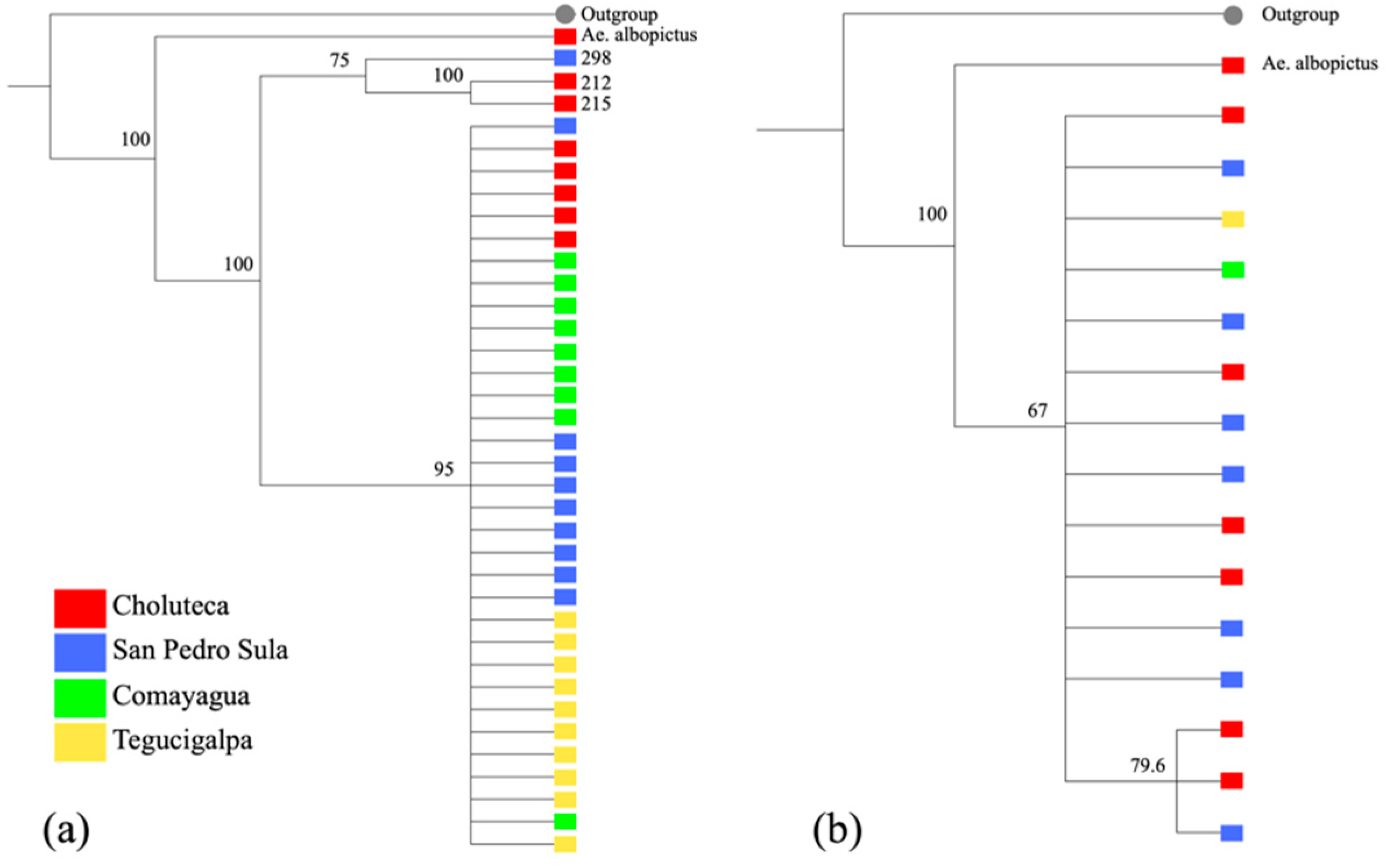
2.2. Phylogenetic Analysis Based on COI

Two phylogenetic analyses were performed with the COI and ITS rDNA sequences obtained in this study (Figure 1). The cladogram based on the COI gene showed two well-separated clades with a bootstrap index of 100. The largest clade included 33 individuals collected in the four cities, while a second clade included only three individuals: two from Choluteca and one from San Pedro Sula. There was no separation of individuals based on geography (Figure 1a). The cladogram constructed with 15 ITS2 rDNA sequences also showed two clades but with a smaller bootstrap, and no separation based on geographical distance was evident (Figure 1b).
In addition, a cladogram was constructed with a 450 bp fragment of the COI gene that included 35 sequences obtained in this study and 635 homologous sequences from mosquitoes collected in 48 countries (Figure 2). The tree revealed a clade containing 605 sequences and a smaller clade containing 30 sequences from the following geographic origins: Indonesia (3), Colombia (3), Myanmar (1), Pakistan (1), Bangladesh (1), Kenya (6), India (2), Democratic Republic of Congo (2), El Salvador (9), and Honduras/Choluteca (2). According to this result, there does not seem to be a clear geographical separation for Ae. aegypti.

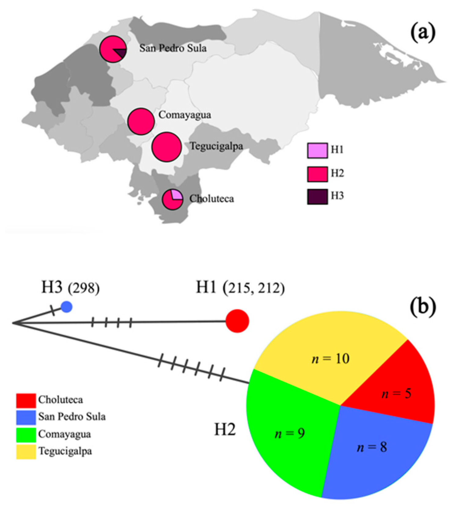
2.3. Mitochondrial COI Haplotype Analysis

Three distinct haplotypes were identified out of 35 mosquitoes collected in four cities (Figure 3). Haplotype 1 (H1) included two mosquitoes collected in Choluteca (specimens 215 and 212), the second haplotype (H2) was the most common with 32 individuals from the four cities, and haplotype 3 (H3) included only one individual collected in San Pedro Sula (specimen 298). The haplotype diversity (Hd) of the entire population was 0.1647.

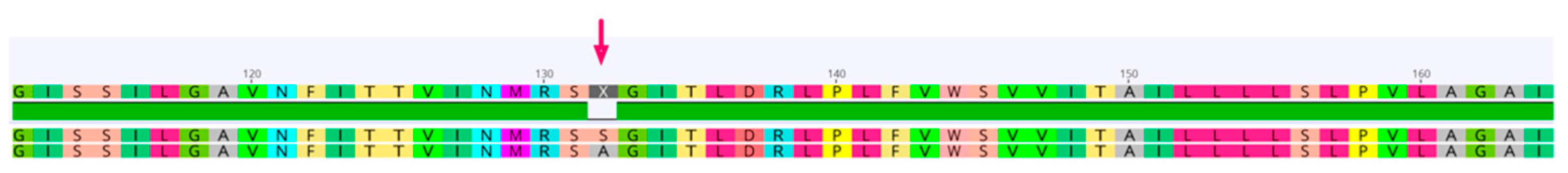
2.4. Amino Acid Sequence Analysis

A unique sequence of 168 amino acids was obtained by translating the 35 sequences of the Ae. aegypti COI gene. There was no inter-individual variation despite the eight polymorphic nucleotides. The partial sequence of the COI protein was “HPGMFIGNDQIYNVIVTAHAFIMIFFMVMPIMIGGFGNWLVPLMLGAPDMAFPRMNNMSFWMLPPSLTLLLSSSMVENGAGTGWTVYPPLSSGTAHAGASVDLAIFSLHLAGISSILGAVNFITTVINMRSSGITLDRLPLFVWSVVITAILLLLSLPVLAGAITMLL”. The number of identical sites was 167 (99.4%), pairwise % identity was 99.97%, and pairwise % positive (BLSM62) was 100%. The homologous sequence of Ae. albopictus showed a single residue difference (serine to alanine) with respect to the protein of Ae. aegypti as shown in Figure 4.

3. Discussion

This study was a pilot aimed at genetically characterizing Ae. aegypti mosquitoes collected in Honduras. Mosquito collections were carried out in four cities located along the so-called logistics corridor of Honduras, which crosses from the Caribbean Sea to the Pacific Ocean. A second objective of the study was to compare the phylogeny of mosquitoes from Honduras with respect to mosquitoes collected in countries on four continents. Our results suggest low genetic diversity in the populations of Ae. aegypti from Honduras, based on a low number of nucleotide differences per site (π = 0.0028, and 99.7% pairwise identity), low number of haplotypes (h = 3), and low haplotype diversity (Hd = 0.16). Large-scale studies have shown that populations of Ae. aegypti differ in genetic diversity according to geographic region [22]. Due to the high allelic richness of mosquito populations in Africa, it is widely accepted that this continent was the region of origin of Ae. aegypti [29]. Less genetic variability outside of Africa suggests that the Americas, Asia, and the Pacific Islands were later colonized by the mosquito [29]. The first massive dispersal out of Africa took place with the transatlantic migratory movements from the Old World to the Americas beginning in the fifteenth century [36], introducing arboviruses such as yellow fever and dengue, which shaped the demography of the Americas during the centuries following the conquest [2,37].
An extensive plan directed by PAHO between 1947 and 1970 aimed to eradicate Ae. aegypti from the American continent with the main objective of combating urban yellow fever through the use of insecticides such as DDT [2]. Despite enormous regional efforts, during the second half of 1960, the mosquito was reintroduced to territories where it had previously been eradicated [12], such as the Central American isthmus [20,23]. This led to a bottleneck effect on mosquito populations [9], with the consequent decrease in its genetic diversity. Two studies evaluating the genetic diversity of Ae. aegypti in Central America have been published recently. Joyce et al., 2018 sequenced a partial region of the COI gene from 84 mosquitoes collected in six geographic regions of El Salvador [20]. The overall nucleotide diversity was 0.015, with 10 haplotypes and a haplotype diversity equal to 0.61. A second study carried out in Panama analyzed 122 mosquitoes collected in 10 provinces, estimating a nucleotide diversity of 0.0096, 13 haplotypes, and Hd = 0.766 [23].
Both reports suggest a moderately higher genetic diversity in mosquito populations in El Salvador and Panama than that found in Honduras. This difference could be the result of the low number of sequences analyzed in this pilot study (n = 36), and another reason behind the low diversity could be that some of the mosquitoes might have been siblings due to the egg collection strategy. In the El Salvador study, a few larvae were collected from each water reservoir [20], and in Panama, only one specimen (larvae or adult mosquito) was collected from each house [23], minimizing the chance of collecting siblings. Furthermore, an entomological collection strategy focused on only four cities and only one neighborhood in each city is also a possible reason behind our results. These are limitations that will have to be overcome in the near future. However, these limitations could explain the low intrapopulation diversity but not the low diversity among the four cities in which the mosquitoes were collected.
Another hypothesis behind the difference between our results and results from neighboring countries could be that mosquitoes from El Salvador and Panama were collected from both urban and rural areas, while mosquitoes from Honduras were collected only from highly urbanized cities, with high connectivity between them. A study based on distribution models of Aedes albopictus in Panama showed that road networks alone better explain the geographic expansion of the mosquito [38]; therefore, the logistics corridor linking the four cities where the Honduran mosquitoes were collected could have favored the homogenization of the populations by passive anthropogenic movement [39]. Moreover, the greater flow of international trade observed in Panama and El Salvador could be responsible for multiple reintroductions of mosquitoes from other geographic regions increasing their genetic diversity [23].
Studies based on different nuclear or mitochondrial molecular markers applied to mosquitoes collected in the Americas [9,10,11,16,18,40] report lower genetic diversity than in some Asian countries [25,26,30,41,42,43,44] and most of Africa [22,31,45]. However, regardless of their country of origin, the vast majority of studies worldwide suggest the existence of two distinct lineages of Ae. aegypti [11,12,16,19,20,24,26,27,30,46,47]. Our results are consistent with the literature. Despite the small sample size, it was possible to detect two strongly supported phylogenetic clades: a dominant clade made up of 33 individuals and a second minority clade made up of the remaining three individuals. This pattern coincides with that reported by Joyce et al., 2018 in El Salvador and is consistent with the theory of multiple reinfestations in the American continent after the 1970s from African populations or the dispersal of mosquitoes that survived the eradication campaign, as suggested by Eskildsen et al., 2018 [23]. A single dominant haplotype in the four cities of Honduras would indicate a single panmictic population. This could indicate that neither the ecological differences nor the distance that separates the four cities would have any significant effect on the configuration of the mosquito populations. On the other hand, a negative Tajima’s D result could be evidence of a recent expansion after the bottleneck suffered fifty years ago. However, it will be necessary to further expand the number of mosquitoes analyzed, the geographical coverage of the country, and the application of neutral markers such as microsatellites to delve into the population structure of Ae. aegypti in the country.
Finally, this study reports for the first time in the country the molecular confirmation of Ae. albopictus. Although this species is present on all continents, occupying the same ecological niches as Ae. aegypti in the Americas [48], neither do the country’s health authorities carry out routine entomological surveillance nor is its relevance as an arbovirus vector known. Future research could contribute to delving into the population dynamics of Ae. albopictus in Honduras.

4. Materials and Methods

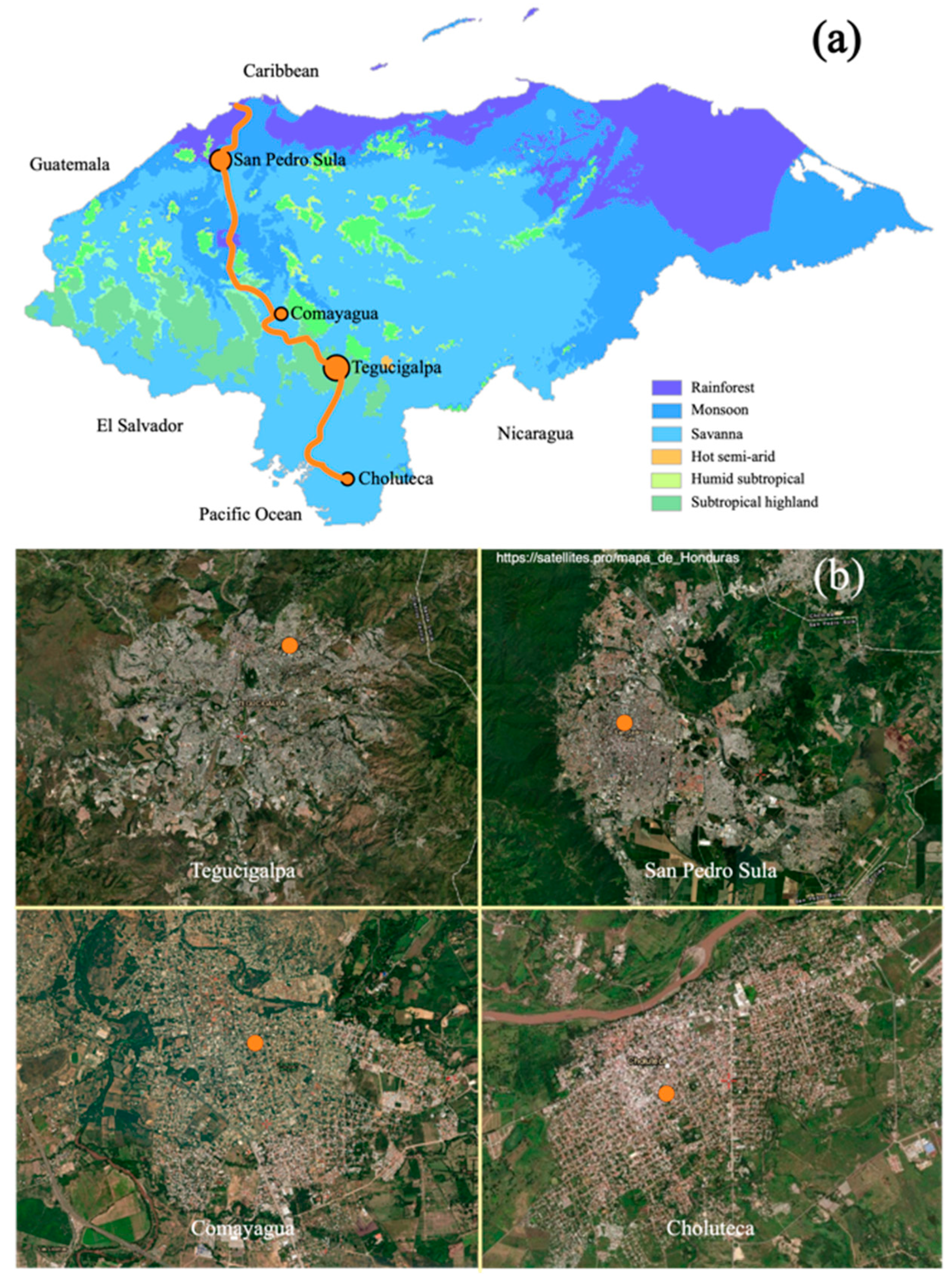
4.1. Study Sites, Mosquito Collection, and Morphological Identification

The study sites selected are used as sampling points in routine entomological surveillance for Ae. aegypti carried out by the Honduran Ministry of Health. Mosquitoes were collected in four cities with a population greater than 150,000: Comayagua (coordinates: 14.455532, −87.636718), Choluteca (13.2979940, −87.189693), Tegucigalpa (14.1080110, −87.188450), and San Pedro Sula (15.5081440, −88.027036) (Figure 5). The four cities are connected by the country’s main road, called “the logistics corridor” or CA-5, which connects the Caribbean Sea with the Pacific Ocean, and through which most of the movement of goods and people in the country takes place. Despite being located in a relatively small country (112,492 km2), the ecological conditions of the four cities are different. San Pedro Sula is located in a valley 83 meters above sea level with Atlantic influence that receives a lot of rain and is bordered by the Merendón mountain range, considered a tropical forest according to the Köppen–Geiger climate classification [49]. Comayagua is located in a valley in the central region of the country, with a savannah-type ecosystem at 594 m.a.s.l. Tegucigalpa, the capital city, rises 990 m.a.s.l. and is surrounded by mountains with subtropical coniferous forest, while Choluteca is located near the coast of the Gulf of Fonseca, with a drier climate at 65 m.a.s.l.
A sentinel site was selected in each city in coordination with the Ministry of Health. Aedes aegypti eggs were collected between January and March 2018 using ovitraps. Briefly, ovitraps were prepared using 10% hay infusion over paper towel as the substrate for oviposition [50]. The ovitraps were distributed in five to ten houses at each site with a distance of 200 meters between them and were visited weekly until eggs were found. Egg-bearing papers with more than 100 eggs were transported to local insectaries in each city where they were hatched and raised to the adult stage. Ovitraps with less than 100 eggs were discarded assuming that a single adult mosquito had laid eggs. In each city, all the larvae were grouped together and then reared to adults.
Standard rearing conditions were used (26–29 °C, with 70–90% relative humidity, and a 12:12 photoperiod) [51]. Adult mosquitoes were confirmed to be Ae. aegypti using morphological characteristics described in taxonomical keys [52]. All female mosquitoes were stored at −20 °C until subsequent molecular analysis in Tegucigalpa.

4.2. DNA Extraction, Gene Amplification, and Sequencing

A subset of ten mosquitoes per site was randomly selected for DNA extraction. DNA was extracted from each specimen following the ReliaPrep™ Blood gDNA Miniprep System (Promega, Madison, WI, USA) protocol. Following the manufacturer’s instructions, mosquito maceration was carried out with a pestle in a 1.5 mL conical tube. Lysis was performed at 56 °C for one hour. DNA was eluted in 80 μL of elution buffer and stored at −20 °C until further use.
Two molecular markers were amplified: the cytochrome c oxidase 1 gene (COI) and the internal transcribed spacer 2 (ITS2 rDNA). COI was amplified with the following primers: LCO1490 (5′-GGT CAA CAA ATC ATA AAG ATA TTG G-3′) and HCO2198 (5′-TAA ACT TCA GGG TGA CCA AAA ATC A-3′) [53]. PCR reactions were carried out in a volume of 50 μL, with 25 μL of Taq Master Mix 2× (Promega, Madison, WI, USA), 2.0 μL of each primer (10 μM), 2 μL of acetylated bovine albumin (BSA) (10 mg/mL), 4 μL of DNA, and nuclease-free water. The PCR program was as follows: 1 cycle at 95 °C for 10 min, 37 cycles at 94 °C for 1 min, 48 °C for 1 min, 72 °C for 1 min, and 1 cycle at 72 °C for 7 min. PCR products were separated by electrophoresis in 1% agarose gels with ethidium bromide.
For ITS2 amplification, PCR reactions were performed using the universal primers 5.8S (5′-ATC ACT CGG CTC GTG GAT CG-3′) and 28S (5′-ATG CTT AAA TTT AGG GGG TAG TC-3′) [54]. Reagent concentrations were as follows: 25 μL of Taq Master Mix 2×, 2 μL of each primer 10 μM, 2 μL of DNA, and water for a total reaction volume of 50 μL. PCR amplifications were performed with the following conditions: 94 °C for 2 min, 34 cycles of 94 °C for 30 s, 57 °C for 30 s, 72 °C for 30 s, and a final extension step at 72 °C for 10 min.
The amplification products of both loci (COI and ITS2 rDNA) were sequenced on both strands using the same primers for the PCR. Sequencing services were provided by Psomagen®. The sequences were edited with the Geneious® 9.1.7 software and deposited into the NCBI GenBank. All sequences were submitted as queries to NCBI through the BLAST tool [55] under default parameters to identify the most similar sequences in the GenBank nucleotide collection.

4.3. Data Analysis

Thirty-six partial sequences of the mitochondrial COI gene of Ae. aegypti were analyzed. For sequence analysis, only those showing a %HQ index greater than 98.5% after trimming were used. Sequences were aligned with the Geneious algorithm of the Geneious® 9.1.7 software. A homologous sequence from Anopheles albimanus was included in the alignment as an outgroup. Sequences with insertions or stop codons that might suggest nuclear mitochondrial pseudogenes were searched for (NUMT) [56]. Phylogenetic analysis used the Tamura–Nei distance model, the Neighbor-Joining method, and a bootstrap of 500. A cladogram (rooted tree layout) was constructed including a homologous sequence of Ae. albopictus obtained in this study. The length of the nucleotide sequences, the number and percentage of identical sites, and the pairwise % identity were calculated. MEGA v11.0 software [57,58] with 500 bootstrap replicates was used to calculate the overall mean distance, standard error (S.E.), number of segregating sites (S), nucleotide diversity (π), and Tajima test statistic (D). Sequences of the ITS2 rDNA region were also analyzed, and a cladogram was constructed.
Two sets of analyses similar to those described above were performed using homologous COI sequences from Ae. aegypti downloaded from GenBank and obtained from mosquitoes collected in other countries. The first analysis included 669 sequences from 48 countries on five continents. The second analysis included 217 sequences from seven countries in the central region of America (Mexico, Guatemala, El Salvador, Honduras, Costa Rica, Panama, and Colombia). In both analyses, the sequences were edited to keep exactly the same size (450 bp and 352 bp, respectively). A phylogenetic cladogram (circular tree layout) was constructed using 669 homologous sequences (36 obtained in this study and 633 downloaded from Genbank), using the Tamura–Nei distance model, the Neighbor-Joining method, and a bootstrap of 500 replicates with a homologous sequence of Anopheles albimanus as an outgroup.
The number of haplotypes (h) and haplotype diversity (Hd) were calculated according to the nucleotide sequences with Dna SP software v. 6.12.03 [59]. Alignment sequences were imported, and parameters were adjusted for mitochondrial DNA with genetic code for Drosophila mtDNA. Haplotype data were generated using the Roehl data file function and default parameters.
Nucleotide sequences obtained in this study were translated using the correct open reading frame (ORF) using the invertebrate mitochondrial genetic code in the Geneious® 9.1.7 software. Residue alignment was performed, and the number of identical sites, pairwise % identity, and pairwise % positive (BLSM62) were calculated.

5. Conclusions

This is, to our knowledge, the first study carried out in Honduras that analyzes the genetic diversity of Ae. aegypti. The results of this pilot seem to indicate the presence of two mosquito lineages and three haplotypes (one dominant haplotype) with low genetic diversity and no apparent population structure, which coincides with what has been reported previously in the Central American region. Future studies that expand the number of mosquitoes and the number of cities will shed more light on the evolutionary history of Ae. aegypti in Central America more than half a century after its reintroduction.

Author Contributions

Conceptualization, D.E. and G.F.; methodology, D.E., O.U., B.O. and G.F.; mosquito collection, D.E. and O.U.; validation, G.F. and D.E.; formal analysis, G.F.; investigation, D.E., O.U., B.O. and G.F.; resources, G.F.; data curation, D.E. and G.F.; writing—original draft preparation, G.F.; project administration, D.E.; funding acquisition, G.F. All authors have read and agreed to the published version of the manuscript.

Funding

This research received no external funding.

Institutional Review Board Statement

Not applicable.

Informed Consent Statement

All individuals included in this section have consented to the acknowledgement.

Data Availability Statement

Not applicable.

Acknowledgments

The authors would like to thank the support given by the staff of the Ministry of Health of Honduras, particularly to the Entomological Surveillance Units in Choluteca, Comayagua, Tegucigalpa, and San Pedro Sula. Thanks to Osman Archaga for his support in laboratory activities.

Conflicts of Interest

The authors declare no conflict of interest.

References

  1. Crawford, J.E.; Alves, J.M.; Palmer, W.J.; Day, J.P.; Sylla, M.; Ramasamy, R.; Surendran, S.N.; Black, W.C.T.; Pain, A.; Jiggins, F.M. Population genomics reveals that an anthropophilic population of Aedes aegypti mosquitoes in West Africa recently gave rise to American and Asian populations of this major disease vector. BMC Biol. 2017, 15, 16. [Google Scholar] [CrossRef] [PubMed] [Green Version]
  2. Brathwaite Dick, O.; San Martin, J.L.; Montoya, R.H.; del Diego, J.; Zambrano, B.; Dayan, G.H. The history of dengue outbreaks in the Americas. Am. J. Trop. Med. Hyg. 2012, 87, 584–593. [Google Scholar] [CrossRef] [PubMed] [Green Version]
  3. Espinal, M.A.; Andrus, J.K.; Jauregui, B.; Waterman, S.H.; Morens, D.M.; Santos, J.I.; Horstick, O.; Francis, L.A.; Olson, D. Emerging and Reemerging Aedes-Transmitted Arbovirus Infections in the Region of the Americas: Implications for Health Policy. Am. J. Public Health 2019, 109, 387–392. [Google Scholar] [CrossRef]
  4. Zambrano, L.I.; Rodriguez, E.; Espinoza-Salvado, I.A.; Rodriguez-Morales, A.J. Dengue in Honduras and the Americas: The epidemics are back! Travel Med. Infect Dis. 2019, 31, 101456. [Google Scholar] [CrossRef] [PubMed]
  5. Guzman, M.G.; Kouri, G. Dengue and dengue hemorrhagic fever in the Americas: Lessons and challenges. J. Clin. Virol. 2003, 27, 1–13. [Google Scholar] [CrossRef]
  6. Pan American Health Organization. PLISA Health Information Platform for the Americas. Available online: https://www3.paho.org/data/index.php/en/mnu-topics/indicadores-dengue-en/dengue-nacional-en/252-dengue-pais-ano-en.html (accessed on 21 April 2022).
  7. Pialoux, G.; Gauzere, B.A.; Jaureguiberry, S.; Strobel, M. Chikungunya, an epidemic arbovirosis. Lancet Infect Dis. 2007, 7, 319–327. [Google Scholar] [CrossRef]
  8. Gubler, D.J.; Vasilakis, N.; Musso, D. History and Emergence of Zika Virus. J. Infect Dis. 2017, 216, S860–S867. [Google Scholar] [CrossRef] [Green Version]
  9. Sherpa, S.; Rioux, D.; Goindin, D.; Fouque, F.; Francois, O.; Despres, L. At the Origin of a Worldwide Invasion: Unraveling the Genetic Makeup of the Caribbean Bridgehead Populations of the Dengue Vector Aedes aegypti. Genome Biol. Evol. 2018, 10, 56–71. [Google Scholar] [CrossRef] [Green Version]
  10. Steffler, L.M.; Dolabella, S.S.; Ribolla, P.E.; Dreyer, C.S.; Araujo, E.D.; Oliveira, R.G.; Martins, W.F.; La Corte, R. Genetic variability and spatial distribution in small geographic scale of Aedes aegypti (Diptera: Culicidae) under different climatic conditions in Northeastern Brazil. Parasit. Vectors 2016, 9, 530. [Google Scholar] [CrossRef] [Green Version]
  11. Jaimes-Duenez, J.; Arboleda, S.; Triana-Chavez, O.; Gomez-Palacio, A. Spatio-temporal distribution of Aedes aegypti (Diptera: Culicidae) mitochondrial lineages in cities with distinct dengue incidence rates suggests complex population dynamics of the dengue vector in Colombia. PLoS Negl. Trop Dis. 2015, 9, e0003553. [Google Scholar] [CrossRef]
  12. Monteiro, F.A.; Schama, R.; Martins, A.J.; Gloria-Soria, A.; Brown, J.E.; Powell, J.R. Genetic diversity of Brazilian Aedes aegypti: Patterns following an eradication program. PLoS Negl. Trop Dis. 2014, 8, e3167. [Google Scholar] [CrossRef] [Green Version]
  13. Maitra, A.; Cunha-Machado, A.S.; Souza Leandro, A.; Costa, F.M.D.; Scarpassa, V.M. Exploring deeper genetic structures: Aedes aegypti in Brazil. Acta Trop. 2019, 195, 68–77. [Google Scholar] [CrossRef] [PubMed]
  14. Souza, K.R.; Ribeiro, G., Jr.; Silva dos Santos, C.G.; de Lima, E.C.; Melo, P.R.; Reis, M.G.; Blanton, R.E.; Silva, L.K. Vector control measures failed to affect genetic structure of Aedes aegypti in a sentinel metropolitan area of Brazil. Acta Trop. 2013, 128, 598–605. [Google Scholar] [CrossRef]
  15. de Lourdes Munoz, M.; Mercado-Curiel, R.F.; Diaz-Badillo, A.; Perez Ramirez, G.; Black, W.C.T. Gene flow pattern among Aedes aegypti populations in Mexico. J. Am. Mosq. Control Assoc. 2013, 29, 1–18. [Google Scholar] [CrossRef]
  16. Paupy, C.; Le Goff, G.; Brengues, C.; Guerra, M.; Revollo, J.; Barja Simon, Z.; Herve, J.P.; Fontenille, D. Genetic structure and phylogeography of Aedes aegypti, the dengue and yellow-fever mosquito vector in Bolivia. Infect. Genet. Evol. 2012, 12, 1260–1269. [Google Scholar] [CrossRef] [PubMed]
  17. Duenas, J.C.; Llinas, G.A.; Panzetia-Dutari, G.M.; Gardenal, C.N. Two different routes of colonization of Aedes aegypti in Argentina from neighboring countries. J. Med. Entomol. 2009, 46, 1344–1354. [Google Scholar] [CrossRef]
  18. Lima, R.S., Jr.; Scarpassa, V.M. Evidence of two lineages of the dengue vector Aedes aegypti in the Brazilian Amazon, based on mitochondrial DNA ND4 gene sequences. Genet. Mol. Biol. 2009, 32, 414–422. [Google Scholar] [CrossRef] [Green Version]
  19. Scarpassa, V.M.; Cardoza, T.B.; Cardoso Junior, R.P. Population genetics and phylogeography of Aedes aegypti (Diptera: Culicidae) from Brazil. Am. J. Trop. Med. Hyg. 2008, 78, 895–903. [Google Scholar] [CrossRef] [PubMed] [Green Version]
  20. Joyce, A.L.; Torres, M.M.; Torres, R.; Moreno, M. Genetic variability of the Aedes aegypti (Diptera: Culicidae) mosquito in El Salvador, vector of dengue, yellow fever, chikungunya and Zika. Parasit. Vectors 2018, 11, 637. [Google Scholar] [CrossRef] [Green Version]
  21. Garcia-Franco, F.; Munoz Mde, L.; Lozano-Fuentes, S.; Fernandez-Salas, I.; Garcia-Rejon, J.; Beaty, B.J.; Black, W.C.T. Large genetic distances among Aedes aegypti populations along the South Pacific coast of Mexico. Am. J. Trop. Med. Hyg. 2002, 66, 594–598. [Google Scholar] [CrossRef] [Green Version]
  22. Gloria-Soria, A.; Ayala, D.; Bheecarry, A.; Calderon-Arguedas, O.; Chadee, D.D.; Chiappero, M.; Coetzee, M.; Elahee, K.B.; Fernandez-Salas, I.; Kamal, H.A.; et al. Global genetic diversity of Aedes aegypti. Mol. Ecol. 2016, 25, 5377–5395. [Google Scholar] [CrossRef] [PubMed] [Green Version]
  23. Eskildsen, G.A.; Rovira, J.R.; Smith, O.; Miller, M.J.; Bennett, K.L.; McMillan, W.O.; Loaiza, J. Maternal invasion history of Aedes aegypti and Aedes albopictus into the Isthmus of Panama: Implications for the control of emergent viral disease agents. PLoS ONE 2018, 13, e0194874. [Google Scholar] [CrossRef] [PubMed] [Green Version]
  24. Kawada, H.; Futami, K.; Higa, Y.; Rai, G.; Suzuki, T.; Rai, S.K. Distribution and pyrethroid resistance status of Aedes aegypti and Aedes albopictus populations and possible phylogenetic reasons for the recent invasion of Aedes aegypti in Nepal. Parasit. Vectors 2020, 13, 213. [Google Scholar] [CrossRef] [PubMed]
  25. Khater, E.I.M.; Baig, F.; Kamal, H.A.; Powell, J.R.; Saleh, A.A. Molecular Phylogenetics and Population Genetics of the Dengue Vector Aedes aegypti From the Arabian Peninsula. J. Med. Entomol. 2021, 58, 2161–2176. [Google Scholar] [CrossRef]
  26. Dharmarathne, H.; Weerasena, O.; Perera, K.; Galhena, G.H. Genetic characterization of Aedes aegypti (Diptera: Culicidae) in Sri Lanka based on COI gene. J. Vector Borne Dis. 2020, 57, 153–160. [Google Scholar] [CrossRef]
  27. Bracco, J.E.; Capurro, M.L.; Lourenco-de-Oliveira, R.; Sallum, M.A. Genetic variability of Aedes aegypti in the Americas using a mitochondrial gene: Evidence of multiple introductions. Mem. Inst. Oswaldo Cruz 2007, 102, 573–580. [Google Scholar] [CrossRef] [Green Version]
  28. Moore, M.; Sylla, M.; Goss, L.; Burugu, M.W.; Sang, R.; Kamau, L.W.; Kenya, E.U.; Bosio, C.; Munoz Mde, L.; Sharakova, M.; et al. Dual African origins of global Aedes aegypti s.l. populations revealed by mitochondrial DNA. PLoS Negl. Trop. Dis. 2013, 7, e2175. [Google Scholar] [CrossRef] [Green Version]
  29. Bennett, K.L.; Shija, F.; Linton, Y.M.; Misinzo, G.; Kaddumukasa, M.; Djouaka, R.; Anyaele, O.; Harris, A.; Irish, S.; Hlaing, T.; et al. Historical environmental change in Africa drives divergence and admixture of Aedes aegypti mosquitoes: A precursor to successful worldwide colonization? Mol. Ecol. 2016, 25, 4337–4354. [Google Scholar] [CrossRef] [Green Version]
  30. Kumar, M.S.; Kalimuthu, M.; Selvam, A.; Mathivanan, A.; Paramasivan, R.; Kumar, A.; Gupta, B. Genetic structure and connectivity among Aedes aegypti populations within Madurai city in Southern India. Infect. Genet. Evol. 2021, 95, 105031. [Google Scholar] [CrossRef]
  31. Abuelmaali, S.A.; Jamaluddin, J.A.F.; Noaman, K.; Allam, M.; Abushama, H.M.; Elnaiem, D.E.; Ishak, I.H.; Wajidi, M.F.F.; Jaal, Z.; Abu Kassim, N.F. Distribution and Genetic Diversity of Aedes aegypti Subspecies across the Sahelian Belt in Sudan. Pathogens 2021, 10, 78. [Google Scholar] [CrossRef]
  32. Calvez, E.; Guillaumot, L.; Millet, L.; Marie, J.; Bossin, H.; Rama, V.; Faamoe, A.; Kilama, S.; Teurlai, M.; Mathieu-Daude, F.; et al. Genetic Diversity and Phylogeny of Aedes aegypti, the Main Arbovirus Vector in the Pacific. PLoS Negl. Trop. Dis. 2016, 10, e0004374. [Google Scholar] [CrossRef] [PubMed] [Green Version]
  33. Naim, D.M.; Kamal, N.Z.M.; Mahboob, S. Population structure and genetic diversity of Aedes aegypti and Aedes albopictus in Penang as revealed by mitochondrial DNA cytochrome oxidase I. Saudi J. Biol. Sci. 2020, 27, 953–967. [Google Scholar] [CrossRef] [PubMed]
  34. Carvajal, T.M.; Ogishi, K.; Yaegeshi, S.; Hernandez, L.F.T.; Viacrusis, K.M.; Ho, H.T.; Amalin, D.M.; Watanabe, K. Fine-scale population genetic structure of dengue mosquito vector, Aedes aegypti, in Metropolitan Manila, Philippines. PLoS Negl. Trop. Dis. 2020, 14, e0008279. [Google Scholar] [CrossRef] [PubMed]
  35. Tedjou, A.N.; Kamgang, B.; Yougang, A.P.; Njiokou, F.; Wondji, C.S. Update on the geographical distribution and prevalence of Aedes aegypti and Aedes albopictus (Diptera: Culicidae), two major arbovirus vectors in Cameroon. PLoS Negl. Trop. Dis. 2019, 13, e0007137. [Google Scholar] [CrossRef] [PubMed] [Green Version]
  36. Powell, J.R.; Tabachnick, W.J. History of domestication and spread of Aedes aegypti—A review. Mem. Inst. Oswaldo Cruz 2013, 108 (Suppl. 1), 11–17. [Google Scholar] [CrossRef]
  37. Sabbatani, S.; Fiorino, S. Yellow fever. Infez. Med. 2007, 15, 129–141. [Google Scholar]
  38. Miller, M.J.; Loaiza, J.R. Geographic expansion of the invasive mosquito Aedes albopictus across Panama—Implications for control of dengue and Chikungunya viruses. PLoS Negl. Trop. Dis. 2015, 9, e0003383. [Google Scholar] [CrossRef] [Green Version]
  39. Fonzi, E.; Higa, Y.; Bertuso, A.G.; Futami, K.; Minakawa, N. Human-Mediated Marine Dispersal Influences the Population Structure of Aedes aegypti in the Philippine Archipelago. PLoS Negl. Trop. Dis. 2015, 9, e0003829. [Google Scholar] [CrossRef] [Green Version]
  40. Ponce, P.; Munoz-Tobar, S.; Carrazco-Montalvo, A.; Villota, S.D.; Coloma, J.; Wang, C.; Holechek, S.; Cevallos, V. Two Haplotypes of Aedes aegypti Detected by ND4 Mitochondrial Marker in Three Regions of Ecuador. Insects 2021, 12, 200. [Google Scholar] [CrossRef]
  41. Lv, R.C.; Zhu, C.; Wang, C.H.; Ai, L.L.; Lv, H.; Zhang, B.; Li, C.M.; An, J.; Wang, P.G.; Hu, D.; et al. Genetic diversity and population structure of Aedes aegypti after massive vector control for dengue fever prevention in Yunnan border areas. Sci. Rep. 2020, 10, 12731. [Google Scholar] [CrossRef]
  42. Shi, Q.M.; Zhang, H.D.; Wang, G.; Guo, X.X.; Xing, D.; Dong, Y.D.; Xiao, L.; Gao, J.; Liu, Q.M.; Sun, A.J.; et al. The genetic diversity and population structure of domestic Aedes aegypti (Diptera: Culicidae) in Yunnan Province, southwestern China. Parasit. Vectors 2017, 10, 292. [Google Scholar] [CrossRef] [PubMed] [Green Version]
  43. Yugavathy, N.; Kim-Sung, L.; Joanne, S.; Vythilingam, I. Genetic variation of the mitochondrial genes, CO1 and ND5, in Aedes aegypti from various regions of peninsular Malaysia. Trop. Biomed. 2016, 33, 543–560. [Google Scholar] [PubMed]
  44. Duong, C.V.; Kang, J.H.; Nguyen, V.V.; Bae, Y.J. Genetic Diversity and Population Structure of the Asian Tiger Mosquito (Aedes albopictus) in Vietnam: Evidence for Genetic Differentiation by Climate Region. Genes 2021, 12, 1579. [Google Scholar] [CrossRef]
  45. Campos, M.; Ward, D.; Morales, R.F.; Gomes, A.R.; Silva, K.; Sepulveda, N.; Gomez, L.F.; Clark, T.G.; Campino, S. Surveillance of Aedes aegypti populations in the city of Praia, Cape Verde: Zika virus infection, insecticide resistance and genetic diversity. Parasit. Vectors 2020, 13, 481. [Google Scholar] [CrossRef]
  46. Brown, J.E.; McBride, C.S.; Johnson, P.; Ritchie, S.; Paupy, C.; Bossin, H.; Lutomiah, J.; Fernandez-Salas, I.; Ponlawat, A.; Cornel, A.J.; et al. Worldwide patterns of genetic differentiation imply multiple ‘domestications’ of Aedes aegypti, a major vector of human diseases. Proc. Biol. Sci. 2011, 278, 2446–2454. [Google Scholar] [CrossRef] [PubMed] [Green Version]
  47. Garjito, T.A.; Widiarti, W.; Hidajat, M.C.; Handayani, S.W.; Mujiyono, M.; Prihatin, M.T.; Ubaidillah, R.; Sudomo, M.; Satoto, T.B.T.; Manguin, S.; et al. Homogeneity and Possible Replacement of Populations of the Dengue Vectors Aedes aegypti and Aedes albopictus in Indonesia. Front Cell Infect. Microbiol. 2021, 11, 705129. [Google Scholar] [CrossRef]
  48. Garcia-Rejon, J.E.; Navarro, J.C.; Cigarroa-Toledo, N.; Baak-Baak, C.M. An Updated Review of the Invasive Aedes albopictus in the Americas; Geographical Distribution, Host Feeding Patterns, Arbovirus Infection, and the Potential for Vertical Transmission of Dengue Virus. Insects 2021, 12, 967. [Google Scholar] [CrossRef]
  49. Beck, H.E.; Zimmermann, N.E.; McVicar, T.R.; Vergopolan, N.; Berg, A.; Wood, E.F. Present and future Koppen-Geiger climate classification maps at 1-km resolution. Sci. Data 2018, 5, 180214. [Google Scholar] [CrossRef] [Green Version]
  50. Polson, A.K.; Curtis, C.M.S.O.J.; Chantha, N.; Rawlins, S. The Use of Ovitraps Baited with Hay Infusion as a Surveillance Tool for Aedes aegypti Mosquitoes in Cambodia; WHO Regional Office for South-East Asia: New Delhi, India, 2002. [Google Scholar]
  51. Proyecto ZIKA AIRS, Z. Manual Práctico para el Funcionamiento de un Insectario: Procedimientos de Cría de la Especie Aedes aegypti y Principios Básicos de Bioseguridad; Abt Associates: Rockville, MD, USA, 2019; p. 91. [Google Scholar]
  52. Rueda, L. Pictorial keys for the identification of mosquitoes (Diptera: Culicidae) associated with Dengue Virus Transmission. Zootaxa 2004, 589. [Google Scholar] [CrossRef]
  53. Folmer, O.; Black, M.; Hoeh, W.; Lutz, R.; Vrijenhoek, R. DNA primers for amplification of mitochondrial cytochrome c oxidase subunit I from diverse metazoan invertebrates. Mol. Mar. Biol. Biotechnol. 1994, 3, 294–299. [Google Scholar]
  54. Djadid, N.D.; Gholizadeh, S.; Aghajari, M.; Zehi, A.H.; Raeisi, A.; Zakeri, S. Genetic analysis of rDNA-ITS2 and RAPD loci in field populations of the malaria vector, Anopheles stephensi (Diptera: Culicidae): Implications for the control program in Iran. Acta Trop. 2006, 97, 65–74. [Google Scholar] [CrossRef] [PubMed]
  55. Altschul, S.F.; Gish, W.; Miller, W.; Myers, E.W.; Lipman, D.J. Basic local alignment search tool. J. Mol. Biol. 1990, 215, 403–410. [Google Scholar] [CrossRef]
  56. Behura, S.K.; Lobo, N.F.; Haas, B.; de Bruyn, B.; Lovin, D.D.; Shumway, M.F.; Puiu, D.; Romero-Severson, J.; Nene, V.; Severson, D.W. Complete sequences of mitochondria genomes of Aedes aegypti and Culex quinquefasciatus and comparative analysis of mitochondrial DNA fragments inserted in the nuclear genomes. Insect Biochem. Mol. Biol. 2011, 41, 770–777. [Google Scholar] [CrossRef] [Green Version]
  57. Stecher, G.; Tamura, K.; Kumar, S. Molecular Evolutionary Genetics Analysis (MEGA) for macOS. Mol. Biol. Evol. 2020, 37, 1237–1239. [Google Scholar] [CrossRef] [PubMed]
  58. Tamura, K.; Stecher, G.; Kumar, S. MEGA11: Molecular Evolutionary Genetics Analysis Version 11. Mol. Biol. Evol. 2021, 38, 3022–3027. [Google Scholar] [CrossRef] [PubMed]
  59. Rozas, J.; Ferrer-Mata, A.; Sanchez-Del Barrio, J.C.; Guirao-Rico, S.; Librado, P.; Ramos-Onsins, S.E.; Sanchez-Gracia, A. DnaSP 6: DNA Sequence Polymorphism Analysis of Large Data Sets. Mol. Biol. Evol. 2017, 34, 3299–3302. [Google Scholar] [CrossRef]
Figure 1. Phylogenetic cladogram of partial sequences of the Aedes aegypti (a) COI gene, and (b) ITS2 ribosomal region, constructed using the Neighbor-Joining method with a bootstrap of 500 replicates. Each color represents a city where the mosquitoes were collected.
Pathogens 11 00620 g001
Figure 2. Cladogram (circular tree layout) of partial sequences of the COI gene of Aedes aegypti collected in 48 countries. The Neighbor-Joining method was used with a bootstrap of 500 replicates. Each color represents a world region where the mosquitoes were collected. The black arrows indicate the sequences obtained in this study, and the light blue arrows indicate the sequences reported in El Salvador by Joyce et al., 2018 [20].
Pathogens 11 00620 g002
Figure 3. (a) Map of Honduras showing three mitochondrial haplotypes (H1–H3) of Ae. qegypti by city. The size of the chart is proportional to the number of individuals per haplotype; (b) haplotype network based on 35 mitochondrial DNA COI sequences of Ae. qegypti collected in four cities of Honduras. Perpendicular bars indicate the number of nucleotide polymorphisms between haplotypes.
Pathogens 11 00620 g003
Figure 4. Amino acid alignment of a partial sequence of Cytochrome oxidase 1 protein from Aedes aegypti (top) and Ae. albopictus (bottom). The red arrow shows the only position where there is a substitution (serine for alanine).
Pathogens 11 00620 g004
Figure 5. (a) Map of Honduras showing the location of four cities where the mosquitoes were collected. The orange line represents the road CA-5 connecting the cities. The map shows the country’s ecoregions according to the Köppen–Geiger climate classification. Adapted from [49]; (b) satellite images of the four cities indicating the geographic point (orange dot) where the eggs of the mosquitoes were collected. Adapted from Google.com/maps (accessed on 4 May 2022).
Pathogens 11 00620 g005
Table 1. Genetic variability parameters estimated for partial sequences of the COI gene of Aedes aegypti.
StatisticsHondurasRegion 1World 2
Length (bp)505352450
Number of sequences35217670
Identical sites (%)493 (97.6%)288 (81.8%)323 (71.8%)
Pairwise % Identity99.7%98.9%99.3%
Overall mean distance0.000.010.01
1 Sequences of mosquitoes collected in Mexico, Guatemala, El Salvador, Honduras, Costa Rica, Panama, and Colombia. 2 Sequences of mosquitoes collected in 48 countries on five continents.
Publisher’s Note: MDPI stays neutral with regard to jurisdictional claims in published maps and institutional affiliations.

