
Rickettsiales in Italy
by

Cristoforo Guccione
1, 
Claudia Colomba
1,2,
Manlio Tolomeo
1,
Marcello Trizzino
2,
Chiara Iaria
3 and
Antonio Cascio
1,2,*
1
Department of Health Promotion, Mother and Child Care, Internal Medicine and Medical Specialties- University of Palermo, 90127 Palermo, Italy
2
Infectious and Tropical Disease Unit, AOU Policlinico “P. Giaccone”, 90127 Palermo, Italy
3
Infectious Diseases Unit, ARNAS Civico-Di Cristina-Benfratelli Hospital, 90127 Palermo, Italy
*
Author to whom correspondence should be addressed.
Pathogens 2021, 10(2), 181; https://doi.org/10.3390/pathogens10020181
Submission received: 31 December 2020
/
Revised: 3 February 2021
/
Accepted: 4 February 2021
/
Published: 8 February 2021
(This article belongs to the Collection Updates on Rickettsia and Coxiella)
Abstract
:There is no updated information on the spread of Rickettsiales in Italy. The purpose of our study is to take stock of the situation on Rickettsiales in Italy by focusing attention on the species identified by molecular methods in humans, in bloodsucking arthropods that could potentially attack humans, and in animals, possible hosts of these Rickettsiales. A computerized search without language restriction was conducted using PubMed updated as of December 31, 2020. The Preferred Reporting Items for Systematic Reviews and Meta-Analyses (PRISMA) methodology was followed. Overall, 36 species of microorganisms belonging to Rickettsiales were found. The only species identified in human tissues were Anaplasma phagocytophilum, Rickettsia conorii, R. conorii subsp. israelensis, R. monacensis, R. massiliae, and R. slovaca. Microorganisms transmissible by bloodsucking arthropods could cause humans pathologies not yet well characterized. It should become routine to study the pathogens present in ticks that have bitten a man and at the same time that molecular studies for the search for Rickettsiales can be performed routinely in people who have suffered bites from bloodsucking arthropods.
1. Introduction
Rickettsiales is an order of α-proteobacteria characterized by intracellular tropism with a wide variety of hosts. They are small, gram-negative bacteria that reside free in the host cell cytoplasm, and some of them can be transmitted to human hosts by arthropod vectors such as ticks, lice, fleas, and mites. As suggested by Szokoli et al. we considered included in this order only 3 families: Rickettsiaceae, Anaplasmataceae, and Candidatus Midichloriaceae. Rickettsiales encompass human and animal pathogens as well a lot of endosymbiont of arthropods, helminths, and algae with various, pathogenic or not manifestation in the host. The family Rickettsiaceae includes 2 genera: Rickettsia and Orientia. A modern classification based on whole-genome analysis divides the species of the genus Rickettsia in four groups: spotted fever group (R. rickettsii, R. conorii, R. parkeri, and several others), typhus group (R. prowazekii and R. typhi), ancestral group (R. bellii and R. canadensis, not known to be pathogenic), and transitional group (R. akari, R. australis, and R. felis) [1,2,3] Orientia tsutsugamushi is the etiologic agent of scrub typhus, a rickettsiosis that is widespread in Asia, the islands of the western Pacific and Indian Oceans, and foci in northern Australia [4]. The family Anaplasmataceae includes the genera Ehrlichia, Anaplasma, Wolbachia, and Neorickettsia. Only the members of the first two genera have been associated to human diseases. The genus Ehrlichia includes six species: E. canis, E. chaffeensis, E. ewingii, E. muris, E. ovis, and E. ruminantium. The genus Anaplasma includes A. marginale, A. centrale, A. ovis, A. mesaeterum, A. platys, and A. phagocytophilum; only the last is associated to human diseases. The family Candidatus Midichloriaceae does not include any bacteria associated to human disease.
Almost all the cases of human rickettsial diseases in Italy are cases of Mediterranean spotted fever (MSF) caused by R. conorii transmitted by the brown dog tick Rhipicephalus sanguineus. In Italy, about 400 cases of MSF are reported every year, most of which in people residing in Sicily, Sardinia and Southern Italy with a lethality of less than 3% [5] However, other pathologies such as Tibola/Debonel (Tick Borne Lymphadenopathy/Dermacentor Borne Necrosis Erythema and Lymphadenopathy [6]) and many other Rickettsia spp. or subspecies have been identified in recent years in humans, vector arthropods and animals [6]. Other rickettsioses that have been historically documented in Italy are murine typhus and epidemic typhus [5]. Since 1950, only sporadic cases of murine typhus have been reported, and Italy currently appears to be free of epidemic typhus. As in other European countries, imported cases of rickettsial pox, African tick-bite fever (ATBF), and scrub typhus have been reported [5].
The purpose of this study is to take stock of the situation on Rickettsiales in Italy by focusing attention on the genera until now identified by molecular methods in humans, in bloodsucking arthropods that could potentially attack humans, and in animals possible hosts of these Rickettsiales. Our research has therefore mainly focused on the genera Rickettsia, Anaplasma, Ehrlichia, and Orientia as these are the ones notoriously associated with human pathology until now.
2. Materials and Methods
For the writing of this review a computerized search without language restriction was conducted using PubMed. The search was performed combining the terms “Ricketts * AND Italy”, “Ehrlichi * AND Italy” and “Anaplasma AND Italy”, Orientia AND Italy”. The Preferred Reporting Items for Systematic Reviews and Meta-Analyses (PRISMA) methodology was followed [7]. Only studies that provided data about Rickettsiales identified by molecular methods in Italy were included in the review. All molecular methods which reached the species level were considered. A flow chart summarizing the literature research approach is reported in Figure 1.
3. Results
A total of 818 papers were retrieved by our search, of these 220 were duplicate and removed; the remains were assessed through their title and abstract and so other 273 were excluded; the selected 325 articles were assessed for eligibility through full text analysis and 168 were excluded as reported in Figure 1; finally, 157 published from 1997 to 2021 studies were included in this review.
The results of our search could be divided in four sections and are analytically reported in Table 1, Table 2, Table 3 and Table 4.
A total of 36 different Rickettsiales species belonging to genus Anaplasma, Ehrlichia and Rickettsia; never Orientia spp. were reported in Italy. 32 of them were identified in arthropods, 9 in animal samples, and 10 in human samples (Table 1, Table 2, Table 3 and Table 4).
3.1. Rickettsiales and Arthropod Vectors
Rickettsiales were identified in 29 species of arthropods, most of them were Ixodidae ticks, and 4 species of fleas. The reports present in the scientific literature are resumed in Table 1 and Table 2. Table 1 offer a view centered on the microorganism, for each Rickettsiales we report the known association with arthropods and from where it was collected. Indeed, Table 2 offers a point of view centered on the arthropods and for each we report which microorganism and host were associated.
3.2. Rickettsiales Identified in Animals
Rickettsiales have been identified 179 times in various animal infections, most of which were Anaplasma spp. especially in livestock, and R. conorii and E. canis especially in companion animals. Fifteen species of mammals with or without symptoms were found infected with Rickettsiales most of them where A. phagocytophilum and A. platy. Symptomatic animals were most often pets, with fever and blood count abnormalities (CBC) being the most frequently observed clinical findings; while asymptomatic animals were more often livestock. In Table 3 are resumed the findings in animal samples with clinical manifestations and the number of animals found positive for each Rickettsiales. When the original study was done on asymptomatic animals, with the aim of screening, we report also the number of total tested animals and the percentage of prevalence; when the studies was more than one, we report the highest and lowest percentage.
3.3. Rickettsiales Involved in Human Disease
Rickettsiales were detected 29 times in samples from human patients: 6 cases of anaplasmosis, and 23 cases of rickettsiosis. Rickettsiales species identified from human sample and their clinical manifestation are resumed in Table 4. Rickettsia spp. associated with MSF were R. conorii, R. conorii subsp. israelensis, R. conorii subsp. indica, R. massiliae, R. slovaca, and R. monacensis. Rickettsia spp. associated with TIBOLA/DEBONEL were R. slovaca, and R. massiliae. R. africae was identified only once in a traveler from Zimbabwe. R. aeschlimannii was associated to a case of acute hepatitis. A. phagocytophilum was identified in 6 cases of human illness.
Symptoms mostly associated with MSF were fever, maculopapular rash, and the presence of a necrotic eschar in site of the tick bite “tache noire” in French black spot. Cases of MSF caused by R. conorii subsp. israelensis were more severe, the rash was petechial and the tache noire was not always present. TIBOLA was characterized by the presence of an eschar in the scalp, and enlargement of suboccipital or neck lymph nodes; the eschar in the scalp typically resulted in an area of alopecia.
All the Italian case reports, with the identification of a Rickettsiales with molecular method, until species level are reported in Table 4 with the clinical manifestations and number of cases.
4. Discussion
The purpose of this article was to analyze all Rickettsiales identified in Italy and which could potentially cause disease in humans and to suggest doctors check whether Rickettsiales that infect arthropods or the animals they parasite can cause disease in man.
In this section, the findings of the single Rickettsiales species are analytically discussed.
4.1. Anaplasma spp.
Anaplasma spp. identified in Italy were A. marginale, A. ovis, A. platy and A. phagocytophilum, A. centrale, and A. bovis. The latter two were found only in sample from animals [98,99,100]. The other four, with the exception of A. platy, found only in ticks, were identified both in ticks and fleas. A. marginale and A. ovis were not a common detection the first was found in the ticks Haemaphysalis punctata and Rhipicephalus turanicus [93], and in the flea Xenopsylla cheopis [9]; the second in the ticks Ha. punctata [10], Rhipicephalus bursa [11] and in two fleas X. cheopis and Ctenocephalides canis [9].
The majority of largest report are about A. phagocytophilum, found in fleas as X. cheopis [9] and ticks belonging to Ha. punctata [10], Hyalomma marginatum from migratory birds [11], different species of Rhipicephalus like Rh. Bursa [13], Rh. turanicus [13,27] and Rh. Sanguineus [13,26,96]; while it was very often found in Ixodes ticks, of these the most common was I. ricinus. Ixodes spp. is the most diffused tick genera in Italy, it is present almost in every Italian region and climatic areas, from island to continental Italy and in both Tyrrhenian and Adriatic coast. Ixodes spp. was found infected with almost all Rickettsiales, from the most to the less common, also with the apparent foreign R. africae [36] and R. felis [48], usually most common in fleas than ticks. Furthermore, I. ricinus is the only tick in which Candidatus Ehrlichia walkerii was found in Italy [12,16,23,33]. I. ricinus was not found mostly on one animal than another, however, seems that it the only tick studied in Italy to be infected when feeding on lizards [78]. I. ventalloi is a tick collected from small animals and found infected both with Ehrlichia, Anaplasma and Rickettsia in Sicily [35,75] and Tuscany [50]; it has also been found in southern Italy to feed on humans [51]. I. acuminatus and I. festai are rare and have been found infected with some Anaplasma spp. [13,32]. Lastly, A. platy is a common detection in animal samples, and it was detected less frequently in arthropods; it was found in Hy. marginatum [11] from migratory bird, I. hexagonus [12] and in same tick belonging to Rhipicephalus [11,29,30,31]. Furthermore, co-infection by A. phagocytophilum and R. monacensis was detected in I. ricinus [22]. No animals are an evident favorite host for Anaplasma infected ticks. Studies about animal infection with Anaplasma spp. are prevalently screening ones conducted on livestock; however, there were also studies about symptomatic animals; A. phagocytophilum was identified in horses with flu-like presentation and in some cases with anemia, thrombocytopenia, jaundice, anorexia and leukocytosis [26,96,118,120,121]; A. phagocytophilum and A. ovis were identified in sheep with a poor general health condition [104] A. phagocytophilum was also identified in cows with acute anaplasmosis and presentation that includes hypo-galactia, mucosal paleness, fever and depression [98]. Other cases were diagnosed in pets, mostly infected by A. phagocytophilum and A. platy [28,29,102,103,108,112,126] less often infected by A. ovis, and A. marginale in screening studies in asymptomatic dogs [102]. A. phagocytophilum was found both in cats and dogs in which depression, fever, weakness and CBC abnormalities like thrombocytopenia, leukocytosis and neutrophilia were described [26,96,107,108,110,111,143] A. platy was found twice in cats [136], but there were no differences in the clinical presentation between cats and dogs; A. platy infects platelets and classically causes also thrombocytopenia, and monocytosis or neutropenia [29,96,126,127,128,129,130,131,132,134,135,136]. A. phagocytophilum was found both in cats and dogs, but more commonly in cats than A. platy; less common are the severe thrombocytopenia, and the symptoms were more non-specific.
Human granulocytic anaplasmosis (HGA) caused by A. phagocytophilum in Europe is not uncommon since the first identification of human illness linked to it in Slovenia in 1997, and human positivity before in 1995. Furthermore, serological surveys show that the illness could be underreported and a good number of asymptomatic patients do not have a diagnosis of anaplasmosis [164]. In humans the most common clinical presentation of anaplasmosis is febrile illness, with fever, weakness and sometimes CBC abnormalities [165] without rash or eschar in the site of tick bite. Differently to rickettsiosis, the clinical course can be subacute and persist for months. In Italy, cases of HGA were diagnosed in northeastern Italy, Sardinia and Sicily; of note the case of a patient misdiagnosed for months and treated also for depression before the correct diagnosis was achieved [163,164,165].
4.2. Ehrlichia spp.
In Italy Ehrlichia spp. has never been identified in human samples. Worldwide, Ehrlichia spp. is more often associated with canine pathology. In United States E. chaffeensis is the agent human monocytic ehrlichiosis and E. ewingii, a canine pathogen, cause of human illness only in immunodeficient or immunosuppressed patients [166].
In arthropods, three Ehrlichia spp. were identified in Italy, once E. ovina in a tick collected from a healthy sheep [10], more often E. canis both in ticks and fleas and Candidatus Ehrlichia walkerii, found in I. ricinus only in the northernmost regions [12,16,23,33]. E. canis was more commonly found in Rhipicephalus [12,27,34,46], Haemaphysalis [27,32,33], Hyalomma [34] and Dermacentor [27,34]; only once in Ixodes ticks, namely in I. ventalloi collected from a cat [35]. Haemaphysalis ticks carry prevalently Ehrlichia and Anaplasma; the genus is not very common, more often found in South and insular Italy. Three tick genera were found infected: Ha. punctata, Ha. sulcata more frequently, and only once Ha. inermis [51], nonetheless this latter is the only of the three collected from humans. Ha. punctata, the commonest species, seems to prefer the livestock and carry often Anaplasma [10,32,93] and Ehrlichia [10,32,97]. Ha. sulcata was found only in Sardinia and carries only two species: E. canis [27,34] and R. hoogstraalii [45,46]. Not frequently, Ehrlichia was found also in ticks non endemic in Italy, collected from migratory birds like Amblyomma spp. [36], Hyalomma rufipes [36] and Hy. marginatum [36,67]. No animal host preference is evident for ticks infected by Ehrlichia.
E. canis, identified in Italy only in samples from dogs, is the etiological agent of canine monocytic ehrlichiosis (CME), typically characterized by fever, depression, anorexia lymph adenomegaly, splenomegaly, hemorrhagic tendencies, pale mucosa, weight loss, ophthalmologic lesions, neurologic disorders, CBC abnormalities like anemia, leukopenia with lymphocytosis, hypoalbuminemia with hyperglobulinemia and increase in alanine aminotransferase, alkaline phosphatase and C-reactive protein [111,126,132,133,134,136,139,140,141,142]. Of note, E. canis in Venezuela has been identified in blood of humans with clinical signs compatible with human monocytic ehrlichiosis [167,168]. Furthermore, E. ruminantium, known as ruminant pathogen; has been recently considered an emergent pathogen for human after the report of three deaths associated with it in Africa [169].
4.3. Rickettsia spp.
4.3.1. R. africae
It is common in Sub-Saharan Africa and South Africa; in Italy it is a recent finding. Indeed, it was found in ticks endemic of African continent, like Amblyomma and Hyalomma, more often removed from migratory birds [19,36,39] and less often from terricolous animals like sheep and cattle [38,40]. R. africae has been identified also in I. ricinus removed from migratory birds in Italy [36]. Amblyomma has been recently introduced in Italy. Recent studies have documented that this tick can reproduce and could be became endemic also in Italy [37].
The human illness associated to R. africae is the African Tick Bite Fever (ATBF), similar to MSF but milder and without maculopapular rash; sometimes the eschars may be two. Occasionally, it can cause neuropathy [170]. R. africae has been identified in Italy in a woman returning from Zimbabwe, with fever, tache noire and rash in the limb ipsilateral to the eschar; the symptomatology was identified as a sacral syndrome, evident in the same side of the eschar [161].
4.3.2. R. aeschlimannii
It is often identified in Hyalomma ticks removed in small and big animals [32,34,36,38,41,43,44,45,46,48] and less commonly in Hyalomma ticks removed from humans [32,42,51], less common it was identified in other ticks as Amblyomma [36,41], D. marginatus [42], I. ricinus [42,52,53], and R. turanicus [53]. Its main host, Hyalomma, is an African tick typically found when feeding on migratory birds [36,39,41], nonetheless it is common to find these ticks in terricolous animal like sheep, wild boar, or other. It is usually found in Italy in the Tyrrhenian coast on the route of migratory birds. The species of Hyalomma found infected in Italy were Hy. marginatum, Hy. rufipes, Hy. lusitanicum, Hy. detritum, Hy. sulcata, Hy. truncatum. R. aeschlimannii was found mainly in Tyrrhenian Italy, on the route of migratory birds. Furthermore, R. aeschlimannii was identified in A. marmoreum [41], another African tick, removed from migratory birds. The first findings of R. aeschlimannii was in a Hyalomma tick in Morocco, Zimbabwe, Mali and Niger in 1996 [171,172]. The first report of human infection dates back to 2000 in a French traveler returning from Morocco; clinical findings were fever, tache noire, and elevated serum liver enzymes; the only Italian case was reported in a man with a strong increase in hepatic enzymes [162]. In the above case, R. aeschlimannii was identified in the liver biopsy. PCR on whole blood was negative, differently to the case reported in France. Of note, R. aeschlimannii was also identified in the skin of a Greek patient with a single skin manifestation similar to “erythema chronicum migrans” of Lyme disease [173].
4.3.3. R. conorii
It is the Rickettsia spp. classically associated with MSF. R. conorii subsp. israelensis and R. conorii subsp. indica have also been associated with MSF in Italy.
R. conorii has been identified in domestic and wild animals, in domestic dogs, and in wild in a road killed otter [145]. R. conorii in dogs has been associated with illness in dogs, with fever, anemia, and thrombocytopenia being the main symptoms, sometimes associated with lethargy [106,112,132,144].
Generally, the clinical symptoms of MSF caused by R. conorii begin 4 to 10 days following the tick bite and the signs of the disease may be fever (95%–100%), flu-like symptoms (78%), sore head and muscle aches (64%), skin rash within 6 to 10 days (87%–96%), and eschar (tache noire), blackish ulcero-necrotic area at the site of the tick bite (52%–77%). In most subjects, the rash is maculo-papular and also affects the soles of the feet and palms of the hands. The typical signs of these rickettsioses, with the formation of papules, petechiae and rash, are a direct consequence of the colonization and damage of the vascular endothelium by these pathogens. MSF may be complicated by cardiac symptoms (coronary artery ectasia, myocarditis and atrial fibrillation), ocular symptoms (uveitis, retinal vasculitis and retinopathy), neurological symptoms (cerebral infarction, meningoencephalitis have been reported and, sensorineural hearing loss), pancreatic involvement, splenic rupture and acute renal failure, and by hemophagocytic syndrome [146,148,149,150,151,152,153,174,175,176,177,178,179,180,181,182]
MSF caused by R. conorii subsp. israelensis is a more severe disease than R. conorii’s one; the rash is often petechial and the tache noire is almost always absent. Many complications have been reported like neurological involvement [155]. R. conorii subsp. indica was identified only once from an inoculation eschar sample of MSF patient in Sicily [154].
4.3.4. R. helvetica
R. helvetica has been identified mountainous territory, more often in northern Italy and in areas far from the coast. It was identified in I. ricinus removed from small animals [22,55,62,67,72,74,76], deers [62,76], vegetation [64,65,66,68,70,71,73] and human [14,23,51,77]. It was also identified in I. festai [32,34,45] I. acuminatus [50], I. ventalloi [50,51,75] and I. trianguliceps [62]. However, the geographical distribution of these last three Ixodes is different: I. acuminatus was found in central-north Italy, far from the coast. I. festai only in Sardinia and I. trianguliceps in the eastern alps. Only once it was found in R. sanguineus collected in vegetation [68]
In humans, R. helvetica infection presents as a mild disease associated with fever, headache, and myalgia but not with a cutaneous rash. In Italy only one human case of disease caused R. helvetica presenting with fever, headache, myalgia and arthralgia was diagnosed only by serology [183]. However, R. helvetica has been identified in Sweden in two case of meningitis, in one of these R. helvetica was identified in the cerebrospinal fluid [182,184].
4.3.5. R. massiliae
R. massiliae belongs to the spotted fever group rickettsiae, and is distributed worldwide. The ticks in which R. massiliae was more commonly identified in Italy were R. sanguineus [32,42,43,45,46,47,48,49,56,67,79], and R. turanicus [42,43,47,48,53]. Less commonly it was found in I. ricinus [48,52,53], never this happened in Sicily or in Sardinia.
The first human case of R. massiliae infection was diagnosed in a Sicilian patient with MSF; the second case was in a patient in southern France who had MSF complicated by acute loss of vision; and the third case was in a woman in Argentina who had fever, a palpable purpuric rash, and tache noire. Two cases of TIBOLA/DEBONEL caused by R. massiliae have been described in Italy: one in in north Italy, the other in Sicily [157,158].
4.3.6. R. monacensis
In contrast with the other species, most common in the south and insular regions, R. monacensis is most common in the inland. I. ricinus [22,48,49,52,54,55,64,66,68,69,70,71,72,74,75,76,78,80,81,82,84] and I. ventalloi [75] were found infect by this species. Less often R. monacensis was found in D. marginatus [51,53], Ha. punctata [48,80], R. sanguineus [48,68,75], and R. turanicus [53]. Furthermore, R. monacensis was detected inside Crataerina pallida, a hematophagous diptera [54]. Sometimes R. monacensis has been found coinfecting a tick, and another time with R. tamurae [62]. In humans, R. monacensis may cause MSF-like illness as described by Jado et al. [185] in Spain. In Italy, it has been identified only once, in the eschar biopsy of an anaeructive MSF in Sardinia [160].
4.3.7. R. slovaca and Other Agents of TIBOLA/DEBONEL/SENLAT
The first identification of R. slovaca, and the related illness, was in France in 1996 from a woman bitten by a D. marginatus in the scalp; the woman complained of fatigue, lymphadenopathy, fever, eschar with erythematous halo and no rash; later, also R. raoultii and R. rioja were associated with this syndrome [186,187,188].
Dermacentor ticks infected with the above rickettsiae were found prevalently in Tyrrhenian coast and western alps and have a period of activity cold season (from late fall to mid spring [186]). The tick species found infected were D. reticulatus [89] and D. marginatus [32,34,40,42,43,45,46,51,61,72,80,83,84,85,86,87,88,90,92], the first was found only once in the western alps on a wild boar, the second was more commonly found. Other ticks involved in ecology of R. slovaca are Ha. punctata [40], Hy. sulcata [34], I. ricinus [48,80,87], I. hexagonus [48], and R. sanguineus [42,51]. Wild boar appears to be the favorite host for infected Dermacentor spp., nonetheless R. slovaca was not found in ticks collected more often from one animal than others.
R. raoultii [46,51,61,72,83,84,85,86,87] and R. rioja [88], were both found in Italy in D. marginatus and, R. raoultii in other ticks like Ixodes spp. [48,52,62], Rhipicephalus spp. [46] and Hyalomma spp. [41].
For TIBOLA and DEBONEL, was proposed by the Marseille group the name SENLAT (scalp eschar and neck lymphadenopathy after tick bite) to bring together the clinical manifestation without etiological differentiation. Indeed, others tick-borne pathogen than R. slovaca as R. massiliae, Bartonella henselae and Borrelia burgdorferi have been associated with this syndrome [157,158,189,190].
4.3.8. R. felis
R. felis is typically found in Ctenocephalides felis [9,35,58,59,60], the common flea of the cat. Of note, C. felis can parasite also other mammals like dogs or foxes. R. felis in Italy has been sometimes identified in I. hexagonus [48] and in R. turanicus [61], but never in humans. The disease caused by R. felis is similar to murine typhus, with fever, myalgia, headache, and rash [189,190]; the eschar may be present. Severe complication, like meningoencephalitis, may occur [191]. R. felis has also been identified in a cutaneous swab of a Senegalese 8-month-old girl with “yaaf”, a febrile illness associated with a cutaneous eruption [192].
The only Italian case of R. felis infection occurred in a traveler from Nepal and was confirmed with indirect fluorescent antibody tests in 2015. The patient complained headache, fever, nausea and vomiting, a raising in liver enzymes was also observed. Interestingly Nepal’s altitude is not well suitable for ticks or fleas, the patients report multiple attack by aquatic leeches, removed with water and salt [193]. The most recent review worldwide that describe the diffusion of R. felis was published in 2016 [194]. In consideration of the spread of flea infection found in Italy, it is possible that the disease may be present in Italy even if it is generally not sought.
4.3.9. Other Rickettsia spp.
Other Rickettsia spp. identified in arthropods in Italy were R. belli [54] R. hoogstraalii [32,45,78], R. limoniae [70], R. peacockii [51], R. rhipicephali [79], R. sp. Strain S [40], R. sp. strainTwKm01 [53], Candidatus R. barbariae [32,43,47], Candidatus R. siciliensis [89], Candidatus R. mendelii [74], R. honei [40], R. tamurae [42], R. rioja [88], R. limoniae [70], R. raoultii [41,46,48,51,52,61,62,72,83,84,85,86,87]. R. belli, interestingly, was identified in Crataerina pallida, an Hippoboscidae hematophagous dipter [54]. None of the above Rickettsia spp. has ever been associated with human disease all over the world.
R. sibirica mongolotimoniae and R. akari have never been identified in Italy. R. sibirica mongolotimoniae is etiological agent of Lymphangitis Associated Rickettsiosis (LAR) [195]. It is frequently associated with Hyalomma spp., ticks widely distributed across the Tyrrhenian coast of Italy. Since the discovery in 1996 of a case of human illness associated with it, it has been documented in France, Spain and Greece and other country. The disease could be present also in Italy and for this reason it is under surveillance according to the report of European Centre for Disease Prevention [196].
R. akari is the agent of rickettsial pox and is transmitted by the Lyponyssoides sanguineus, the house-mouse-mite. Cases of rickettsial pox have been reported from all continents. R. akari infection presents with a triad of fever, vesicular rash, and eschar. Between the first and fourth day of fever a papulovesicular eruption occurs on many parts of the body except the palms of the hands and soles of the feet. The eruption is nonpruritic and resolves without leaving scars. In Italy, R. akari has never been identified in humans, in mite or in animal [186].
R. prowazekii, the agent of louse-borne typhus. This disease occurs in colder regions of central and eastern Africa, central and South America, and Asia. In recent years, most outbreaks have taken place in Burundi, Ethiopia and Rwanda. Typhus fever occurs in conditions of overcrowding and poor hygiene, such as in prisons and refugee camps. Cases of louse-borne typhus in Italy were reported before World War II. R. prowazekii has never been identified in Italy by molecular methods. Symptoms of epidemic typhus begin within 2 weeks after contact with infected body lice. Signs and symptoms may include: headache, confusion, fever and chills, rapid breathing, cough, vomiting, muscle aches, and rash. R. prowazekii can remain dormant for years or even decades in patients who recover from the primary infection. In certain individuals, stress or waning immunity are likely to reactivate this persistent infection, and cause a recrudescent form of typhus known as Brill-Zinsser disease [197]. A case of seroconversion to R. prowazekii in a homeless person has been reported in France in 2005 [198]. The current migratory flows from Africa to Italy require us to pay attention to this disease which could reactivate in people exhausted by the travel and the discomfort suffered in the prison camps.
R. typhi, the agent of flea-borne typhus. It occurs in tropical and subtropical climates around the world including areas of the United States. Symptoms of flea-borne typhus begin within 2 weeks after contact with infected fleas. Signs and symptoms may include: Fever and chills, body aches and muscle pain, vomiting, cough, and rash that typical occurs around day 5 of illness. Since 1950, only sporadic cases of murine typhus have been reported, and R. typhi has never been identified in Italy by molecular methods. However, a case murine typhus diagnosed only by serology in a 75-year-old woman presenting with spotted fever followed by acute renal failure and septic shock was recently described in south Italy [199].
4.4. Orientia spp.
Orientia tsutsugamushi is the etiologic agent of scrub typhus, a rickettsiosis that is widespread in Asia, the islands of the western Pacific and Indian Oceans, and foci in northern Australia. It is transmitted by the bites of larval trombiculid mites (chiggers) of the genus Leptotrombidium. Recent evidences from Africa, France, the Middle East, and South America, have led to the supposition that scrub typhus should no longer be considered restricted to Asia and Western Pacific [200]. Besides, cases of travel-associated scrub typhus have been reported from Europe, North America, and Japan [201]. Symptoms of scrub typhus usually begin within 10 days of being bitten. Signs and symptoms generally include: headache, fever and chills, muscle pain, a black eschar in the site of the chigger bite, enlarged lymph nodes and maculopapular rash [202]. In Italy, Orientia spp. has never been identified neither in man nor in animals nor in mites.
5. Conclusions
Rickettsiales found in humans in Italy were: R. aeschlimannii, R. africae, R. massiliae, R. monacensis, R. slovaca, R. conorii, R. conorii subsp. israelensis, R. conorii subsp. indica and A. phagocytophilum. MSF and TIBOLA and HGA were the most frequent clinical manifestations. E. canis, A. platy and A. phagocytophilum were the most frequently identified Rickettsiales found in dogs and cattle, respectively. Other Rickettsiales identified were: A. bovis, A. ovis, A. marginale, A. centrale, A. platy, E. ovina, Candidatus N. mikurensis, Candidatus R. siciliensis, Candidatus R. barbariae, Candidatus. R. mendelii, R. hoogstraalii, R. limoniae, R. peacockii, R. rhipicephali, R. sp. Strain S, R. sp. strainTwKm01, R. belli, R. tamurae, R. rioja, R. limoniae, R. raoultii, R. honei; some of them, even if it has not yet been demonstrated, could in the future be shown to be capable of causing in humans not yet well characterized syndromic pictures. That’s why molecular studies for the search for Rickettsiales should be routinely performed in people who have been bitten by bloodsucking arthropods.
Author Contributions
Conceptualization, A.C.; methodology, A.C.; software, C.G.; validation, A.C., C.C. and C.I.; formal analysis, C.G.; investigation, C.G.; resources, C.C, C.G.; data curation, C.C., C.G., M.T. (Manlio Tolomeo) and M.T. (Marcello Trizzino); writing—original draft preparation, C.G.; writing—review and editing, C.C., C.I., C.G., M.T. (Manlio Tolomeo) and M.T. (Marcello Trizzino); visualization, C.C., C.I. and M.T. (Manlio Tolomeo); supervision, A.C.; project administration, A.C.; funding acquisition, A.C. All authors have read and agree to the published version of the manuscript.
Funding
This research received no external funding.
Institutional Review Board Statement
Not applicable.
Informed Consent Statement
Not applicable.
Data Availability Statement
No new data were created or analyzed in this study. Data sharing is not applicable to this article.
Conflicts of Interest
The authors declare no conflict of interest.
References
- Szokoli, F.; Castelli, M.; Sabaneyeva, E.; Schrallhammer, M.; Krenek, S.; Doak, T.G.; Berendonk, T.U.; Petroni, G. Disentangling the Taxonomy of Rickettsiales and Description of Two Novel Symbionts (“Candidatus Bealeia Paramacronuclearis” and “Candidatus Fokinia Cryptica”) Sharing the Cytoplasm of the Ciliate Protist Paramecium Biaurelia. Appl. Environ. Microbiol. 2016, 82, 7236–7247. [Google Scholar] [CrossRef] [Green Version]
- Weinert, L.A.; Werren, J.H.; Aebi, A.; Stone, G.N.; Jiggins, F.M. Evolution and Diversity of Rickettsia Bacteria. Bmc Biol. 2009, 7, 1–15. [Google Scholar] [CrossRef]
- Bechah, Y.; Capo, C.; Mege, J.L.; Raoult, D. Epidemic typhus. Lancet Infect. Dis. 2008, 8, 417–426. [Google Scholar] [CrossRef]
- Banerjee, A.; Kulkarni, S. Orientia Tsutsugamushi: The Dangerous yet Neglected Foe from the East. Int. J. Med Microbiol. 2021, 311, 151467. [Google Scholar] [CrossRef]
- Ciceroni, L.; Pinto, A.; Ciarrocchi, S.; Ciervo, A. Current knowledge of rickettsial diseases in Italy. Ann. N. Y. Acad. Sci. 2006, 1078, 143–149. [Google Scholar] [CrossRef] [PubMed]
- Parola, P.; Raoult, D. Ticks and Tickborne Bacterial Diseases in Humans: An Emerging Infectious Threat. Clin. Infect. Dis. 2001, 32, 897–928. [Google Scholar] [CrossRef]
- Shamseer, L.; Moher, D.; Clarke, M.; Ghersi, D.; Liberati, A.; Petticrew, M.; Shekelle, P.; Stewart, L.A.; PRISMA-P Group. Preferred reporting items for systematic review and meta-analysis protocols (PRISMA-P) 2015: Elaboration and explanation. BMJ 2016, 354, i4086. [Google Scholar] [CrossRef] [PubMed] [Green Version]
- de La Fuente, J.; Torina, A.; Naranjo, V.; Caracappa, S.; Vicente, J.; Mangold, A.J.; Vicari, D.; Alongi, A.; Scimeca, S.; Kocan, K.M. Genetic Diversity of Anaplasma Marginale Strains from Cattle Farms in the Province of Palermo, Sicily. J. Vet. Med. Ser. B Infect. Dis. Vet. Public Health 2005, 52, 226–229. [Google Scholar] [CrossRef]
- Torina, A.; Blanda, V.; Antoci, F.; Scimeca, S.; D’Agostino, R.; Scariano, E.; Piazza, A.; Galluzzo, P.; Giudice, E.; Caracappa, S. A Molecular Survey of Anaplasma Spp., Rickettsia Spp., Ehrlichia Canis and Babesia Microti in Foxes and Fleas from Sicily. Transbound. Emerg. Dis. 2013, 60, 125–130. [Google Scholar] [CrossRef] [PubMed]
- Giangaspero, A.; Marangi, M.; Papini, R.; Paoletti, B.; Wijnveld, M.; Jongejan, F. Theileria Sp. OT3 and Other Tick-Borne Pathogens in Sheep and Ticks in Italy: Molecular Characterization and Phylogeny. Ticks Tick-Borne Dis. 2015, 6, 75–83. [Google Scholar] [CrossRef] [PubMed]
- Chisu, V.; Zobba, R.; Lecis, R.; Sotgiu, F.; Masala, G.; Foxi, C.; Pisu, D.; Alberti, A. GroEL Typing and Phylogeny of Anaplasma Species in Ticks from Domestic and Wild Vertebrates. Ticks Tick-Borne Dis. 2018, 9, 31–36. [Google Scholar] [CrossRef]
- Zanet, S.; Battisti, E.; Pepe, P.; Ciuca, L.; Colombo, L.; Trisciuoglio, A.; Ferroglio, E.; Cringoli, G.; Rinaldi, L.; Maurelli, M.P. Tick-Borne Pathogens in Ixodidae Ticks Collected from Privately-Owned Dogs in Italy: A Country-Wide Molecular Survey. BMC Vet. Res. 2020, 16, 1–10. [Google Scholar] [CrossRef] [PubMed] [Green Version]
- Aureli, S.; Foley, J.E.; Galuppi, R.; Rejmanek, D.; Bonoli, C.; Tampieri, M.P. Anaplasma Phagocytophilum in Ticks from Parks in the Emilia-Romagna Region of Northern Italy. Vet. Ital. 2012, 48, 413–423. [Google Scholar] [PubMed]
- Baráková, I.; Derdáková, M.; Carpi, G.; Rosso, F.; Collini, M.; Tagliapietra, V.; Ramponi, C.; Hauffe, H.C.; Rizzoli, A. Genetic and Ecologic Variability among Anaplasma Phagocytophilum Strains, Northern Italy. Emerg. Infect. Dis. 2014, 20, 1082–1085. [Google Scholar] [CrossRef]
- Aureli, S.; Galuppi, R.; Ostanello, F.; Foley, J.E.; Bonoli, C.; Rejmanek, D.; Rocchi, G.; Orlandi, E.; Tampieri, M.P. Abundance of Questing Ticks and Molecular Evidence for Pathogens in Ticks in Three Parks of Emilia-Romagna Region of Northern Italy. Ann. Agric. Environ. Med. 2015, 22, 459–466. [Google Scholar] [CrossRef] [PubMed]
- Brouqui, P.; Sanogo, Y.O.; Caruso, G.; Merola, F.; Raoult, D. Candidatus Ehrlichia Walkerii: A New Ehrlichia Detected in Ixodes Ricinus Tick Collected from Asymptomatic Humans in Northern Italy. Ann. N. Y. Acad. Sci. 2003, 990, 134–140. [Google Scholar] [CrossRef]
- Carpi, G.; Bertolotti, L.; Pecchioli, E.; Cagnacci, F.; Rizzoli, A. Anaplasma Phagocytophilum GroEL Gene Heterogeneity in Ixodes Ricinus Larvae Feeding on Roe Deer in Northeastern Italy. Vector Borne Zoonotic Dis. 2009, 9, 179–184. [Google Scholar] [CrossRef] [Green Version]
- Cinco, M.; Padovan, D.; Murgia, R.; Heldtander, M.; Olsson Engvall, E. Detection of HGE Agent-like Ehrlichia in Ixodes Ricinus Ticks in Northern Italy by PCR. Wien. Klin. Wochenschr. 1998, 110, 898–900. [Google Scholar]
- Di Domenico, M.; Pascucci, I.; Curini, V.; Cocco, A.; Dall’Acqua, F.; Pompilii, C.; Cammà, C. Detection of Anaplasma Phagocytophilum Genotypes That Are Potentially Virulent for Human in Wild Ruminants and Ixodes Ricinus in Central Italy. Ticks Tick-Borne Dis. 2016, 7, 782–787. [Google Scholar] [CrossRef] [Green Version]
- Mannelli, A.; Boggiatto, G.; Grego, E.; Cinco, M.; Murgia, R.; Stefanelli, S.; de Meneghi, D.; Rosati, S. Acarological Risk of Exposure to Agents of Tick-Borne Zoonoses in the First Recognized Italian Focus of Lyme Borreliosis. Epidemiol. Infect. 2003, 131, 1139–1147. [Google Scholar] [CrossRef]
- Mantelli, B.; Pecchioli, E.; Hauffe, H.C.; Rosà, R.; Rizzoli, A. Prevalence of Borrelia Burgdorferi s.l. and Anaplasma Phagocytophilum in the Wood Tick Ixodes Ricinus in the Province of Trento, Italy. Eur. J. Clin. Microbiol. Infect. Dis. 2006, 25, 737–739. [Google Scholar] [CrossRef]
- Morganti, G.; Gavaudan, S.; Canonico, C.; Ravagnan, S.; Olivieri, E.; Diaferia, M.; Marenzoni, M.L.; Antognoni, M.T.; Capelli, G.; Silaghi, C.; et al. Molecular Survey on Rickettsia Spp., Anaplasma Phagocytophilum, Borrelia Burgdorferi Sensu Lato, and Babesia Spp. in Ixodes Ricinus Ticks Infesting Dogs in Central Italy. Vector Borne Zoonotic Dis. 2017, 17, 743–748. [Google Scholar] [CrossRef]
- Sanogo, Y.O.; Parola, P.; Shpynov, S.; Camicas, J.L.; Brouqui, P.; Caruso, G.; Raoult, D. Genetic Diversity of Bacterial Agents Detected in Ticks Removed from Asymptomatic Patients in Northeastern Italy. Ann. N. Y. Acad. Sci. 2003, 990, 182–190. [Google Scholar] [CrossRef] [PubMed]
- Veronesi, F.; Galuppi, R.; Tampieri, M.P.; Bonoli, C.; Mammoli, R.; Fioretti, D.P. Prevalence of Anaplasma Phagocytophilum in Fallow Deer (Dama Dama) and Feeding Ticks from an Italy Preserve. Res. Vet. Sci. 2011, 90, 40–43. [Google Scholar] [CrossRef] [PubMed]
- Díaz-Sánchez, S.; Hernández-Jarguín, A.; Torina, A.; de Mera, I.G.F.; Blanda, V.; Caracappa, S.; Gortazar, C.; de la Fuente, J. Characterization of the Bacterial Microbiota in Wild-Caught Ixodes Ventalloi. Ticks Tick-Borne Dis. 2019, 10, 336–343. [Google Scholar] [CrossRef] [PubMed]
- Alberti, A.; Addis, M.F.; Sparagano, O.; Zobba, R.; Chessa, B.; Cubeddu, T.; Parpaglia, M.L.P.; Ardu, M.; Pittau, M. Anaplasma Phagocytophilum, Sardinia, Italy. Emerg. Infect. Dis. 2005, 11, 1322–1324. [Google Scholar] [CrossRef]
- Satta, G.; Chisu, V.; Cabras, P.; Fois, F.; Masala, G. Pathogens and Symbionts in Ticks: A Survey on Tick Species Distribution and Presence of Tick-Transmitted Micro-Organisms in Sardinia, Italy. J. Med Microbiol. 2011, 60, 63–68. [Google Scholar] [CrossRef] [PubMed] [Green Version]
- De La Fuente, J.; Torina, A.; Naranjo, V.; Nicosia, S.; Alongi, A.; la Mantia, F.; Kocan, K.M. Molecular Characterization of Anaplasma Platys Strains from Dogs in Sicily, Italy. Bmc Vet. Res. 2006, 2, 1–5. [Google Scholar] [CrossRef] [Green Version]
- Ramos, R.A.N.; Latrofa, M.S.; Giannelli, A.; Lacasella, V.; Campbell, B.E.; Dantas-Torres, F.; Otranto, D. Detection of Anaplasma Platys in Dogs and Rhipicephalus Sanguineus Group Ticks by a Quantitative Real-Time PCR. Vet. Parasitol. 2014, 205, 285–288. [Google Scholar] [CrossRef] [PubMed]
- Sparagano, O.A.E.; de Vos, A.P.; Paoletti, B.; Cammà, C.; de Santis, P.; Otranto, D.; Giangaspero, A. Molecular Detection of Anaplasma Platys in Dogs Using Polymerase Chain Reaction and Reverse Line Blot Hybridization. J. Vet. Diagn. Investig. 2003, 15, 527–534. [Google Scholar] [CrossRef] [Green Version]
- Latrofa, M.S.; Dantas-Torres, F.; Giannelli, A.; Otranto, D. Molecular Detection of Tick-Borne Pathogens in Rhipicephalus Sanguineus Group Ticks. Ticks Tick-Borne Dis. 2014, 5, 943–946. [Google Scholar] [CrossRef]
- Chisu, V.; Foxi, C.; Mannu, R.; Satta, G.; Masala, G. A Five-Year Survey of Tick Species and Identification of Tick-Borne Bacteria in Sardinia, Italy. Ticks Tick-Borne Dis. 2018, 9, 678–681. [Google Scholar] [CrossRef] [PubMed]
- Koutaro, M.; Santos, A.S.; Dumler, J.S.; Brouqui, P. Distribution of “Ehrlichia Walkeri” in Ixodes Ricinus (Acari: Ixodidae) from the Northern Part of Italy. J. Med Entomol. 2005, 42, 82–85. [Google Scholar] [CrossRef]
- Chisu, V.; Loi, F.; Foxi, C.; Chessa, G.; Masu, G.; Rolesu, S.; Masala, G. Coexistence of Tick-Borne Pathogens in Ticks Collected from Their Hosts in Sardinia: An Update. Acta Parasitol. 2020. [Google Scholar] [CrossRef] [PubMed]
- Persichetti, M.F.; Solano-Gallego, L.; Serrano, L.; Altet, L.; Reale, S.; Masucci, M.; Pennisi, M.G. Detection of Vector-Borne Pathogens in Cats and Their Ectoparasites in Southern Italy. Parasites Vectors 2016, 9, 1–7. [Google Scholar] [CrossRef] [Green Version]
- Toma, L.; Mancini, F.; di Luca, M.; Cecere, J.G.; Bianchi, R.; Khoury, C.; Quarchioni, E.; Manzia, F.; Rezza, G.; Ciervo, A. Detection of Microbial Agents in Ticks Collected from Migratory Birds in Central Italy. Vector Borne Zoonotic Dis. 2014, 14, 199–205. [Google Scholar] [CrossRef] [Green Version]
- Pintore, E.; Olivieri, E.; Floriano, A.M.; Sassera, D.; Sanna, N.; Garippa, G. First Detection of Amblyomma Variegatum and Molecular Finding of Rickettsia Africae in Sardinia, Italy. Ticks Tick-Borne Dis. 2021, 12, 101561. [Google Scholar] [CrossRef] [PubMed]
- Pascucci, I.; di Domenico, M.; Capobianco Dondona, G.; di Gennaro, A.; Polci, A.; Capobianco Dondona, A.; Mancuso, E.; Cammà, C.; Savini, G.; Cecere, J.G.; et al. Assessing the Role of Migratory Birds in the Introduction of Ticks and Tick-Borne Pathogens from African Countries: An Italian Experience. Ticks Tick-Borne Dis. 2019, 10, 101272. [Google Scholar] [CrossRef]
- Rollins, R.E.; Schaper, S.; Kahlhofer, C.; Frangoulidis, D.; Strauß, A.F.T.; Cardinale, M.; Springer, A.; Strube, C.; Bakkes, D.K.; Becker, N.S.; et al. Ticks (Acari: Ixodidae) on Birds Migrating to the Island of Ponza, Italy, and the Tick-Borne Pathogens They Carry. Ticks Tick-Borne Dis. 2021, 12, 10–14. [Google Scholar] [CrossRef]
- Beninati, T.; Genchi, C.; Torina, A.; Caracappa, S.; Bandi, C.; Lo, N. Rickettsiae in Ixodid Ticks, Sicily [8]. Emerg. Infect. Dis. 2005, 11, 509–511. [Google Scholar] [CrossRef] [PubMed]
- Battisti, E.; Urach, K.; Hodžić, A.; Fusani, L.; Hufnagl, P.; Felsberger, G.; Ferroglio, E.; Duscher, G.G. Zoonotic Pathogens in Ticks from Migratory Birds, Italy. Emerg. Infect. Dis. 2020, 26, 2886–2988. [Google Scholar] [CrossRef]
- Blanda, V.; Torina, A.; la Russa, F.; D’Agostino, R.; Randazzo, K.; Scimeca, S.; Giudice, E.; Caracappa, S.; Cascio, A.; de la Fuente, J. A Retrospective Study of the Characterization of Rickettsia Species in Ticks Collected from Humans. Ticks Tick-Borne Dis. 2017, 8, 610–614. [Google Scholar] [CrossRef] [PubMed]
- Chisu, V.; Masala, G.; Foxi, C.; Socolovschi, C.; Raoult, D.; Parola, P. Rickettsia Conorii Israelensis in Rhipicephalus Sanguineus Ticks, Sardinia, Italy. Ticks Tick-Borne Dis. 2014, 5, 446–448. [Google Scholar] [CrossRef]
- Chisu, V.; Zobba, R.; Foxi, C.; Pisu, D.; Masala, G.; Alberti, A. Molecular Detection and GroEL Typing of Rickettsia Aeschlimannii in Sardinian Ticks. Parasitol. Res. 2016, 115, 3323–3328. [Google Scholar] [CrossRef] [PubMed]
- Chisu, V.; Leulmi, H.; Masala, G.; Piredda, M.; Foxi, C.; Parola, P. Detection of Rickettsia Hoogstraalii, Rickettsia Helvetica, Rickettsia Massiliae, Rickettsia Slovaca and Rickettsia Aeschlimannii in Ticks from Sardinia, Italy. Ticks Tick-Borne Dis. 2017, 8, 347–352. [Google Scholar] [CrossRef] [PubMed]
- Chisu, V.; Foxi, C.; Masala, G. First Molecular Detection of the Human Pathogen Rickettsia Raoultii and Other Spotted Fever Group Rickettsiae in Ixodid Ticks from Wild and Domestic Mammals. Parasitol. Res. 2018, 117, 3421–3429. [Google Scholar] [CrossRef] [PubMed]
- Mura, A.; Masala, G.; Tola, S.; Satta, G.; Fois, F.; Piras, P.; Rolain, J.M.; Raoult, D.; Parola, P. First Direct Detection of Rickettsial Pathogens and a New Rickettsia, “Candidatus Rickettsia Barbariae”, in Ticks from Sardinia, Italy. Clin. Microbiol. Infect. 2008, 14, 1028–1033. [Google Scholar] [CrossRef] [Green Version]
- Pascucci, I.; di Domenico, M.; Curini, V.; Cocco, A.; Averaimo, D.; D’alterio, N.; Cammà, C. Diversity of Rickettsia in Ticks Collected in Abruzzi and Molise Regions (Central Italy). Microorganisms 2019, 7, 696. [Google Scholar] [CrossRef] [PubMed] [Green Version]
- Scarpulla, M.; Barlozzari, G.; Marcario, A.; Salvato, L.; Blanda, V.; de Liberato, C.; D’Agostini, C.; Torina, A.; Macrì, G. Molecular Detection and Characterization of Spotted Fever Group Rickettsiae in Ticks from Central Italy. Ticks Tick-Borne Dis. 2016, 7, 1052–1056. [Google Scholar] [CrossRef] [Green Version]
- Tomassone, L.; Grego, E.; Auricchio, D.; Iori, A.; Giannini, F.; Rambozzi, L. Lyme Borreliosis Spirochetes and Spotted Fever Group Rickettsiae in Ixodid Ticks from Pianosa Island, Tuscany Archipelago, Italy. Vector Borne Zoonotic Dis. 2013, 13, 84–91. [Google Scholar] [CrossRef] [Green Version]
- Otranto, D.; Dantas-Torres, F.; Giannelli, A.; Latrofa, M.S.; Cascio, A.; Cazzin, S.; Ravagnan, S.; Montarsi, F.; Zanzani, S.A.; Manfredi, M.T.; et al. Ticks Infesting Humans in Italy and Associated Pathogens. Parasites Vectors 2014, 7, 1–9. [Google Scholar] [CrossRef] [PubMed] [Green Version]
- Castro, L.R.; Gabrielli, S.; Iori, A.; Cancrini, G. Molecular Detection of Rickettsia, Borrelia, and Babesia Species in Ixodes Ricinus Sampled in Northeastern, Central, and Insular Areas of Italy. Exp. Appl. Acarol. 2015, 66, 443–452. [Google Scholar] [CrossRef] [Green Version]
- Mancini, F.; Ciccozzi, M.; Lo Presti, A.; Cella, E.; Giovanetti, M.; Di Luca, M.; Toma, L.; Bianchi, R.; Khoury, C.; Rezza, G.; et al. Characterization of spotted fever group Rickettsiae in ticks from a city park of Rome, Italy. Ann. Ist. Super Sanita 2015, 51, 284–290. [Google Scholar] [CrossRef]
- Cerutti, F.; Modesto, P.; Rizzo, F.; Cravero, A.; Jurman, I.; Costa, S.; Giammarino, M.; Mandola, M.L.; Goria, M.; Radovic, S.; et al. The Microbiota of Hematophagous Ectoparasites Collected from Migratory Birds. PLoS ONE 2018, 13, 1–16. [Google Scholar] [CrossRef]
- Stefanetti, V.; Morganti, G.; Veronesi, F.; Gavaudan, S.; Capelli, G.; Ravagnan, S.; Antognoni, M.T.; Bianchi, F.; Passamonti, F. Exposure of Owned Dogs and Feeding Ticks to Spotted Fever Group Rickettsioses in Central Italy. Vector Borne Zoonotic Dis. 2018, 18, 704–708. [Google Scholar] [CrossRef] [PubMed]
- Trotta, M.; Nicetto, M.; Fogliazza, A.; Montarsi, F.; Caldin, M.; Furlanello, T.; Solano-Gallego, L. Detection of Leishmania Infantum, Babesia Canis, and Rickettsiae in Ticks Removed from Dogs Living in Italy. Ticks Tick-Borne Dis. 2012, 3, 294–297. [Google Scholar] [CrossRef]
- Giammanco, G.M.; Mansueto, S.; Ammatuna, P.; Vitale, G. Israeli Spotted Fever Rickettsia in Sicilian Rhipicephalus Sanguineus Ticks. Emerg. Infect. Dis. 2003, 9, 892–893. [Google Scholar] [CrossRef] [PubMed]
- Capelli, G.; Montarsi, F.; Porcellato, E.; Maioli, G.; Furnari, C.; Rinaldi, L.; Oliva, G.; Otranto, D. Occurrence of Rickettsia Felis in Dog and Cat Fleas (Ctenocephalides Felis) from Italy. Parasites Vectors 2009, 2, 1–4. [Google Scholar] [CrossRef] [Green Version]
- Giudice, E.; di Pietro, S.; Alaimo, A.; Blanda, V.; Lelli, R.; Francaviglia, F.; Caracappa, S.; Torina, A.; la Russa, F.; D’Agostino, R.; et al. Emerging Feline Vector-Borne Pathogens in Italy. Ticks Tick-Borne Dis. 2014, 26, 1–9. [Google Scholar] [CrossRef] [Green Version]
- Maioli, G.; Horta, M.C.; Ogrzewalska, M.; Capelli, G.; Souza, S.O.; Richtzenhain, L.J.; Labruna, M.B. First Detection of Rickettsia Felis in Ctenocephalides Felis Fleas from Italy. Clin. Microbiol. Infect. 2009, 15, 222–223. [Google Scholar] [CrossRef] [PubMed] [Green Version]
- Raele, D.A.; Galante, D.; Pugliese, N.; Salandra, G.L.; Cafiero, M.A. Spotted Fever Group Rickettsiae Associated with Ixodid Ticks in Wild Environment in Southern Italy. Microbiologyopen 2018, 7, e00527. [Google Scholar] [CrossRef] [Green Version]
- Baráková, I.; Derdáková, M.; Selyemová, D.; Chvostáč, M.; Špitalská, E.; Rosso, F.; Collini, M.; Rosà, R.; Tagliapietra, V.; Girardi, M.; et al. Tick-Borne Pathogens and Their Reservoir Hosts in Northern Italy. Ticks Tick-Borne Dis. 2018, 9, 164–170. [Google Scholar] [CrossRef]
- Beltrame, A.; Laroche, M.; Degani, M.; Perandin, F.; Bisoffi, Z.; Raoult, D.; Parola, P. Tick-Borne Pathogens in Removed Ticks Veneto, Northeastern Italy: A Cross-Sectional Investigation. Travel Med. Infect. Dis. 2018, 26, 58–61. [Google Scholar] [CrossRef]
- Beninati, T.; Lo, N.; Noda, H.; Esposito, F.; Rizzoli, A.; Favia, G.; Genchi, C. First Detection of Spotted Fever Group Rickettsiae in Ixodes Ricinus from Italy. Emerg. Infect. Dis. 2002, 8, 983–986. [Google Scholar] [CrossRef]
- Bertolotti, L.; Tomassone, L.; Tramuta, C.; Grego, E.; Amore, G.; Ambrogi, C.; Nebbia, P.; Mannelli, A. Borrelia Lusitaniae and Spotted Fever Group Rickettsiae in Ixodes Ricinus (Acari: Ixodidae) in Tuscany, Central Italy. J. Med Entomol. 2006, 43, 159–165. [Google Scholar] [CrossRef] [PubMed] [Green Version]
- Capelli, G.; Ravagnan, S.; Montarsi, F.; Ciocchetta, S.; Cazzin, S.; Porcellato, E.; Babiker, A.M.; Cassini, R.; Salviato, A.; Cattoli, G.; et al. Occurrence and Identification of Risk Areas of Ixodes Ricinus-Borne Pathogens: A Cost-Effectiveness Analysis in North-Eastern Italy. Parasites Vectors 2012, 5, 61. [Google Scholar] [CrossRef] [Green Version]
- Chisu, V.; Foxi, C.; Masu, G.; D’Amaddio, B.; Masala, G. Detection of Potentially Pathogenic Bacteria from Ixodes Ricinus Carried by Pets in Tuscany, Italy. Vet. Rec. Open 2020, 7, 1–6. [Google Scholar] [CrossRef]
- Corrain, R.; Drigo, M.; Fenati, M.; Menandro, M.L.; Mondin, A.; Pasotto, D.; Martini, M. Study on Ticks and Tick-Borne Zoonoses in Public Parks in Italy. Zoonoses Public Health 2012, 59, 468–476. [Google Scholar] [CrossRef] [PubMed]
- da Rold, G.; Ravagnan, S.; Soppelsa, F.; Porcellato, E.; Soppelsa, M.; Obber, F.; Citterio, C.V.; Carlin, S.; Danesi, P.; Montarsi, F.; et al. Ticks Are More Suitable than Red Foxes for Monitoring Zoonotic Tick-Borne Pathogens in Northeastern Italy. Parasites Vectors 2018, 11, 1–10. [Google Scholar] [CrossRef] [PubMed] [Green Version]
- Floris, R.; Yurtman, A.N.; Margoni, E.F.; Mignozzi, K.; Boemo, B.; Altobelli, A.; Cinco, M. Detection and Identification of Rickettsia Species in the Northeast of Italy. Vector-Borne Zoonotic Dis. 2008, 8, 777–782. [Google Scholar] [CrossRef] [PubMed]
- Garcia-Vozmediano, A.; Krawczyk, A.I.; Sprong, H.; Rossi, L.; Ramassa, E.; Tomassone, L. Ticks Climb the Mountains: Ixodid Tick Infestation and Infection by Tick-Borne Pathogens in the Western Alps. Ticks Tick-Borne Dis. 2020, 11, 101489. [Google Scholar] [CrossRef]
- Maioli, G.; Pistone, D.; Bonilauri, P.; Pajoro, M.; Barbieri, I.; Patrizia, M.; Vicari, N.; Dottori, M. Ethiological Agents of Rickettsiosis and Anaplasmosis in Ticks Collected in Emilia-Romagna Region (Italy) during 2008 and 2009. Exp. Appl. Acarol. 2012, 57, 199–208. [Google Scholar] [CrossRef]
- Millet, I.; Ragionieri, M.; Tomassone, L.; Trentin, C.; Mannelli, A. Assessment of the Exposure of People to Questing Ticks Carrying Agents of Zoonoses in Aosta Valley, Italy. Vet. Sci. 2019, 6, 28. [Google Scholar] [CrossRef] [PubMed] [Green Version]
- Pistone, D.; Pajoro, M.; Novakova, E.; Vicari, N.; Gaiardelli, C.; Viganò, R.; Luzzago, C.; Montagna, M.; Lanfranchi, P. Ticks and Bacterial Tick-Borne Pathogens in Piemonte Region, Northwest Italy. Exp. Appl. Acarol. 2017, 73, 477–491. [Google Scholar] [CrossRef]
- Tomassone, L.; Ceballos, L.A.; Ragagli, C.; Martello, E.; de Sousa, R.; Stella, M.C.; Mannelli, A. Importance of Common Wall Lizards in the Transmission Dynamics of Tick-Borne Pathogens in the Northern Apennine Mountains, Italy. Microb. Ecol. 2017, 74, 961–968. [Google Scholar] [CrossRef]
- Pajoro, M.; Pistone, D.; Boccazzi, I.V.; Mereghetti, V.; Bandi, C.; Fabbi, M.; Scattorin, F.; Sassera, D.; Montagna, M. Molecular Screening for Bacterial Pathogens in Ticks (Ixodes Ricinus) Collected on Migratory Birds Captured in Northern Italy. Folia Parasitol. 2018, 65, 4–9. [Google Scholar] [CrossRef]
- Scarpulla, M.; Barlozzari, G.; Salvato, L.; de Liberato, C.; Lorenzetti, R.; Macrì, G. Rickettsia Helvetica in Human-Parasitizing and Free-Living Ixodes Ricinus from Urban and Wild Green Areas in the Metropolitan City of Rome, Italy. Vector-Borne Zoonotic Dis. 2018, 18, 404–407. [Google Scholar] [CrossRef] [PubMed] [Green Version]
- Pennisi, M.G.; Persichetti, M.F.; Serrano, L.; Altet, L.; Reale, S.; Gulotta, L.; Solano-Gallego, L. Ticks and Associated Pathogens Collected from Cats in Sicily and Calabria (Italy). Parasites Vectors 2015, 8. [Google Scholar] [CrossRef] [PubMed] [Green Version]
- Geurden, T.; Becskei, C.; Six, R.H.; Maeder, S.; Latrofa, M.S.; Otranto, D.; Farkas, R. Detection of Tick-Borne Pathogens in Ticks from Dogs and Cats in Different European Countries. Ticks Tick-Borne Dis. 2018, 9, 1431–1436. [Google Scholar] [CrossRef] [PubMed]
- Ebani, V.V.; Bertelloni, F.; Turchi, B.; Filogari, D.; Cerri, D. Molecular Survey of Tick-Borne Pathogens in Ixodid Ticks Collected from Hunted Wild Animals in Tuscany, Italy. Asian Pac. J. Trop. Med. 2015, 8, 714–717. [Google Scholar] [CrossRef] [Green Version]
- Battisti, E.; Zanet, S.; Boraso, F.; Minniti, D.; Giacometti, M.; Duscher, G.G.; Ferroglio, E. Survey on Tick-Borne Pathogens in Ticks Removed from Humans in Northwestern Italy. Vet. Parasitol. Reg. Stud. Rep. 2019, 18, 100352. [Google Scholar] [CrossRef]
- Mori, E.; Pisanu, B.; Zozzoli, R.; Solano, E.; Olivieri, E.; Sassera, D.; Montagna, M. Arthropods and Associated Pathogens from Native and Introduced Rodents in Northeastern Italy. Parasitol. Res. 2018, 117, 3237–3243. [Google Scholar] [CrossRef] [PubMed]
- Martello, E.; Selmi, M.; Ragagli, C.; Ambrogi, C.; Stella, M.C.; Mannelli, A.; Tomassone, L. Rickettsia Slovaca in Immature Dermacentor Marginatus and Tissues from Apodemus Spp. in the Northern Apennines, Italy. Ticks Tick-Borne Dis. 2013, 4, 518–521. [Google Scholar] [CrossRef]
- Martello, E.; Mannelli, A.; Grego, E.; Ceballos, L.A.; Ragagli, C.; Stella, M.C.; Tomassone, L. Borrelia Burgdorferi Sensu Lato and Spotted Fever Group Rickettsiae in Small Rodents and Attached Ticks in the Northern Apennines, Italy. Ticks Tick-Borne Dis. 2019, 10, 862–867. [Google Scholar] [CrossRef] [PubMed]
- Selmi, M.; Martello, E.; Bertolotti, L.; Bisanzio, D.; Tomassone, L. Rickettsia Slovaca and Rickettsia Raoultii in Dermacentor Marginatus Ticks Collected on Wild Boars in Tuscany, Italy. J. Med. Entomol. 2009, 46, 1490–1493. [Google Scholar] [CrossRef] [PubMed]
- Selmi, M.; Ballardini, M.; Salvato, L.; Ricci, E. Rickettsia Spp. in Dermacentor Marginatus Ticks: Analysis of the Host-Vector-Pathogen Interactions in a Northern Mediterranean Area. Exp. Appl. Acarol. 2017, 72, 79–91. [Google Scholar] [CrossRef] [PubMed]
- Sgroi, G.; Iatta, R.; Lia, R.P.; D’Alessio, N.; Manoj, R.R.S.; Veneziano, V.; Otranto, D. Spotted Fever Group Rickettsiae in Dermacentor Marginatus from Wild Boars in Italy. Transbound. Emerg. Dis. 2020, 1–10. [Google Scholar] [CrossRef] [PubMed]
- Garcia-Vozmediano, A.; Giglio, G.; Ramassa, E.; Nobili, F.; Rossi, L.; Tomassone, L. Dermacentor Marginatus and Dermacentor Reticulatus, and Their Infection by SFG Rickettsiae and Francisella-Like Endosymbionts, in Mountain and Periurban Habitats of Northwestern Italy. Vet. Sci. 2020, 7, 157. [Google Scholar] [CrossRef]
- Eremeeva, M.E.; Stromdahl, E.Y. Short Report: New Spotted Fever Group Rickettsia in a Rhipicephalus Turanicus Tick Removed from a Child in Eastern Sicily, Italy. Am. J. Trop. Med. Hyg. 2011, 84, 99–101. [Google Scholar] [CrossRef]
- Masala, G.; Chisu, V.; Satta, G.; Socolovschi, C.; Raoult, D.; Parola, P. Rickettsia Slovaca from Dermacentor Marginatus Ticks in Sardinia, Italy. Ticks Tick-Borne Dis. 2012, 3, 393–395. [Google Scholar] [CrossRef] [PubMed]
- Mancini, F.; Vescio, M.F.; Toma, L.; Di Luca, M.; Severini, F.; Cacciò, S.M.; Mariano, C.; Nicolai, G.; Masci, V.L.; Fausto, A.M.; et al. Detection of Tick-Borne Pathogens in Ticks Collected in the Suburban Area of Monte Romano, Lazio Region, Central Italy. Ann. Ist. Super Sanità 2019, 55, 143–150. [Google Scholar] [CrossRef]
- Selmi, M.; Bertolotti, L.; Tomassone, L.; Mannelli, A. Rickettsia Slovaca in Dermacentor Marginatus and Tick-Borne Lymphadenopathy, Tuscany, Italy. Emerg. Infect. Dis. 2008, 14, 817–820. [Google Scholar] [CrossRef]
- de La Fuente, J.; Massung, R.F.; Wong, S.J.; Chu, F.K.; Lutz, H.; Meli, M.; von Loewenich, F.D.; Grzeszczuk, A.; Torina, A.; Caracappa, S.; et al. Sequence Analysis of the Msp4 Gene of Anaplasma Phagocytophilum Strains. J. Clin. Microbiol. 2005, 43, 1309–1317. [Google Scholar] [CrossRef] [Green Version]
- Santino, I.; Cammarata, E.; Franco, S.; Galdiero, F.; Oliva, B.; Sessa, R.; Cipriani, P.; Tempera, G.; del Piano, M. Multicentric Study of Seroprevalence of Borrelia Burgdorferi and Anaplasma Phagocytophila in High-Risk Groups in Regions of Central and Southern Italy. Int. J. Immunopathol. Pharmacol. 2004, 17, 219–223. [Google Scholar] [CrossRef]
- Santino, I.; Iori, A.; Sessa, R.; Sulli, C.; Favia, G.; Piano, M. Borrelia Burgdorferi s.l. and Ehrlichia Chaffeensis in the National Park of Abruzzo. Fems Microbiol Lett. 1998, 164, 1–6. [Google Scholar] [CrossRef] [PubMed] [Green Version]
- Alberti, A.; Zobba, R.; Chessa, B.; Addis, M.F.; Sparagano, O.; Parpaglia, M.L.P.; Cubeddu, T.; Pintori, G.; Pittau, M. Equine and Canine Anaplasma Phagocytophilum Strains Isolated on the Island of Sardinia (Italy) Are Phylogenetically Related to Pathogenic Strains from the United States. Appl. Environ. Microbiol. 2005, 71, 6418–6422. [Google Scholar] [CrossRef] [Green Version]
- Mancini, F.; di Luca, M.; Toma, L.; Vescio, F.; Bianchi, R.; Khoury, C.; Marini, L.; Rezza, G.; Ciervo, A. Prevalence of Tick-Borne Pathogens in an Urban Park in Rome, Italy. Ann. Agric. Environ. Med. 2014, 21, 723–727. [Google Scholar] [CrossRef] [Green Version]
- Carelli, G.; Decaro, N.; Lorusso, A.; Elia, G.; Lorusso, E.; Mari, V.; Ceci, L.; Buonavoglia, C. Detection and Quantification of Anaplasma Marginale DNA in Blood Samples of Cattle by Real-Time PCR. Vet Microbiol 2007, 124, 107–114. [Google Scholar] [CrossRef]
- Zobba, R.; Anfossi, A.G.; Parpaglia, M.L.P.; Dore, G.M.; Chessa, B.; Spezzigu, A.; Rocca, S.; Visco, S.; Pittau, M.; Alberti, A. Molecular Investigation and Phylogeny of Anaplasma Spp. in Mediterranean Ruminants Reveal the Presence of Neutrophil-Tropic Strains Closely Related to A. Platys. Appl. Environ. Microbiol. 2014, 80, 271–280. [Google Scholar] [CrossRef] [PubMed] [Green Version]
- Georges, K.; Loria, G.R.; Riili, S.; Greco, A.; Caracappa, S.; Jongejan, F.; Sparagano, O. Detection of Haemoparasites in Cattle by Reverse Line Blot Hybridisation with a Note on the Distribution of Ticks in Sicily. Vet. Parasitol. 2001, 99, 273–286. [Google Scholar] [CrossRef]
- De La Fuente, J.; Torina, A.; Caracappa, S.; Tumino, G.; Furlá, R.; Almazán, C.; Kocan, K.M. Serologic and Molecular Characterization of Anaplasma Species Infection in Farm Animals and Ticks from Sicily. Vet. Parasitol. 2005, 133, 357–362. [Google Scholar] [CrossRef]
- Torina, A.; Alongi, A.; Naranjo, V.; Scimeca, S.; Nicosia, S.; di Marco, V.; Caracappa, S.; Kocan, K.M.; de La Fuente, J. Characterization of Anaplasma Infections in Sicily, Italy. Ann. N. Y. Acad. Sci. 2008, 1149, 90–93. [Google Scholar] [CrossRef]
- Torina, A.; Alongi, A.; Naranjo, V.; Estrada-Peña, A.; Vicente, J.; Scimeca, S.; Marino, A.M.F.; Salina, F.; Caracappa, S.; de La Fuente, J. Prevalence and Genotypes of Anaplasma Species and Habitat Suitability for Ticks in a Mediterranean Ecosystem. Appl. Environ. Microbiol. 2008, 74, 7578–7584. [Google Scholar] [CrossRef] [PubMed] [Green Version]
- Torina, A.; Galindo, R.C.; Vicente, J.; di Marco, V.; Russo, M.; Aronica, V.; Fiasconaro, M.; Scimeca, S.; Alongi, A.; Caracappa, S.; et al. Characterization of Anaplasma Phagocytophilum and A. Ovis Infection in a Naturally Infected Sheep Flock with Poor Health Condition. Trop. Anim. Health Prod. 2010, 42, 1327–1331. [Google Scholar] [CrossRef] [PubMed] [Green Version]
- Torina, A.; Caracappa, S. Anaplasmosis in Cattle in Italy. Vet. Res. Commun. 2007, 31, 73–78. [Google Scholar] [CrossRef]
- Torina, A.; Vicente, J.; Alongi, A.; Scimeca, S.; Turlá, R.; Nicosia, S.; di Marco, V.; Caracappa, S.; de La Fuente, J. Observed Prevalence of Tick-Borne Pathogens in Domestic Animals in Sicily, Italy during 2003-2005. Zoonoses Public Health 2007, 54, 8–15. [Google Scholar] [CrossRef]
- de Arcangeli, S.; Balboni, A.; Serafini, F.; Battilani, M.; Dondi, F. Anaplasma Phagocytophilum Infection in Thrombocytopenic Dogs. Vet. Ital. 2018, 54, 73–78. [Google Scholar] [CrossRef]
- Dondi, F.; Russo, S.; Agnoli, C.; Mengoli, N.; Balboni, A.; Alberti, A.; Battilani, M. Clinicopathological and Molecular Findings in a Case of Canine Anaplasma Phagocytophilum Infection in Northern Italy. Sci. World J. 2014, 2014. [Google Scholar] [CrossRef] [Green Version]
- Ebani, V.V.; Bertelloni, F.; Turchi, B.; Cerri, D. Serological and Molecular Survey of Anaplasma Phagocytophilum in Italian Hunting Dogs. Ann. Agric. Environ. Med. 2013, 20, 289–292. [Google Scholar] [PubMed]
- Manna, L.; Alberti, A.; Pavone, L.M.; Scibelli, A.; Staiano, N.; Gravino, A.E. First Molecular Characterization of a Granulocytic Ehrlichia Strain Isolated from a Dog in South Italy. Vet. J. 2004, 167, 224–227. [Google Scholar] [CrossRef] [PubMed]
- Solano-Gallego, L.; Trotta, M.; Razia, L.; Furlanello, T.; Caldin, M. Molecular Survey of Ehrlichia Canis and Anaplasma Phagocytophilum from Blood of Dogs in Italy. Ann. N. Y. Acad. Sci. 2006, 1078, 515–518. [Google Scholar] [CrossRef]
- Traversa, D.; di Cesare, A.; Simonato, G.; Cassini, R.; Merola, C.; Diakou, A.; Halos, L.; Beugnet, F.; Frangipane di Regalbono, A. Zoonotic Intestinal Parasites and Vector-Borne Pathogens in Italian Shelter and Kennel Dogs. Comp. Immunol. Microbiol. Infect. Dis. 2017, 51, 69–75. [Google Scholar] [CrossRef] [PubMed]
- Ebani, V.V.; Cerri, D.; Fratini, F.; Ampola, M.; Andreani, E. Anaplasma Phagocytophilum Infection in a Fallow Deer (Dama Dama) Population in a Preserve of Central Italy. New Microbiol. 2007, 30, 161–165. [Google Scholar] [PubMed]
- Ebani, V.V.; Verin, R.; Fratini, F.; Poli, A.; Cerri, D. Molecular Survey of Anaplasma Phagocytophilum and Ehrlichia Canis in Red Foxes (Vulpes Vulpes) from Central Italy. J. Wildl. Dis. 2011, 47, 699–703. [Google Scholar] [CrossRef] [PubMed]
- Battisti, E.; Zanet, S.; Khalili, S.; Trisciuoglio, A.; Hertel, B.; Ferroglio, E. Molecular Survey on Vector-Borne Pathogens in Alpine Wild Carnivorans. Front. Vet. Sci. 2020, 7, 1–9. [Google Scholar] [CrossRef] [Green Version]
- Ebani, V.V.; Rocchigiani, G.; Nardoni, S.; Bertelloni, F.; Vasta, V.; Papini, R.A.; Verin, R.; Poli, A.; Mancianti, F. Molecular Detection of Tick-Borne Pathogens in Wild Red Foxes (Vulpes Vulpes) from Central Italy. Acta Trop. 2017, 172, 197–200. [Google Scholar] [CrossRef]
- Zobba, R.; ben Said, M.; Belkahia, H.; Pittau, M.; Cacciotto, C.; Pinna Parpaglia, M.L.; Messadi, L.; Alberti, A. Molecular Epidemiology of Anaplasma Spp. Related to A. Phagocytophilum in Mediterranean Small Ruminants. Acta Trop. 2020, 202, 105286. [Google Scholar] [CrossRef]
- Alberti, A.; Sparagano, O.A.E. Molecular Diagnosis of Granulocytic Anaplasmosis and Infectious Cyclic Thrombocytopenia by PCR-RFLP. Ann. N. Y. Acad. Sci. 2006, 1081, 371–378. [Google Scholar] [CrossRef]
- Giudice, E.; Giannetto, C.; Torina, A.; Gianesella, M. Anaplasma Phagocytophilum Intragranulocytic Morulae in Aborting Sheep: A Herd Case in Sicily. Transbound. Emerg. Dis. 2011, 58, 263–267. [Google Scholar] [CrossRef]
- Laus, F.; Veronesi, F.; Passamonti, F.; Paggi, E.; Cerquetella, M.; Hyatt, D.; Tesei, B.; Fioretti, D.P. Prevalence of Tick-Borne Pathogens in Horses from Italy. J. Vet. Med Sci. 2013, 75, 715–720. [Google Scholar] [CrossRef] [Green Version]
- Lillini, E.; Macrì, G.; Proietti, G.; Scarpulla, M. New Findings on Anaplasmosis Caused by Infection with Anaplasma Phagocytophilum. Ann. N. Y. Acad. Sci. 2006, 1081, 360–370. [Google Scholar] [CrossRef]
- Passamonti, F.; Veronesi, F.; Cappelli, K.; Capomaccio, S.; Giacomo, C.; Luisa, M.M.; Daniela, P.F.; Andrea, V.S.; Mauro, C. Anaplasma Phagocytophilum in Horses and Ticks: A Preliminary Survey of Central Italy. Comp. Immunol. Microbiol. Infect. Dis. 2010, 33, 73–83. [Google Scholar] [CrossRef]
- Ebani, V.V.; Rocchigiani, G.; Bertelloni, F.; Nardoni, S.; Leoni, A.; Nicoloso, S.; Mancianti, F. Molecular Survey on the Presence of Zoonotic Arthropod-Borne Pathogens in Wild Red Deer (Cervus Elaphus). Comp. Immunol. Microbiol. Infect. Dis. 2016, 47, 77–80. [Google Scholar] [CrossRef]
- Rosso, F.; Tagliapietra, V.; Baráková, I.; Derdáková, M.; Konečný, A.; Hauffe, H.C.; Rizzoli, A. Prevalence and Genetic Variability of Anaplasma Phagocytophilum in Wild Rodents from the Italian Alps. Parasites Vectors 2017, 10, 293. [Google Scholar] [CrossRef] [PubMed]
- Beninati, T.; Piccolo, G.; Rizzoli, A.; Genchi, C.; Bandi, C. Anaplasmataceae in Wild Rodents and Roe Deer from Trento Province (Northern Italy). Eur. J. Clin. Microbiol. Infect. Dis. Off. Publ. Eur. Soc. Clin. Microbiol. 2006, 25, 677–678. [Google Scholar] [CrossRef]
- Ramos, R.A.N.; Giannelli, A.; Lia, R.P.; Brianti, E.; Tarallo, V.D.; Breitshwerdt, E.B.; Dantas-Torres, F.; Stanneck, D.; Otranto, D. Incidence of Cercopithifilaria Bainae in Dogs and Probability of Co-Infection with Other Tick-Borne Pathogens. PLoS ONE 2014, 9, 9–11. [Google Scholar] [CrossRef] [Green Version]
- Antognoni, M.T.; Veronesi, F.; Morganti, G.; Mangili, V.; Fruganti, G.; Miglio, A. Natural Infection of Anaplasma Platys in Dogs from Umbria Region (Central Italy). Vet Ital. 2014, 50, 49–56. [Google Scholar] [CrossRef] [PubMed]
- de Caprariis, D.; Dantas-Torres, F.; Capelli, G.; Mencke, N.; Stanneck, D.; Breitschwerdt, E.B.; Otranto, D. Evolution of Clinical, Haematological and Biochemical Findings in Young Dogs Naturally Infected by Vector-Borne Pathogens. Vet. Microbiol. 2011, 149, 206–212. [Google Scholar] [CrossRef] [PubMed]
- De Tommasi, A.S.; Otranto, D.; Dantas-Torres, F.; Capelli, G.; Breitschwerdt, E.B.; de Caprariis, D. Are Vector-Borne Pathogen Co-Infections Complicating the Clinical Presentation in Dogs? Parasites Vectors 2013, 6. [Google Scholar] [CrossRef] [PubMed] [Green Version]
- De Tommasi, A.S.; Baneth, G.; Breitschwerdt, E.B.; Stanneck, D.; Dantas-Torres, F.; Otranto, D.; de Caprariis, D. Anaplasma Platys in Bone Marrow Megakaryocytes of Young Dogs. J. Clin. Microbiol. 2014, 52, 2231–2234. [Google Scholar] [CrossRef] [Green Version]
- Latrofa, M.S.; Dantas-Torres, F.; de Caprariis, D.; Cantacessi, C.; Capelli, G.; Lia, R.P.; Breitschwerdt, E.B.; Otranto, D. Vertical Transmission of Anaplasma Platys and Leishmania Infantum in Dogs during the First Half of Gestation. Parasites Vectors 2016, 9, 1–6. [Google Scholar] [CrossRef] [PubMed] [Green Version]
- René-Martellet, M.; Lebert, I.; Chêne, J.; Massot, R.; Leon, M.; Leal, A.; Badavelli, S.; Chalvet-Monfray, K.; Ducrot, C.; Abrial, D.; et al. Diagnosis and Incidence Risk of Clinical Canine Monocytic Ehrlichiosis under Field Conditions in Southern Europe. Parasites Vectors 2015, 8, 1–10. [Google Scholar] [CrossRef] [Green Version]
- Solano-Gallego, L.; Caprì, A.; Pennisi, M.G.; Caldin, M.; Furlanello, T.; Trotta, M. Acute Febrile Illness Is Associated with Rickettsia Spp Infection in Dogs. Parasites Vectors 2015, 8, 1–10. [Google Scholar] [CrossRef] [Green Version]
- Trotta, M.; Fogliazza, A.; Furlanello, T.; Solano-Gallego, L. A Molecular and Serological Study of Exposure to Tick-Borne Pathogens in Sick Dogs from Italy. Clin. Microbiol. Infect. 2009, 15, 62–63. [Google Scholar] [CrossRef] [PubMed] [Green Version]
- Zobba, R.; Anfossi, A.G.; Visco, S.; Sotgiu, F.; Dedola, C.; Pinna Parpaglia, M.L.; Battilani, M.; Pittau, M.; Alberti, A. Cell Tropism and Molecular Epidemiology of Anaplasma Platys-like Strains in Cats. Ticks Tick-Borne Dis. 2015, 6, 272–280. [Google Scholar] [CrossRef] [PubMed]
- Hofmann-Lehmann, R.; Wagmann, N.; Meli, M.L.; Riond, B.; Novacco, M.; Joekel, D.; Gentilini, F.; Marsilio, F.; Pennisi, M.G.; Lloret, A.; et al. Detection of ‘Candidatus Neoehrlichia Mikurensis’ and Other Anaplasmataceae and Rickettsiaceae in Canidae in Switzerland and Mediterranean Countries. Schweiz. Arch. Fur Tierheilkd. 2016, 158, 691–700. [Google Scholar] [CrossRef] [PubMed] [Green Version]
- Ebani, V.V.; Guardone, L.; Marra, F.; Altomonte, I.; Nardoni, S.; Mancianti, F. Arthropod-Borne Pathogens in Stray Cats from Northern Italy: A Serological and Molecular Survey. Animals 2020, 10, 2334. [Google Scholar] [CrossRef]
- Santoro, M.; Veneziano, V.; D’Alessio, N.; di Prisco, F.; Lucibelli, M.G.; Borriello, G.; Cerrone, A.; Dantas-Torres, F.; Latrofa, M.S.; Otranto, D.; et al. Molecular Survey of Ehrlichia Canis and Coxiella Burnetii Infections in Wild Mammals of Southern Italy. Parasitol. Res. 2016, 115, 4427–4431. [Google Scholar] [CrossRef] [PubMed] [Green Version]
- Solano-Gallego, L.; Trotta, M.; Caldin, M.; Furlanello, T. Molecular Survey of Rickettsia spp. in Sick Dogs in Italy. Zoonoses Public Hlth. 2008, 55, 521–525. [Google Scholar] [CrossRef]
- Locatelli, C.; Stefanello, D.; Riscazzi, G.; Borgonovo, S.; Comazzi, S. Pulmonary Hypertension Associated with Ehrlichia Canis Infection in a Dog. Vet. Rec. 2012, 170, 676. [Google Scholar] [CrossRef]
- Otranto, D.; Testini, G.; Dantas-Torres, F.; Latrofa, M.S.; de Paiva Diniz, P.P.V.; de Caprariis, D.; Lia, R.P.; Mencke, N.; Stanneck, D.; Capelli, G.; et al. Diagnosis of Canine Vector-Borne Diseases in Young Dogs: A Longitudinal Study. J. Clin. Microbiol. 2010, 48, 3316–3324. [Google Scholar] [CrossRef] [Green Version]
- Cortese, L.; Terrazzano, G.; Piantedosi, D.; Sica, M.; Prisco, M.; Ruggiero, G.; Ciaramella, P. Prevalence of Anti-Platelet Antibodies in Dogs Naturally Co-Infected by Leishmania Infantum and Ehrlichia Canis. Vet. J. 2011, 188, 118–121. [Google Scholar] [CrossRef]
- Spada, E.; Proverbio, D.; Galluzzo, P.; della Pepa, A.; Perego, R.; Bagnagatti De Giorgi, G.; Ferro, E. Molecular Study on Selected Vector-Borne Infections in Urban Stray Colony Cats in Northern Italy. J. Feline Med. Surg. 2014, 16, 684–688. [Google Scholar] [CrossRef]
- Solano-Gallego, L.; Kidd, L.; Trotta, M.; di Marco, M.; Caldin, M.; Furlanello, T.; Breitschwerdt, E. Febrile Illness Associated with Rickettsia Conorii Infection in Dogs from Sicily. Emerg. Infect. Dis. 2006, 12, 1985–1988. [Google Scholar] [CrossRef]
- Santoro, M.; D’alessio, N.; Cerrone, A.; Lucibelli, M.G.; Borriello, G.; Aloise, G.; Auriemma, C.; Riccone, N.; Galiero, G. The Eurasian Otter (Lutra Lutra) as a Potential Host for Rickettsial Pathogens in Southern Italy. PLoS ONE 2017, 12, 3–11. [Google Scholar] [CrossRef] [Green Version]
- Caroleo, S.; Longo, C.; Pirritano, D.; Nisticò, R.; Valentino, P.; Iocco, M.; Santangelo, E.; Amantea, B. A Case of Acute Quadriplegia Complicating Mediterranean Spotted Fever. Clin. Neurol. Neurosurg. 2007, 109, 463–465. [Google Scholar] [CrossRef]
- Madeddu, G.; Fiore, V.; Mancini, F.; Caddeo, A.; Ciervo, A.; Babudieri, S.; Masala, G.; Bagella, P.; Nunnari, G.; Rezza, G.; et al. Mediterranean Spotted Fever-like Illness in Sardinia, Italy: A Clinical and Microbiological Study. Infection 2016, 44, 733–738. [Google Scholar] [CrossRef]
- Colomba, C.; Imburgia, C.; Trizzino, M.; Titone, L. First Case of Mediterranean Spotted Fever-Associated Rhabdomyolysis Leading to Fatal Acute Renal Failure and Encephalitis. Int. J. Infect. Dis. 2014, 26, 12–13. [Google Scholar] [CrossRef] [Green Version]
- Saporito, L.; Giammanco, G.M.; Rubino, R.; Ingrassia, D.; Spicola, D.; Titone, L.; Colomba, C. Severe Mediterranean Spotted Fever Complicated by Acute Renal Failure and Herpetic Oesophagitis. J. Med. Microbiol. 2010, 59, 990–992. [Google Scholar] [CrossRef] [Green Version]
- Colomba, C.; Siracusa, L.; Trizzino, M.; Gioè, C.; Giammanco, A.; Cascio, A. Myocarditis in Mediterranean Spotted Fever: A Case Report and a Review of the Literature. Jmm. Case Rep. 2016, 3, 1–4. [Google Scholar] [CrossRef]
- Colomba, C.; Saporito, L.; Siracusa, L.; Giammanco, G.; Bonura, S.; Titone, L. La Febbre Bottonosa Del Mediterraneo in Età Pediatrica e in Età Adulta: Due Realtà Cliniche Differenti Nella Stessa Malattia. Infez. Med. 2011, 19, 248–253. [Google Scholar]
- Colomba, C.; Siracusa, L.; Madonia, S.; Saporito, L.; Bonura, C.; de Grazia, S.; Giammanco, G.M. A Case of Spotted Fever Rickettsiosis in a Human Immunodeficiency Virus-Positive Patient. J. Med Microbiol. 2013, 62, 1363–1364. [Google Scholar] [CrossRef] [Green Version]
- Colomba, C.; Saporito, L.; Polara, V.F.; Rubino, R.; Titone, L. Mediterranean Spotted Fever: Clinical and Laboratory Characteristics of 415 Sicilian Children. Bmc Infect. Dis. 2006, 6, 1–5. [Google Scholar] [CrossRef]
- Torina, A.; de Mera, I.G.F.; Alongi, A.; Mangold, A.J.; Blanda, V.; Scarlata, F.; di Marco, V.; de la Fuente, J. Rickettsia Conorii Indian Tick Typhus Strain and r. Slovaca in Humans, Sicily. Emerg. Infect. Dis. 2012, 18, 1008–1010. [Google Scholar] [CrossRef] [Green Version]
- Colomba, C.; Trizzino, M.; Giammanco, A.; Bonura, C.; di Bona, D.; Tolomeo, M.; Cascio, A. Israeli Spotted Fever in Sicily. Description of Two Cases and Minireview. Int. J. Infect. Dis. 2017, 61, 7–12. [Google Scholar] [CrossRef]
- Giammanco, G.M.; Vitale, G.; Mansueto, S.; Capra, G.; Caleca, M.P.; Ammatuna, P. Presence of Rickettsia Conorii Subsp. Israelensis, the Causative Agent of Israeli Spotted Fever, in Sicily, Italy, Ascertained in a Retrospective Study. J. Clin. Microbiol. 2005, 43, 6027–6031. [Google Scholar] [CrossRef] [Green Version]
- Eldin, C.; Virgili, G.; Attard, L.; Edouard, S.; Viale, P.; Raoult, D.; Parola, P. Rickettsia Massiliae Infection after a Tick Bite on the Eyelid. Travel Med. Infect. Dis. 2018, 26, 66–68. [Google Scholar] [CrossRef]
- Cascio, A.; Torina, A.; Valenzise, M.; Blanda, V.; Camarda, N.; Bombaci, S.; Iaria, C.; De Luca, F.; Wasniewska, M. Scalp eschar and neck lymphadenopathy caused by Rickettsia massiliae. Emerg. Infect. Dis. 2013, 19, 836–837. [Google Scholar] [CrossRef] [Green Version]
- Vitale, G.; Mansueto, S.; Rolain, J.M.; Raoult, D. Rickettsia Massiliae Human Isolation. Emerg. Infect. Dis. 2006, 12, 174–175. [Google Scholar] [CrossRef]
- Madeddu, G.; Mancini, F.; Caddeo, A.; Ciervo, A.; Babudieri, S.; Maida, I.; Fiori, M.L.; Rezza, G.; Mura, M.S. Rickettsia Monacensis as Cause of Mediterranean Spotted Fever-like Illness, Italy. Emerg. Infect. Dis. 2012, 18, 702–704. [Google Scholar] [CrossRef]
- Zammarchi, L.; Farese, A.; Trotta, M.; Amantini, A.; Raoult, D.; Bartoloni, A. Rickettsia Africae Infection Complicated with Painful Sacral Syndrome in an Italian Traveller Returning from Zimbabwe. Int. J. Infect. Dis. 2014, 29, 194–196. [Google Scholar] [CrossRef] [Green Version]
- Tosoni, A.; Mirijello, A.; Ciervo, A.; Mancini, F.; Rezza, G.; Damiano, F.; Cauda, R.; Gasbarrini, A.; Addolorato, G. Human Rickettsia Aeschlimannii Infection: First Case with Acute Hepatitis and Review of the Literature. Eur. Rev. Med. Pharmacol. Sci. 2016, 20, 2630–2633. [Google Scholar]
- Mastrandrea, S.; Mura, M.S.; Tola, S.; Patta, C.; Tanda, A.; Porcu, R.; Masala, G. Two Cases of Human Granulocytic Ehrlichiosis in Sardinia, Italy Confirmed by PCR. Ann. N. Y. Acad. Sci. 2006, 1078, 548–551. [Google Scholar] [CrossRef]
- De la Fuente, J.; Torina, A.; Naranjo, V.; Caracappa, S.; di Marco, V.; Alongi, A.; Russo, M.; Maggio, A.; Kocan, K.M. Infection with Anaplasma Phagocytophilum in a Seronegative Patient in Sicily, Italy: Case Report. Ann. Clin. Microbiol. Antimicrob. 2005, 4, 3–5. [Google Scholar] [CrossRef] [Green Version]
- Beltrame, A.; Ruscio, M.; Arzese, A.; Rorato, G.; Negri, C.; Londero, A.; Crapis, M.; Scudeller, L.; Viale, P. Human Granulocytic Anaplasmosis in Northeastern Italy. Ann. N. Y. Acad. Sci. 2006, 1078, 106–109. [Google Scholar] [CrossRef]
- Ismail, N.; McBride, J.W. Tick-Borne Emerging Infections: Ehrlichiosis and Anaplasmosis. Clin. Lab. Med. 2017, 37, 317–340. [Google Scholar] [CrossRef]
- Matei, I.A.; Estrada-Peña, A.; Cutler, S.J.; Vayssier-Taussat, M.; Varela-Castro, L.; Potkonjak, A.; Zeller, H.; Mihalca, A.D. A Review on the Eco-Epidemiology and Clinical Management of Human Granulocytic Anaplasmosis and Its Agent in Europe. Parasites Vectors 2019, 12, 1–19. [Google Scholar] [CrossRef]
- Perez, M.; Bodor, M.; Zhang, C.; Xiong, Q.; Rikihisa, Y. Human Infection with Ehrlichia Canis Accompanied by Clinical Signs in Venezuela. Ann. N. Y. Acad. Sci. 2006, 1078, 110–117. [Google Scholar] [CrossRef]
- Allsopp, M.T.; Louw, M.; Meyer, E.C. Ehrlichia Ruminantium: An Emerging Human Pathogen? Ann. N. Y. Acad. Sci. 2005, 1063, 358–360. [Google Scholar] [CrossRef]
- Frean, J.; Grayson, W. South African Tick Bite Fever: An Overview. Dermatopathology 2019, 6, 70–76. [Google Scholar] [CrossRef]
- Raoult, D.; Fournier, P.E.; Abboud, P.; Caron, F. First Documented Human Rickettsia Aeschlimannii Infection. Emerg. Infect. Dis. 2002, 8, 748–749. [Google Scholar] [CrossRef]
- Beati, L.; Meskini, M.; Thiers, B.; Raoult, D. Rickettsia Aeschlimannii Sp. Nov., a New Spotted Fever Group Rickettsia Associated with Hyalomma Marginatum Ticks. Int. J. Syst. Bacteriol. 1997, 47, 548–554. [Google Scholar] [CrossRef] [Green Version]
- Germanakis, A.; Chochlakis, D.; Angelakis, E.; Tselentis, Y.; Psaroulaki, A. Rickettsia Aeschlimannii Infection in a Man, Greece. Emerg. Infect. Dis. 2013, 19, 1176–1177. [Google Scholar] [CrossRef]
- Colomba, C.; Saporito, L.; Colletti, P.; Mazzola, G.; Rubino, R.; Pampinella, D.; Titone, L. Atrial Fibrillation in Mediterranean Spotted Fever. J. Med. Microbiol. 2008, 57, 1424–1426. [Google Scholar] [CrossRef]
- Cascio, A.; Dones, P.; Romano, A.; Titone, L. Clinical and Laboratory Findings of Boutonneuse Fever in Sicilian Children. Eur. J. Pediatr. 1998, 157, 482–486. [Google Scholar] [CrossRef]
- Cascio, A.; Giordano, S.; Dones, P.; Venezia, S.; Iaria, C.; Ziino, O. Haemophagocytic Syndrome and Rickettsial Diseases. J. Med. Microbiol. 2011, 60, 537–542. [Google Scholar] [CrossRef] [Green Version]
- Cascio, A.; Colomba, C. I macrolidi nella terapia della febbre bottonosa del Mediterraneo in età pediatrica. Le Infezioni in Medicina 2002, 3, 145–150. [Google Scholar]
- Cascio, A.; Colomba, C.; Antinori, S.; Paterson, D.L.; Titone, L. Clarithromycin versus azithromycin in the treatment of Mediterranean spotted fever in children: A randomized controlled trial. Clin. Infect. Dis. 2002, 34, 154–158. [Google Scholar] [CrossRef]
- Cascio, A.; Colomba, C.; Rosa, D.D.; Salsa, L.; Martino, L.D.; Titone, L. Efficacy and Safety of Clarithromycin as Treatment for Mediterranean Spotted Fever in Children: A Randomized Controlled Trial. Clin. Infect. Dis. 2002, 33, 409–411. [Google Scholar] [CrossRef] [PubMed]
- Mansueto, P.; Vitale, G.; Cascio, A.; Seidita, A.; Pepe, I.; Carroccio, A.; di Rosa, S.; Rini, G.B.; Cillari, E.; Walker, D.H. New insight into immunity and immunopathology of Rickettsial diseases. Clin. Dev. Immunol. 2012, 2012, 1–26. [Google Scholar] [CrossRef] [PubMed]
- Cascio, A.; Gervasi, F.; Giordano, S.; Palazzolo, B.; Salsa, L. Plasma Levels of Tumor Necrosis Factor-Alpha and Interferon-Gamma in Sicilian Children with Mediterranean Spotted Fever. Int. J. Clin. Lab. Res. 1997, 27, 135–138. [Google Scholar] [CrossRef]
- Iaria, C.; Colomba, C.; Di Carlo, P.; Scarlata, F.; Tolomeo, M.; Cascio, A. Rickettsia typhi and Haemophagocytic Syndrome. Am. J. Trop. Med. Hyg. 2017, 97, 1632. [Google Scholar] [CrossRef] [PubMed] [Green Version]
- Cascio, A.; Pernice, L.M.; Barberi, G.; Delfino, D.; Biondo, C.; Beninati, C.; Mancuso, G.; Rodriguez-Morales, A.J.; Iaria, C. Secondary Hemophagocytic Lymphohistiocytosis in Zoonoses. A Systematic Review. Eur. Rev. Med. Pharmacol. Sci. 2012, 16, 1324–1337. [Google Scholar] [PubMed]
- Fournier, P.E.; Allombert, C.; Supputamongkol, Y.; Caruso, G.; Brouqui, P.; Raoult, D. Aneruptive Fever Associated with Antibodies to Rickettsia Helvetica in Europe and Thailand. J. Clin. Microbiol. 2004, 42, 816–818. [Google Scholar] [CrossRef] [Green Version]
- Nilsson, K.; Elfving, K.; Påhlson, C. Rickettsia Helvetica in Patient with Meningitis, Sweden, 2006. Emerg. Infect. Dis. 2010, 16, 490–492. [Google Scholar] [CrossRef]
- Nilsson, K. Septicaemia with Rickettsia Helvetica in a Patient with Acute Febrile Illness, Rash and Myasthenia. J. Infect. 2009, 58, 79–82. [Google Scholar] [CrossRef]
- Jado, I.; Oteo, J.A.; Aldámiz, M.; Gil, H.; Escudero, R.; Ibarra, V.; Portu, J.; Portillo, A.; Lezaun, M.J.; García-Amil, C.; et al. Rickettsia monacensis and human disease, Spain. Emerg. Infect. Dis. 2007, 13, 1405–1407. [Google Scholar] [CrossRef]
- Oteo, J.A.; Portillo, A. Tick-Borne Rickettsioses in Europe. Ticks Tick-Borne Dis. 2012, 3, 271–278. [Google Scholar] [CrossRef] [PubMed]
- Angelakis, E.; Pulcini, C.; Waton, J.; Imbert, P.; Socolovschi, C.; Edouard, S.; Dellamonica, P.; Raoult, D. Scalp Eschar and Neck Lymphadenopathy Caused by Bartonella Henselae after Tick Bite. Clin. Infect. Dis. 2010, 50, 549–551. [Google Scholar] [CrossRef] [Green Version]
- Raoult, D.; Berbis, P.; Roux, V.; Xu, W.; Maurin, M. A New Tick-Transmitted Disease Due to Rickettsia Slovaca. Lancet 1997, 350, 112–113. [Google Scholar] [CrossRef]
- Hun, L.; Troyo, A. An Update on the Detection and Treatment of Rickettsia Felis. Res. Rep. Trop. Med. 2012, 47. [Google Scholar] [CrossRef] [Green Version]
- Angelakis, E.; Mediannikov, O.; Parola, P.; Raoult, D. Rickettsia Felis: The Complex Journey of an Emergent Human Pathogen. Trends Parasitol. 2016, 32, 554–564. [Google Scholar] [CrossRef]
- Mawuntu, A.H.P.; Johar, E.; Anggraeni, R.; Feliana, F.; Bernadus, J.B.B.; Safari, D.; Yudhaputri, F.A.; Dhenni, R.; Dewi, Y.P.; Kato, C.; et al. Rickettsia Felis Identified in Two Fatal Cases of Acute Meningoencephalitis. PLoS Negl. Trop. Dis. 2020, 14, 1–10. [Google Scholar] [CrossRef] [Green Version]
- Mediannikov, O.; Fenollar, F.; Bassene, H.; Tall, A.; Sokhna, C.; Trape, J.F.; Raoult, D. Description of “ Yaaf”, the Vesicular Fever Caused by Acute Rickettsia Felis Infection in Senegal. J. Infect. 2013, 66, 536–540. [Google Scholar] [CrossRef]
- Sulis, G.; Rodari, P.; Caligaris, S.; Tomasoni, L.R.; Castelli, F.; Gulletta, M. A Case of Rickettsia Felis Infection Imported from Nepal. J. Travel Med. 2015, 22, 276–278. [Google Scholar] [CrossRef] [PubMed] [Green Version]
- Portillo, A.; Santibáñez, S.; García-Álvarez, L.; Palomar, A.M.; Oteo, J.A. Rickettsioses in Europe. Microbes Infect. 2015, 17, 834–838. [Google Scholar] [CrossRef] [PubMed]
- Zeller, H.; van Bortel, W.; Stefanoff, P.; Parda, N.; Polkowska, A.; Anda, P.; Rizzoli, A.; Escudero, R.; Kocianová, E.; Donoso-Mantke, O.; et al. Epidemiological Situation of Rickettsioses in EU/EFTA Countries Epidemiological Situation of Rickettsioses in EU/EFTA Countries with Contributions From; European Centre for Disease Prevention and Control: Stockholm, Sweden, 2013. [Google Scholar]
- Bechah, Y.; Paddock, C.D.; Capo, C.; Mege, J.L.; Raoult, D. Adipose tissue serves as a reservoir for recrudescent Rickettsia prowazekii infection in a mouse model. PLoS ONE. 2010, 5, e8547. [Google Scholar] [CrossRef]
- Badiaga, S.; Brouqui, P.; Raoult, D. Autochthonous epidemic typhus associated with Bartonella quintana bacteremia in a homeless person. Am. J. Trop. Med. Hyg. 2005, 72, 638–639. [Google Scholar] [CrossRef] [PubMed]
- Luciani, F.; Cione, E.; Corsonello, A.; Guido, F.; de Santis, S.; Cannataro, R.; Perri, M.; Caroleo, M.C.; Cannataro, A.M. Spotted Fever from Rickettsia Typhi in an Older Woman: A Case Report from a Geographic Area Where It Would Not Be Expected. Int. J. Infect. Dis. 2014, 27, 10–12. [Google Scholar] [CrossRef] [Green Version]
- Jiang, J.; Richards, A.L. Scrub Typhus: No Longer Restricted to the Tsutsugamushi Triangle. Trop. Med. Infect. Dis. 2018, 3, 11. [Google Scholar] [CrossRef] [Green Version]
- Jensenius, M.; Fournier, P.E.; Raoult, D. Rickettsioses and the international traveler. Clin. Infect. Dis. 2004, 39, 1493–1499. [Google Scholar] [CrossRef] [PubMed] [Green Version]
Figure 1.
Process of selection of articles for the review according to PRISMA methodology [7]. *: Bibliography.
Figure 1.
Process of selection of articles for the review according to PRISMA methodology [7]. *: Bibliography.


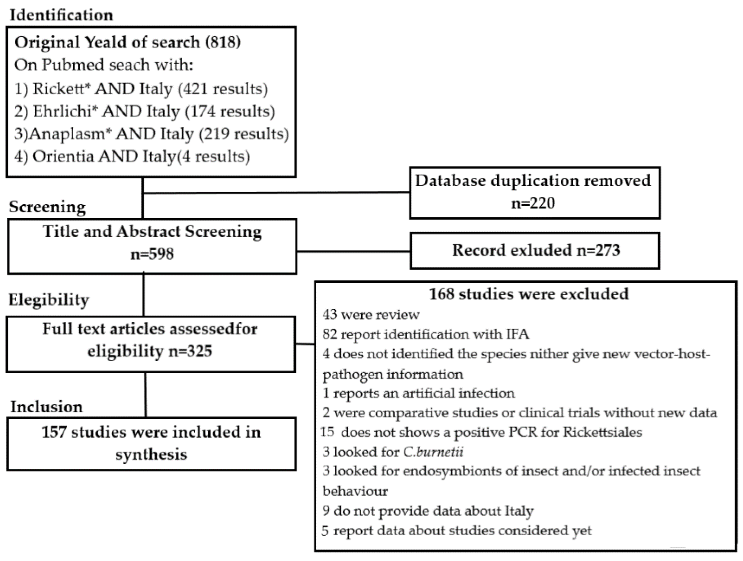
Table 1.
Species of Rickettsiales, arthropods from which they were identified and corresponding hosts.
Table 1.
Species of Rickettsiales, arthropods from which they were identified and corresponding hosts.
| Rickettsiales | Arthropod | Collected From | Reference |
| Anaplasma marginale | Haemaphysalis punctata | Cattle | [8] |
| Rhipicephalus turanicus | Cattle | [8] | |
| Xenopsylla cheopis | Fox | [8] | |
| A. ovis | Ctenocephalides canis | Fox | [9] |
| Haemaphysalis punctata | Sheep | [10] | |
| Rhipicephalus bursa | Goat, mouflon | [11] | |
| Rhipicephalus sanguineus s.l. | Dog | [12] | |
| Xenopsylla cheopis | Fox | [9] | |
| A. phagocytophilum | Haemaphysalis punctata | Sheep | [10] |
| Ixodes ricinus | Bird, cat, dog, fallow deer, free life, horse, human, red deer, rodent, roe deer, sheep, vegetation | [12,13,14,15,16,17,18,19,20,21,22,23,24] | |
| Ixodes ventalloi | Vegetation | [25] | |
| Hyalomma marginatum | Migratory birds | [11] | |
| Ixodes acuminatus | Dog | [13] | |
| Rhipicephalus sanguineus | Dog | [13,26] | |
| Rhipicephalus turanicus | Dog, horse, sheep, goat | [13,27] | |
| Rhipicephalus bursa | Dog | [13] | |
| Xenopsylla cheopis | Fox | [9] | |
| A. platy | Ixodes hexagonus | Dog | [12,28] |
| Hyalomma marginatum | Wild boar | [11] | |
| Rhipicephalus bursa | Goat | [11] | |
| Rhipicephalus sanguineus | Dog | [29,30] | |
| Rhipicephalus sp. II | Dog | [31] | |
| Anaplasma spp. | Haemaphysalis punctata | Mouflon | [32] |
| Ixodes festai | Hedgehog | [32] | |
| Rhipicephalus bursa | Mouflon, cattle, fox | [32] | |
| Rhipicephalus sanguineus s.l. | Cat, fox, goat, marten, mouflon | [32] | |
| Candidatus Ehrlichia walkerii | Ixodes ricinus | Dog, goat, human, sheep, vegetation, | [12,16,23,33] |
| E. ovina | Haemaphysalis punctata | Sheep | [10] |
| E. canis | Cediopsylla inaequalis | Fox | [9] |
| Dermacentor marginatus | Mouflon, wild boar | [27,34] | |
| Haemaphysalis punctata | Mouflon | [32] | |
| Haemaphysalis sulcata | Goat, mouflon | [27,34] | |
| Hyalomma marginatum | Sheep, swine | [34,35] | |
| Ixodes ventalloi | Cat | [35] | |
| Rhipicephalus bursa | Deer, sheep, goat | [32,34] | |
| Rhipicephalus sanguineus s.l. | Dog, fox, sheep | [12,32,34] | |
| Rhipicephalus sanguineus | Dog | [27] | |
| Xenopsylla cheopis | Fox | [9] | |
| Ehrlichia spp. | Amblyomma spp. | Migratory birds | [36] |
| Hyalomma marginatum | Migratory birds | [36] | |
| Hyalomma rufipes | Migratory birds | [36] | |
| Rickettsia africae | Amblyomma marginatus | Sheep | [37] |
| Amblyomma variegatum | Migratory birds | [38,39] | |
| Hyalomma marginatum | Cattle | [40] | |
| Hyalomma rufipes | Migratory birds | [41] | |
| Hyalomma spp. | Migratory birds | [36] | |
| Ixodes ricinus | Migratory birds | [36] | |
| R. aeschlimannii | Amblyomma marmoreum | Migratory birds | [41] |
| Dermacentor marginatus | Human | [41] | |
| Hyalomma marginatum | Bird, cattle, dog, free life, goat, hedgehog, migratory birds, horse, human, mouflon, red deer, sheep, vegetation | [32,34,36,38,40,42,43,44,45,46,47,48,49,50] | |
| Hyalomma lusitanicum | Human, free life | [42,46,51] | |
| Hyalomma rufipes | Migratory birds | [36,38,39,41] | |
| Hyalomma truncatum | Migratory birds | [38] | |
| Hyalomma detritum | Vegetation | [50] | |
| Ixodes ricinus | Free life, human | [42,52,53] | |
| Rhipicephalus turanicus | Free life | [53] | |
| R. barbarie (candidatus) | Rhipicephalus sanguineus s.l. | Dog, fox, goat | [32] |
| Rhipicephalus turanicus | Goat, sheep | [43,47] | |
| R. belli | Hippoboscidae Crataerina pallida | Bird | [54] |
| R. conorii | Ixodes ricinus | Free life | [53] |
| Rhipicephalus turanicus | Brown Hare, cattle, free life, human | [40,42,48,53] | |
| Rhipicephalus sanguineus | Dog, free life | [49,55,56] | |
| Rhipicephalus sanguineus s.l. | Human | [42] | |
| R. conorii subsp. israelensis | Rhipicephalus sanguineus | Dog | [43,57] |
| Rhipicephalus sanguineus s.l. | Dog, fox, goat | [32] | |
| R. felis | Ctenocephalides felis | Dog, cat, fox | [9,35,58,59,60] |
| Ixodes hexagonus | Fox, hedgehog | [48] | |
| Rhipicephalus turanicus | Sheep | [61] | |
| R. helvetica | Ixodes ricinus | Bird, cat, dog, fox, free life, human, lizard, migratory birds, red deer, rodent, roe deer, vegetation | [22,23,48,51,52,55,61,62,63,64,65,66,67,68,69,70,71,72,73,74,75,76,77,78] |
| Ixodes ventalloi | Bird, cat, human | [50,51,75] | |
| Ixodes festai | Cat, hedgehog | [45,46,67] | |
| Ixodes acuminatus | Cat, red partridge | [50] | |
| Ixodes trianguliceps | Rodent | [62] | |
| Rhipicephalus sanguineus | Vegetation | [68] | |
| R. honei | Hyalomma marginatum | Cattle | [40] |
| R. hoogstraalii | Haemaphysalis punctata | Mouflon, sheep | [45,46] |
| Haemaphysalis sulcata | Mouflon, sheep | [45,46] | |
| Ixodes ricinus | Lizard | [78] | |
| R. IRS3 | Ixodes ricinus | Migratory birds, vegetation | [64,74] |
| Ixodes ventalloi | Cat | [50] | |
| R. limoniae | Ixodes ricinus | Vegetation | [70] |
| R. massiliae | Ixodes ricinus | Free life, human | [48,52,53] |
| Rhipicephalus turanicus | Brown hare, cattle, free life, goat, human | [42,43,47,48,53] | |
| Rhipicephalus sanguineus s.l. | Dog, fox, goat, human, sheep | [32,36,42,46,48] | |
| Rhipicephalus sanguineus | Dog, fox, human, cat | [43,45,49,56,79] | |
| R. mendelii (candidatus) | Ixodes ricinus | Migratory birds | [74] |
| R. monacensis | Dermacentor marginatus | Free life, human | [51,53] |
| Haemaphysalis punctata | Chamois, fallow deer | [48,80] | |
| Hippoboscidae Crataerina pallida | Bird | [54] | |
| Ixodes ricinus | Bear, cat, chamois, dog, fallow deer, free life, goat, hare, human, lizard, migratory birds, red deer, rodent, roe deer, vegetation, wild boar, wolf | [22,48,51,52,53,55,62,64,66,68,69,70,71,72,74,75,76,77,78,80,81,82] | |
| Rhipicephalus sanguineus s.l. | Cat, dog | [48,75] | |
| Rhipicephalus turanicus | Free life | [53] | |
| Rhipicephalus sanguineus | Vegetation | [68] | |
| R. tamurae | Ixodes ricinus | Human, red deer | [62] |
| R. peacockii | Dermacentor marginatus | Human | [51] |
| R. raoultii | Dermacentor marginatus | Deer, human, rodent, wild boar | [45,51,61,72,83,84,85,86,87] |
| Hyalomma spp. | Migratory birds | [41] | |
| Ixodes hexagonus | Badger | [48] | |
| Ixodes ricinus | Free life, red deer | [52,62] | |
| Rhipicephalus sanguineus s.l. | Dog | [46] | |
| R. rhipicephali | Rhipicephalus sanguineus | Cat | [79] |
| R. rioja | Dermacentor marginatus | Vegetation, wild boar | [88] |
| Candidatus R. siciliensis | Rhipicephalus turanicus | Human | [89] |
| R. slovaca | Dermacentor marginatus | Cattle, chamois, deer, dog, human, red deer, rodent, roe deer, sheep, swine, vegetation, wild boar | [32,34,39,42,43,45,46,48,51,61,72,80,83,84,85,86,87,88,90] |
| Dermacentor reticulatus | Wild Boar | [88] | |
| Haemaphysalis punctata | Cattle | [40] | |
| Hyalomma sulcata | Dog, sheep | [34] | |
| Ixodes ricinus | Human, red deer, wild boar | [48,80,87] | |
| Rhipicephalus sanguineus s.l. | Human | [42] | |
| Rhipicephalus sanguineus | Human | [51] | |
| R. sp strain S | Hyalomma marginatum | Cattle | [42] |
| R. sp. strain TwKm01 | Ixodes ricinus | Free life | [53] |
| Rhipicephalus turanicus | Free life | [53] | |
| Rickettsia spp. | Haemaphysalis inermis | Human | [51] |
| Rhipicephalus annulatus | Cattle | [91] | |
| Rhipicephalus bursa | Cattle | [91] | |
| Rhipicephalus turanicus | Fox | [49] |
Table 2.
Arthropod species found infected with Rickettsiales in Italy.
| Arthropods | Rickettsiales | Arthropods Collected From | Reference |
|---|---|---|---|
| Amblyomma marginatus | R. africae | Sheep | [37] |
| Amblyomma marmoreum | R. aeschlimannii | Migratory birds | [41] |
| Amblyomma spp. | Ehrlichia spp. | Migratory birds | [36] |
| R. aeschlimannii | Migratory birds | [36] | |
| Amblyomma variegatum | R. africae | Migratory birds | [38,39] |
| Cediopsylla inaequalis | E. canis | Fox | [9] |
| Ctenocephalides canis | A. ovis | Fox | [9] |
| Ctenocephalides felis | R. felis | Cat, dog, fox | [9,35,58,59] |
| Dermacentor marginatus | E. canis | Mouflon, wild boar | [27,34] |
| R. aeschlimannii | Human | [42] | |
| R. monacensis | Free life, human | [51,53] | |
| R. peacockii | Human | [51] | |
| R. raoultii | Deer, human, rodent, wild boar | [23,46,51,83,84,85,86,87] | |
| R. rioja | Vegetation, wild boar | [88] | |
| R. slovaca | Cattle, chamois, deer, dog, human, red deer, rodent, roe deer, sheep, swine, vegetation, wild boar | [32,34,40,42,43,46,48,51,61,72,80,83,84,85,86,87,88,90,92] | |
| Dermacentor reticulatus | R. slovaca | Wild Boar | [88] |
| Haemaphysalis inermis | Rickettsia spp. | Human | [51] |
| Haemaphysalis punctata | A. marginale | Cattle | [8,93] |
| A. ovis | Sheep | [10] | |
| A. phagocytophilum | Sheep | [10] | |
| Anaplasma spp. | Mouflon | [32] | |
| E. canis | Mouflon | [32] | |
| E. ovina | Sheep | [10] | |
| R. hoogstraalii | Mouflon, sheep | [32,45] | |
| R. monacensis | Chamois, fallow deer | [48,80] | |
| R. slovaca | Cattle | [40] | |
| Hippoboscidae Crataerina pallida | R. belli | Bird | [54] |
| R. monacensis | Bird | [54] | |
| Hyalomma detritum | R. aeschlimannii | Vegetation | [50] |
| Hyalomma lusitanicum | R. aeschlimannii | Human, free life | [42,46,51] |
| Hyalomma marginatum | A. platy | Migratory birds | [11] |
| A. phagocytophilum | Migratory birds | [11] | |
| E. canis | Sheep, swine | [34] | |
| Ehrlichia spp. | Migratory birds | [36] | |
| R. africae | Cattle | [40] | |
| R. aeschlimannii | Bird, cattle, dog, free life, goat, hedgehog, horse, human, migratory birds, mouflon, red deer, sheep, vegetation | [32,34,36,38,40,42,43,44,45,46,47,48,50] | |
| R. honei | Cattle | [40] | |
| R. sp strain S | Cattle | [40] | |
| Hyalomma rufipes | Ehrlichia spp. | Migratory birds | [36] |
| R. africae | Migratory birds | [41] | |
| R. aeschlimannii | Migratory birds | [36,38,39,41] | |
| Hyalomma spp. | R. raoultii | Migratory birds | [41] |
| Hyalomma sulcata | R. slovaca | Dog, sheep | [34] |
| Hyalomma truncatum | R. aeschlimannii | Migratory birds | [38] |
| Ixodes acuminatus | A. phagocytophilum | Dog | [13] |
| R. helvetica | Cat, red partridge | [50] | |
| Ixodes hexagonus | A. platy | Dog | [12] |
| R. felis | Fox, hedgehog | [48] | |
| R. raoultii | Badger | [48] | |
| R. slovaca | Badger | [48] | |
| Ixodes festai | Anaplasma spp. | Hedgehog | [32] |
| R. helvetica | Cat, hedgehog | [32,34,45] | |
| Ixodes ricinus | A. phagocytophilum | Bird, cat, dog, fallow deer, free life, horse, human, red deer, sheep, vegetation | [12,13,15,16,17,19,20,21,22,23,24,62,94,95] |
| Candidatus Ehrlichia walkerii | Dog, goat, human, sheep, vegetation, human | [12,16,23,33] | |
| R. africae | Migratory birds | [36] | |
| R. aeschlimannii | Free life, human | [42,52,53] | |
| R. conorii | Free life | [53] | |
| R. helvetica | Bird, cat, dog, fox, free life, human, lizard, migratory birds, red deer, roe deer, vegetation | [23,51,52,55,61,62,63,64,65,66,67,68,69,70,71,72,73,74,76,78] | |
| R. hoogstraalii | Lizard | [78] | |
| R. IRS3 | Migratory birds, vegetation | [64,74] | |
| R. limoniae | Vegetation | [70] | |
| R. massiliae | Free life, human | [48,52,53] | |
| Candidatus R. mendelii | Migratory birds | [74] | |
| R. monacensis | Bear, cat, chamois, dog, fallow deer, free life, goat, hare, human, lizard, migratory birds, red deer, roe deer, rodent, vegetation, wild boar, wolf | [22,48,49,51,52,53,55,64,66,68,69,70,71,72,74,75,76,78,80,81,82,84] | |
| R. tamurae | Human, red deer | [62] | |
| R. raoultii | Free life, red deer | [52,62] | |
| R. slovaca | Human, red deer, wild boar | [48,80,87] | |
| R. sp. strain TwKm01 | Free life | [53] | |
| Ixodes trianguliceps | R. helvetica | Rodent | [62] |
| Ixodes ventalloi | A. phagocytophilum | Vegetation | [25] |
| E. canis | Cat | [35] | |
| R. helvetica | Bird, cat, human | [50,51,75] | |
| R. IRS3 | Cat | [50] | |
| R. monacensis | Cat | [75] | |
| Rhipicephalus annulatus | Rickettsia spp. | Cattle | [91] |
| Rhipicephalus bursa | A. phagocytophilum | Dog | [11] |
| A. platy | Goat | [11] | |
| Anaplasma spp. | Mouflon | [11] | |
| A. ovis | Goat, mouflon | [11] | |
| Anaplasma spp. | Cattle, fox, goat | [11] | |
| E. canis | Deer, goat, sheep | [32,34] | |
| Rickettsia spp. | Cattle | [91] | |
| Rhipicephalus sanguineus | A. phagocytophilum | Dog | [13,96] |
| A. platy | Dog | [29,30] | |
| E. canis | Dog | [27] | |
| R. conorii | Dog, free life | [49,55,56] | |
| R. conorii subsp. israelensis | Dog | [43,57] | |
| R. helvetica | Vegetation | [68] | |
| R. massiliae | Cat, fox, human | [43,45,47,49,56,79] | |
| R. rhipicephali | Cat | [79] | |
| R. monacensis | Vegetation | [68] | |
| R. slovaca | Human | ||
| Rhipicephalus sanguineus s.l | A. ovis | Dog | [12] |
| Anaplasma spp. | Cat, fox, goat, marten, mouflon | [32] | |
| E. canis | Dog, fox, sheep | [12,32,34] | |
| Candidatus R. barbariae | Dog, fox, human | [32] | |
| R. conorii | Human | [42] | |
| R. conorii subsp. israelensis | Dog, fox, goat | [32] | |
| R. massiliae | Dog, fox, goat, human, sheep | [32,34,42,46,48] | |
| R. monacensis | Cat, dog | [48,75] | |
| R. raoultii | Dog | [46] | |
| R. slovaca | Human | [42] | |
| Rhipicephalus sp. II | A. platy | Dog | [31] |
| Rhipicephalus turanicus | A. marginale | Cattle | [8] |
| A. phagocytophilum | Dog, horse, sheep, goat | [13,27] | |
| R. aeschlimannii | Free life | [53] | |
| Candidatus R. barbariae | Goat, sheep | [43] | |
| R. conorii | Brown Hare, cattle, free life, human | [40,42,48,53] | |
| R. felis | Sheep | [61] | |
| R. massiliae | Brown Hare, cattle, free life, goat, human | [42,43,47,48,53] | |
| R. monacensis | Free life | [53] | |
| Candidatus R. siciliensis | Human | [89] | |
| R. sp. strain TwKm01 | Free life | [53] | |
| Rickettsia spp. | Fox | [97] | |
| Xenopsylla cheopis | A. marginale | Fox | [9] |
| A. ovis | Fox | [9] | |
| A. phagocytophilum | Fox | [9] | |
| E. canis | Fox | [9] |
Table 3.
Rickettsiales identified in wild and domestic animal tissues in Italy.
| Rickettsiales | Animal | Clinical Manifestation | Number of Positive/Tested Animals (%) | Reference |
|---|---|---|---|---|
| Anaplasma bovis | Cattle | No symptoms | 1/51 (1.9%) | [98] |
| Sheep | No symptoms | 3/20 (15%) | [99] | |
| A. centrale | Cattle | Acute anaplasmosis: hypo-galactia, mucosal paleness, depression high temperature (40–45 °C), anemia, thrombocytopenia, erythrocytic inclusion | 26/270 (8–21%) | [98,100] |
| A. marginale | Cattle | No symptoms | 535/2500 (2.3–76,4%) | [8,93,98,99,100,101,102,103,104,105] |
| Dog | No symptoms | 2/46 (4.3%) | [102] | |
| Goat | No symptoms | 95/184 (27–85%) | [99,102,106] | |
| Horse | No symptoms | 26/134 (19.4%) | [102] | |
| Rodent | No symptoms | 3/69 (4.3%) | [102] | |
| Sheep | No symptoms | 35/286 (12.2%) | [102] | |
| A. ovis | Cattle | No symptoms | 22/374 (5.9%) | [102] |
| Dog | No symptoms | 2/46 (4.3%) | [102] | |
| Goat | No symptoms | 113/468 (14.9–85%) | [99,101,102] | |
| Horse | No symptoms | 23/134 (17.1%) | [102] | |
| Rodent | No symptoms | 23/69 (33%) | [102] | |
| Sheep | No symptoms | 164/716 (11–81.8%) | [10,93,99,101,102,103,104] | |
| A. phagocytophilum | Cattle | No symptoms | 233/984 (2–88%) | [99,100,101,102,105,106] |
| Cat | Lymphadenomegaly, pale mucous, stomatitis, sign of respiratory involvement | 47/360 (1–31.9%) | [101,107] | |
| Chamois | No symptoms | 6/9 (66.6%) | [19] | |
| Dog | Fever, acute lameness to right forelimb, depression, jaundice, dysorexia, leukocytosis, neutrophilia, thrombocytopenia, mild anemia | 15 | [26,96,102,107,108,109,110,111,112] | |
| Donkey | No symptoms | 3/3 (100%) | [93] | |
| Fallow deer | No symptoms | 42/80 (40%) | [24,113] | |
| Fox | No symptoms | 18/277 (0.8–10.8%) | [114,115,116] | |
| Goat | No symptoms | 55/203 (16.9–72%) | [103,117] | |
| Horse | Hyperthermia, anemia, anorexia, jaundice, myalgia, reluctance to move, thrombocytopenia, leukocytosis | 45 | [26,96,103,118,119,120,121,122] | |
| Red deer | No symptoms | 66/119 (54–59%) | [116,123] | |
| Rodent | No symptoms | 56/2259 (3–5.3%) | [12,103,124] | |
| Roe deer | No symptoms | 32/116 (19–75%) | [103,116,125] | |
| Sheep | Screening or poor health condition | 166/1496 (0.1–81%) | [10,99,103,104,117,118,119] | |
| A. platy | Dog | Depression, myalgia, anorexia, fever, epistaxis, rough coat, reluctance to move, diarrhea, lymphadenomegaly, weight loss, pale mucous membranes, high hepatic enzymes, splenomegaly, ascites, thrombocytopenia, anemia, mono-cytosis, eosinophilia, neutropenia or neutrophilia hypoalbuminemia; evidence of vertical transmission | 181 | [26,29,30,56,96,103,112,126,127,128,129,130,131,132,133,134,135,136] |
| Cat | Thrombocytopenia, anemia, or leukopenia/ leukocytosis | 14 | [135] | |
| Candidatus Neoehrlichia mikurensis | Rodent | No symptoms | 1/34 (2.9%) | [125] |
| Ehrlichia canis | Cat | No symptoms | 2/85 (2.3%) | [19] |
| Fox | No symptoms | 113/225 (52–56%) | [137,138] | |
| Dog | Pulmonary hypertension, fever, anemia, tongue ulcer, lymphadenopathy, polyclonal gammopathy, weight loss, anorexia, dermatitis, epistaxis | 78 | [111,126,132,133,134,136,139,140,141,142] | |
| Gray wolves | No symptoms | 3/6 (50%) | [138] | |
| E. ovina | Sheep | No symptoms | 1/87 (1.1%) | [10] |
| Ehrlichia spp. | Cat | lymphadenopathy, pale mucous, stomatitis, sign of respiratory involvement | 14/260 (5.3%) | [143] |
| Rickettsia conorii | Dog | Fever, anemia, thrombocytopenia, leukocytosis, hunched posture, abdominal pain, orchitis, splenomegaly, lymphadenopathy, vomiting, diarrhea, hyperglobulinemia, elevated liver enzyme | 27 | [106,112,133,144] |
| Eurasian Otter | Carcass | 1/1 (100%) | [145] | |
| Rickettsia spp. | Cat | limp adenomegaly, pale mucous, stomatitis, sign of respiratory involvement | 83/260 (31.9%) | [143] |
Table 4.
Rickettsiales identified by molecular methods in clinically ill patients in Italy.
| Species | Fever | TacheNoire | Rash | Clinical Notes | Number of Cases | Reference |
|---|---|---|---|---|---|---|
| Rickettsiaconorii | Yes | Yes | Yes | MSF, Sepsis, respiratory insufficiency and quadriplegia | 1 | [146] |
| Yes | Yes | Yes | MSF, PCR positive on eschar while negative on whole blood | 1 | [147] | |
| Yes | Yes (two) | Yes | MSF, Rhabdomyolysis, acute kidney injury and Encephalitis | 1 | [148] | |
| Yes | Yes | Yes | MSF, acute kidney injury and herpetic esophagitis | 1 | [149] | |
| Yes | Yes | Yes | MSF, Myocarditis-sepsis induced multi organ failure | 1 | [150] | |
| Yes | Yes | Yes | MSF | 5 | [151] | |
| Yes | Yes | Yes | MSF, HIV patient | 1 | [152] | |
| Yes | Yes | Yes | MSF | 1 | [153] | |
| R. conorii subsp. indica | Yes | Yes | Yes | MSF | 1 | [154] |
| R. conorii subsp. israelensis | Yes | No | Yes | Petechial rash, severe sepsis and multi organ failure | 1 | [155] |
| Yes | No | Yes | Dysarthria, dysdiadochokinesis, mild neck stiffness, meningism | 1 | [155] | |
| Yes | Yes | Yes | 1 | [156] | ||
| R. massiliae | Yes | Yes | No | TIBOLA, bite on eyelid, PCR positive on lesion and swab | 1 | [157] |
| Yes | Yes | No | TIBOLA; palpable liver, PCR positive on eschar | 1 | [158] | |
| Yes | Yes | Yes | MSF | 1 | [159] | |
| R. monacensis | Yes | Yes | No | MSF, PCR positive on tache noir and negative in whole blood | 1 | [160] |
| R. africae | Yes | Yes | Only ipsilateral limb | Sacral syndrome, traveler from Zimbabwe | 1 | [161] |
| R. slovaca | No | No | No | asymptomatic | 1 | [92] |
| Yes | Yes | No | TIBOLA, alopecia and painful lymph node | 1 | [92] | |
| No | Yes | No | TIBOLA, myalgia, weariness and painful lymph node | 1 | [92] | |
| No | Yes | No | TIBOLA, itching | 1 | [92] | |
| No | Yes | No | TIBOLA, painful cervical lymph node | 1 | [92] | |
| Yes | Yes | Yes | MSF-like | 1 | [154] | |
| R.aeschlimannii | Yes | Yes | No | Hepatitis, PCR positive on liver biopsies | 1 | [162] |
| Anaplasmaphagocytophilum | Yes | No | Yes | Atypical pneumonia, leukopenia and thrombocytopenia, high liver enzyme | 1 | [163] |
| Yes | No | Yes | Oral erythema, edema of labium, leukopenia and thrombocytopenia | 1 | [163] | |
| Yes | No | No | 6-month illness-misdiagnosis | 1 | [164] | |
| Yes | No | No | Myalgia | 1 | [165] | |
| Yes | No | No | Nuchal rigidity and myalgia | 1 | [165] | |
| No | No | No | Asymptomatic | 1 | [165] |
Publisher’s Note: MDPI stays neutral with regard to jurisdictional claims in published maps and institutional affiliations. |
© 2021 by the authors. Licensee MDPI, Basel, Switzerland. This article is an open access article distributed under the terms and conditions of the Creative Commons Attribution (CC BY) license (http://creativecommons.org/licenses/by/4.0/).
Share and Cite
MDPI and ACS Style
Guccione, C.; Colomba, C.; Tolomeo, M.; Trizzino, M.; Iaria, C.; Cascio, A. Rickettsiales in Italy. Pathogens 2021, 10, 181. https://doi.org/10.3390/pathogens10020181
AMA Style
Guccione C, Colomba C, Tolomeo M, Trizzino M, Iaria C, Cascio A. Rickettsiales in Italy. Pathogens. 2021; 10(2):181. https://doi.org/10.3390/pathogens10020181
Chicago/Turabian StyleGuccione, Cristoforo, Claudia Colomba, Manlio Tolomeo, Marcello Trizzino, Chiara Iaria, and Antonio Cascio. 2021. "Rickettsiales in Italy" Pathogens 10, no. 2: 181. https://doi.org/10.3390/pathogens10020181
Note that from the first issue of 2016, this journal uses article numbers instead of page numbers. See further details here.