
Lutein Has a Positive Impact on Brain Health in Healthy Older Adults: A Systematic Review of Randomized Controlled Trials and Cohort Studies

Abstract
:1. Introduction
2. Materials and Methods
2.1. Systematic Review Protocol and Registration
2.2. Search Strategy
2.3. Detail of Included Studies
2.3.1. Types of Study
2.3.2. Participants
2.4. Main Outcomes
2.5. Data Extraction
3. Results
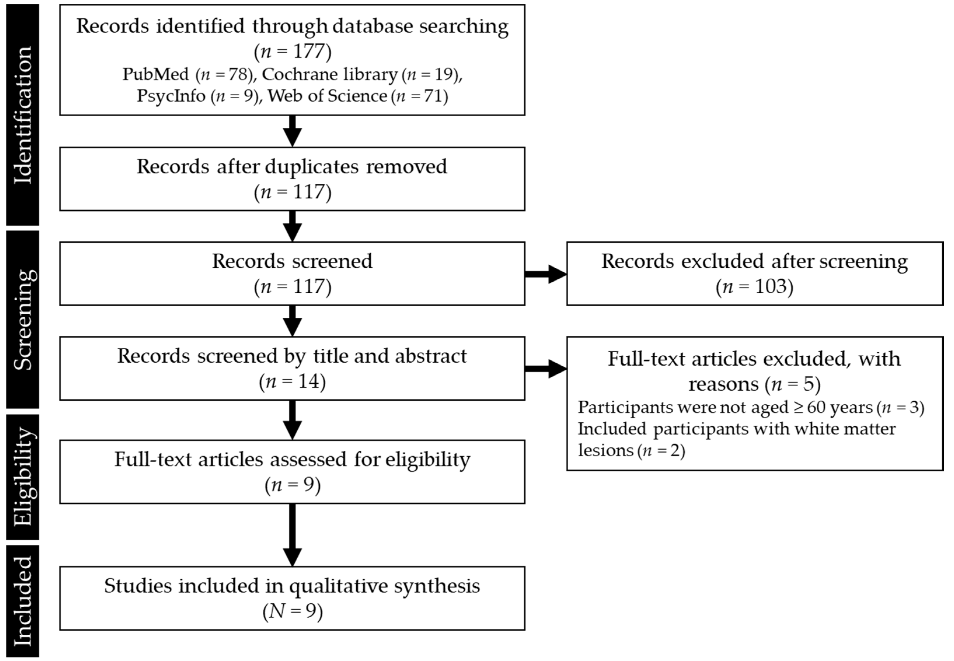
3.1. Search Results
3.2. Included Studies
3.3. Participants
3.4. MPOD
3.5. Outcomes and Imaging Methods
3.6. Quality Assessment
4. Discussion
5. Conclusions
Supplementary Materials
Author Contributions
Funding
Institutional Review Board Statement
Informed Consent Statement
Data Availability Statement
Acknowledgments
Conflicts of Interest
References
- Carrillo, J.Á.; Zafrilla, M.P.; Marhuenda, J. Cognitive function and consumption of fruit and vegetable polyphenols in a young population: Is there a relationship? Foods 2019, 8, 507. [Google Scholar] [CrossRef] [Green Version]
- Jiang, X.; Huang, J.; Song, D.; Deng, R.; Wei, J.; Zhang, Z. Increased consumption of fruit and vegetables is related to a reduced risk of cognitive impairment and dementia: Meta-analysis. Front. Aging Neurosci. 2017, 9, 18. [Google Scholar] [CrossRef] [PubMed] [Green Version]
- Mottaghi, T.; Amirabdollahian, F.; Haghighatdoost, F. Fruit and vegetable intake and cognitive impairment: A systematic review and meta-analysis of observational studies. Eur. J. Clin. Nutr. 2018, 72, 1336–1344. [Google Scholar] [CrossRef] [PubMed]
- Morris, M.C.; Evans, D.A.; Tangney, C.C.; Bienias, J.L.; Wilson, R.S. Associations of vegetable and fruit consumption with age-related cognitive change. Neurology 2006, 67, 1370–1376. [Google Scholar] [CrossRef] [PubMed] [Green Version]
- Nooyens, A.C.J.; Bueno-De-Mesquita, H.B.; Van Boxtel, M.P.J.; Van Gelder, B.M.; Verhagen, H.; Verschuren, W.M.M. Fruit and vegetable intake and cognitive decline in middle-aged men and women: The doetinchem cohort study. Br. J. Nutr. 2011, 106, 752–761. [Google Scholar] [CrossRef] [Green Version]
- Kang, J.H.; Ascherio, A.; Grodstein, F. Fruit and vegetable consumption and cognitive decline in aging women. Ann. Neurol. 2005, 57, 713–720. [Google Scholar] [CrossRef]
- Péneau, S.; Galan, P.; Jeandel, C.; Ferry, M.; Andreeva, V.; Hercberg, S.; Kesse-Guyot, E.; Vogt, L.; Escande, M.; Sérot, J.M.; et al. Fruit and vegetable intake and cognitive function in the SU.VI.MAX 2 prospective study. Am. J. Clin. Nutr. 2011, 94, 1295–1303. [Google Scholar] [CrossRef]
- Lee, S.; Kim, E.Y.; Shin, C. Changes in brain volume associated with vegetable intake in a general population. J. Am. Coll. Nutr. 2019, 38, 506–512. [Google Scholar] [CrossRef]
- Yabuzaki, J. Carotenoids database: Structures, chemical fingerprints and distribution among organisms. Database 2017, 2017, 1–11. [Google Scholar] [CrossRef] [Green Version]
- Khoo, H.E.; Prasad, K.N.; Kong, K.W.; Jiang, Y.; Ismail, A. Carotenoids and their isomers: Color pigments in fruits and vegetables. Molecules 2011, 16, 1710–1738. [Google Scholar] [CrossRef]
- Perrone, S.; Tei, M.; Longini, M.; Buonocore, G. The multiple facets of lutein: A call for further investigation in the perinatal period. Oxid. Med. Cell. Longev. 2016, 2016, 5381540. [Google Scholar] [CrossRef] [PubMed] [Green Version]
- Perera, C.O.; Yen, G.M. Functional properties of carotenoids in human health. Int. J. Food Prop. 2007, 10, 201–230. [Google Scholar] [CrossRef]
- Johnson, E.J. Role of lutein and zeaxanthin in visual and cognitive function throughout the lifespan. Nutr. Rev. 2014, 72, 605–612. [Google Scholar] [CrossRef] [PubMed]
- Raman, G.; Haslam, D.; Avendano, E.; Johnson, E.J. Lutein/zeaxanthin intake and visual outcomes in adults with healthy eyes: Qualitative gap analysis. Cogent Med. 2019, 6, 1683939. [Google Scholar] [CrossRef]
- Hajizadeh-Sharafabad, F.; Ghoreishi, Z.; Maleki, V.; Tarighat-Esfanjani, A. Mechanistic insights into the effect of lutein on atherosclerosis, vascular dysfunction, and related risk factors: A systematic review of in vivo, ex vivo and in vitro studies. Pharmacol. Res. 2019, 149, 104477. [Google Scholar] [CrossRef]
- Niranjana, R.; Gayathri, R.; Mol, S.N.; Sugawara, T.; Hirata, T.; Miyashita, K.; Ganesan, P. Carotenoids modulate the hallmarks of cancer cells. J. Funct. Foods 2015, 18, 968–985. [Google Scholar] [CrossRef]
- Gong, X.; Smith, J.; Swanson, H.; Rubin, L. Carotenoid lutein selectively inhibits breast cancer cell growth and potentiates the effect of chemotherapeutic agents through ROS-mediated mechanisms. Molecules 2018, 23, 905. [Google Scholar] [CrossRef] [PubMed] [Green Version]
- Nouchi, R.; Suiko, T.; Kimura, E.; Takenaka, H.; Murakoshi, M.; Uchiyama, A.; Aono, M.; Kawashima, R. Effects of lutein and astaxanthin intake on the improvement of cognitive functions among healthy adults: A systematic review of randomized controlled trials. Nutrients 2020, 12, 617. [Google Scholar] [CrossRef] [Green Version]
- Erdman, J.; Smith, J.; Kuchan, M.; Mohn, E.; Johnson, E.; Rubakhin, S.; Wang, L.; Sweedler, J.; Neuringer, M. Lutein and brain function. Foods 2015, 4, 547–564. [Google Scholar] [CrossRef] [Green Version]
- Stringham, N.T.; Holmes, P.V.; Stringham, J.M. Effects of macular xanthophyll supplementation on brain-derived neurotrophic factor, pro-inflammatory cytokines, and cognitive performance. Physiol. Behav. 2019, 211, 112650. [Google Scholar] [CrossRef]
- Zamroziewicz, M.K.; Paul, E.J.; Zwilling, C.E.; Johnson, E.J.; Kuchan, M.J.; Cohen, N.J.; Barbey, A.K. Parahippocampal cortex mediates the relationship between lutein and crystallized intelligence in healthy, older adults. Front. Aging Neurosci. 2016, 8, 297. [Google Scholar] [CrossRef] [PubMed] [Green Version]
- Lindbergh, C.A.; Lv, J.; Zhao, Y.; Mewborn, C.M.; Puente, A.N.; Terry, D.P.; Renzi-Hammond, L.M.; Hammond, B.R.; Liu, T.; Miller, L.S. The effects of lutein and zeaxanthin on resting state functional connectivity in older Caucasian adults: A randomized controlled trial. Brain Imaging Behav. 2020, 14, 668–681. [Google Scholar] [CrossRef] [PubMed]
- Moher, D.; Liberati, A.; Tetzlaff, J.; Altman, D.G.; Altman, D.; Antes, G.; Atkins, D.; Barbour, V.; Barrowman, N.; Berlin, J.A.; et al. Preferred reporting items for systematic reviews and meta-analyses: The PRISMA statement. Ann. Intern. Med. 2009, 151, 264–269. [Google Scholar] [CrossRef] [Green Version]
- Lindbergh, C.A.; Renzi-Hammond, L.M.; Hammond, B.R.; Terry, D.P.; Mewborn, C.M.; Puente, A.N.; Miller, L.S. Lutein and zeaxanthin influence brain function in older adults: A randomized controlled trial. J. Int. Neuropsychol. Soc. 2018, 24, 77–90. [Google Scholar] [CrossRef]
- Mewborn, C.M.; Lindbergh, C.A.; Hammond, B.R.; Renzi-Hammond, L.M.; Miller, L.S. The effects of lutein and zeaxanthin supplementation on brain morphology in older adults: A randomized, controlled trial. J. Aging Res. 2019, 2019. [Google Scholar] [CrossRef] [PubMed]
- Ceravolo, S.A.; Hammond, B.R.; Oliver, W.; Clementz, B.; Miller, L.S.; Renzi-Hammond, L.M. Dietary carotenoids lutein and zeaxanthin change brain activation in older adult participants: A randomized, double-masked, placebo-controlled trial. Mol. Nutr. Food Res. 2019, 63, 1801051. [Google Scholar] [CrossRef] [Green Version]
- Mewborn, C.M.; Terry, D.P.; Renzi-Hammond, L.M.; Hammond, B.R.; Miller, L.S. Relation of retinal and serum lutein and zeaxanthin to white matter integrity in older adults: A diffusion tensor imaging study. Arch. Clin. Neuropsychol. 2018, 33, 861–874. [Google Scholar] [CrossRef]
- Mewborn, C.M.; Lindbergh, C.A.; Robinson, T.L.; Gogniat, M.A.; Terry, D.P.; Jean, K.R.; Hammond, B.R.; Renzi-Hammond, L.M.; Miller, L.S. Lutein and zeaxanthin are positively associated with visual–spatial functioning in older adults: An fMRI study. Nutrients 2018, 10, 458. [Google Scholar] [CrossRef] [Green Version]
- Lindbergh, C.A.; Mewborn, C.M.; Hammond, B.R.; Renzi-Hammond, L.M.; Curran-Celentano, J.M.; Miller, L.S. Relationship of lutein and zeaxanthin levels to neurocognitive functioning: An fMRI study of older adults. J. Int. Neuropsychol. Soc. 2017, 23, 11–22. [Google Scholar] [CrossRef] [Green Version]
- Oliver, W.; Renzi-Hammond, L.M.; Thorne, S.A.; Clementz, B.; Miller, L.S.; Hammond, B.R., Jr. Neural activation during visual attention differs in individuals with high versus low macular pigment density. Mol. Nutr. Food Res. 2019, 63. [Google Scholar] [CrossRef]
- Bone, R.A.; Landrum, J.T.; Dixon, Z.; Chen, Y.; Llerena, C.M. Lutein and Zeaxanthin in the eyes, serum and diet of human subjects. Exp. Eye Res. 2000, 71, 239–245. [Google Scholar] [CrossRef] [PubMed]
- Vishwanathan, R.; Schalch, W.; Johnson, E.J. Macular pigment carotenoids in the retina and occipital cortex are related in humans. Nutr. Neurosci. 2016, 19, 95–101. [Google Scholar] [CrossRef]
- Nouchi, R.; Kawashima, R. Improving cognitive function from children to old age: A systematic review of recent smart ageing intervention studies. Adv. Neurosci. 2014, 2014, 1–15. [Google Scholar] [CrossRef] [Green Version]
- Ozawa, Y.; Sasaki, M.; Takahashi, N.; Kamoshita, M.; Miyake, S.; Tsubota, K. Neuroprotective effects of lutein in the retina. Curr. Pharm. Des. 2012, 18, 51–56. [Google Scholar] [CrossRef] [PubMed] [Green Version]
- Stringham, J.M.; Johnson, E.J.; Hammond, B.R. Lutein across the lifespan: From childhood cognitive performance to the aging eye and brain. Curr. Dev. Nutr. 2019, 3, 1–8. [Google Scholar] [CrossRef] [PubMed] [Green Version]
- Craft, N.E.; Haitema, T.B.; Garnett, K.M.; Fitch, K.A.; Dorey, C.K. Carotenoid, tocopherol, and retinol concentrations in elderly human brain. J. Nutr. Health Aging 2004, 8, 156–162. [Google Scholar] [PubMed]
- Vishwanathan, R.; Kuchan, M.J.; Sen, S.; Johnson, E.J. Lutein and preterm infants with decreased concentrations of brain carotenoids. J. Pediatr. Gastroenterol. Nutr. 2014, 59, 659–665. [Google Scholar] [CrossRef] [PubMed]
- Lamport, D.J.; Pal, D.; Moutsiana, C.; Field, D.T.; Williams, C.M.; Spencer, J.P.E.; Butler, L.T. The effect of flavanol-rich cocoa on cerebral perfusion in healthy older adults during conscious resting state: A placebo controlled, crossover, acute trial. Psychopharmacology 2015, 232, 3227–3234. [Google Scholar] [CrossRef] [Green Version]
- Bookheimer, S.Y.; Renner, B.A.; Ekstrom, A.; Li, Z.; Henning, S.M.; Brown, J.A.; Jones, M.; Moody, T.; Small, G.W. Pomegranate juice augments memory and fMRI activity in middle-aged and older adults with mild memory complaints. Evidence-Based Complement. Altern. Med. 2013, 2013, 1–14. [Google Scholar] [CrossRef]
- Calabrese, F.; Rossetti, A.C.; Racagni, G.; Gass, P.; Riva, M.A.; Molteni, R. Brain-derived neurotrophic factor: A bridge between inflammation and neuroplasticity. Front. Cell. Neurosci. 2014, 8, 430. [Google Scholar] [CrossRef]
- Kowiański, P.; Lietzau, G.; Czuba, E.; Waśkow, M.; Steliga, A.; Moryś, J. BDNF: A key factor with multipotent impact on brain signaling and synaptic plasticity. Cell. Mol. Neurobiol. 2018, 38, 579–593. [Google Scholar] [CrossRef] [PubMed]
- Canas, J.A.; Lochrie, A.; McGowan, A.G.; Hossain, J.; Schettino, C.; Balagopal, P.B. Effects of Mixed Carotenoids on Adipokines and Abdominal Adiposity in Children: A Pilot Study. J. Clin. Endocrinol. Metab. 2017, 102, 1983–1990. [Google Scholar] [CrossRef] [PubMed] [Green Version]
- Eisenhauer, B.; Natoli, S.; Liew, G.; Flood, V.M. Lutein and zeaxanthin—Food sources, bioavailability and dietary variety in age-related macular degeneration protection. Nutrients 2017, 9, 120. [Google Scholar] [CrossRef]
- Perry, A.; Rasmussen, H.; Johnson, E.J. Xanthophyll (lutein, zeaxanthin) content in fruits, vegetables and corn and egg products. J. Food Compos. Anal. 2009, 22, 9–15. [Google Scholar] [CrossRef]
- Müller, V.I.; Cieslik, E.C.; Laird, A.R.; Fox, P.T.; Radua, J.; Mataix-Cols, D.; Tench, C.R.; Yarkoni, T.; Nichols, T.E.; Turkeltaub, P.E.; et al. Ten simple rules for neuroimaging meta-analysis. Neurosci. Biobehav. Rev. 2018, 84, 151–161. [Google Scholar] [CrossRef]


| Lead Author; Year; Country | Study Design; Duration | Sample Size (Female) | Age (Years) (Mean ± SD) | Health Status | Intervention (Timing or Method) | Control (Contents) | Imaging Method |
|---|---|---|---|---|---|---|---|
| Lindbergh; 2018; Georgia [24] | A single-site, double-blind RCT; 12 months | 44 (26) P: 14 A: 30 | P: 70.43 ± 5.43 A: 72.43 ± 6.48 | Community-dwelling older adults; good overall health | Consumed one pill per day with a meal (L: 10 mg + Z: 2 mg/placebo) | Placebo (n/R) | fMRI |
| Lindbergh; 2020; Georgia [22] | A single-site, double-blind RCT; 12 months | 48 (28) P: 14 A: 34 | P: 70.43 ± 5.43 A: 73.06 ± 6.48 | Community-dwelling older adults; good overall health; older adults without dementia | Consumed pills per day with a meal (L: 10 mg + Z: 2 mg/placebo) | Placebo (n/R) | fMRI |
| Mewborn; 2019; Georgia [25] | A single-site, double-blind RCT; 12 months | 47 (27) P: 14 A: 33 | P: 72.4 ± 6.27 A: 70.4 ± 5.43 | Community-dwelling older adults; good overall health; older adults without dementia (CDR = 0.5, P: 05; A: 12.1%) | Took one tablet from the bottle daily with a meal (L: 10 mg + Z: 2 mg/placebo) | Placebo (n/R) | sMRI |
| Ceravolo; 2019; Georgia [26] | A single-site, double-blind RCT; 12 months | 50 P: 15 (4) A: 35 (18) | P: 72.51 ± 6.24 A: 70.87 ± 5.50 | Community-dwelling older adults; good overall health; older adults without dementia (included CDR = 0.5) | Received either 10 mg of L + 2 mg of Z per day | Placebo (n/R) | EEG |
| Mewborn; 2018a; Georgia [27] | Cross-sectional study as part of a larger RCT; one-shot | O: 54 (31) Y: 38 (17) | O: 71.87 ± 6.05 Y: 20.58 ± 2.02 | Healthy men and women; older adults without dementia | No intervention | -- | sMRI |
| Mewborn; 2018b; Georgia [28] | Cross-sectional study as part of a larger RCT; one-shot | 51 (30) | 71.75 ± 6.16 | Community-dwelling older adults; good overall health; older adults without dementia | No intervention | -- | fMRI |
| Lindbergh; 2017; Georgia [29] | Cross-sectional study; one-shot | 43 (25) | 71.55 ± 5.84 | Community-dwelling older adults; good overall health | No intervention | -- | fMRI |
| Zamroziewicz; 2016; Illinois [21] | Cross-sectional study; one-shot | 76 (50) | 69 ± 3 | Healthy men and women; older adults without dementia | No intervention | -- | sMRI |
| Oliver; 2019; Georgia [30] | Case-control design study as part of a larger cross-sectional study; one-shot | O: 42 (26) Y: 43 (20) | O: 72.36 ± 6.58 Y: 20.79 ± 2.16 | Older adults without dementia (CDR = 0.5, Y: N/A; O: 9.5%) | No intervention | The stimuli presented and the task Instructions were controlled | EEG |
| Lead Author; Year | Group | Subgroup | MPOD | Serum Nutrients Lutein | |||
|---|---|---|---|---|---|---|---|
| Baseline | Post | (μmol/mL) | p Values | Effect Size | |||
| M (SD) | M (SD) | M (SD) | (t-test) | (Cohen’s d) | |||
| Lindbergh; 2018 [24] | Placebo | 0.44 (0.14) | 0.44 (0.19) | 0.961 | 0.03 | ||
| Supplement | 0.54 (0.19) | 8.80 (2.16) | 0.016 | 0.95 | |||
| Lindbergh; 2020 [22] | Placebo | 0.44 (0.14) | 0.44 (0.19) | 0.961 | 0.03 | ||
| Supplement | 0.50 (0.21) | 0.57 (0.23) | 0.008 | 0.98 | |||
| Mewborn; 2019 [25] | Placebo | Responder | 0.45 (0.20) | 0.69 (0.23) | |||
| Non-responder | 0.57 (0.17) | 0.51 (0.17) | |||||
| Supplement | Responder | 0.39 (0.16) | 0.594 (0.16) | ||||
| Non-responder | 0.50 (0.99) | 0.37 (0.18) | |||||
| Ceravolo; 2019 [26] | Placebo | 0.47 (0.17) | - | Non-significant | |||
| Supplement | 0.52 (0.18) | 0.58 (0.23) | <0.03 | ||||
| Mewborn; 2018a [27] | Younger adults | 0.43 (0.16) | |||||
| Older adults | 0.50 (0.17) | ||||||
| Mewborn; 2018b [28] | 0.50 (0.18) | ||||||
| Lindbergh; 2017 [29] | 0.51 (0.18) | ||||||
| Zamroziewicz; 2016 [21] | 454 (275) | ||||||
| Oliver; 2019 [30] | Younger adults | 0.43 (0.17) | |||||
| Older adults | 0.50 (0.19) | ||||||
| Lead Author; Year | Imaging Method | Results |
|---|---|---|
| Lindbergh; 2018 [24] | fMRI while participants were engaged in a verbal learning task | An enhanced BOLD signal in select ROIs, including left dorsolateral prefrontal cortex and anterior cingulate cortex |
| Lindbergh; 2020 [22] | Resting-state fMRI | An enhanced correlation of default mode network to other functional networks |
| Mewborn; 2019 [25] | MRI (T1-weighted, DTI) | Did not appear to influence age-related reductions for frontal and medial-temporal gray and white matter (exploratory analyses: individuals who showed greater increases in MPOD had less reduction in global and prefrontal gray matter volume than supplement “non-responders”) |
| Ceravolo; 2019 [26] | EEG while participants looked at stimuli to elicit the steady-state visual evoked potentials | Supplementation with L and Z changed both the power at the drive frequencies and the signal-to-noise ratio at those frequencies changed |
| Mewborn; 2018a [27] | MRI (DTI) | Higher L and Z concentrations predicted better white matter integrity in older adults |
| Mewborn; 2018b [28] | fMRI while participants were engaged in a judgment of line orientation task | Higher concentrations of L and Z decreased BOLD signal during task performance in key areas related to visual-spatial perception, decision-making, processing, and motor coordination |
| Lindbergh; 2017 [29] | fMRI while participants were engaged in a verbal learning task | MPOD was associated with activity in regions involved in language processing/serum L and Z predicted activity in regions involved in somatosensory functions |
| Zamroziewicz; 2016 [21] | MRI (T1-weighted) | Gray matter thickness only in the right parahippocampal cortex mediated the relationship between serum lutein and crystallized intelligence |
| Oliver; 2019 [30] | EEG while participants were engaged in an attentionally taxing task | MPOD covaried with visual attention |
Publisher’s Note: MDPI stays neutral with regard to jurisdictional claims in published maps and institutional affiliations. |
© 2021 by the authors. Licensee MDPI, Basel, Switzerland. This article is an open access article distributed under the terms and conditions of the Creative Commons Attribution (CC BY) license (https://creativecommons.org/licenses/by/4.0/).
Share and Cite
Yagi, A.; Nouchi, R.; Butler, L.; Kawashima, R. Lutein Has a Positive Impact on Brain Health in Healthy Older Adults: A Systematic Review of Randomized Controlled Trials and Cohort Studies. Nutrients 2021, 13, 1746. https://doi.org/10.3390/nu13061746
Yagi A, Nouchi R, Butler L, Kawashima R. Lutein Has a Positive Impact on Brain Health in Healthy Older Adults: A Systematic Review of Randomized Controlled Trials and Cohort Studies. Nutrients. 2021; 13(6):1746. https://doi.org/10.3390/nu13061746
Chicago/Turabian StyleYagi, Ayano, Rui Nouchi, Laurie Butler, and Ryuta Kawashima. 2021. "Lutein Has a Positive Impact on Brain Health in Healthy Older Adults: A Systematic Review of Randomized Controlled Trials and Cohort Studies" Nutrients 13, no. 6: 1746. https://doi.org/10.3390/nu13061746