Article

A Protein/Lipid Preload Attenuates Glucose-Induced Endothelial Dysfunction in Individuals with Abnormal Glucose Tolerance

by
Domenico Tricò
1,2,*,
Lorenzo Nesti
3,
Silvia Frascerra
3,
Simona Baldi
3,
Alessandro Mengozzi
3 and
Andrea Natali
3
1
Department of Surgical, Medical and Molecular Pathology and Critical Care Medicine, University of Pisa, Via Savi 10, 56126 Pisa, Italy
2
Institute of Life Sciences, Sant’Anna School of Advanced Studies, Via Santa Cecilia 3, 56127 Pisa, Italy
3
Department of Clinical and Experimental Medicine, University of Pisa, Via Roma 67, 56126 Pisa, Italy
*
Author to whom correspondence should be addressed.
Nutrients 2020, 12(7), 2053; https://doi.org/10.3390/nu12072053
Submission received: 25 May 2020 / Revised: 24 June 2020 / Accepted: 8 July 2020 / Published: 10 July 2020
(This article belongs to the Special Issue Nutrition on Endothelial Function)

Abstract

:
Postprandial hyperglycemia interferes with vascular reactivity and is a strong predictor of cardiovascular disease. Macronutrient preloads reduce postprandial hyperglycemia in subjects with impaired glucose tolerance (IGT) or type 2 diabetes (T2D), but the effect on endothelial function is unknown. Therefore, we examined whether a protein/lipid preload can attenuate postprandial endothelial dysfunction by lowering plasma glucose responses in subjects with IGT/T2D. Endothelial function was assessed by the reactive hyperemia index (RHI) at fasting, 60 min and 120 min during two 75 g oral glucose tolerance tests (OGTTs) preceded by either water or a macronutrient preload (i.e., egg and parmesan cheese) in 22 volunteers with IGT/T2D. Plasma glucose, insulin, glucagon-like peptide-1 (GLP-1), glucose-dependent insulinotropic polypeptide (GIP), glucagon, free fatty acids, and amino acids were measured through each test. RHI negatively correlated with fasting plasma glucose. During the control OGTT, RHI decreased by 9% and its deterioration was associated with the rise in plasma glucose. The macronutrient preload attenuated the decline in RHI and markedly reduced postprandial glycemia. The beneficial effect of the macronutrient preload on RHI was proportional to the improvement in glucose tolerance and was associated with the increase in plasma GLP-1 and arginine levels. In conclusion, a protein/lipid macronutrient preload attenuates glucose-induced endothelial dysfunction in individuals with IGT/T2D by lowering plasma glucose excursions and by increasing GLP-1 and arginine levels, which are known regulators of the nitric oxide vasodilator system.

1. Introduction

Despite the global improvement in diabetes care and the large availability of novel therapies, type 2 diabetes (T2D) and the prediabetic condition remain associated with increased cardiovascular risk [1]. This excess risk can be partly attributed to the adverse effects of hyperglycemia and glucose-induced oxidative stress on normal vascular biology [2,3,4,5]. Vascular endothelial cells secrete several mediators that participate in the regulation of vascular tone, platelet aggregation, coagulation, and fibrinolysis. The loss of one major physiologic property of the endothelium, which is the ability to induce vascular relaxation in response to transient ischemia, is commonly used to identify the condition of “endothelial dysfunction”, an early driver of atherosclerosis and cardiovascular disease [6].
Postprandial hyperglycemia occurs early in the progression of T2D and is a stronger predictor of cardiovascular disease than fasting hyperglycemia [7,8]. The acute increase in plasma glucose levels in response to carbohydrate ingestion rapidly and transiently suppresses endothelium-mediated vasodilation in a dose-dependent fashion [8,9,10]. Proposed mechanisms of endothelial dysfunction related to postprandial hyperglycemia and diabetes involve alterations in arginine [11], branched chain amino acid (BCAA) [12], and free fatty acid (FFA) metabolism [13], and changes in gut hormones such as the glucagon-like peptide-1 (GLP-1) [14]. In metabolically healthy individuals, the negative effect of postprandial hyperglycemia on endothelial function is counterbalanced by a parallel increase in insulin-induced nitric oxide (NO) production by endothelial cells. This compensatory mechanism, however, is reduced in insulin-resistant states [15]. A better understanding of the factors modulating vascular dilation in the absorptive phase and the identification of non-pharmacological strategies that positively impact on postprandial endothelial dysfunction are needed to improve the management of prediabetes and T2D.
We [16,17,18] and others [19,20,21,22,23] have recently demonstrated that a protein/lipid preload, ingested shortly before an oral glucose load, markedly reduces postprandial glucose responses by delaying oral glucose absorption and by increasing plasma amino acids, gut hormones, and glucose-stimulated insulin secretion [24,25]. Hence, we hypothesized that a similar nutritional approach could prevent the deterioration of endothelial function induced by glucose ingestion in individuals with prediabetes or T2D. To test this hypothesis, in the present study we assessed fasting and postprandial endothelial function after oral glucose loading preceded by water or a protein/lipid preload in subjects with abnormal glucose tolerance (AGT). We also measured plasma substrates and hormones to dissect the underlying physiological mechanisms.

2. Materials and Methods

2.1. Subjects

A total of 35 volunteers were recruited among students, fellows and patients attending the outpatient clinic of the Unit of Clinical Nutrition and Dietetics at the University of Pisa (Italy). The inclusion criteria were age 18–65 years, body mass index (BMI) 18–35 kg/m2, both men and women. Individuals with chronic or acute diseases (other than diet-controlled T2D and overweight/obesity), taking medications influencing endothelial function or glucose metabolism, and pregnant women were excluded. Individuals with T2D were recruited if recently diagnosed (≤5 years) and adequately controlled with diet alone (glycated hemoglobin 48–58 mmol/mol for at least six months). For the purpose of this study, former smokers were included if they had quit smoking for at least five years, while active smokers were excluded (n = 5). This resulted in a study population of 30 subjects.
Information regarding medical history, drug use, and smoking status was collected using standardized self-reported questionnaires. Brachial blood pressure was measured three times in subjects seated for at least 10 min, and the last two measurements were averaged for analysis. Subjects were classified as having normal glucose tolerance (NGT) or AGT, according to the current diagnostic criteria [26]. The latter group included individuals with either impaired glucose tolerance (IGT) or T2D.
The study was approved by the local Human Ethics Committee (Comitato Etico di Area Vasta Nord Ovest, or CEAVNO, clinical trial number: NCT02342834, approval code 13053_NATALI) and conducted in accordance with the principles expressed in the Declaration of Helsinki. All subjects provided written informed consent before enrollment.

2.2. Study Design

An outline of the study protocol is depicted in Figure 1. In this open, cross-over, randomized controlled trial, we measured fasting and postprandial endothelial function in individuals with AGT during two oral glucose tolerance tests (OGTTs) preceded by either water or a non-carbohydrate macronutrient preload, in a random order. Endothelial function was also measured during a control OGTT preceded by water in people with NGT. Further details of the protocol and metabolic data from the original cohort (n = 35) have been previously reported [16,27].

2.3. Metabolic Tests

Metabolic procedures were performed after an overnight fast (12 h) on two days separated by 2–4 weeks. Participants were asked to maintain their habitual lifestyle and to refrain from alcohol, caffeine, and exercise for at least 24 h prior to each visit. On each study day, volunteers were admitted to our Clinical Research Unit at 8 am. The tests were performed in a quiet room with controlled temperature of 20–22 °C. A 20-gauge polyethylene cannula was inserted into a superficial vein of the upper limb for blood sampling. Thirty minutes before glucose ingestion (time −30 to −25 min), volunteers were randomized to consume either 500 mL water (control study) or a small macronutrient preload consisting of 50 g parmesan cheese, one boiled egg, and 300 mL water (preload study). The preload was rich in protein and fat and virtually free from carbohydrates (23 g protein, 17 g fat, 2 g carbohydrate, for a total of ~250 Kcal). A glucose drink consisting of 150 mL of 50% glucose solution (wt/vol) was consumed at time 0. Blood samples were collected during each test at time −40, −30, −20, −10, 0, 15, 30, 45, 60, 90, and 120 min to measure plasma glucose, insulin, GLP-1, GIP, glucagon, free fatty acids (FFA), and amino acids (AA).

2.4. Peripheral Arterial Tonometry (PAT)

Endothelial function was assessed at fasting (time −60), at 60 min and at 120 min during each OGTT using an EndoPAT device (EndoPAT 2000, Itamar Medical Ltd., Caesarea, Israel), according to standard procedures [28,29,30]. This device records changes in the digital pulse waveform elicited by a downstream hyperemic response to transient ischemia, which are largely dependent on proper endothelial function [29]. Room temperature was maintained as stable throughout the study. Peripheral arterial tonometry (PAT) probes were placed on both forefingers for continuous recording of the PAT signal. An inflatable blood pressure cuff was placed on the dominant upper arm. After a 5-min equilibration period, the cuff was inflated to suprasystolic pressures for 5 min. Then the cuff was deflated, while PAT recording continued for 5 min. The reactive hyperemia index (RHI) was calculated automatically by the EndoPAT software (software version 3.1.2, Itamar Medical Ltd., Caesarea, Israel) as the ratio between post- and pre-occlusion amplitudes of the PAT signal, normalized to the contralateral finger [28,29,30]. In addition, the EndoPAT technique allows the estimate of arterial stiffness by calculating the augmentation index (AI) from the analysis of the pulse-wave contour. AI is automatically calculated as the ratio of the difference between the late (P2) and early (P1) systolic peaks of the waveform relative to the early peak (P2–P1/P1), expressed as a percentage. To adjust for differences in heart rate, EndoPAT also records heart rate during the measurement and provides an AI normalized to heart rate of 75 bpm (AI@75).

2.5. Analytical Methods and Calculations

Plasma glucose was measured at the bedside by the glucose-oxidase technique (Beckman Glucose Analyzer II, Beckman Instruments, Fullerton, CA, USA). Insulin and C-peptide assays were performed by electrochemiluminescence on a COBAS e411 instrument (Roche, Indianapolis, IN, USA). Plasma GLP-1, GIP and glucagon were assayed using a multiplex immunoassay (Biorad Laboratories, Hercules, CA, USA). Plasma FFA were assayed by standard spectrophotometric methods on a Synchron Clinical System CX4 (Beckman Instruments). Plasma amino acids were measured in all AGT subjects with available blood samples (n = 10) using a reverse-phase, high-performance liquid chromatography system (HPLC), as previously described [27]. For the purposes of this study, arginine, branched chain amino acids (BCAA; i.e., leucine, isoleucine, and valine) and total amino acids data were analyzed. To estimate insulin sensitivity, we calculated the Homeostatic Model Assessment for Insulin Resistance (HOMA-IR) index, the Matsuda index, the Oral Glucose Insulin Sensitivity index (OGIS), and the hepatic insulin resistance index (HIRI) [31,32].

2.6. Statistical Analysis

Group differences were analyzed by Kruskal-Wallis test for continuous variables and Fisher exact test for categorical variables, followed by post-hoc pairwise comparisons as appropriate. In AGT, repeated measures were analyzed by Wilcoxon signed-rank test or by mixed models including the variable of interest, time, and the interaction between variable and time as fixed effects and subject as random effect. To account for potential sex-related differences in men and women, sex and interaction factors were also added to multivariable models.
Variables with a skewed distribution were log-transformed before mixed model analysis. Bivariate correlations were tested using Kendall’s correlation. Three subjects had missing data (RHI at 120 min during one study visit due to technical reasons) and therefore were excluded from correlation analyses. Areas under the curve (AUC) were calculated using the trapezoidal rule. Data are presented as mean ± SD or median [interquartile range], unless otherwise stated. RHI data during the OGTT are presented as percentage changes from baseline to adjust for the inter- and intra-individual variability of baseline values. Based on previous data [28], a sample size of 22 subjects was calculated to provide 80% power to detect a difference of at least 5% in RHI at the end of the OGTT between the preload and control study, deemed clinically significant (α = 0.05, two-sided). All tests were conducted at a two-sided α level of 0.05. Analyses were performed using JMP Pro software version 13.2.1 (SAS Institute, Cary, NC, USA).

3. Results

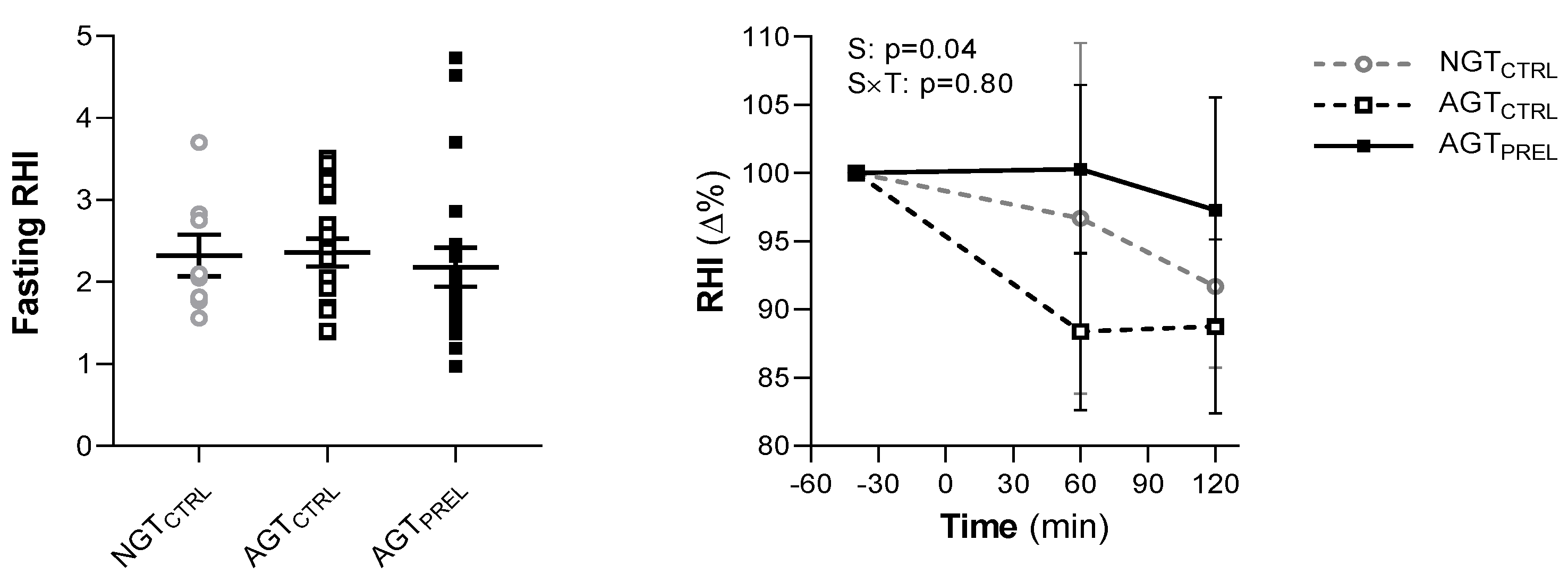
3.1. Fasting and Post-Glucose Endothelial Function

The clinical and metabolic characteristics of study participants stratified by glucose tolerance status are shown in Table 1.
At fasting, the average RHI was 2.35 ± 0.70. Five individuals had an RHI lower than 1.67, indicative of endothelial dysfunction [28], including four people with AGT and one with NGT (p = 0.56 for group differences). RHI was similar between men and women (2.36 ± 0.70 vs. 2.34 ± 0.74, respectively, p = 0.94) and across groups of glucose tolerance (Figure 2). RHI was not associated with age, BMI, or systolic and diastolic blood pressure. Among metabolic variables, RHI negatively correlated with fasting plasma glucose (r = −0.29, p = 0.04) and HOMA-IR (r = −0.29, p = 0.04), and positively correlated with Matsuda index (r = 0.39, p = 0.048). RHI was not significantly associated with fasting plasma insulin (r = −0.25, p = 0.07), nor with OGIS, HIRI, GLP-1, GIP, glucagon, FFA, or total AA.
After glucose ingestion, we observed a significant decrease in RHI, with a median 9% reduction from baseline values at 120 min in all participants (p = 0.02; p = 0.74 for group difference) (Figure 2). The time course of RHI was similar across glucose tolerance groups (p = 0.72; group × time interaction: p = 0.60) and by sex (p = 0.54; sex × time interaction: p = 0.23). As expected, all measured hormones and metabolites were affected by the OGTT (Figure 3). In repeated measure analyses, RHI values during the OGTT were associated with plasma glucose (β = −0.03, p = 0.015) and insulin levels (β = −0.10, p = 0.01). The RHI was not related to plasma GLP-1, GIP, glucagon, FFA, or AA levels throughout the OGTT.

3.2. Effect of Nutrients on Post-Glucose Endothelial Dysfunction

On a separate day, in random order, endothelial function was assessed during an OGTT preceded by the protein/lipid macronutrient preload in individuals with AGT. Fasting RHI measurements were similar between the two study days (p = 0.45; coefficient of variation 26.4%) (Figure 2).
Compared with the control OGTT, the postprandial reduction of RHI was significantly attenuated by nutrient ingestion (Figure 2), without sex differences. In parallel, plasma glucose excursion was markedly reduced by nutrients (AUC 982 ± 186 vs. 1213 ± 203 mmol × min/L, p < 0.0001), while plasma insulin responses were similar between the two studies (AUC 38.0 [25.9–60.2] vs. 38.9 [31.5–50.7] nmol × min/L, p = 0.40) (Figure 3). The time courses of all other measured hormones and metabolites showed significant differences compared with the control OGTT (Figure 3).
The attenuation of postprandial endothelial dysfunction induced by the macronutrient preload was proportional to the effect on plasma glucose excursions (r = −0.52, p = 0.02) (Figure 4). In fact, RHI was reduced less in individuals whose glucose tolerance improved more after preload consumption. Furthermore, smaller or positive changes in RHI were associated with increased plasma GLP-1 (r = 0.47, p = 0.04) and arginine (r = 0.64, p = 0.04) levels during the preload study (Figure 4). Changes in other hormones and metabolites did not correlate with changes in RHI between the control and preload OGTT.

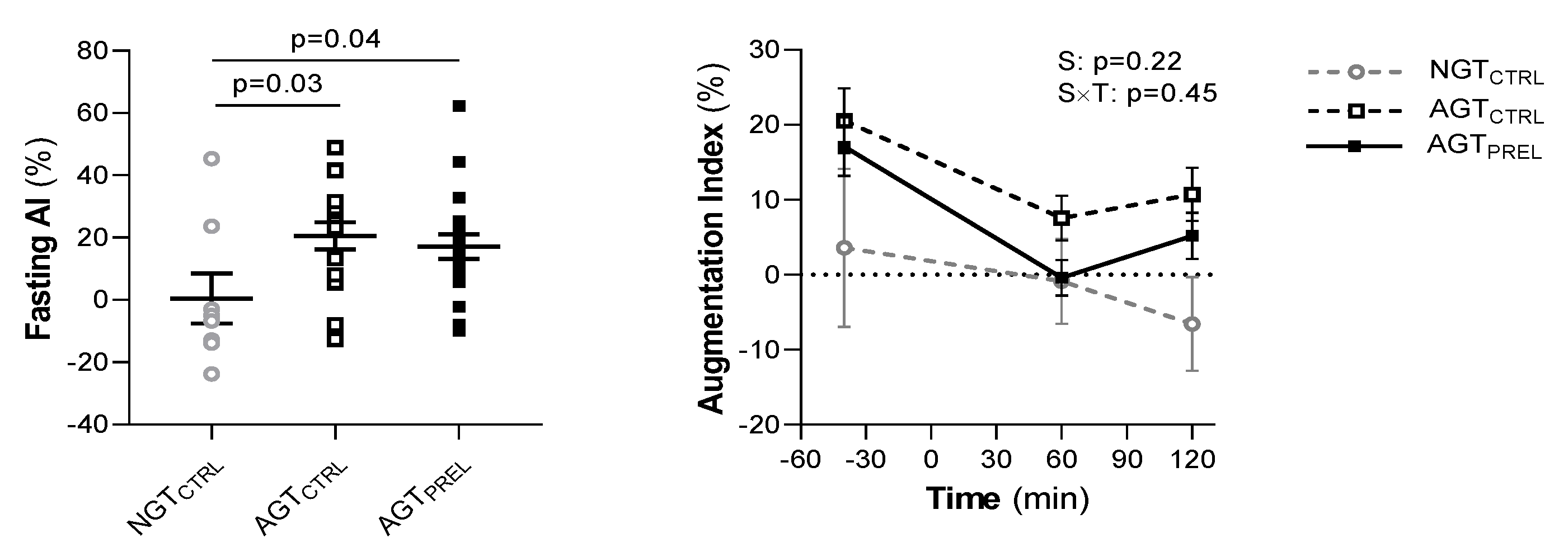
3.3. Fasting and Post-Glucose Arterial Stiffness

At fasting, the AI was significantly lower in NGT than AGT (p = 0.03, Figure 5) and positively correlated with age (r = 0.51, p = 0.01). The AI decreased in all subjects during the control OGTT (p < 0.0001 for 120 min vs. baseline values) (Figure 5). Its reduction was associated with plasma glucose (β = −3.4, p = 0.04) and tended to be greater in AGT than NGT (p = 0.07; group × time interaction: p = 0.18). Fasting and post-glucose AI were similar in men and women and not associated with other measured clinical or metabolic variables. In AGT, the AI was similar between the two study days at fasting (p > 0.99; coefficient of variation 37.1%) and its post-glucose decline was not attenuated by the macronutrient preload (Figure 5).
Glucose-induced changes in heart rate and AI@75 are shown in Figure S1 (Supplementary Materials). During the OGTT, the heart rate increased and the AI@75 decreased to a similar extent in NGT and IGT. Heart rate and AI@75 responses to glucose were not affected by the macronutrient preload (Figure S1).

4. Discussion

In this cross-over, randomized clinical trial, we demonstrated that a protein/lipid preload is able to prevent the acute endothelial dysfunction produced by glucose ingestion in individuals with IGT and early T2D. This effect of the macronutrient preload is likely to be explained by the more favorable glucose-to-insulin ratio and by the increase in plasma GLP-1 and arginine levels, both of which can upregulate the NO vasodilator system. To our knowledge, this is the first study that examined the effect of a high-protein/high-lipid macronutrient preload on postprandial endothelial dysfunction, revealing its potential mediators in a well-characterized cohort of individuals with IGT and well-controlled T2D. Our results have been obtained in an experimental setting that is easily transposable into real-life. Indeed, glucose loads are commonly provided by sugar-sweetened beverages (e.g., cola, orange juice), which contain on average 30–40 g carbohydrate (with no protein and fat) and do exert detrimental effects on endothelial function [33]. Further, there is compelling evidence that tailoring the sequence of nutrient consumption in the context of a mixed meal, so as to eat protein- and fat-rich food before carbohydrate, can improve postprandial glucose control to a similar extent compared with a preload approach [21,23,25]. Therefore, one might speculate that such a simple dietary strategy, if applied into a real-life setting, could have protective effects also on endothelial function in patients with prediabetes or manifest T2D.
In agreement with previous studies using different techniques to explore endothelial function [2,3,8,9,10], we found that RHI is acutely impaired by a physiologic plasma glucose rise in both NGT and AGT subjects. Although there is no conclusive demonstration that RHI is entirely dependent on the release of NO from endothelial cells, an elegant study has shown that at least 50% of this response depends on NO bioavailability [34]. RHI, in addition, has been validated against other procedures (i.e., flow mediated dilation, or FMD) for which a role of NO has been demonstrated [35] and correlates with erectile dysfunction [36], a clinical condition that is closely related to NO pathway impairment. With these limitations, it is plausible to hypothesize that an interference of the postprandial glucose increase with NO bioavailability explains the results of this study.
Crucial for endothelium-dependent vasodilation, NO is produced from L-arginine via the constitutively active, calcium-calmodulin dependent enzyme endothelial nitric oxide synthase (eNOS) [37]. Both chronic and acute hyperglycemia are known to induce uncoupling of eNOS from NO production in favor of superoxide. Moreover, NO availability is further reduced by superoxide combination with NO to form the highly reactive oxygen species peroxinitrite [38], which in turn further impairs eNOS activity starting a vicious cycle [39]. Also NADPH oxidases-membrane-bound enzyme complexes are considered an additional source of reactive oxygen species in endothelial cells, particularly under conditions of hyperglycemia [40], and asymmetric dimethylarginine, a naturally occurring product of amino acid metabolism that can bind eNOS [41], further interferes with NO availability.
We previously demonstrated that a high-protein nutrient preload is able to reduce the plasma glucose response to an OGTT largely by delaying gastric emptying [16,25]. In this view, it is of interest that other dietary manipulations able to delay gastric emptying have been reported to reduce the postprandial decline in conductance arteries’ endothelial function as measured by FMD [42]. Other glucose-lowering mechanisms activated by nutrient preloads include increased insulin secretion relative to plasma glucose and decreased insulin clearance, whose effects are partly counterbalanced by increased glucagon release and less suppressed endogenous glucose production [16,25].
Since carbohydrate ingestion increases simultaneously, and proportionally, both plasma glucose and insulin levels, it is challenging to dissect the role of hyperglycemia vs. hyperinsulinemia on endothelial function [10]. In healthy individuals, insulin activates eNOS-mediated production of NO and therefore exerts vasodilatory effects both on large conduit arteries [43] and resistance arterioles [44]. This hemodynamic effect is attenuated in conditions of insulin resistance or chronic hyperinsulinemia [15,45]. Our intervention markedly reduced glucose levels effecting neither insulin levels nor insulin sensitivity. We can therefore explain the beneficial effect of the preload on postprandial RHI with the reduction in plasma glucose levels and the amelioration of the post-OGTT glucose-to-insulin ratio; a mechanism we have previously demonstrated in non-physiologic experimental conditions [46].
In our study, we also observed a positive correlation of RHI with nutrient-induced plasma GLP-1 rise. This gut hormone is released after a meal and has extra-glycemic positive effects on the cardiovascular system [14]. The infusion of exogenous GLP-1 has been shown to ameliorate endothelial function in T2D, possibly through an improvement in the endothelial antioxidant properties and a decrease of oxidative stress [14,47]. These effects are both indirect (i.e., insulin-mediated) and direct on the endothelium, which expresses specific receptors for GLP-1. In support of our findings, Tanaka et al. [48] demonstrated that RHI is unchanged 2 h after a mixed meal in 17 T2D patients treated with a single injection of exenatide, a short-acting GLP-1 receptor agonist, while it decreases by 15% without exenatide.
The improvement in endothelial function induced by the macronutrient preload might also be due to the greater availability of substrates entering the circulation following digestion and absorption of nutrients, such as amino and fatty acids. Among the measured substrates, only the increase in plasma arginine after the high-protein preload was correlated with the improvement in post-glucose endothelial function. Arginine plays a central role in the biosynthesis of NO, being its direct precursor, and an increased arginine bioavailability may facilitate NO production particularly in the stimulated condition of post-ischemic vasodilation. In fact, a reduction in arginine availability through increased arginase activity impairs NO production in T2D [11,49], while arginase inhibition ameliorates endothelial function [50]. Furthermore, the lack of the glucose-induced decline of plasma arginine when fructose—rather than glucose—is ingested [10] or when a small amount of protein is co-ingested with glucose [51] has been hypothesized to be responsible for the lesser impact of the oral load on endothelial function. Finally, the increase in plasma arginine might also counterbalance the rise in asymmetric dimethylarginine (ADMA) observed after glucose ingestion in subjects with AGT [52]. Dietary fats are known to exert a negative impact on postprandial endothelial function [53]. In our study, however, plasma FFA were similarly suppressed during the OGTTs preceded by water or nutrients and did not correlate with changes in RHI.
Measures of endothelial function are to some extent dependent on arterial stiffness and blood pressure, particularly when measuring acute responses [54,55]. Indeed, our estimates of stiffness (AI and AI@75) showed a decline after glucose consumption that was consistent with previous observations using mixed meals [56] and was not affected by the macronutrient preload. Although we did not measure blood pressure during the OGTTs, it has been observed by Pham et al. [57] that blood pressure responses to oral glucose are not different when glucose is preceded by a high-protein load. Most importantly, the RHI is automatically corrected for changes in systemic hemodynamics at each time point, being calculated as the post-to-pre occlusion PAT signal ratio in the occluded arm, relative to the same ratio in the control arm [28,29,30]. For these reasons, we consider changes in arterial stiffness and blood pressure unlikely to explain the observed changes in RHI.
This study has some limitations, such as the small sample size and the lack of repeated blood pressure measurements. Given that RHI is similarly reduced during the OGTT across glucose tolerance groups, nutrient preloads may also exert beneficial effects in NGT, which have not been verified. NGT had lower age and blood pressure than AGT. Both factors could influence endothelial function, and this should be considered when interpreting group differences. Also, our study was not powered to assess differences between the two groups of individuals with prediabetes and early diagnosed, diet-controlled diabetes. These groups were substantially homogeneous, representing two consecutive phases in the continuum of diabetes progression, and therefore combined data were analyzed and presented. The homogeneity of our population and the small sample size might also explain the lack of correlation between RHI and known factors associated with endothelial dysfunction (such as age, BMI and blood pressure), which were all demonstrated in large, population-based cohorts [30]. Participants were not characterized in terms of plasma lipid profile and waist circumference, which may also influence endothelial function. Our study design did not include a protein-only or a mixed-meal control group; thus, we cannot fully dissect the impact of protein alone or of the preload strategy on endothelial function. Preliminary evidence supports the sustained metabolic benefits of preload strategies and their neutral impact on body weight (likely as a consequence of reduced appetite after protein consumption) [17,20]. Due to the lack of longitudinal data, however, we could not examine the long-term efficacy of the preload strategy on endothelial function and its effects on body weight. Although the inclusion of both men and women strengthens the external validity of the present study, differences related to the sex and to different phases of menstrual cycle in pre-menopausal women may introduce variability. Moreover, despite the fact that AI is widely used in clinics and research as an indirect measure of arterial stiffness, it measures pulse wave reflections rather than vascular stiffness and can be influenced by several factors [58,59,60]. Finally, although the dependence of postprandial endothelial dysfunction on hyperglycemia-induced NO impairment relies on a strong biological rationale and is indirectly supported by our findings, NO availability and its metabolic signaling was not directly investigated in this study.

5. Conclusions

In conclusion, a protein/lipid macronutrient preload ameliorates acute glucose-induced endothelial dysfunction in individuals with early AGT. This effect is possibly mediated by an improved glucose-to-insulin ratio, as well as higher GLP-1 and arginine bioavailability. Our results support the efficacy of this dietary strategy to improve glucose control and prevent the deterioration of the endothelial vasodilator function induced by the hyperglycemic environment.

Supplementary Materials

The following are available online at https://www.mdpi.com/2072-6643/12/7/2053/s1, Figure S1: Heart rate and augmentation index normalized to heart rate of 75 bpm (AI@75).

Author Contributions

D.T. and A.N. conceptualization; D.T., L.N., S.F., S.B., A.M. and A.N. data curation; D.T., L.N. and A.N. formal analysis; D.T. and A.N. funding acquisition; D.T., S.F., S.B. and A.N. investigation; D.T. and A.N. methodology; A.N. project administration; A.N. supervision; A.N. validation; D.T. visualization; D.T., L.N., A.M. and A.N. writing—original draft; D.T., L.N., S.F., S.B., A.M. and A.N. writing—review & editing. All authors have read and agreed to the published version of the manuscript.

Funding

This work was supported by grants from the University of Pisa (Fondi di Ateneo). DT is supported by the European Foundation for the Study of Diabetes through a Rising Star Fellowship and the Future Leaders Mentorship Programme for Clinical Diabetologists.

Acknowledgments

We would like to thank the volunteers enrolled in this trial and all the personnel of the Nutrition, Metabolism and Atherosclerosis Laboratory at the University of Pisa.

Conflicts of Interest

The authors declare no conflict of interest.

Abbreviations

AAAmino acid
AGTAbnormal glucose tolerance
AIAugmentation index
AI@75 AI normalized to heart rate of 75 bpm
AUCArea under the curve
BCAA Branched-chain amino acid
eNOSEndothelial nitric oxide synthase
FFA Free fatty acid
FMDFlow mediated dilation
GIPGlucose-dependent insulinotropic polypeptide
GLP-1 Glucagon-like peptide-1
HbA1cGlycated hemoglobin
HIRIHepatic insulin resistance index
HOMA-IRHomeostatic model assessment for insulin resistance
IGTImpaired glucose tolerance
NGTNormal glucose tolerance
NO Nitric oxide
OGISOral glucose insulin sensitivity
OGTTOral glucose tolerance test
RHI Reactive hyperemia index
T2DType 2 diabetes

References

  1. Heintjes, E.M.; Houben, E.; Beekman-Hendriks, W.L.; Lighaam, E.; Cremers, S.M.; Penning van Beest, F.J.A.; Stehouwer, C.D.A.; Herings, R.M.C. Trends in mortality, cardiovascular complications, and risk factors in type 2 diabetes. Neth. J. Med. 2019, 77, 317–329. [Google Scholar] [PubMed]
  2. Williams, S.B.; Goldfine, A.B.; Timimi, F.K.; Ting, H.H.; Roddy, M.-A.; Simonson, D.C.; Creager, M.A. Acute hyperglycemia attenuates endothelium-dependent vasodilation in humans in vivo. Circulation 1998, 97, 1695–1701. [Google Scholar] [CrossRef] [PubMed] [Green Version]
  3. Su, Y.; Liu, X.-M.; Sun, Y.-M.; Jin, H.-B.; Fu, R.; Wang, Y.-Y.; Wu, Y.; Luan, Y. The relationship between endothelial dysfunction and oxidative stress in diabetes and prediabetes. Int. J. Clin. Pract. 2008, 62, 877–882. [Google Scholar] [CrossRef] [PubMed]
  4. Loader, J.; Montero, D.; Lorenzen, C.; Watts, R.; Méziat, C.; Reboul, C.; Stewart, S.; Walther, G. Acute hyperglycemia impairs vascular function in healthy and cardiometabolic diseased subjects. Arterioscler. Thromb. Vasc. Biol. 2015, 35, 2060–2072. [Google Scholar] [CrossRef] [Green Version]
  5. Mann, B.K.; Bhandohal, J.S.; Hong, J. An overall glance of evidence supportive of one-hour and two-hour postload plasma glucose levels as predictors of long-term cardiovascular events. Int. J. Endocrinol. 2019, 2019, 6048954. [Google Scholar] [CrossRef]
  6. Vita, J.A.; Treasure, C.B.; Nabel, E.G.; McLenachan, J.M.; Fish, R.D.; Yeung, A.C.; Vekshtein, V.I.; Selwyn, A.P.; Ganz, P. Coronary vasomotor response to acetylcholine relates to risk factors for coronary artery disease. Circulation 1990, 81, 491–497. [Google Scholar] [CrossRef] [PubMed] [Green Version]
  7. DECODE Study Group. Glucose tolerance and cardiovascular mortality. Arch. Intern. Med. 2001, 161, 397. [Google Scholar] [CrossRef] [PubMed]
  8. Costantino, S.; Paneni, F.; Battista, R.; Castello, L.; Capretti, G.; Chiandotto, S.; Tanese, L.; Russo, G.; Pitocco, D.; Lanza, G.A.; et al. Impact of glycemic variability on chromatin remodeling, oxidative stress, and endothelial dysfunction in patients with type 2 diabetes and with target HbA 1c levels. Diabetes 2017, 66, 2472–2482. [Google Scholar] [CrossRef] [Green Version]
  9. Kawano, H.; Motoyama, T.; Hirashima, O.; Hirai, N.; Miyao, Y.; Sakamoto, T.; Kugiyama, K.; Ogawa, H.; Yasue, H. Hyperglycemia rapidly suppresses flow-mediated endothelium- dependent vasodilation of brachial artery. J. Am. Coll. Cardiol. 1999, 34, 146–154. [Google Scholar] [CrossRef] [Green Version]
  10. Mah, E.; Noh, S.K.; Ballard, K.; Matos, M.E.; Volek, J.S.; Bruno, R.S. Postprandial hyperglycemia impairs vascular endothelial function in healthy men by inducing lipid peroxidation and increasing asymmetric dimethylarginine: Arginine. J. Nutr. 2011, 141, 1961–1968. [Google Scholar] [CrossRef]
  11. Romero, M.; Iddings, J.; Platt, D.H.; Ali, M.I.; Cederbaum, S.D.; Stepp, D.W.; Caldwell, R.B.; Caldwell, R.W. Diabetes-induced vascular dysfunction involves arginase I. Am. J. Physiol. Circ. Physiol. 2012, 302, H159–H166. [Google Scholar] [CrossRef] [Green Version]
  12. Zhenyukh, O.; González-Amor, M.; Rodrigues-Diez, R.R.; Esteban, V.; Ruiz-Ortega, M.; Salaices, M.; Mas, S.; Briones, A.M.; Egido, J. Branched-chain amino acids promote endothelial dysfunction through increased reactive oxygen species generation and inflammation. J. Cell. Mol. Med. 2018, 22, 4948–4962. [Google Scholar] [CrossRef] [PubMed]
  13. Vogel, R.A.; Corretti, M.C.; Plotnick, G.D. Effect of a single high-fat meal on endothelial function in healthy subjects. Am. J. Cardiol. 1997, 79, 350–354. [Google Scholar] [CrossRef]
  14. Ceriello, A.; Esposito, K.; Testa, R.; Bonfigli, A.; Marra, M.; Giugliano, D. The possible protective role of glucagon-like peptide 1 on endothelium during the meal and evidence for an “endothelial resistance” to glucagon-like peptide 1 in diabetes. Diabetes Care 2011, 34, 697–702. [Google Scholar] [CrossRef] [PubMed] [Green Version]
  15. Muniyappa, R.; Sowers, J.R. Role of insulin resistance in endothelial dysfunction. Rev. Endocr. Metab. Disord. 2013, 14, 5–12. [Google Scholar] [CrossRef]
  16. Tricò, D.; Baldi, S.; Tulipani, A.; Frascerra, S.; Macedo, M.P.; Mari, A.; Ferrannini, E.; Natali, A. Mechanisms through which a small protein and lipid preload improves glucose tolerance. Diabetology 2015, 58, 2503–2512. [Google Scholar] [CrossRef]
  17. Tricò, D.; Filice, E.; Trifirò, S.; Natali, A. Manipulating the sequence of food ingestion improves glycemic control in type 2 diabetic patients under free-living conditions. Nutr. Diabetes 2016, 6, e226. [Google Scholar] [CrossRef] [PubMed] [Green Version]
  18. Tricò, D.; Filice, E.; Baldi, S.; Frascerra, S.; Mari, A.; Natali, A. Sustained effects of a protein and lipid preload on glucose tolerance in type 2 diabetes patients. Diabetes Metab. 2016, 42, 242–248. [Google Scholar] [CrossRef]
  19. Jakubowicz, D.; Froy, O.; Ahren, B.; Boaz, M.; Landau, Z.; Bar-Dayan, Y.; Ganz, T.; Barnea, M.; Wainstein, J. Incretin, insulinotropic and glucose-lowering effects of whey protein pre-load in type 2 diabetes: A randomised clinical trial. Diabetology 2014, 57, 1807–1811. [Google Scholar] [CrossRef]
  20. Ma, J.; Jesudason, D.R.; Stevens, J.E.; Keogh, J.; Jones, K.L.; Clifton, P.M.; Horowitz, M.; Rayner, C.K. Sustained effects of a protein ‘preload’ on glycaemia and gastric emptying over 4 weeks in patients with type 2 diabetes: A randomized clinical trial. Diabetes Res. Clin. Pract. 2015, 108, e31–e34. [Google Scholar] [CrossRef]
  21. Shukla, A.P.; Iliescu, R.G.; Thomas, C.; Aronne, L.J. Food order has a significant impact on postprandial glucose and insulin levels: Table 1. Diabetes Care 2015, 38, e98–e99. [Google Scholar] [CrossRef] [Green Version]
  22. Wu, T.; Little, T.; Bound, M.J.; Borg, M.; Zhang, X.; Deacon, C.F.; Horowitz, M.; Jones, K.L.; Rayner, C.K. A protein preload enhances the glucose-lowering efficacy of vildagliptin in type 2 diabetes. Diabetes Care 2016, 39, 511–517. [Google Scholar] [CrossRef] [Green Version]
  23. Kuwata, H.; Iwasaki, M.; Shimizu, S.; Minami, K.; Maeda, H.; Seino, S.; Nakada, K.; Nosaka, C.; Murotani, K.; Kurose, T.; et al. Meal sequence and glucose excursion, gastric emptying and incretin secretion in type 2 diabetes: A randomised, controlled crossover, exploratory trial. Diabetology 2015, 59, 453–461. [Google Scholar] [CrossRef] [PubMed] [Green Version]
  24. Tricò, D.; Natali, A. Modulation of postprandial glycemic responses by noncarbohydrate nutrients provides novel approaches to the prevention and treatment of type 2 diabetes. Am. J. Clin. Nutr. 2017, 106, 701–702. [Google Scholar] [CrossRef] [PubMed] [Green Version]
  25. Nesti, L.; Mengozzi, A.; Tricò, D. Impact of nutrient type and sequence on glucose tolerance: Physiological insights and therapeutic implications. Front. Endocrinol. 2019, 10, 144. [Google Scholar] [CrossRef] [PubMed]
  26. American Diabetes Association 2. Classification and diagnosis of diabetes: Standards of medical care in diabetes—2020. Diabetes Care 2019, 43, S14–S31. [Google Scholar] [CrossRef] [Green Version]
  27. Tricò, D.; Frascerra, S.; Baldi, S.; Mengozzi, A.; Nesti, L.; Mari, A.; Natali, A. The insulinotropic effect of a high-protein nutrient preload is mediated by the increase of plasma amino acids in type 2 diabetes. Eur. J. Nutr. 2018, 58, 2253–2261. [Google Scholar] [CrossRef]
  28. Bonetti, P.O.; Pumper, G.M.; Higano, S.T.; Holmes, D.R.; Kuvin, J.T.; Lerman, L.O. Noninvasive identification of patients with early coronary atherosclerosis by assessment of digital reactive hyperemia. J. Am. Coll. Cardiol. 2004, 44, 2137–2141. [Google Scholar] [CrossRef] [Green Version]
  29. Axtell, A.L.; Gomari, F.A.; Cooke, J.P. Assessing endothelial vasodilator function with the Endo-PAT 2000. J. Vis. Exp. 2010. [Google Scholar] [CrossRef] [Green Version]
  30. Venturi, E.; Pinnola, S.; Morizzo, C.; Boldrini, B.; Rossi, M.; Trifirò, S.; Tricò, D.; Natali, A.; The SUMMIT study group. Clinical phenotype and microvascular dynamics of subjects with endothelial dysfunction as assessed by peripheral tonometry. Microcirculation 2016, 23, 230–239. [Google Scholar] [CrossRef]
  31. Mari, A.; Pacini, G.; Murphy, E.; Ludvik, B.; Nolan, J.J. A model-based method for assessing insulin sensitivity from the oral glucose tolerance test. Diabetes Care 2001, 24, 539–548. [Google Scholar] [CrossRef] [PubMed] [Green Version]
  32. Abdul-Ghani, M.A.; Matsuda, M.; Balas, B.; DeFronzo, R.A. Muscle and liver insulin resistance indexes derived from the oral glucose tolerance test. Diabetes Care 2006, 30, 89–94. [Google Scholar] [CrossRef] [PubMed] [Green Version]
  33. Loader, J.; Meziat, C.; Watts, R.; Lorenzen, C.; Sigaudo-Roussel, D.; Stewart, S.; Reboul, C.; Meyer, G.; Walther, G. Effects of sugar-sweetened beverage consumption on microvascular and macrovascular function in a healthy population. Arter. Thromb. Vasc. Biol. 2017, 37, 1250–1260. [Google Scholar] [CrossRef] [PubMed] [Green Version]
  34. Nohria, A.; Gerhard-Herman, M.; Creager, M.A.; Hurley, S.; Mitra, D.; Ganz, P. Role of nitric oxide in the regulation of digital pulse volume amplitude in humans. J. Appl. Physiol. 2006, 101, 545–548. [Google Scholar] [CrossRef] [PubMed] [Green Version]
  35. Green, D.J.; Dawson, E.A.; Groenewoud, H.M.; Jones, H.; Thijssen, D.H.J. Is flow-mediated dilation nitric oxide mediated? Hypertension 2014, 63, 376–382. [Google Scholar] [CrossRef] [Green Version]
  36. Kovac, J.R.; Gomez, L.; Smith, R.P.; Coward, R.M.; Gonzales, M.A.; Khera, M.; Lamb, L.J.; Lipshultz, L.I. Measurement of endothelial dysfunction via peripheral arterial tonometry predicts vasculogenic erectile dysfunction. Int. J. Impot. Res. 2014, 26, 218–222. [Google Scholar] [CrossRef] [Green Version]
  37. Meza, C.A.; La Favor, J.D.; Kim, D.-H.; Hickner, R.C. Endothelial dysfunction: Is there a hyperglycemia-induced Imbalance of NOX and NOS? Int. J. Mol. Sci. 2019, 20, 3775. [Google Scholar] [CrossRef] [Green Version]
  38. Stuehr, D.; Pou, S.; Rosen, G.M. Oxygen reduction by nitric-oxide synthases. J. Biol. Chem. 2001, 276, 14533–14536. [Google Scholar] [CrossRef] [Green Version]
  39. Tanaka, J.; Qiang, L.; Banks, A.S.; Welch, C.L.; Matsumoto, M.; Kitamura, T.; Ido-Kitamura, Y.; Depinho, R.A.; Accili, M. Foxo1 links hyperglycemia to LDL oxidation and endothelial nitric oxide synthase dysfunction in vascular endothelial cells. Diabetes 2009, 58, 2344–2354. [Google Scholar] [CrossRef] [Green Version]
  40. Gorin, Y.; Block, K. Nox as a target for diabetic complications. Clin. Sci. 2013, 125, 361–382. [Google Scholar] [CrossRef] [Green Version]
  41. Sibal, L.; Agarwal, S.C.; Home, P.D.; Boger, R.H. The role of Asymmetric Dimethylarginine (ADMA) in endothelial dysfunction and cardiovascular disease. Curr. Cardiol. Rev. 2010, 6, 82–90. [Google Scholar] [CrossRef]
  42. Thazhath, S.S.; Wu, T.; Bound, M.J.; Checklin, H.L.; Jones, K.L.; Willoughby, S.; Horowitz, M.; Rayner, C.K. Changes in meal composition and duration affect postprandial endothelial function in healthy humans. Am. J. Physiol. Liver Physiol. 2014, 307, G1191–G1197. [Google Scholar] [CrossRef] [Green Version]
  43. Westerbacka, J.; Vehkavaara, S.; Bergholm, R.; Wilkinson, I.; Cockcroft, J.; Yki-Jarvinen, H. Marked resistance of the ability of insulin to decrease arterial stiffness characterizes human obesity. Diabetes 1999, 48, 821–827. [Google Scholar] [CrossRef]
  44. Zheng, C.; Liu, Z. Vascular function, insulin action, and exercise: An intricate interplay. Trends Endocrinol. Metab. 2015, 26, 297–304. [Google Scholar] [CrossRef] [Green Version]
  45. Olver, T.D.; Grunewald, Z.I.; Ghiarone, T.; Restaino, R.M.; Sales, A.R.K.; Park, L.K.; Thorne, P.K.; Ganga, R.R.; Emter, C.A.; Lemon, P.W.R.; et al. Persistent insulin signaling coupled with restricted PI3K activation causes insulin-induced vasoconstriction. Am. J. Physiol. Circ. Physiol. 2019, 317, H1166–H1172. [Google Scholar] [CrossRef] [PubMed]
  46. Natali, A.; Baldi, S.; Vittone, F.; Muscelli, E.; Casolaro, A.; Morgantini, C.; Palombo, C.; Ferrannini, E. Effects of glucose tolerance on the changes provoked by glucose ingestion in microvascular function. Diabetology 2008, 51, 862–871. [Google Scholar] [CrossRef] [PubMed] [Green Version]
  47. Nystrom, T.; Gutniak, M.K.; Zhang, Q.; Zhang, F.; Holst, J.J.; Ahren, B.; Sjöholm, Å. Effects of glucagon-like peptide-1 on endothelial function in type 2 diabetes patients with stable coronary artery disease. Am. J. Physiol. Metab. 2004, 287, E1209–E1215. [Google Scholar] [CrossRef] [PubMed]
  48. Torimoto, K.; Okada, Y.; Mori, H.; Otsuka, T.; Kawaguchi, M.; Matsuda, M.; Kuno, F.; Sugai, K.; Sonoda, S.; Hajime, M.; et al. Effects of exenatide on postprandial vascular endothelial dysfunction in type 2 diabetes mellitus. Cardiovasc. Diabetol. 2015, 14, 25. [Google Scholar] [CrossRef] [PubMed] [Green Version]
  49. Thengchaisri, N. Upregulation of arginase by H2O2 impairs endothelium-dependent nitric oxide-mediated dilation of coronary arterioles. Arter. Thromb. Vasc. Biol. 2006, 26, 2035–2042. [Google Scholar] [CrossRef] [PubMed] [Green Version]
  50. Mahdi, A.; Kövamees, O.; Checa, A.; Wheelock, C.E.; Von Heijne, M.; Alvarsson, M.; Pernow, J. Arginase inhibition improves endothelial function in patients with type 2 diabetes mellitus despite intensive glucose-lowering therapy. J. Intern. Med. 2018, 284, 388–398. [Google Scholar] [CrossRef]
  51. McDonald, J.D.; Mah, E.; Chitchumroonchokchai, C.; Reverri, E.J.; Li, J.; Volek, J.S.; Villamena, F.A.; Bruno, R.S. Co-ingestion of whole eggs or egg whites with glucose protects against postprandial hyperglycaemia-induced oxidative stress and dysregulated arginine metabolism in association with improved vascular endothelial function in prediabetic men. Br. J. Nutr. 2018, 120, 901–913. [Google Scholar] [CrossRef]
  52. Konukoglu, D.; Fırtına, S.; Serin, O.; Konukoǧlu, D.; Firtina, S. The relationship between plasma asymmetrical dimethyl-l-arginine and inflammation and adhesion molecule levels in subjects with normal, impaired, and diabetic glucose tolerance. Metabolism 2008, 57, 110–115. [Google Scholar] [CrossRef] [PubMed]
  53. Steer, P.; Sarabi, D.M.; Karlström, B.; Basu, S.; Berne, C.; Vessby, B.; Lind, L.; Karlstrm, B. The effect of a mixed meal on endothelium-dependent vasodilation is dependent on fat content in healthy humans. Clin. Sci. 2003, 105, 81–87. [Google Scholar] [CrossRef] [PubMed] [Green Version]
  54. Bock, J.; E Hughes, W.; Casey, D.P. Age-associated differences in central artery responsiveness to sympathoexcitatory stimuli. Am. J. Hypertens. 2019, 32, 564–569. [Google Scholar] [CrossRef] [PubMed]
  55. Zhou, J.; Li, Y.-S.; Chien, S. Shear stress–initiated signaling and its regulation of endothelial function. Arter. Thromb. Vasc. Biol. 2014, 34, 2191–2198. [Google Scholar] [CrossRef] [Green Version]
  56. Taylor, J.L.; Curry, T.B.; Matzek, L.J.; Joyner, M.J.; Casey, D.P. Acute effects of a mixed meal on arterial stiffness and central hemodynamics in healthy adults. Am. J. Hypertens. 2013, 27, 331–337. [Google Scholar] [CrossRef] [Green Version]
  57. Pham, H.; Holen, I.S.; Phillips, L.; Hatzinikolas, S.; Huynh, L.Q.; Wu, T.; Hausken, T.; Rayner, C.K.; Horowitz, M.; Jones, K.L. The effects of a whey protein and guar gum-containing preload on gastric emptying, glycaemia, small intestinal absorption and blood pressure in healthy older subjects. Nutrients 2019, 11, 2666. [Google Scholar] [CrossRef] [Green Version]
  58. Mitchell, G.F.; Lacourcière, Y.; Arnold, J.M.O.; Dunlap, M.E.; Conlin, P.R.; Izzo, J.L. Changes in aortic stiffness and augmentation index after acute converting enzyme or vasopeptidase inhibition. Hypertension 2005, 46, 1111–1117. [Google Scholar] [CrossRef] [Green Version]
  59. Cheng, L.-T.; Tang, L.-J.; Cheng, L.; Huang, H.-Y.; Wang, T. Limitation of the augmentation index for evaluating arterial stiffness. Hypertens. Res. 2007, 30, 713–722. [Google Scholar] [CrossRef] [Green Version]
  60. Sakurai, M.; Yamakado, T.; Kurachi, H.; Kato, T.; Kuroda, K.; Ishisu, R.; Okamoto, S.; Isaka, N.; Nakano, T.; Ito, M. The relationship between aortic augmentation index and pulse wave velocity: An invasive study. J. Hypertens. 2007, 25, 391–397. [Google Scholar] [CrossRef]
Figure 1. Outline of the study protocol. Endothelial function was assessed by the reactive hyperemia index (RHI) using an EndoPAT device at fasting, 60 min and 120 min during two 75 g oral glucose tolerance tests (OGTTs) preceded by water or a protein/lipid preload. Plasma glucose, insulin, glucagon-like peptide-1 (GLP-1), glucose-dependent insulinotropic polypeptide (GIP), glucagon, free fatty acids, and amino acids were measured.
Nutrients 12 02053 g001
Figure 2. Fasting endothelial function assessed by the reactive hyperemia index (RHI) and its percentage changes (Δ%) during two 75 g oral glucose tolerance tests preceded by water (CTRL) or a protein/lipid preload (PREL) in subjects with normal glucose tolerance (NGT) or abnormal glucose tolerance (AGT). Data are mean ± SEM. Baseline differences between the three groups were tested by Kruskal–Wallis test followed by post-hoc pairwise comparisons. In AGT, repeated measures were analyzed by mixed models including study (S), time (T), and an interaction term (S × T) as fixed effects and subject as random effect. p value for time effect is <0.05.
Nutrients 12 02053 g002
Figure 3. Plasma concentrations of glucose, insulin, glucagon-like peptide-1 (GLP-1), glucose-dependent insulinotropic polypeptide (GIP), glucagon, free fatty acids (FFA), arginine, branched-chain amino acids (BCAA), and total amino acids during two 75 g oral glucose tolerance tests preceded by water (CTRL) or a protein/lipid preload (PREL) in subjects with normal glucose tolerance (NGT) or abnormal glucose tolerance (AGT). Data are mean ± SEM. In AGT, repeated measures were analyzed by mixed models including study (S), time, and an interaction term (S × T) as fixed effects and subject as random effect. P values are not shown for time effects (<0.05 for all variables).
Nutrients 12 02053 g003
Figure 4. Correlations between differences in RHI percentage changes at the end of the preload vs. control study (ΔRHIΔ%) and changes in the areas under the curve (AUC) of plasma glucose (ΔglucoseAUC), glucagon-like peptide-1 (ΔGLP-1AUC), and arginine (ΔarginineAUC) in subjects with abnormal glucose tolerance.
Nutrients 12 02053 g004
Figure 5. Fasting arterial stiffness assessed by the augmentation index (AI) and its change during two 75 g oral glucose tolerance tests preceded by water (CTRL) or a protein/lipid preload (PREL) in subjects with normal glucose tolerance (NGT) or abnormal glucose tolerance (AGT). Data are mean ± SEM. Baseline differences between the three groups were tested by Kruskal–Wallis test followed by post-hoc pairwise comparisons. In AGT, repeated measures were analyzed by mixed models including study (S), time (T), and an interaction term (S × T) as fixed effects and subject as random effect. p value for time effect is <0.05.
Nutrients 12 02053 g005
Table 1. Clinical and metabolic characteristics of study participants.
AGTNGTp
N228-
Age (years)50.0 ± 14.231.8 ± 11.90.004
Sex (men/women; n (%))14/8 (63.6/36.4)4/4 (50.0/50.0)0.68
Body Mass Index (kg/m2)27.4 ± 5.526.6 ± 5.10.64
Systolic Blood Pressure (mmHg)121 ± 9109 ± 100.006
Diastolic Blood Pressure (mmHg)78 ± 869 ± 80.01
Heart Rate (bpm)63 ± 859 ± 100.45
Non-smokers/Ex-smokers (n (%))17/5 (77.3/22.7)7/1 (87.5/12.5)0.99
Fasting Plasma Glucose (mmol/L)6.0 ± 1.05.1 ± 0.50.02
2-h Plasma Glucose (mmol/L)10.7 ± 2.66.3 ± 1.1<0.0001
Plasma Glucose AUC (mmol × min/L)1,213 ± 203852 ± 1050.0002
Glucose tolerance (IGT/T2D; n (%))13/9 [59/41]--
HbA1c (%)6.1 ± 0.65.3 ± 0.20.002
Fasting Plasma Insulin (pmol/L)79 [42–120]52 [27–75]0.09
2-h Plasma Insulin (pmol/L)398 [299–703]192 [141–425]0.02
Plasma Insulin AUC (nmol × min/L)38.9 [31.5–50.7]37.9 [30.6–59.8]0.96
HOMA-IR (unit)3.0 [1.4–4.7]1.7 [0.8–2.7]0.049
Matsuda Index (unit)5.3 [3.4–9.6]9.5 [5.1–14.1]0.07
OGIS Index (unit)357 [313–406]416 [386–455]0.003
HIRI (unit)3.5 [2.6–4.4]3.1 [2.4–6.5]0.99
Data are mean  ±  SD or median [interquartile range] for normally or non-normally distributed variables, respectively. Differences were tested using Mann-Whitney test. Abbreviations: AGT, Abnormal Glucose Tolerance; AUC, Area Under the Curve; HbA1c, Glycated Hemoglobin; HIRI, Hepatic Insulin Resistance Index; HOMA-IR, Homeostatic Model Assessment for Insulin Resistance; IGT, Impaired Glucose Tolerance; NGT, Normal Glucose Tolerance; OGIS, Oral Glucose Insulin Sensitivity index; T2D, type 2 diabetes.

