Current Knowledge about the Effect of Nutritional Status, Supplemented Nutrition Diet, and Gut Microbiota on Hepatic Ischemia-Reperfusion and Regeneration in Liver Surgery

 , , and
Abstract
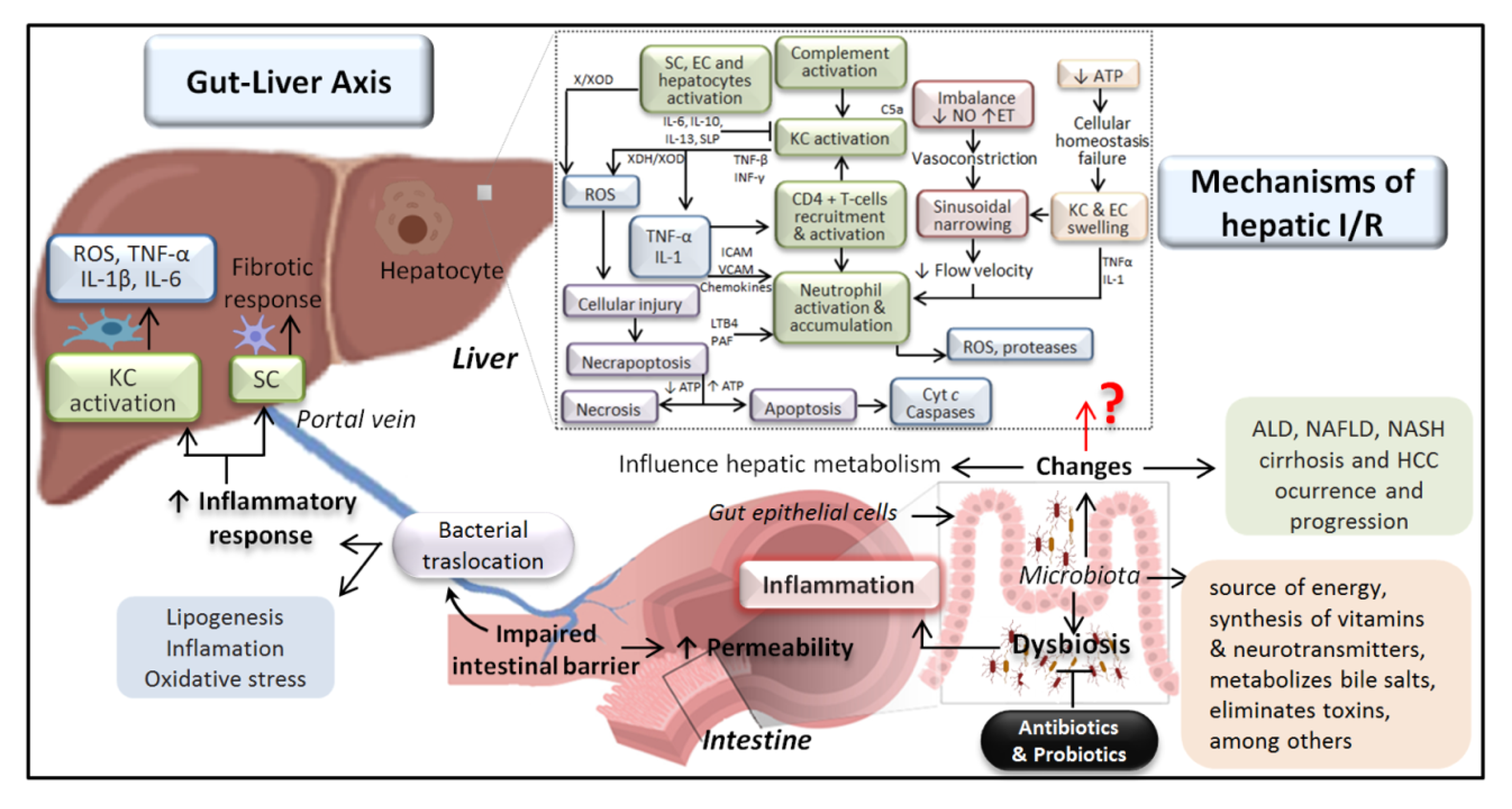
:1. Introduction
2. Starvation Effects on I/R Injury Associated with Liver Surgery
2.1. Studies of Short-Term Starvation (12–24 h)
2.2. Studies of Long-Term Starvation (Two to Seven Days)
3. Nutritional Support by Nutraceuticals and Functional Foods on Liver Surgery under Hepatic Ischemia-Reperfusion
3.1. Plant-Derived Supplements and Other Food Additives
3.2. Vitamins
3.3. Fish and Rosa Mosqueta Oils
3.4. Fatty Acids, Arginine, and Nucleotides
3.5. Branched-Chain Amino Acid
3.6. Probiotics
4. Gut Microbiota and Hepatic Ischemia Reperfusion in Liver Surgery
5. Future Perspectives and Conclusions
Author Contributions
Funding
Conflicts of Interest
Abbreviations
| AKT | Protein kinase B |
| ALA | α-linolenic acid |
| ALD | Alcoholic liver disease |
| AMPK | AMP-activated protein kinase |
| ATP | Adenosine triphosphate |
| BCAA | Branched-chain amino acid |
| BCL | B-cell lymphoma |
| BHB | β-hydroxybutyric acid |
| CAT | Catalase |
| Co A | Coenzyme A |
| DHA | Docosahexaenoic acid |
| EPA | Eicosapentaenoic acid |
| ESPEN | European Society for Parenteral and Enteral Nutrition |
| FOXO1 | Forkhead box protein O1 |
| GSH | Glutathione |
| HBeAg | Hepatitis B virus e-antigen |
| HCC | Hepatocellular carcinoma |
| HMGB1 | High mobility group box 1 |
| HO-1 | Heme oxygenase 1 |
| HWP | Hydrolyzed whey peptide |
| I/R | Ischemia-reperfusion |
| IFNγ | Interferon-gamma |
| IL | Interleukin |
| LC-PUFAs | Long-chain PUFAs |
| LSEC | Liver sinusoidal endothelial cells |
| LT | Liver transplantation |
| NAFLD | Nonalcoholic fatty liver disease |
| NASH | Nonalcoholic steatohepatitis |
| NF-κB | Nuclear factor kappa-light-chain-enhancer of activated B cells |
| NKT | Natural killer T |
| NLRP3 | Nucleotide oligomerization domain-like receptor family, pyrin domain containing protein 3 |
| NPO | Nil per os |
| Nqo1 | NAD(P)H quinone dehydrogenase 1 |
| Nrf2 | Nuclear factor erythroid-derived 2-related factor 2 |
| PA | Pantothenic acid |
| PH | Partial hepatectomy |
| PUFAs | Polyunsaturated fatty acids |
| Sirt1 | Sirtuin 1 |
| SOD | Superoxide dismutase |
| TGF-β | Tumor growth factor beta |
| TNF-α | Tumor necrosis factor alpha |
| TRF | Time restricted feeding |
References
- Peralta, C.; Jiménez-Castro, M.B.; Gracia-Sancho, J. Hepatic ischemia and reperfusion injury: Effects on the liver sinusoidal milieu. J. Hepatol. 2013, 59, 1094–1106. [Google Scholar] [CrossRef] [Green Version]
- Fu, P.; Li, W. Nitric oxide in liver ischemia-reperfusion injury. In Liver Pathophysiology; Muriel, P., Ed.; Elsevier Inc.: London, UK, 2017; Volume 8, pp. 125–127. [Google Scholar]
- Selzner, N.; Rudiger, H.; Graf, R.; Clavien, P. Protective strategies against ischemic injury of the liver. Gastroenterology 2003, 125, 917–936. [Google Scholar] [CrossRef]
- Jaeschke, H. Molecular mechanisms of hepatic ischemia-reperfusion injury and preconditioning. Am. J. Physiol. Gastrointest. Liver Physiol. 2003, 284, G15–G26. [Google Scholar] [CrossRef]
- Montalvo-Jave, E.E.; Escalante-Tattersfield, T.; Ortega-Salgado, J.A.; Piña, E.; Geller, D.A. Factors in the pathophysiology of the liver ischemia-reperfusion injury. J. Surg. Res. 2008, 147, 153–159. [Google Scholar] [CrossRef] [PubMed] [Green Version]
- Gracia-Sancho, J.; Villarreal, G., Jr.; Zhang, Y.; Yu, J.X.; Liu, Y.; Tullius, S.G.; García-Cardeña, G. Flow cessation triggers endothelial dysfunction during organ cold storage conditions: Strategies for pharmacologic intervention. Transplantation 2010, 90, 142–149. [Google Scholar] [CrossRef] [PubMed] [Green Version]
- Gracia-Sancho, J.; Casillas-Ramírez, A.; Peralta, C. Molecular pathways in protecting the liver from ischaemia/reperfusion injury: A 2015 update. Clin. Sci. 2015, 129, 345–362. [Google Scholar] [CrossRef] [PubMed]
- Ramalho, F.; Alfany-Fernandez, I.; Casillas-Ramírez, A.; Massip-Salcedo, M.; Serafín, A.; Rimola, A.; Arroyo, V.; Rodes, J.; Rosello-Catafau, J.; Peralta, C. Are angiotensin II receptor antagonists useful strategies in steatotic and nonsteatotic livers in conditions of partial hepatectomy under ischemia-reperfusion? J. Pharmacol. Exp. Ther. 2009, 329, 130–140. [Google Scholar] [CrossRef]
- Ploeg, R.J.; D’Alessandro, A.M.; Knechtle, S.J.; Stegall, M.D.; Pirsch, J.D.; Hoffmann, R.M.; Sasaki, T.; Sollinger, H.W.; Belzer, F.O.; Kalayoglu, M. Risk factors for primary dysfunction after liver transplantation—A multivariate analysis. Transplantation 1993, 55, 807–813. [Google Scholar] [CrossRef]
- Behrns, K.E.; Tsiotos, G.G.; DeSouza, N.F.; Krishna, M.K.; Ludwig, J.; Nagorney, D.M. Hepatic steatosis as a potential risk factor for major hepatic resection. J. Gastrointest. Surg. 1998, 2, 292–298. [Google Scholar] [CrossRef]
- Selzner, M.; Clavien, P.A. Fatty liver in liver transplantation and surgery. Semin. Liver Dis. 2001, 21, 105–113. [Google Scholar] [CrossRef]
- D’Alessandro, A.M.; Kalayoglu, M.; Sollinger, H.W.; Hoffmann, R.M.; Reed, A.; Knechtle, S.J.; Pirsch, J.D.; Hafez, G.R.; Lorentzen, D.; Belzer, F.O. The predictive value of donor liver biopsies for the development of primary nonfunction after orthotopic liver transplantation. Transplantation 1991, 51, 157–163. [Google Scholar] [CrossRef] [PubMed]
- Adam, R.; Reynes, M.; Johann, M.; Morino, M.; Astarcioglu, I.; Kafetzis, I.; Castaing, D.; Bismuth, H. The outcome of steatotic grafts in liver transplantation. Transplant Proc. 1991, 23, 1538–1540. [Google Scholar] [PubMed]
- Todo, S.; Demetris, A.J.; Makowka, L.; Teperman, L.; Podesta, L.; Shaver, T.; Tzakis, A.; Starzl, T.E. Primary nonfunction of hepatic allografts with preexisting fatty infiltration. Transplantation 1989, 47, 903–905. [Google Scholar] [CrossRef] [PubMed] [Green Version]
- Belghiti, J.; Hiramatsu, K.; Benoist, S.; Massault, P.; Sauvanet, A.; Farges, O. Seven hundred forty-seven hepatectomies in the 1990s: An update to evaluate the actual risk of liver resection. J. Am. Coll. Surg. 2000, 191, 38–46. [Google Scholar] [CrossRef]
- Safwan, M.; Collins, K.M.; Abouljoud, M.S.; Salgia, R. Outcome of liver transplantation in patients with prior bariatric surgery. Liver Transplant. 2017, 23, 1415–1421. [Google Scholar] [CrossRef] [PubMed]
- Saïdi, S.A.; Abdelkafi, S.; Jbahi, S.; Van Pelt, J.; El-Feki, A. Temporal changes in hepatic antioxidant enzyme activities after ischemia and reperfusion in a rat liver ischemia model: Effect of dietary fish oil. Hum. Exp. Toxicol. 2015, 34, 249–259. [Google Scholar] [CrossRef]
- Silva, R.M.; Malafaia, O.; Torres, O.J.; Czeczko, N.G.; Marinho Junior, C.H.; Kozlowski, R.K. Evaluation of liver regeneration diet supplemented with omega-3 fatty acids: Experimental study in rats. Rev. Col. Bras. Cir. 2015, 42, 393–397. [Google Scholar] [CrossRef] [Green Version]
- Caraceni, P.; Nardo, B.; Domenicali, M.; Turi, P.; Vici, M.; Simoncini, M.; De Maria, N.; Trevisani, F.; Van Thiel, D.H.; Derenzini, M.; et al. Ischemia-reperfusion injury in rat fatty liver: Role of nutritional status. Hepatology 1999, 29, 1139–1146. [Google Scholar] [CrossRef]
- Gasbarrini, A.; Borle, A.B.; Farghali, H.; Caraceni, P.; Van Thiel, D. Fasting enhances the effects of anoxia on ATP, Cai2+ and cell injury in isolated rat hepatocytes. Biochim. Biophys. Acta 1993, 1178, 9–19. [Google Scholar] [CrossRef]
- Bradford, B.U.; Marotto, M.; Lemasters, J.J.; Thurman, R.G. New, simple models to evaluate zone-specific damage due to hypoxia in the perfused rat liver: Time course and effect of nutritional state. J. Pharmacol. Exp. Ther. 1986, 236, 263–268. [Google Scholar]
- Tanigawa, K.; Kim, Y.M.; Lancaster, J.R., Jr.; Zar, H.A. Fasting augments lipid peroxidation during reperfusion after ischemia in the perfused rat liver. Crit. Care Med. 1999, 27, 401–406. [Google Scholar] [CrossRef] [PubMed]
- Jimenez-Castro, M.B.; Casillas-Ramirez, A.; Massip-Salcedo, M.; Elias-Miro, M.; Serafin, A.; Rimola, A.; Rodes, J.; Peralta, C. Cyclic adenosine 3′,5′-monophosphate in rat steatotic liver transplantation. Liver Transplant. 2011, 17, 1099–1110. [Google Scholar]
- Hammad, A.; Kaido, T.; Uemoto, S. Perioperative nutritional therapy in liver transplantation. Surg. Today 2015, 45, 271–283. [Google Scholar] [CrossRef] [PubMed]
- Chiarla, C.; Giovannini, I.; Giuliante, F.; Ardito, F.; Vellone, M.; De Rose, A.M.; Nuzzo, G. Parenteral nutrition in liver resection. J. Nutr. Metab. 2012, 2012, 508103. [Google Scholar] [CrossRef] [PubMed]
- Sonnenburg, J.L.; Bäckhed, F. Diet-microbiota interactions as moderators of human metabolism. Nature 2016, 535, 56–64. [Google Scholar] [CrossRef] [PubMed]
- Wahlström, A.; Sayin, S.I.; Marschall, H.U.; Bäckhed, F. Intestinal Crosstalk between Bile Acids and Microbiota and Its Impact on Host Metabolism. Cell Metab. 2016, 24, 41–50. [Google Scholar] [CrossRef] [Green Version]
- Moschen, A.R.; Kaser, S.; Tilg, H. Non-alcoholic steatohepatitis: A microbiota-driven disease. Trends Endocrinol. Metab. 2013, 24, 537–545. [Google Scholar] [CrossRef]
- Acharya, C.; Sahingur, S.E.; Bajaj, J.S. Microbiota, cirrhosis, and the emerging oral-gut-liver axis. JCI Insight 2017, 2, 94416. [Google Scholar] [CrossRef]
- Abu-Shanab, A.; Quigley, E.M. The role of the gut microbiota in nonalcoholic fatty liver disease. Nat. Rev. Gastroenterol. Hepatol. 2010, 7, 691–701. [Google Scholar] [CrossRef]
- Ahuja, M.; Schwartz, D.M.; Tandon, M.; Son, A.; Zeng, M.; Swaim, W.; Eckhaus, M.; Hoffman, V.; Cui, Y.; Xiao, B.; et al. Orai1-Mediated Antimicrobial Secretion from Pancreatic Acini Shapes the Gut Microbiome and Regulates Gut Innate Immunity. Cell Metab. 2017, 25, 635–646. [Google Scholar] [CrossRef] [Green Version]
- Hine, C.; Harputlugil, E.; Zhang, Y.; Ruckenstuhl, C.; Lee, B.C.; Brace, L.; Longchamp, A.; Treviño-Villarreal, J.H.; Mejia, P.; Ozaki, C.K.; et al. Endogenous hydrogen sulfide production is essential for dietary restriction benefits. Cell 2015, 160, 132–144. [Google Scholar] [CrossRef] [PubMed] [Green Version]
- Miyauchi, T.; Uchida, Y.; Kadono, K.; Hirao, H.; Kawasoe, J.; Watanabe, T.; Ueda, S.; Jobara, K.; Kaido, T.; Okajima, H.; et al. Preventive Effect of Antioxidative Nutrient-Rich Enteral Diet Against Liver Ischemia and Reperfusion Injury. JPEN J. Parenter. Enteral Nutr. 2019, 43, 133–144. [Google Scholar] [CrossRef] [PubMed] [Green Version]
- Mitchell, J.R.; Verweij, M.; Brand, K.; van de Ven, M.; Goemaere, N.; van den Engel, S.; Chu, T.; Forrer, F.; Müller, C.; de Jong, M.; et al. Short-term dietary restriction and fasting precondition against ischemia reperfusion injury in mice. Aging Cell 2010, 9, 40–53. [Google Scholar] [CrossRef] [PubMed] [Green Version]
- Domenicali, M.; Caraceni, P.; Vendemiale, G.; Grattagliano, I.; Nardo, B.; Dall’Agata, M.; Santoni, B.; Trevisani, F.; Cavallari, A.; Altomare, E.; et al. Food deprivation exacerbates mitochondrial oxidative stress in rat liver exposed to ischemia-reperfusion injury. J. Nutr. 2001, 131, 105–110. [Google Scholar] [CrossRef] [PubMed] [Green Version]
- Van Ginhoven, T.M.; Mitchell, J.R.; Verweij, M.; Hoeijmakers, J.H.; Ijzermans, J.N.; de Bruin, R.W. The use of preoperative nutritional interventions to protect against hepatic ischemia-reperfusion injury. Liver Transplant. 2009, 15, 1183–1191. [Google Scholar] [CrossRef]
- Rickenbacher, A.; Jang, J.H.; Limani, P.; Ungethüm, U.; Lehmann, K.; Oberkofler, C.E.; Weber, A.; Graf, R.; Humar, B.; Clavien, P.A. Fasting protects liver from ischemic injury through Sirt1-mediated downregulation of circulating HMGB1 in mice. J. Hepatol. 2014, 61, 301–308. [Google Scholar] [CrossRef]
- Awad, S.; Varadhan, K.K.; Ljungqvist, O.; Lobo, D.N. A meta-analysis of randomized controlled trials on preoperative oral carbohydrate treatment in elective surgery. Clin. Nutr. 2013, 32, 34–44. [Google Scholar] [CrossRef]
- Maltby, J.R.; Sutherland, A.D.; Sale, J.P.; Shaffer, E.A. Preoperative oral fluids: Is a five-hour fast justified prior to elective surgery? Anesth. Analg. 1986, 65, 1112–1116. [Google Scholar] [CrossRef] [Green Version]
- Page, A.J.; Ejaz, A.; Spolverato, G.; Zavadsky, T.; Grant, M.C.; Galante, D.J.; Wick, E.C.; Weiss, M.; Makary, M.A.; Wu, C.L.; et al. Enhanced recovery after surgery protocols for open hepatectomy—Physiology, immunomodulation, and implementation. J. Gastrointest. Surg. 2015, 19, 387–399. [Google Scholar] [CrossRef]
- Ljungqvist, O.; Nygren, J.; Thorell, A. Modulation of post-operative insulin resistance by pre-operative carbohydrate loading. Proc. Nutr. Soc. 2002, 61, 329–336. [Google Scholar] [CrossRef]
- Soop, M.; Nygren, J.; Myrenfors, P.; Thorell, A.; Ljungqvist, O. Preoperative oral carbohydrate treatment attenuates immediate postoperative insulin resistance. Am. J. Physiol. Endocrinol. Metab. 2001, 280, E576–E583. [Google Scholar] [CrossRef] [PubMed]
- Hausel, J.; Nygren, J.; Lagerkranser, M.; Hellström, P.M.; Hammarqvist, F.; Almström, C.; Lindh, A.; Thorell, A.; Ljungqvist, O. A carbohydrate-rich drink reduces preoperative discomfort in elective surgery patients. Anesth. Analg. 2001, 93, 1344–1350. [Google Scholar] [CrossRef] [PubMed] [Green Version]
- Eshuis, W.J.; Hermanides, J.; van Dalen, J.W.; van Samkar, G.; Busch, O.R.; van Gulik, T.M.; DeVries, J.H.; Hoekstra, J.B.; Gouma, D.J. Early postoperative hyperglycemia is associated with postoperative complications after pancreatoduodenectomy. Ann. Surg. 2011, 253, 739–744. [Google Scholar] [CrossRef] [PubMed]
- Pruim, J.; van Woerden, W.F.; Knol, E.; Klompmaker, I.J.; de Bruijn, K.M.; Persijn, G.G.; Slooff, M.J. Donor data in liver grafts with primary non-function--a preliminary analysis by the European Liver Registry. Transplant Proc. 1989, 21, 2383–2384. [Google Scholar] [PubMed]
- Miyauchi, T.; Uchida, Y.; Kadono, K.; Hirao, H.; Kawasoe, J.; Watanabe, T.; Ueda, S.; Okajima, H.; Terajima, H.; Uemoto, S. Up-regulation of FOXO1 and reduced inflammation by β-hydroxybutyric acid are essential diet restriction benefits against liver injury. Proc. Natl. Acad. Sci. USA 2019, 116, 13533–13542. [Google Scholar] [CrossRef] [Green Version]
- Papegay, B.; Stadler, M.; Nuyens, V.; Kruys, V.; Boogaerts, J.G.; Vamecq, J. Short fasting does not protect perfused ex vivo rat liver against ischemia-reperfusion. On the importance of a minimal cell energy charge. Nutrition 2017, 35, 21–27. [Google Scholar] [CrossRef]
- Qin, J.; Zhou, J.; Dai, X.; Zhou, H.; Pan, X.; Wang, X.; Zhang, F.; Rao, J.; Lu, L. Short-term starvation attenuates liver ischemia-reperfusion injury (IRI) by Sirt1-autophagy signaling in mice. Am. J. Transl. Res. 2016, 8, 3364–3375. [Google Scholar]
- Zhan, C.; Dai, X.; Shen, G.; Lu, X.; Wang, X.; Lu, L.; Qian, X.; Rao, J. Preoperative short-term fasting protects liver injury in patients undergoing hepatectomy. Ann. Transl. Med. 2018, 6, 449. [Google Scholar] [CrossRef]
- Mauro, C.R.; Tao, M.; Yu, P.; Treviño-Villerreal, J.H.; Longchamp, A.; Kristal, B.S.; Ozaki, C.K.; Mitchell, J.R. Preoperative dietary restriction reduces intimal hyperplasia and protects from ischemia-reperfusion injury. J. Vasc. Surg. 2016, 63, 500–509. [Google Scholar] [CrossRef] [Green Version]
- De Meijer, V.E.; Kalish, B.T.; Puder, M.; Ijzermans, J.N. Systematic review and meta-analysis of steatosis as a risk factor in major hepatic resection. Br. J. Surg. 2010, 97, 1331–1339. [Google Scholar] [CrossRef]
- Bachellier, P.; Rosso, E.; Pessaux, P.; Oussoultzoglou, E.; Nobili, C.; Panaro, F.; Jaeck, D. Risk factors for liver failure and mortality after hepatectomy associated with portal vein resection. Ann. Surg. 2011, 253, 173–179. [Google Scholar] [CrossRef] [PubMed] [Green Version]
- Álvarez-Mercado, A.I.; Bujaldon, E.; Gracia-Sancho, J.; Peralta, C. The Role of Adipokines in Surgical Procedures Requiring Both Liver Regeneration and Vascular Occlusion. Int. J. Mol. Sci. 2018, 19, 3395. [Google Scholar] [CrossRef] [PubMed] [Green Version]
- López-Velázquez, J.A.; Silva-Vidal, K.V.; Ponciano-Rodríguez, G.; Chávez-Tapia, N.C.; Arrese, M.; Uribe, M.; Méndez-Sánchez, N. The prevalence of nonalcoholic fatty liver disease in the Americas. Ann. Hepatol. 2014, 13, 166–178. [Google Scholar] [CrossRef]
- Vasco, M.; Paolillo, R.; Schiano, C.; Sommese, L.; Cuomo, O.; Napoli, C. Compromised nutritional status in patients with end-stage liver disease: Role of gut microbiota. Hepatobiliary Pancreat. Dis. Int. 2018, 17, 290–300. [Google Scholar] [CrossRef] [PubMed]
- Chandrasekara, A.; Josheph Kumar, T. Roots and Tuber Crops as Functional Foods: A Review on Phytochemical Constituents and Their Potential Health Benefits. Int. J. Food Sci. 2016, 2016, 3631647. [Google Scholar] [CrossRef] [PubMed] [Green Version]
- Bakshi, N.; Singh, K. Nutrition assessment in patients undergoing liver transplant. Indian J. Crit. Care Med. 2014, 18, 672–681. [Google Scholar] [CrossRef] [Green Version]
- Yang, H.J.; Tang, L.M.; Zhou, X.J.; Qian, J.; Zhu, J.; Lu, L.; Wang, X.H. Ankaflavin ameliorates steatotic liver ischemia-reperfusion injury in mice. Hepatobiliary Pancreat. Dis. Int. 2015, 14, 619–625. [Google Scholar] [CrossRef]
- Yücel, A.; Aydogan, M.S.; Ucar, M.; Sarıcı, K.B.; Karaaslan, M.G. Effects of Apocynin on Liver Ischemia-Reperfusion Injury in Rats. Transplant Proc. 2019, 51, 1180–1183. [Google Scholar] [CrossRef]
- Kim, H.; Hong, M.K.; Choi, H.; Moon, H.S.; Lee, H.J. Chemopreventive effects of korean red ginseng extract on rat hepatocarcinogenesis. J. Cancer 2015, 6, 1–8. [Google Scholar] [CrossRef] [Green Version]
- Ucar, M.; Aydogan, M.S.; Vardı, N.; Parlakpınar, H. Protective Effect of Dexpanthenol on Ischemia-Reperfusion-Induced Liver Injury. Transplant Proc. 2018, 50, 3135–3143. [Google Scholar] [CrossRef]
- Reynolds, P.S.; Fisher, B.J.; McCarter, J.; Sweeney, C.; Martin, E.J.; Middleton, P.; Ellenberg, M.; Fowler, E.; Brophy, D.F.; Fowler, A.A., 3rd; et al. Interventional vitamin C: A strategy for attenuation of coagulopathy and inflammation in a swine multiple injuries model. J. Trauma Acute Care Surg. 2018, 85, S57–S67. [Google Scholar] [CrossRef] [PubMed]
- Dossi, C.G.; González-Mañán, D.; Romero, N.; Silva, D.; Videla, L.A.; Tapia, G.S. Anti-oxidative and anti-inflammatory effects of Rosa Mosqueta oil supplementation in rat liver ischemia-reperfusion. Food Funct. 2018, 9, 4847–4857. [Google Scholar] [CrossRef] [PubMed]
- Yao, H.; Fu, X.; Zi, X.; Jia, W.; Qiu, Y. Perioperative oral supplementation with fish oil promotes liver regeneration following partial hepatectomy in mice via AMPK activation. Mol. Med. Rep. 2018, 17, 3905–3911. [Google Scholar] [CrossRef] [PubMed]
- Montenegro, W.S.; Malafaia, O.; Nassif, P.A.; Moreira, L.B.; Prestes, M.A.; Kume, M.H.; Jurkonis, L.B.; Cella, I.F. Evaluation of liver regeneration with use of diet supplemented with L-arginine. Acta Cir. Bras. 2014, 29, 603–607. [Google Scholar] [CrossRef] [PubMed] [Green Version]
- Magalhães, C.R.; Malafaia, O.; Torres, O.J.; Moreira, L.B.; Tefil, S.C.; Pinherio Mda, R.; Harada, B.A. Liver regeneration with l-glutamine supplemented diet: Experimental study in rats. Rev. Col. Bras. Cir. 2014, 41, 117–121. [Google Scholar] [CrossRef]
- Akbari, M.; Celik, S.U.; Kocaay, A.F.; Cetinkaya, O.A.; Demirer, S. Omega-3 fatty acid supplementation does not influence liver regeneration in rats after partial hepatectomy. Clin. Exp. Hepatol. 2018, 4, 253–259. [Google Scholar] [CrossRef]
- Uno, H.; Furukawa, K.; Suzuki, D.; Shimizu, H.; Ohtsuka, M.; Kato, A.; Yoshitomi, H.; Miyazaki, M. Immunonutrition suppresses acute inflammatory responses through modulation of resolvin E1 in patients undergoing major hepatobiliary resection. Surgery 2016, 160, 228–236. [Google Scholar] [CrossRef]
- Russell, K.; Zhang, H.G.; Gillanders, L.K.; Bartlett, A.S.; Fisk, H.L.; Calder, P.C.; Swan, P.J.; Plank, L.D. Preoperative immunonutrition in patients undergoing liver resection: A prospective randomized trial. World J. Hepatol. 2019, 11, 305–317. [Google Scholar] [CrossRef]
- Kamo, N.; Kaido, T.; Hamaguchi, Y.; Uozumi, R.; Okumura, S.; Kobayashi, A.; Shirai, H.; Yagi, S.; Okajima, H.; Uemoto, S. Impact of Enteral Nutrition with an Immunomodulating Diet Enriched with Hydrolyzed Whey Peptide on Infection After Liver Transplantation. World J. Surg. 2018, 42, 3715–3725. [Google Scholar] [CrossRef]
- Nii, A.; Utsunomiya, T.; Shimada, M.; Ikegami, T.; Ishibashi, H.; Imura, S.; Morine, Y.; Ikemoto, T.; Sasaki, H.; Kawashima, A. Hydrolyzed whey peptide-based diet ameliorates hepatic ischemia-reperfusion injury in the rat nonalcoholic fatty liver. Surg. Today 2014, 44, 2354–2360. [Google Scholar] [CrossRef]
- Mendes-Braz, M.; Elias-Miró, M.; Kleuser, B.; Fayyaz, S.; Jiménez-Castro, M.B.; Massip-Salcedo, M.; Gracia-Sancho, J.; Ramalho, F.S.; Rodes, J.; Peralta, C. The effects of glucose and lipids in steatotic and non-steatotic livers in conditions of partial hepatectomy under ischaemia-reperfusion. Liver Int. 2014, 34, e271–e289. [Google Scholar] [CrossRef]
- Nanno, Y.; Toyama, H.; Terai, S.; Mizumoto, T.; Tanaka, M.; Kido, M.; Ajiki, T.; Fukumoto, T. Preoperative Oral Branched-Chain Amino Acid Supplementation Suppresses Intraoperative and Postoperative Blood Lactate Levels in Patients Undergoing Major Hepatectomy. JPEN J. Parenter. Enteral Nutr. 2019, 43, 220–225. [Google Scholar] [CrossRef]
- Beppu, T.; Nitta, H.; Hayashi, H.; Imai, K.; Okabe, H.; Nakagawa, S.; Hashimoto, D.; Chikamoto, A.; Ishiko, T.; Yoshida, M.; et al. Effect of branched-chain amino acid supplementation on functional liver regeneration in patients undergoing portal vein embolization and sequential hepatectomy: A randomized controlled trial. J. Gastroenterol. 2015, 50, 1197–1205. [Google Scholar] [CrossRef] [PubMed]
- Müller, M.J. Malnutrition in cirrhosis. J. Hepatol. 1995, 23 (Suppl. S1), 31–35. [Google Scholar]
- Riggio, O.; Ariosto, F.; Merli, M.; Caschera, M.; Zullo, A.; Balducci, G.; Ziparo, V.; Pedretti, G.; Fiaccadori, F.; Bottari, E.; et al. Short-term oral zinc supplementation does not improve chronic hepatic encephalopathy. Results of a double-blind crossover trial. Dig. Dis. Sci. 1991, 36, 1204–1208. [Google Scholar] [CrossRef]
- Gorelick, P.B.; Counts, S.E.; Nyenhuis, D. Vascular cognitive impairment and dementia. Biochim. Biophys. Acta 2016, 1862, 860–868. [Google Scholar] [CrossRef]
- Gemperlein, K.; Dietrich, D.; Kohlstedt, M.; Zipf, G.; Bernauer, H.S.; Wittmann, C.; Wenzel, S.C.; Müller, R. Polyunsaturated fatty acid production by Yarrowia lipolytica employing designed myxobacterial PUFA synthases. Nat. Commun. 2019, 10, 4055. [Google Scholar] [CrossRef] [Green Version]
- Senkal, M.; Mumme, A.; Eickhoff, U.; Geier, B.; Späth, G.; Wulfert, D.; Joosten, U.; Frei, A.; Kemen, M. Early postoperative enteral immunonutrition: Clinical outcome and cost-comparison analysis in surgical patients. Crit. Care Med. 1997, 25, 1489–1496. [Google Scholar] [CrossRef] [Green Version]
- Barbul, A.; Fishel, R.S.; Shimazu, S.; Wasserkrug, H.L.; Yoshimura, N.N.; Tao, R.C.; Efron, G. Intravenous hyperalimentation with high arginine levels improves wound healing and immune function. J. Surg. Res. 1985, 38, 328–334. [Google Scholar] [CrossRef]
- Bifari, F.; Nisoli, E. Branched-chain amino acids differently modulate catabolic and anabolic states in mammals: A pharmacological point of view. Br. J. Pharmacol. 2017, 174, 1366–1377. [Google Scholar] [CrossRef] [Green Version]
- Malaguarnera, G.; Giordano, M.; Nunnari, G.; Bertino, G.; Malaguarnera, M. Gut microbiota in alcoholic liver disease: Pathogenetic role and therapeutic perspectives. World J. Gastroenterol. 2014, 20, 16639–16648. [Google Scholar] [CrossRef] [PubMed]
- Jones, S.E.; Versalovic, J. Probiotic Lactobacillus reuteri biofilms produce antimicrobial and anti-inflammatory factors. BMC Microbiol. 2009, 9, 35. [Google Scholar] [CrossRef] [PubMed] [Green Version]
- Borruel, N.; Carol, M.; Casellas, F.; Antolín, M.; de Lara, F.; Espín, E.; Naval, J.; Guarner, F.; Malagelada, J.R. Increased mucosal tumour necrosis factor alpha production in Crohn’s disease can be downregulated ex vivo by probiotic bacteria. Gut 2002, 51, 659–664. [Google Scholar] [CrossRef] [PubMed] [Green Version]
- McCarthy, J.; O’Mahony, L.; O’Callaghan, L.; Sheil, B.; Vaughan, E.E.; Fitzsimons, N.; Fitzgibbon, J.; O’Sullivan, G.C.; Kiely, B.; Collins, J.K.; et al. Double blind, placebo controlled trial of two probiotic strains in interleukin 10 knockout mice and mechanistic link with cytokine balance. Gut 2003, 52, 975–980. [Google Scholar] [CrossRef] [PubMed]
- Sheth, A.A.; Garcia-Tsao, G. Probiotics and liver disease. J. Clin. Gastroenterol. 2008, 42 (Suppl. S2), S80–S84. [Google Scholar] [CrossRef] [PubMed]
- Rayes, N.; Seehofer, D.; Theruvath, T.; Schiller, R.A.; Langrehr, J.M.; Jonas, S.; Bengmark, S.; Neuhaus, P. Supply of pre- and probiotics reduces bacterial infection rates after liver transplantation—A randomized, double-blind trial. Am. J. Transplant 2005, 5, 125–130. [Google Scholar] [CrossRef]
- Stadlbauer, V.; Mookerjee, R.P.; Hodges, S.; Wright, G.A.; Davies, N.A.; Jalan, R. Effect of probiotic treatment on deranged neutrophil function and cytokine responses in patients with compensated alcoholic cirrhosis. J. Hepatol. 2008, 48, 945–951. [Google Scholar] [CrossRef]
- Sharma, P.; Sharma, B.C.; Puri, V.; Sarin, S.K. An open-label randomized controlled trial of lactulose and probiotics in the treatment of minimal hepatic encephalopathy. Eur. J. Gastroenterol. Hepatol. 2008, 20, 506–511. [Google Scholar] [CrossRef]
- Bajaj, J.S.; Saeian, K.; Christensen, K.M.; Hafeezullah, M.; Varma, R.R.; Franco, J.; Pleuss, J.A.; Krakower, G.; Hoffmann, R.G.; Binion, D.G. Probiotic yogurt for the treatment of minimal hepatic encephalopathy. Am. J. Gastroenterol. 2008, 103, 1707–1715. [Google Scholar] [CrossRef]
- Stickel, F.; Hoehn, B.; Schuppan, D.; Seitz, H.K. Review article: Nutritional therapy in alcoholic liver disease. Aliment. Pharmacol. Ther. 2003, 18, 357–373. [Google Scholar] [CrossRef]
- Halsted, C.H. Nutrition and alcoholic liver disease. Semin. Liver Dis. 2004, 24, 289–304. [Google Scholar] [CrossRef] [PubMed]
- Elwyn, D.H.; Kinney, J.M.; Askanazi, J. Energy expenditure in surgical patients. Surg. Clin. N. Am. 1981, 61, 545–556. [Google Scholar] [CrossRef]
- Cabré, E.; Periago, J.L.; Abad-Lacruz, A.; González-Huix, F.; González, J.; Esteve-Comas, M.; Fernández-Bañares, F.; Planas, R.; Gil, A.; Sánchez-Medina, F.; et al. Plasma fatty acid profile in advanced cirrhosis: Unsaturation deficit of lipid fractions. Am. J. Gastroenterol. 1990, 85, 1597–1604. [Google Scholar] [CrossRef]
- Hasse, J.M.; Blue, L.S.; Liepa, G.U.; Goldstein, R.M.; Jennings, L.W.; Mor, E.; Husberg, B.S.; Levy, M.F.; Gonwa, T.A.; Klintmalm, G.B. Early enteral nutrition support in patients undergoing liver transplantation. JPEN J. Parenter. Enteral Nutr. 1995, 19, 437–443. [Google Scholar] [CrossRef] [PubMed]
- Lochs, H.; Plauth, M. Liver cirrhosis: Rationale and modalities for nutritional support—The European Society of Parenteral and Enteral Nutrition consensus and beyond. Curr. Opin. Clin. Nutr. Metab. Care 1999, 2, 345–349. [Google Scholar] [CrossRef]
- Fan, S.T.; Lo, C.M.; Lai, E.C.; Chu, K.M.; Liu, C.L.; Wong, J. Perioperative nutritional support in patients undergoing hepatectomy for hepatocellular carcinoma. N. Engl. J. Med. 1994, 331, 1547–1552. [Google Scholar] [CrossRef] [Green Version]
- Morgan, M.Y.; Madden, A.M.; Jennings, G.; Elia, M.; Fuller, N.J. Two-component models are of limited value for the assessment of body composition in patients with cirrhosis. Am. J. Clin. Nutr. 2006, 84, 1151–1162. [Google Scholar] [CrossRef] [Green Version]
- Plauth, M.; Cabré, E.; Riggio, O.; Assis-Camilo, M.; Pirlich, M.; Kondrup, J.; Ferenci, P.; Holm, E.; Vom Dahl, S.; DGEM (German Society for Nutritional Medicine); et al. ESPEN Guidelines on Enteral Nutrition: Liver disease. Clin. Nutr. 2006, 25, 285–294. [Google Scholar] [CrossRef]
- DiCecco, S.R.; Francisco-Ziller, N. Nutrition in alcoholic liver disease. Nutr. Clin. Pract. 2006, 21, 245–254. [Google Scholar] [CrossRef]
- Weimann, A.; Braga, M.; Harsanyi, L.; Laviano, A.; Ljungqvist, O.; Soeters, P.; Jauch, K.W.; Kemen, M.; Hiesmayr, J.M.; DGEM (German Society for Nutritional Medicine); et al. ESPEN Guidelines on Enteral Nutrition: Surgery including organ transplantation. Clin. Nutr. 2006, 25, 224–244. [Google Scholar] [CrossRef]
- Sarin, S.K.; Pande, A.; Schnabl, B. Microbiome as a therapeutic target in alcohol-related liver disease. J. Hepatol. 2019, 70, 260–272. [Google Scholar] [CrossRef] [PubMed] [Green Version]
- Fulde, M.; Hornef, M.W. Maturation of the enteric mucosal innate immune system during the postnatal period. Immunol. Rev. 2014, 260, 21–34. [Google Scholar] [CrossRef] [PubMed]
- Kamada, N.; Chen, G.Y.; Inohara, N.; Núñez, G. Control of pathogens and pathobionts by the gut microbiota. Nat. Immunol. 2013, 14, 685–690. [Google Scholar] [CrossRef] [PubMed]
- Ijssennagger, N.; Belzer, C.; Hooiveld, G.J.; Dekker, J.; van Mil, S.W.; Müller, M.; Kleerebezem, M.; van der Meer, R. Gut microbiota facilitates dietary heme-induced epithelial hyperproliferation by opening the mucus barrier in colon. Proc. Natl. Acad. Sci. USA 2015, 112, 10038–10043. [Google Scholar] [CrossRef] [Green Version]
- Reinhardt, C.; Bergentall, M.; Greiner, T.U.; Schaffner, F.; Ostergren-Lundén, G.; Petersen, L.C.; Ruf, W.; Bäckhed, F. Tissue factor and PAR1 promote microbiota-induced intestinal vascular remodelling. Nature 2012, 483, 627–631. [Google Scholar] [CrossRef] [Green Version]
- Yano, J.M.; Yu, K.; Donaldson, G.P.; Shastri, G.G.; Ann, P.; Ma, L.; Nagler, C.R.; Ismagilov, R.F.; Mazmanian, S.K.; Hsiao, E.Y. Indigenous bacteria from the gut microbiota regulate host serotonin biosynthesis. Cell 2015, 161, 264–276. [Google Scholar] [CrossRef] [Green Version]
- Neuman, H.; Debelius, J.W.; Knight, R.; Koren, O. Microbial endocrinology: The interplay between the microbiota and the endocrine system. FEMS Microbiol. Rev. 2015, 39, 509–521. [Google Scholar] [CrossRef] [Green Version]
- Canfora, E.E.; Jocken, J.W.; Blaak, E.E. Short-chain fatty acids in control of body weight and insulin sensitivity. Nat. Rev. Endocrinol. 2015, 11, 577–591. [Google Scholar] [CrossRef]
- Yatsunenko, T.; Rey, F.E.; Manary, M.J.; Trehan, I.; Dominguez-Bello, M.G.; Contreras, M.; Magris, M.; Hidalgo, G.; Baldassano, R.N.; Anokhin, A.P.; et al. Human gut microbiome viewed across age and geography. Nature 2012, 486, 222–227. [Google Scholar] [CrossRef]
- Devlin, A.S.; Fischbach, M.A. A biosynthetic pathway for a prominent class of microbiota-derived bile acids. Nat. Chem. Biol. 2015, 11, 685–690. [Google Scholar] [CrossRef] [Green Version]
- Haiser, H.J.; Gootenberg, D.B.; Chatman, K.; Sirasani, G.; Balskus, E.P.; Turnbaugh, P.J. Predicting and manipulating cardiac drug inactivation by the human gut bacterium Eggerthella lenta. Science 2013, 341, 295–298. [Google Scholar] [CrossRef] [Green Version]
- Plaza-Díaz, J.; Álvarez-Mercado, A.I.; Ruiz-Marín, C.M.; Reina-Pérez, I.; Pérez-Alonso, A.J.; Sánchez-Andujar, M.B.; Torné, P.; Gallart-Aragón, T.; Sánchez-Barrón, M.T.; Reyes Lartategui, S.; et al. Association of breast and gut microbiota dysbiosis and the risk of breast cancer: A case-control clinical study. BMC Cancer 2019, 19, 495. [Google Scholar] [CrossRef] [PubMed] [Green Version]
- Álvarez-Mercado, A.I.; Navarro-Oliveros, M.; Robles-Sánchez, C.; Plaza-Díaz, J.; Sáez-Lara, M.J.; Muñoz-Quezada, S.; Fontana, L.; Abadía-Molina, F. Microbial Population Changes and Their Relationship with Human Health and Disease. Microorganisms 2019, 7, 68. [Google Scholar] [CrossRef] [PubMed] [Green Version]
- Tripathi, A.; Debelius, J.; Brenner, D.A.; Karin, M.; Loomba, R.; Schnabl, B.; Knight, R. The gut-liver axis and the intersection with the microbiome. Nat. Rev. Gastroenterol. Hepatol. 2018, 15, 397–411. [Google Scholar] [CrossRef] [PubMed]
- Chen, P.; Schnabl, B. Host-microbiome interactions in alcoholic liver disease. Gut Liver 2014, 8, 237–241. [Google Scholar] [CrossRef] [PubMed] [Green Version]
- Yang, A.M.; Inamine, T.; Hochrath, K.; Chen, P.; Wang, L.; Llorente, C.; Bluemel, S.; Hartmann, P.; Xu, J.; Koyama, Y.; et al. Intestinal fungi contribute to development of alcoholic liver disease. J. Clin. Investig. 2017, 127, 2829–2841. [Google Scholar] [CrossRef] [PubMed] [Green Version]
- Gabbard, S.L.; Lacy, B.E.; Levine, G.M.; Crowell, M.D. The impact of alcohol consumption and cholecystectomy on small intestinal bacterial overgrowth. Dig. Dis. Sci. 2014, 59, 638–644. [Google Scholar] [CrossRef]
- Kirpich, I.A.; Solovieva, N.V.; Leikhter, S.N.; Shidakova, N.A.; Lebedeva, O.V.; Sidorov, P.I.; Bazhukova, T.A.; Soloviev, A.G.; Barve, S.S.; McClain, C.J.; et al. Probiotics restore bowel flora and improve liver enzymes in human alcohol-induced liver injury: A pilot study. Alcohol 2008, 42, 675–682. [Google Scholar] [CrossRef] [Green Version]
- Casafont Morencos, F.; de las Heras Castaño, G.; Martín Ramos, L.; López Arias, M.J.; Ledesma, F.; Pons Romero, F. Small bowel bacterial overgrowth in patients with alcoholic cirrhosis. Dig. Dis. Sci. 1996, 41, 552–556. [Google Scholar] [CrossRef]
- Michail, S.; Lin, M.; Frey, M.R.; Fanter, R.; Paliy, O.; Hilbush, B.; Reo, N.V. Altered gut microbial energy and metabolism in children with non-alcoholic fatty liver disease. FEMS Microbiol. Ecol. 2015, 91, 1–9. [Google Scholar] [CrossRef]
- Raman, M.; Ahmed, I.; Gillevet, P.M.; Probert, C.S.; Ratcliffe, N.M.; Smith, S.; Greenwood, R.; Sikaroodi, M.; Lam, V.; Crotty, P.; et al. Fecal microbiome and volatile organic compound metabolome in obese humans with nonalcoholic fatty liver disease. Clin. Gastroenterol. Hepatol. 2013, 11, 868–875. [Google Scholar] [CrossRef] [PubMed]
- Boursier, J.; Mueller, O.; Barret, M.; Machado, M.; Fizanne, L.; Araujo-Perez, F.; Guy, C.D.; Seed, P.C.; Rawls, J.F.; David, L.A.; et al. The severity of nonalcoholic fatty liver disease is associated with gut dysbiosis and shift in the metabolic function of the gut microbiota. Hepatology 2016, 63, 764–775. [Google Scholar] [CrossRef] [PubMed] [Green Version]
- Del Chierico, F.; Nobili, V.; Vernocchi, P.; Russo, A.; De Stefanis, C.; Gnani, D.; Furlanello, C.; Zandonà, A.; Paci, P.; Capuani, G.; et al. Gut microbiota profiling of pediatric nonalcoholic fatty liver disease and obese patients unveiled by an integrated meta-omics-based approach. Hepatology 2017, 65, 451–464. [Google Scholar] [CrossRef] [PubMed]
- Qin, N.; Yang, F.; Li, A.; Prifti, E.; Chen, Y.; Shao, L.; Guo, J.; Le Chatelier, E.; Yao, J.; Wu, L.; et al. Alterations of the human gut microbiome in liver cirrhosis. Nature 2014, 513, 59–64. [Google Scholar] [CrossRef]
- Chen, Y.; Ji, F.; Guo, J.; Shi, D.; Fang, D.; Li, L. Dysbiosis of small intestinal microbiota in liver cirrhosis and its association with etiology. Sci. Rep. 2016, 6, 34055. [Google Scholar] [CrossRef]
- Yu, L.X.; Schwabe, R.F. The gut microbiome and liver cancer: Mechanisms and clinical translation. Nat. Rev. Gastroenterol. Hepatol. 2017, 14, 527–539. [Google Scholar] [CrossRef]
- Ren, Y.D.; Ye, Z.S.; Yang, L.Z.; Jin, L.X.; Wei, W.J.; Deng, Y.Y.; Chen, X.X.; Xiao, C.X.; Yu, X.F.; Xu, H.Z.; et al. Fecal microbiota transplantation induces hepatitis B virus e-antigen (HBeAg) clearance in patients with positive HBeAg after long-term antiviral therapy. Hepatology 2017, 65, 1765–1768. [Google Scholar] [CrossRef]
- Ferrere, G.; Wrzosek, L.; Cailleux, F.; Turpin, W.; Puchois, V.; Spatz, M.; Ciocan, D.; Rainteau, D.; Humbert, L.; Hugot, C.; et al. Fecal microbiota manipulation prevents dysbiosis and alcohol-induced liver injury in mice. J. Hepatol. 2017, 66, 806–815. [Google Scholar] [CrossRef]
- Marra, F.; Svegliati-Baroni, G. Lipotoxicity and the gut-liver axis in NASH pathogenesis. J. Hepatol. 2018, 68, 280–295. [Google Scholar] [CrossRef]
- Soderborg, T.K.; Clark, S.E.; Mulligan, C.E.; Janssen, R.C.; Babcock, L.; Ir, D.; Young, B.; Krebs, N.; Lemas, D.J.; Johnson, L.K.; et al. The gut microbiota in infants of obese mothers increases inflammation and susceptibility to NAFLD. Nat. Commun. 2018, 9, 4462. [Google Scholar] [CrossRef] [Green Version]
- Cogger, V.C.; Mohamad, M.; Solon-Biet, S.M.; Senior, A.M.; Warren, A.; O’Reilly, J.N.; Tung, B.T.; Svistounov, D.; McMahon, A.C.; Fraser, R.; et al. Dietary macronutrients and the aging liver sinusoidal endothelial cell. Am. J. Physiol. Heart Circ. Physiol. 2016, 310, H1064–H1070. [Google Scholar] [CrossRef] [PubMed]
- Friedman, S.L.; Neuschwander-Tetri, B.A.; Rinella, M.; Sanyal, A.J. Mechanisms of NAFLD development and therapeutic strategies. Nat. Med. 2018, 24, 908–922. [Google Scholar] [CrossRef] [PubMed]
- Harte, A.L.; da Silva, N.F.; Creely, S.J.; McGee, K.C.; Billyard, T.; Youssef-Elabd, E.M.; Tripathi, G.; Ashour, E.; Abdalla, M.S.; Sharada, H.M.; et al. Elevated endotoxin levels in non-alcoholic fatty liver disease. J. Inflamm. 2010, 7, 15. [Google Scholar] [CrossRef] [PubMed] [Green Version]
- Brun, P.; Castagliuolo, I.; Di Leo, V.; Buda, A.; Pinzani, M.; Palù, G.; Martines, D. Increased intestinal permeability in obese mice: New evidence in the pathogenesis of nonalcoholic steatohepatitis. Am. J. Physiol. Gastrointest. Liver Physiol. 2007, 292, G518–G525. [Google Scholar] [CrossRef] [Green Version]
- Wu, J.; Meng, Z.; Jiang, M.; Zhang, E.; Trippler, M.; Broering, R.; Bucchi, A.; Krux, F.; Dittmer, U.; Yang, D.; et al. Toll-like receptor-induced innate immune responses in non-parenchymal liver cells are cell type-specific. Immunology 2010, 129, 363–374. [Google Scholar] [CrossRef]
- Björkholm, B.; Bok, C.M.; Lundin, A.; Rafter, J.; Hibberd, M.L.; Pettersson, S. Intestinal microbiota regulate xenobiotic metabolism in the liver. PLoS ONE 2009, 4, e6958. [Google Scholar] [CrossRef]
- Chuang, H.L.; Huang, Y.T.; Chiu, C.C.; Liao, C.D.; Hsu, F.L.; Huang, C.C.; Hou, C.C. Metabolomics characterization of energy metabolism reveals glycogen accumulation in gut-microbiota-lacking mice. J. Nutr. Biochem. 2012, 23, 752–758. [Google Scholar] [CrossRef]
- Wiest, R.; Albillos, A.; Trauner, M.; Bajaj, J.S.; Jalan, R. Targeting the gut-liver axis in liver disease. J. Hepatol. 2017, 67, 1084–1103. [Google Scholar] [CrossRef] [Green Version]
- Wang, W.; Xu, S.; Ren, Z.; Jiang, J.; Zheng, S. Gut microbiota and allogeneic transplantation. J. Transl. Med. 2015, 13, 275. [Google Scholar] [CrossRef]
- Álvarez-Mercado, A.I.; Gulfo, J.; Romero Gómez, M.; Jiménez-Castro, M.B.; Gracia-Sancho, J.; Peralta, C. Use of Steatotic Grafts in Liver Transplantation: Current Status. Liver Transplant. 2019, 25, 771–786. [Google Scholar] [CrossRef]
- Bäckhed, F.; Ding, H.; Wang, T.; Hooper, L.V.; Koh, G.Y.; Nagy, A.; Semenkovich, C.F.; Gordon, J.I. The gut microbiota as an environmental factor that regulates fat storage. Proc. Natl. Acad. Sci. USA 2004, 101, 15718–15723. [Google Scholar] [CrossRef] [PubMed] [Green Version]
- Schnabl, B.; Brenner, D.A. Interactions between the intestinal microbiome and liver diseases. Gastroenterology 2014, 146, 1513–1524. [Google Scholar] [CrossRef] [PubMed] [Green Version]
- Martinez, K.B.; Leone, V.; Chang, E.B. Western diets, gut dysbiosis, and metabolic diseases: Are they linked? Gut Microbes 2017, 8, 130–142. [Google Scholar] [CrossRef] [Green Version]
- Fan, J.G.; Kim, S.U.; Wong, V.W. New trends on obesity and NAFLD in Asia. J. Hepatol. 2017, 67, 862–873. [Google Scholar] [CrossRef] [PubMed] [Green Version]
- Hammad, A.; Kaido, T.; Aliyev, V.; Mandato, C.; Uemoto, S. Nutritional Therapy in Liver Transplantation. Nutrients 2017, 9, 1126. [Google Scholar] [CrossRef]
- Zhang, Q.K.; Wang, M.L. The management of perioperative nutrition in patients with end stage liver disease undergoing liver transplantation. Hepatobiliary Surg. Nutr. 2015, 4, 336–344. [Google Scholar]
- Ren, Z.; Jiang, J.; Lu, H.; Chen, X.; He, Y.; Zhang, H.; Xie, H.; Wang, W.; Zheng, S.; Zhou, L. Intestinal microbial variation may predict early acute rejection after liver transplantation in rats. Transplantation 2014, 98, 844–852. [Google Scholar] [CrossRef] [Green Version]
- Grąt, M.; Hołówko, W.; Wronka, K.M.; Grąt, K.; Lewandowski, Z.; Kosińska, I.; Krasnodębski, M.; Wasilewicz, M.; Gałęcka, M.; Szachta, P.; et al. The relevance of intestinal dysbiosis in liver transplant candidates. Transpl. Infect. Dis. 2015, 17, 174–184. [Google Scholar] [CrossRef]
- Bajaj, J.S.; Fagan, A.; Sikaroodi, M.; White, M.B.; Sterling, R.K.; Gilles, H.; Heuman, D.; Stravitz, R.T.; Matherly, S.C.; Siddiqui, M.S.; et al. Liver transplant modulates gut microbial dysbiosis and cognitive function in cirrhosis. Liver Transplant. 2017, 23, 907–914. [Google Scholar] [CrossRef]
- Sun, L.Y.; Yang, Y.S.; Qu, W.; Zhu, Z.J.; Wei, L.; Ye, Z.S.; Zhang, J.R.; Sun, X.Y.; Zeng, Z.G. Gut microbiota of liver transplantation recipients. Sci. Rep. 2017, 7, 3762. [Google Scholar] [CrossRef] [Green Version]
- Bajaj, J.S.; Kakiyama, G.; Cox, I.J.; Nittono, H.; Takei, H.; White, M.; Fagan, A.; Gavis, E.A.; Heuman, D.M.; Gilles, H.C.; et al. Alterations in gut microbial function following liver transplant. Liver Transplant. 2018, 24, 752–761. [Google Scholar] [CrossRef] [PubMed] [Green Version]
- Tian, X.; Yang, Z.; Luo, F.; Zheng, S. Gut microbial balance and liver transplantation: Alteration, management, and prediction. Front. Med. 2018, 12, 123–129. [Google Scholar] [CrossRef] [PubMed]
- Wu, X.; Sun, R.; Chen, Y.; Zheng, X.; Bai, L.; Lian, Z.; Wei, H.; Tian, Z. Oral ampicillin inhibits liver regeneration by breaking hepatic innate immune tolerance normally maintained by gut commensal bacteria. Hepatology 2015, 62, 253–264. [Google Scholar] [CrossRef] [PubMed]
- Liu, H.X.; Rocha, C.S.; Dandekar, S.; Wan, Y.J. Functional analysis of the relationship between intestinal microbiota and the expression of hepatic genes and pathways during the course of liver regeneration. J. Hepatol. 2016, 64, 641–650. [Google Scholar] [CrossRef] [PubMed] [Green Version]
- Adolph, T.E.; Grander, C.; Moschen, A.R.; Tilg, H. Liver-Microbiome Axis in Health and Disease. Trends Immunol. 2018, 39, 712–723. [Google Scholar] [CrossRef] [PubMed]
- Xie, Y.; Chen, H.; Zhu, B.; Qin, N.; Chen, Y.; Li, Z.; Deng, M.; Jiang, H.; Xu, X.; Yang, J.; et al. Effect of intestinal microbiota alteration on hepatic damage in rats with acute rejection after liver transplantation. Microb. Ecol. 2014, 68, 871–880. [Google Scholar] [CrossRef]
- Ito, T.; Nakamura, K.; Kageyama, S.; Korayem, I.M.; Hirao, H.; Kadono, K.; Aziz, J.; Younan, S.; DiNorcia, J., 3rd; Agopian, V.G.; et al. Impact of Rifaximin Therapy on Ischemia/Reperfusion Injury in Liver Transplantation: A Propensity Score-Matched Analysis. Liver Transplant. 2019, in press. [Google Scholar] [CrossRef]
- Nakamura, K.; Kageyama, S.; Ito, T.; Hirao, H.; Kadono, K.; Aziz, A.; Dery, K.J.; Everly, M.J.; Taura, K.; Uemoto, S.; et al. Antibiotic pretreatment alleviates liver transplant damage in mice and humans. J. Clin. Investig. 2019, 129, 3420–3434. [Google Scholar] [CrossRef] [Green Version]
- Jia, J.; Tian, X.; Jiang, J.; Ren, Z.; Lu, H.; He, N.; Xie, H.; Zhou, L.; Zheng, S. Structural shifts in the intestinal microbiota of rats treated with cyclosporine A after orthotropic liver transplantation. Front. Med. 2019, 13, 451–460. [Google Scholar] [CrossRef]
- Jiang, J.W.; Ren, Z.G.; Lu, H.F.; Zhang, H.; Li, A.; Cui, G.Y.; Jia, J.J.; Xie, H.Y.; Chen, X.H.; He, Y.; et al. Optimal immunosuppressor induces stable gut microbiota after liver transplantation. World J. Gastroenterol. 2018, 24, 3871–3883. [Google Scholar] [CrossRef]
- Liu, H.X.; Hu, Y.; Wan, Y.J. Microbiota and bile acid profiles in retinoic acid-primed mice that exhibit accelerated liver regeneration. Oncotarget 2016, 7, 1096–1106. [Google Scholar] [CrossRef] [Green Version]
- Liu, Z.; Li, C.; Huang, M.; Tong, C.; Zhang, X.; Wang, L.; Peng, H.; Lan, P.; Zhang, P.; Huang, N.; et al. Positive regulatory effects of perioperative probiotic treatment on postoperative liver complications after colorectal liver metastases surgery: A double-center and double-blind randomized clinical trial. BMC Gastroenterol. 2015, 15, 34. [Google Scholar] [CrossRef] [PubMed] [Green Version]
- Ren, J.; Hu, D.; Mao, Y.; Yang, H.; Liao, W.; Xu, W.; Ge, P.; Zhang, H.; Sang, X.; Lu, X.; et al. Alteration in gut microbiota caused by time-restricted feeding alleviate hepatic ischaemia reperfusion injury in mice. J. Cell. Mol. Med. 2019, 23, 1714–1722. [Google Scholar] [CrossRef] [PubMed] [Green Version]
- Cheng, E.Y.; Everly, M.J. Trends of Immunosuppression and Outcomes Following Liver Transplantation: An Analysis of the United Network for Organ Sharing Registry. In Clinical Transplants 2014; Everly, M.J., Terasaki, P.I., Eds.; UCLA Immunogenetics Center: Los Angeles, CA, USA, 2015; Volume 2, pp. 13–26. [Google Scholar]
- Ravaioli, M.; Neri, F.; Lazzarotto, T.; Bertuzzo, V.R.; Di Gioia, P.; Stacchini, G.; Morelli, M.C.; Ercolani, G.; Cescon, M.; Chiereghin, A.; et al. Immunosuppression Modifications Based on an Immune Response Assay: Results of a Randomized, Controlled Trial. Transplantation 2015, 99, 1625–1632. [Google Scholar] [CrossRef] [PubMed]
- Jiang, J.W.; Ren, Z.G.; Cui, G.Y.; Zhang, Z.; Xie, H.Y.; Zhou, L. Chronic bile duct hyperplasia is a chronic graft dysfunction following liver transplantation. World J. Gastroenterol. 2012, 18, 1038–1047. [Google Scholar] [CrossRef]
- Zhang, W.; Fung, J. Limitations of current liver transplant immunosuppressive regimens: Renal considerations. Hepatobiliary Pancreat. Dis. Int. 2017, 16, 27–32. [Google Scholar] [CrossRef]
- Humar, A.; Michaels, M.; AST ID Working Group on Infectious Disease Monitoring. American Society of Transplantation recommendations for screening, monitoring and reporting of infectious complications in immunosuppression trials in recipients of organ transplantation. Am. J. Transplant. 2006, 6, 262–274. [Google Scholar] [CrossRef]
- Jia, J.J.; Lin, B.Y.; He, J.J.; Geng, L.; Kadel, D.; Wang, L.; Yu, D.D.; Shen, T.; Yang, Z.; Ye, Y.F.; et al. “Minimizing tacrolimus” strategy and long-term survival after liver transplantation. World J. Gastroenterol. 2014, 20, 11363–11369. [Google Scholar] [CrossRef]
- He, Y.Q.; Gong, L.; Fang, Y.P.; Zhan, Q.; Liu, H.X.; Lu, Y.L.; Guo, G.L.; Lehman-McKeeman, L.; Fang, J.W.; Wan, Y.J. The role of retinoic acid in hepatic lipid homeostasis defined by genomic binding and transcriptome profiling. BMC Genom. 2013, 14, 575. [Google Scholar] [CrossRef] [Green Version]
- Yang, F.; He, Y.Q.; Liu, H.X.; Tsuei, J.; Jiang, X.Y.; Yang, L.; Wang, Z.T.; Wan, Y.J. All-trans retinoic acid regulates hepatic bile acid homeostasis. Biochem. Pharmacol. 2014, 91, 483–489. [Google Scholar] [CrossRef] [Green Version]
- Huang, W.D.; Ma, K.; Zhang, J.; Qatanani, M.; Cuvillier, J.; Liu, J.; Dong, B.N.; Huang, X.F.; Moore, D.D. Nuclear receptordependent bile acid signaling is required for normal liver regeneration. Science 2006, 312, 233–236. [Google Scholar] [CrossRef] [PubMed]
- Liu, H.X.; Ly, I.; Hu, Y.; Wan, Y.J. Retinoic acid regulates cell cycle genes and accelerates normal mouse liver regeneration. Biochem. Pharmacol. 2014, 91, 256–265. [Google Scholar] [CrossRef] [PubMed] [Green Version]
- Melkani, G.C.; Panda, S. Time-restricted feeding for prevention and treatment of cardiometabolic disorders. J. Physiol. 2017, 595, 3691–3700. [Google Scholar] [CrossRef] [PubMed] [Green Version]
- Gonzalez-Flecha, B.; Cutrin, J.; Boveris, A. Time course and mechanism of oxidative stress and tissue damage in rat liver subjected to in vivo ischemia-reperfusion. J. Clin. Investig. 1993, 91, 456–464. [Google Scholar] [CrossRef] [PubMed] [Green Version]
- Kawachi, S.; Hines, I.N.; Laroux, F.S.; Hoffman, J.; Bharwani, S.; Gray, L.; Leffer, D.; Grisham, M.B. Nitric oxide synthase and postischemic liver injury. Biochem. Biophys. Res. Commun. 2000, 276, 851–854. [Google Scholar] [CrossRef]
- Kuboki, S.; Shin, T.; Huber, N.; Eismann, T.; Galloway, E.; Schuster, R.; Blanchard, J.; Edwards, M.J.; Lentsch, A.B. Hepatocyte signaling through CXC chemokine receptor-2 is detrimental to liver recovery after ischemia/reperfusion in mice. Hepatology 2008, 48, 1213–1223. [Google Scholar] [CrossRef] [PubMed] [Green Version]


| Starvation Time | Model | Specie | Main Therapeutic Effects |
|---|---|---|---|
| Short-term: 12 h | Ischemia WIT: 60 min RT: 0, 1, 3, 6, 12 h [46] | Mice | ↓ Liver injury, inflammation, apoptosis ↑ BHB, FOXO1 and HO-1 |
| Short-term: 18 h | Ex vivo Ischemia WIT: 60 min RT: 60 min [47] | Rats | ↑ Liver injury, inflammation, apoptosis ↓ Energetic substrates (ATP, glycogen) |
| Short-term: 24 h | Ischemia WIT: 60 min RT: 6 h [37] | Mice | ↓ Liver injury, inflammation, HMGB1 ↑ Sirt1 activity, autophagy |
| Ischemia WIT: 90 min RT: 6 h [48] | Mice | ↓ Liver injury, inflammation, caspase-3 ↑ Sirt1 activity, autophagy, anti-apoptotic proteins | |
| Ischemia WIT: 60 min RT: 6 h [49] | Humans | ↓ Liver injury, inflammation, oxidative stress ↑ Nrf2, HO-1 and Nqo1 | |
| Long-term: 2–3 days | Ischemia WIT: 60 min RT: 6 h [37] | Mice | ↑ Liver injury, inflammation, HMGB1 |
| Ischemia WIT: 90 min RT: 6 h [48] | Mice | ↓ Liver injury, inflammation, caspase-3 ↑ Sirt1 activity, autophagy, anti-apoptotic proteins | |
| Long-term: 3–7 days | Ischemia WIT: 30 min RT: 24 h [50] | Mice | ↓ Liver injury |
| Drug | Administration | Model | Specie | Main Therapeutic Effects |
|---|---|---|---|---|
| Ankaflavin (food additive) [58] | Gavage (orally) 0.624 mg/kg daily for 1 week | Ischemia, fatty liver WIT: 60 min RT: 3 h | Mice | ↓ Liver injury, steatosis, oxidative stress, apoptosis, inflammatory cytokines (TNF-α, IL-6, IL-1β) |
| Apocynin (organic compound related to vanillin) [59] | Intraperitoneally 20 mg/kg 30 min before surgery | Ischemia WIT: 60 min RT: 60 min | Rats | ↓ Oxidative stress (MPO) ↑ Antioxidant levels (SOD) |
| Korean red ginseng extract [60] | Orally 0.5%, 1%, or 2% for 10 weeks | PH RT: 7 weeks | Rats | ↓ Lipid peroxidation, cytochrome P450 signaling pathway ↑Antioxidant levels (tGSH, GST, GPx), |
| Antioxidative nutrient-rich enteral diet (Polyphenols, Vitamin C and E) [33] | Orally ad libitum for 7 days | Ischemia WIT: 60 min RT: 6 h | Mice | ↓ Liver injury, necrosis, inflammatory cytokines (IL-6, CXCL1), MDA, cell adhesion molecules, neutrophils and macrophage infiltration ↑ Antioxidant levels (SOD1, SOD2) |
| Dexpanthenol (analogue of provitamin B5) [61] | Intraperitoneally 500 mg/kg during the ischemic period | Ischemia WIT: 60 min RT: 60 min | Rats | ↓ Oxidative stress (MPO), histologic tissue damage ↑ Antioxidant levels (SOD, tGSH) |
| Vitamin C [62] | Intravenous 50–200 mg/kg after surgery | Ischemia WIT: 3 × 15 min pringle maneuver with 5 min between occlusion RT: 4 h | Swine | ↓ Inflammatory cytokines (IL-1β, IL-8, TNF-α), procoagulant response (PAI-1, tissue factor) |
| Rosa mosqueta oil [63] | Orally 0.4 mL/g/day for 21 days | Ischemia WIT: 60 min RT: 20 h | Rats | ↓ Liver injury, inflammation, oxidative stress ↑ α-linolenic acid, EPA and DHA fatty acids levels |
| Tilapia fish oil [17] | Gavage (orally) 0.4% body weight for 3 weeks | Ischemia WIT: 30 min RT: 1, 12, and 24 h | Rats | ↓ Liver injury, antioxidant levels (CAT, SOD, GPx), tissue TBARS, histological tissue damage |
| Fish oil [64] | Gavage (orally) 12 mL/kg daily | PH RT: 1, 2, 3, and 5 days | Mice | ↓ Liver injury, total bilirubin ↑ Proliferation, AMPK activation, liver-to-body weight ratio, tight junction, and BSEP protein expression |
| L-arginine [65] | Gavage (orally) 10% in 1 mL/100g of solution 15 min before surgery and 24 h until date of death | PH RT: 24 h, 72 h, and 7 days | Rats | ↑ Alkaline phosphatase No effect in regeneration |
| L-glutamine [66] | Gavage (orally) 1 mL/100g body weight 6 h and 15 min before surgery | PH RT: 24 h, 72 h, and 7 days | Rats | ↑ Regeneration, albumin No effect in liver function |
| Omega-3 fatty acids [67] | Orally 10 mg/kg/day for 28 days | PH RT: 7 days | Rats | ↓ Inflammatory cellular infiltrate No effect in regeneration |
| Omega-3 fatty acids [18] | Gavage (orally) 1 mL/100g (10% v/v) 15 min and 24 h before surgery | PH RT: 24 h, 72 h, and 7 days | Rats | ↓ GGT No effect in regeneration |
| Immunonutrients (EPA, arginine, and nucleotides) [68] | Orally 1000 kcal/day for 5 days before surgery | PH RT: 1, 3, 7, and 14 days | Humans | ↓ Inflammatory response (IL-6), infection, severe complications ↑ Resolving E1 |
| Immunonutrientes (EPA, arginine, and nucleotides) [69] | Orally 3 × 237 mL 1020 kcal, 54 g protein, 12.6 g arginine, 1.3 g nucleotides, 3.3 g EPA/day × 5 days before surgery | PH RT: 1, 3, 5, 7, 10, and 30 days | Humans | No benefits |
| Immunomodulating diet enriched with HWP [70] | Intravenous 20 mL/h 24 h after surgery | LDLT CIT: 132 ± 100 min RT: 0, 1, 2, 3, and 4 weeks | Humans | ↓ Incidence of bacteremia |
| Hydrolyzed whey peptide (HWP) [71] | Orally 4 mL every 6 h after reperfusion | Ischemia, steatotic liver WIT: 30 min RT: 6 and 12 h | Rats | ↓ Liver injury, inflammatory cytokines (TNF-α, IL-6), iNOS, oxidative stress (UCP-2), necrosis ↑ Survival |
| Lipid emulsion [72] | Intravenous 5 mL 4 h after surgery | PH + I/R, steatotic liver WIT: 60 min RT: 12, 24, and 48 h | Rats | ↓ Liver injury, TGF-β ↑ Regeneration (HGF, cyclin A and E), IL-6, ATP, phospholipid levels |
| BCAA [73] | Orally 1000 mg valine, 2000 mg leucine, 1000 mg isoleucine in 500 mL until 2 h before surgery | PH RT: 0 day | Humans | ↓ Lactate levels No effect in morbidity rates |
| BCAA [74] | Orally 4 g BCAA granules with: 952 mg L-isoleuciene, 1904 mg L-leucine, 1144 mg L-valine twice daily for 6 months | PH RT: 1–2 weeks until 1, 3, and 6 months | Humans | ↑ Functional regeneration No effect in infectious, nutritional and immunologic status |
| Drug | Administration | Model | Specie | Main Therapeutic Effects |
|---|---|---|---|---|
| Ampicillin, neomycin sulfate, metronidazole and vancomycin [154] | Orally 1 g/L ampicillin, neomycin sulfate, metronidazole, and 500 mg/L vancomycin for 4 weeks | PH | Mice | ↓ Liver regeneration ↑ IFNγ, IL-12 |
| Gentamicin [157] | Gavage 2 mL daily for 3 weeks | LT CIT: Not indicated RT: 1 week and 2 weeks | Rats | ↓ Liver injury, necrosis, inflammation |
| Rifaximin [158] | Orally 550 mg twice daily for 28 days | LT CIT: 440 min RT: not indicated | Humans | ↓ Liver injury, inflammation, early allograft dysfunction |
| Amoxicillin [159] | Gavage 50 mg/mL for 10 days before LT | LT CIT: 18 h RT: 6 h | Mice | ↓ Liver injury, inflammation, CHOP, mTORC1 activity ↑ PGE2, EP4, autophagy |
| Neomycin, erythromycin and ampicillin-sulbactam [159] | Orally 1 g neomycin, erythromycin 4× and 3 g ampicillin-sulbactam before or on day of LT | LT CIT: not indicated RT: not indicated | Humans | ↓ Liver injury, inflammation, CHOP, early allograft dysfunction ↑ EP4, LC3B, autophagy |
| Cyclosporine A [160] | Intragastrically 2 mg/kg twice daily for 28 days after LT | LT CIT: not indicated RT: 28 days | Rats | ↓ Liver injury, inflammation |
| Tacrolimus [161] | Subcutaneously, 1.0, 0.5, or 0.1 mg/kg every 12 h for 7 days and intragastrically once daily for 8–29 days after LT | LT CIT: not indicated RT: 30 days | Rats | ↓ Liver injury |
| Retinoic acid [162] | Gavage 25 μg/g body weight 48 h before surgery | PH | Mice | ↑ Liver regeneration, FGF21 |
| Probiotics [163] | Orally 2 g/day LP, LA-11, and BL-88, total of 2.6 × 10¹⁴ CFU daily for 6 days before surgery and 10 days after surgery | PH RT: 10 days | Humans | ↓ Infectious complications, septicemia, plasma endotoxin, serum zonulin concentration ↑ Liver barrier |
| Time-restricted feeding [164] | Food restriction: 8–10 h/day, 12 weeks before surgery | Ischemia WIT: 60 min RT: 6, 12, 24 h | Mice | ↓ Liver injury, inflammation, oxidative stress, apoptosis |
© 2020 by the authors. Licensee MDPI, Basel, Switzerland. This article is an open access article distributed under the terms and conditions of the Creative Commons Attribution (CC BY) license (http://creativecommons.org/licenses/by/4.0/).
Share and Cite
Cornide-Petronio, M.E.; Álvarez-Mercado, A.I.; Jiménez-Castro, M.B.; Peralta, C. Current Knowledge about the Effect of Nutritional Status, Supplemented Nutrition Diet, and Gut Microbiota on Hepatic Ischemia-Reperfusion and Regeneration in Liver Surgery. Nutrients 2020, 12, 284. https://doi.org/10.3390/nu12020284
Cornide-Petronio ME, Álvarez-Mercado AI, Jiménez-Castro MB, Peralta C. Current Knowledge about the Effect of Nutritional Status, Supplemented Nutrition Diet, and Gut Microbiota on Hepatic Ischemia-Reperfusion and Regeneration in Liver Surgery. Nutrients. 2020; 12(2):284. https://doi.org/10.3390/nu12020284
Chicago/Turabian StyleCornide-Petronio, María Eugenia, Ana Isabel Álvarez-Mercado, Mónica B. Jiménez-Castro, and Carmen Peralta. 2020. "Current Knowledge about the Effect of Nutritional Status, Supplemented Nutrition Diet, and Gut Microbiota on Hepatic Ischemia-Reperfusion and Regeneration in Liver Surgery" Nutrients 12, no. 2: 284. https://doi.org/10.3390/nu12020284