Immunomodulatory Effects of Glutathione, Garlic Derivatives, and Hydrogen Sulfide


Food Science & Human Nutrition Department, University of Florida, Gainesville, FL 32611, USA
*
Author to whom correspondence should be addressed.
Nutrients 2019, 11(2), 295; https://doi.org/10.3390/nu11020295
Submission received: 21 December 2018
/
Revised: 24 January 2019
/
Accepted: 28 January 2019
/
Published: 30 January 2019
(This article belongs to the Special Issue Dietary Bioactive Compounds and Human Health and Disease)
Abstract
:Glutathione and aged garlic extract are sulfur-containing products that play important protective and regulatory roles within the immune system and in oxidative processes. Hydrogen sulfide (H2S), an endogenous, gaseous, signaling transmitter, has also been shown to be involved in the regulation of inflammation. Recent studies have shown that sulfur-containing compounds from garlic have beneficial effects in attenuating outcomes associated with cardiovascular disease and inflammation by a mechanism that may be related to the H2S signaling pathway. In this review, we summarize the main functions of glutathione (GSH), garlic derivatives and H2S and their role in the immune response and impact on health and disease.
1. Introduction
Glutathione (GSH) is required by the immune system for two important reasons: it protects host immune cells through its antioxidant mechanism and it provides the optimal functioning of lymphocytes and other cells of the immune system [1]. Conditions under which GSH supplementation may be recommended occur in the elderly who have low levels of GSH and in individuals with chronic inflammation [2]. Garlic derivatives and aged garlic extract (AGE), an odorless product containing S-allylcysteine (SAC) and S-allylmercaptocysteine (SAMC), have shown immunomodulatory effects by improving the immune response, resulting in attenuation of the effects of cardiovascular disease and inflammatory-associated processes [3,4]. In addition, others have demonstrated that sulfur compounds from AGE play an important regulatory role in the inflammatory response through the prevention of NF-kB activation, which leads to the suppression of the production of pro-inflammatory cytokines such as TNF-α, IL-6, and IL-1β [5,6].
Hydrogen sulfide (H2S) was discovered to be endogenously produced by cystathionine beta synthetase (CBS), cystathionine gamm-lyase (CSE) and later by 3-mercaptopyruvate transferase (3-MST) under physiological conditions [7,8]. More recently, it has been suggested that H2S be used as a targeted therapy for diseases like cancer, Alzheimer’s disease, atherosclerosis and hypertension [9,10,11]. Low levels of H2S have been associated with the inward hypertrophic remodeling of the microvasculature involved in obesity, while its replenishment aids in the attenuation of this process [12]. H2S also plays an important role in inflammation by regulating the post-translational modification of the NF-κB pathway. However, several studies have shown that H2S can act in both ways by activating and inhibiting the activation of NF-κB, which may depend on experimental conditions as well as the concentration of the exogenous donor [13,14].
The objective of this review is to summarize the main functions of GSH, garlic derivatives and H2S and their role in the immune response and impact on health and disease.
2. Glutathione
Reduced glutathione (GSH) is a tripeptide (glutamate-cysteine-glycine) found in plants and animals, which acts as a major hydrophilic, intracellular antioxidant, protecting cells against endogenous and exogenous toxins including reactive oxygen species (ROS) and radical nitrogen species (RNS). GSH acts as a nucleophile and as a reducing agent by reacting with and eliminating electrophilic or oxidizing species, thus preventing the damage of protein, lipid and nucleic acid molecules [15].
GSH can be obtained through the diet by consuming foods like meat, fish, broccoli, cabbage, garlic, onions, cereals, dairy products and dietary supplements [16,17]. However, this has a miniscule contribution to whole body GSH, since dietary GSH can be hydrolyzed by γ-glutamyltranspeptidase (GGT) in the gastrointestinal tract and it has inefficient cellular transport, hence its bioavailability is low [18]. In contrast, GSH can be synthesized by the human body in large amounts, especially in the liver, as a consequence of two ATP-dependent enzymatic reactions by precursor amino acids cysteine, glutamate and glycine in the trans-sulfuration pathway. The homocysteine/methionine cycle provides a substrate that undergoes the trans-sulfuration pathway to produce GSH. This pathway is mainly controlled by cystathionine β-synthase (CBS) and cystathionine γ-lyase (CSE), two enzymes needed for L-cysteine biosynthesis, both of which require vitamin B6 and are important for maintaining normal levels of GSH in the system [19,20].
The first step of GSH synthesis is controlled by glutamate-cysteine ligase (GCL), which is ATP-dependent and catalyzes a reaction to form γ-glutamylcysteine. This enzyme is a rate-limiting step in the pathway and can be allosterically regulated by feedback inhibition, dependent on GSH and L-cysteine availability. The second step, in which glycine incorporates with γ-glutamylcysteine to form GSH, is catalyzed by glutathione synthetase (GSS) [21]. Although GSS is not rate-limiting in glutathione synthesis, some studies have revealed that GSS and GCL are coordinately regulated and may play a role in overall GSH synthesis capacity in specific tissues or under trauma conditions [22,23,24]. Furthermore, GSH can be metabolized by gamma-glutamyl transferase (GGT) to form glutamate and cysteine-glycine in the γ-glutamyl cycle, by transferring a glutamyl moiety to a series of acceptor molecules such as α-amino acids. This mechanism helps to maintain the intracellular levels of cysteine [25,26].
GSH plays a role as a potent antioxidant in both plants and animals, preventing cellular damage caused by ROS that are being continuously produced by cells, either as the result of electron transfer or as products of enzymatic reactions responsible for cell oxidative and degenerative processes. The process is mediated by GPx, which oxidizes GSH in glutathione disulfide (GSSG), the oxidized form of GSH, and reduces free H2O2 to water and lipid hydroperoxide to stable lipid alcohol. GSSG is further reduced by glutathione reductase (GR) and is dependent on NADPH to regenerate GSH and start the process all over again. GSH can also react non-enzymatically with O2•−, NO, •OH and ONOO−, and neutralize these free radicals [27]. This tripeptide is mainly present in the cytosol in a specific concentration range (~1–10 mM), and kept in a regulated amount within the mitochondria, endoplasmic reticulum (ER), nuclear matrix and peroxisomes. Plasma GSH is usually found in low concentrations (~10–25 µM) because it can be rapidly oxidized in a non-enzymatic reaction to GSSG in the presence of ROS/RNS [28,29,30]. These concentration values will vary according to the type of cell and the compartment that is being analyzed, such as plasma, intercellular or extracellular.
It is known that cysteine, GSH, and H2S are closely linked in metabolic processes. There are factors that may contribute to an increase in GSH levels when H2S levels rise [31]. H2S has been described as playing a role in changing intracellular levels of GSH by increasing the transport of cysteine, helping the redistribution of GSH into mitochondria and contributing to the protection of cells from oxidative stress [32]. H2S has also been shown to act as a cytoprotective molecule against oxidative stress in SH-SY5Y cells undergoing HOCl-induced cytotoxicity, which is comparable to the effect of GSH [33]. In addition, enzymes required for the conversion of methionine into cysteine have been shown to decline with age, affecting GSH synthesis and the consequent reduction of H2S levels in the body [34].
Considering all the beneficial effects of GSH in the immune response, consuming food that can increase GSH levels is a good alternative to GSH dietary supplementation, since GSH is known to have low bioavailability. The intake of sulfur-rich food is a good example of how to increase endogenous GSH levels, since an excess of cysteine and methionine can be stored in the body in the form of GSH or be metabolized and excreted [35]. Chen et al. tested the effect of cruciferous vegetables on colonic GSH levels. In this study, male rats with an induced colon tumor were given a diet supplemented with 10–40% lyophilized cabbage and broccoli for 14 days and levels of colon mucosal GSH were measured. Results demonstrated that GSH levels in the colon significantly increased in the treatment group while in a parallel group, oral GSH supplementation did not affect endogenous GSH levels [36]. In a cell culture study, supplementation with sulforaphane, a cruciferous vegetables inducer, significantly increased total levels of GSH by around 106% and also increased GR (~20%) and GST (~130%) activities in human neuroblastoma cells [37]. In previous studies, coffee intake has been shown to affect GSH levels. Esposito et al. demonstrated that an intake of 5 cups of coffee per day for 7 days increased plasma GSH levels by 16% in healthy individuals without an increase in homocysteine levels [38]. In a similar approach, authors supplemented healthy male individuals with 5 mg/kg/day of pure caffeine (equivalent to approximately 5 cups/day) for 7 days. Data showed an increase of 106% on GSH levels and a reduction of 70% on GSSG levels, which elevated the GSH/GSSG ratio to 249% [39].
3. Garlic (Allium sativum L.)
Garlic (Allium sativum L.) is a member of the Liliaceae family and its consumption has been dated back over 6000 years as a food ingredient with medicinal properties. Garlic contains organosulfur compounds that provide a unique odor and flavor, and potential health benefits [40]. The major organosulfur compounds found in fresh garlic are S-allyl-L-cysteine sulfoxides (alliin) and γ-glutamyl-S-allyl-L-cysteines (sulfoxides). Once it is crushed or cut, these compounds are enzymatically converted into alliin and subsequently to allicin, a highly unstable thiosulfinate. Thereafter, allicin is rapidly decomposed to organosulfur volatiles: diallyl trisulfide (DTS), diallyl disulfide (DADS), diallyl sulfide (DAS) and sulfur dioxide (SO2) [41,42]. Contrarily, the process of aqueous extractions rapidly converts γ-glutamyl-S-allylcysteine to SAC, a water-soluble compound [43].
Different types of garlic supplements are commercially available, and the profile of sulfur-containing compounds varies according to manufacturing processes. Several in vivo and in vitro studies have demonstrated the beneficial effects of garlic consumption, such as antioxidant [44,45,46], cardioprotective [47], anti-inflammatory [46,48] and anti-microbial [49], and these effects are mediated through different mechanisms, depending on the sulfur-containing compound profiling. Among the different supplements, aged garlic extract (AGE) accounts for the most studied product associated with health benefits.
Aged Garlic Extract (AGE)
AGE is a product of the prolonged extraction of fresh garlic soaked in aqueous/ethanol solution at room temperature for 20 months, which is commonly known as an aging process. This process results in an odorless product containing a variety of water-soluble allyl amino acid derivatives, especially SAC and S-allylmercaptocysteine (SAMC), which comprise the majority of the organosulfur compounds, and minor amounts of oil-soluble organosulfur volatiles in addition to other flavonoids and saponins [50]. These major compounds are present in the extract due to its aging process unlike fresh garlic, which is high in allicin and alliin. Due to its high bioavailability, SAC is easily absorbed in the gastrointestinal tract and distributed among different organs where it exerts a protective effect against oxidative processes within and around the cells [51]. In a human pharmacokinetic study, healthy volunteers were given a single dose of 500 mg of AGE in order to measure the levels of SAC in the blood. In this study, SAC was found in the blood after 1 h of oral administration of AGE, and traces were found even after 10 h after consumption [52].
The antioxidant activity is the most known and explored property of AGE and it is related to the presence of the water-soluble organosulfur compounds. In fact, it has been suggested that AGE has more potent antioxidant properties than fresh garlic extract [53]. Constituents of AGE have the ability to scavenge ROS [54,55] and suppress their generation [56], consequently reducing the oxidative damage and mitigating the effect of aging [57]. Recently, AGE has been suggested to alleviate metabolic syndrome-induced cardiovascular risk in rats through its antioxidant property [58].
AGE has also been reported to reduce oxidative stress via activation of the Nrf2-ARE pathway [59,60]. Likewise, AGE has been demonstrated to attenuate the effects of cardiovascular disease by lowering blood cholesterol, triglycerides, blood pressure and inhibiting blood coagulation [61,62]; reduce diabetes [63] and obesity [4]; and prevent diverse types of cancer [64,65] and neurodegenerative disorders such as Alzheimer’s disease [66].
4. Hydrogen Sulfide
Hydrogen sulfide (H2S) is a colorless and flammable toxic gas with a characteristic smell of rotten egg and it can be found in natural gas, volcanic emissions, petroleum and decomposition of organic matter. H2S is believed to originate from hydrothermal vents along the separation lines of tectonic plates. Recent theories have raised the question that life originated from the heat generated by the process of hydrothermal circulation that provides energy mainly from H2S [67]. This gas is a small lipophilic molecule that can penetrate cell membranes without transporters and can cause brain and pulmonary damage when in high concentrations. High oxygen concentration can also increase toxicity of high H2S levels leading to vasoconstriction and inhibition of cellular proliferation [68,69,70]. Additionally, acute exposure to high H2S concentrations has been shown to cause neurodegenerative damage in mice [71].
In 1989, Warenycia and Goodwin [7] first demonstrated the presence of endogenous H2S in the human brain, also showing a selective uptake of exogenous H2S in the brainstem. H2S was discovered to be an endogenous gas produced by two cytosolic enzymes: CBS and CSE. These two enzymes, as mentioned before, are rate limiting steps on the trans-sulfuration pathway having GSH as a final product, and both catalyze multiple H2S-generating reactions utilizing cysteine and homocysteine as substrate. The regulation of this mechanism is highly dependent on substrate availability. A third enzymatic pathway that metabolizes H2S is mediated by cysteine aminotransferase (CAT) and 3-mercaptopyruvate sulfurtransferase (3-MST) in the cysteine catabolism pathway [8,72,73]. These enzymes are expressed in different tissues and also in different cell compartments. CBS and CSE are responsible for metabolizing cysteine and/or homocysteine to produce H2S along with other metabolites such as homolanthionine and cystathionine; whereas 3-MST catabolizes 3-mercaptopyruvate to pyruvate, simultaneously releasing H2S in a process that catalyzes the transfer of sulfane sulfur from persulfides or thiosulfates to an acceptor [74,75,76].
Over the past 25 years, the H2S concept has gone from a toxic gas to a signaling molecule with potential clinical relevance. Hence, H2S has become the third endogenous gaseous signaling transmitter among nitric oxide (NO) and carbon monoxide (CO) [69,77]. Recently, H2S has been described as a target molecule for therapies for specific diseases such as cancer, Alzheimer’s, atherosclerosis and hypertension [9,10,78,79]. Studies have demonstrated that H2S plays a role in a variety of pathways, regulating important mechanisms in our body as a signaling molecule. H2S interacts with a variety of ion channels and receptors, such as KATP, Ca2+, Cl− channels and TRVP1 and TRPA1 receptors, modulating different responses [80]. Moreover, H2S behaves as a cardioprotective molecule involved in different mechanisms [81,82].
It is known that H2S regulates the Keap1-Nrf2 pathway, the major regulatory cytoprotective response against oxidative stress, resulting in an increased expression of AREs. In an animal study, H2S activates Nrf2 pathway through modification in Keap1 protein, leading to activation of several ARE-driven genes that include Cbs, Cse and Sqrdl. H2S has been shown to stabilize Nrf2 through inhibition of Keap1 in mouse embryonic fibroblasts. The same study demonstrated that H2S upregulates the expression of Cbs in a Nrf2-dependent manner [83]. One mechanism proposed is through S-sulfhydration of Keap1 leading to its dissociation from Nrf2, consequently enhancing Nrf2 nuclear translocation and further activation of ARE genes. This was confirmed by increased GSH production [84,85]. S-sulfhydration is a post-translational modification of protein cysteine residues that can either activate or inactive a signaling pathway [86].
H2S has also presented the ability to inhibit the activity of phosphodiesterases (PDEs), which is involved in vascular smooth muscle relaxation, leading to vasodilation and reducing blood pressure [87]. Moreover, this gasotransmitter has been described as related to gastric ulcers, acting as a promoter healing, which is produced by CSE and CBS in the gastric mucosa in response to injury, whereas inhibition of H2S can contribute to gastric injury caused by anti-inflammatory drugs [88,89]. Recently, lower levels of H2S have been associated with microvascular inward hypertrophic remodeling involved in obesity, while its replenishment helps to attenuate this whole process [12].
H2S is considered to be a toxic gas and its administration is considered to be dangerous. Previous data have demonstrated that some food and dietary approaches may boost endogenous H2S levels in a physiological rate and bring the beneficial effects cited above. Constituents of fresh garlic and AGE, as mentioned in this review, have the potential to increase endogenous H2S. Benavides et al. [90] showed that garlic-derived organic polysulfides treatment was able to increase H2S levels in red blood cells, and consequently, modulate vasorelaxation is smooth muscle cells. Likewise, Predmore et al. [91] demonstrated that DATS from fresh garlic had the ability to increase H2S levels in blood and myocardial tissues in mice. Dietary restriction is one alterative that is known to affect endogenous H2S production. In fact, Hine et al. [92] suggested that sulfur amino acid restriction can contribute to increased longevity and stress resistance by increasing the expression of CSE, resulting in the increase of H2S production in a mouse model study. In another study, Wang et al. [93] demonstrated that 30% of dietary restriction was able to increase the expression of CSE and CBS, and endogenous H2S levels in aged rats, consequently, reducing the level of oxidative stress and renal aging.
5. Current Status of Knowledge
5.1. Glutathione and Immunity
GSH is involved in several other cellular events, such as synthesis of proteins and DNA, transport, enzyme activity, nutrient metabolism, cell protection and protein S-glutathionylation [94,95]. Moreover, GSH is essential for the regeneration of other antioxidants, like vitamin E and C [15]. GSH also participates in a series of immune processes, protecting host immune cells through its antioxidant mechanism and providing the optimal functioning of lymphocytes and other cells of the immune system. Endogenous GSH is essential for T-cell proliferation, dendritic cell functions, in which authors demonstrated that cysteine supplementation mediates the redox modeling of Tregs through the reduction of GSH synthesis in isolated dendritic cells from mice. In addition, they found that during T cell activation, GSH transport from the nucleus to the cytoplasm was blocked [96]. GSH is also important for the activity of polymorphonuclear neutrophils (PMN). It was observed that when isolated leukocytes from healthy humans were treated with GSH-oxidizing reagents, phagocytosis was regulated in PMN through the inhibition of the assembly of microtubules and consequent reduction of H2O2 production in GSH homeostasis [97].
The immune system is highly affected by the levels of GSH in the body and even small changes in intracellular GSH levels can affect lymphocyte activity [1]. Hadzic et al. [98] have demonstrated that when depleting GSH from T cells isolated from mice, T cell proliferation and IL-2 production were reduced. Likewise, when GSH levels were restored with N-acetycysteine (NAC), a precursor of L-cysteine, supplementation, T cell proliferation and IL-2 levels were increased. More recently, it has been suggested that GSH supplementation may protect cells from immunological cell damage by inhibiting the complement activation cascade and the binding of antibodies to antigens in isolated glomerular mesangial cells from rats, suggesting the possible use of GSH in the treatment of certain immune disorders [99].
Human clinical studies have revealed that GSH and cysteine levels are decreased in HIV-infected patients, leading to impairment in the functioning of the immune system. Supplementation with NAC (~7 g/day) has been shown to increase the levels of intracellular GSH during HIV infection, improving the immune response and increasing protection against oxidative stress caused by low levels of GSH in those patients [100]. In a recent double-blind study in a group of HIV-infected individuals with low CD4+ cell counts, 3 months of GSH supplementation (1.26 g/day) restored levels of intracellular GSH, consequently alleviating oxidative stress and balancing the production of cytokines by increasing the production of IL-10 and decreasing the levels of IL-12, IL-2, and IFN-γ [101]. Pena et al. [102] investigated the effect of a GSH precursor supplementation in patients with cirrhosis. In their study, subjects receiving intravenous GSH prodrug at a dose of 70 mg/kg 3 times a day for 8 days had a reduction in TNF-α, IL-8 and IL-6 levels. Another study revealed that even moderate GSH depletion is able to downregulate TNF-α dependent on NF-kB activation in cultured hepatocytes, with a major role in GSH/GSSG redox status [103]. Previous data for glutathione and its effects in the immune response are summarized in Table 1.
Nrf2-ARE is the sole pathway that controls the enzymes responsible for GSH production, such as GCL and it also helps to support GSH utilization through several regulating mechanisms. Compounds that can activate the Nrf2-ARE pathway are called Nrf2 activators, such as R-α-lipoic acid, sulforaphane and S-allylcysteine (SAC) from garlic. These compounds can increase the activation of this pathway, consequently leading to increased production of antioxidant products [104,105]. Likewise, the inhibition of Nrf2 response has been shown to cause an accumulation of GSSG and increased cytotoxicity in an animal model study, which indicates that Nrf2 is critical for the maintenance of GSH homeostasis, a mechanism that is important to minimize the effects of oxidative stress and boost the immune response [106].
Several conditions can affect the levels of intracellular GSH, such as smoking, excessive alcohol use, drugs, UV radiation, obesity, age, type 2 diabetes and cardiovascular diseases [107,108,109,110,111,112]. In addition, conditions in which amounts of GSH may be required under appropriate circumstances also occur in elderly people [2,113,114], in individuals infected with tuberculosis [115,116], HIV [117,118], in episodes of sepsis or shock [119] and inflammatory conditions such as colds and flu [120]. In consequence, a decrease in GSH levels can have adverse effects in diverse physiological processes. Studies have demonstrated that immune functions are impaired under low conditions of GSH, such as T cells proliferation and NK activity [115,121]. In situations where GSH synthesis was inhibited by L-buthionine-sulfoximine (BSO), an inhibitor of GCL, DNA synthesis in naïve human CD4+ T cells was impaired, suggesting that T cells require GSH for normal proliferation [122].
5.2. Fresh Garlic Derivatives and Immunity
The immunomodulatory effect of fresh garlic derivatives is evidenced in a variety of studies and in different physiological states. Hung et al. [123] reported that DATS supplementation (1 and 10 mg/kg/day) for 2 weeks promoted the activity of phagocytosis on PBMC from mice with leukemia. It has been documented that purified protein fraction from garlic extract modulates immunity in mice with implanted breast tumors. Results showed that animals treated with different doses of garlic fractions had increased T-cell activation and an increase of intra-tumor infiltration of lymphocytes in all doses [124]. Hassan et al. [125] reported that administration of 20 mg/kg of an isolated fraction of garlic extract caused a significant increase in NK activity in mice. The immunomodulatory effect of fresh garlic compounds has also been evidenced in humans. In a randomized crossover study, the consumption of a single meal containing 5 g of raw crushed garlic increased the gene expression of nuclear factor of activated T cell activating protein with immunoreceptor tyrosine-based activation motif 1 (NFAM1), a protein that is expressed in B cells, T cells, monocytes, which is involved with B cell development and signaling [126].
Fresh garlic constituents also play an important role in inflammation. In a cell culture model using isolated PBMC from healthy individuals, treatment with fresh garlic extract was able to decrease the expression of the pro-inflammatory cytokine, IL-17, in a dose dependent way when stimulated with PHA [48]. Quintero-Fabian et al. [127] investigated the effects of alliin in LPS-stimulated 3T3-L1 adipocytes. Cells incubated with 100 μmol/L of alliin for 24 h followed by LPS stimulation for 1 h, prevented the increase of IL-6, MCP-1 gene expression and protein levels. Authors also identified the up-regulation of an array of genes involved with immune response and a down-regulation of genes involved with tumor development. Allicin supplementation has been shown to protect against alcoholic fatty liver disease by improving inflammatory conditions and antioxidant functions. Results demonstrated that mice with alcoholic fatty liver disease receiving allicin supplementation in two different doses (5 mg/kg/day and 20 mg/kg/day) had a reduction in TNF-α, IL-1β and IL-6 with the 20 mg dose, and a reduction in several biomarkers of oxidative stress [46]. Human colorectal cancer cells supplemented with DADS (2.5–40 μmol/L) showed a decreased phosphorylation and nuclear translocation of NF-κB/p65 protein in a dose dependent way [128]. Recently, Shi et al. [129] demonstrated that oral administration of alliin (500 mg/kg) suppressed LPS-induced AP-1/NF-κB/STAT-1 activation in mice with colitis.
5.3. AGE and Immunity
Several studies have shown the effectiveness of AGE on modulating the immune response, which contributes to its protective effects (summarized in Table 2). In a cell culture model, AGE supplementation was able to increase NK cell activation and cytotoxic T cells from isolated spleen cells from mice. Results also showed increased levels of Il-2, TNF-α and IFN-γ [130]. In addition, AGE has been suggested as a preventive therapy for cancer through its immunomodulatory effects. In a mice study, AGE was able to modify cytokines production into a protective pattern by decreasing the levels of IFN-γ and IL-4 and reducing the number of T regulatory lymphocytes in the spleen [131]. Similarly, mice with implanted fibrosarcoma tumor receiving AGE over 28 days presented induced effective immune responses by increasing IFN-γ levels and both CD4+ and CD8+ T cells when compared to control group, which contributed to significant inhibitory effect on tumor growth and increased the life spans of the mice [132]. Nillert et al. [133] examined the neuroimmunomodulatory effect of AGE on β-amyloid-42-induced cognitive dysfunction in rats. The authors concluded that AGE significantly attenuated neuroinflammation by reducing IL-1β and the activation of microglia. These results indicate that AGE may be useful for reducing neural damage commonly associated with neurological disorders.
The immunomodulatory effect of AGE is also evidenced in human studies where daily administration of AGE (500 mg/day) for 6 months led to an increase in the number of NK cells in patients with advanced colon, liver and pancreatic cancer. This result suggests that AGE confers a protective effect against death due to cancer, considering that control group patients who died rapidly showed a significant decrease in NK cell activity [64]. In addition, supplementation with AGE (2.56 g/day) for 45 days helped to improve immune response by increasing the proliferation of γδ-T cells and NK cells and attenuating the severity of reported symptoms during the cold and flu season in healthy adults [134]. Recently, daily consumption of AGE (3.6 g/day) modified immunity and inflammation by increasing γδ-T cell population and decreasing NKT cell population and IL-6 and TNF-α levels in the serum of adults with obesity [135].
Sulfur compounds present in AGE, especially SAC, have been proposed as a key regulator of the inflammatory response, mainly acting by attenuating NF-kB activation, consequently suppressing the production of pro-inflammatory cytokines such as TNF-α, IL-6, and IL-1β. In a cell culture model, Geng et al. [3] showed that Jurkat T cells treated with SAC in different doses was able to inhibit the NF-κB p50/65 heterodimer activation. Effects are also evidenced in animal studies. In a study using diabetic rats, oral administration of SAC significantly reduced the protein levels of NF-κB, TNF-α and TLR4 in the hippocampus, which can contribute to minimize the severity of neuroinflammation, consequently, attenuating neuronal injury [6]. Likewise, diabetic mice receiving SAC supplementation by drinking water had a reduction in renal levels of IL-6 and TNF-α and suppression of renal NF-κB activity, mRNA expression and protein production, a defensive mechanism that contributes to minimize kidney injury caused by diabetes [5]. Anandasadagopan et al. [136] have recently demonstrated that administration of SAC in rats significantly decreased the expression of NF-κB p65, TNF-α and inducible nitric oxide synthase (iNOS), exerting a protective effect against chromium-induced toxicity. Likewise, AGE has been shown to play an important role in the regulation of the Nrf2-ARE pathway. Hiramatsu et al. found that AGE promoted Nrf2 activation in a dose dependent manner using human endothelial cells, consequently increasing the expression of CGLC and HO-1 [59]. In particular, SAC has also shown potential effects on activating Nrf2 factor in neural cells, a mechanism that may contribute to its cytoprotective effects caused by chronic inflammation and oxidative stress [60].
5.4. Hydrogen Sulfide and Inflammation
In addition to other physiological effects, H2S also plays an important regulatory role in inflammatory and anti-inflammatory processes. H2S has been shown to reduce edema formation by inhibiting leukocyte-endothelial cell adhesion, to promote neutrophil apoptosis and to stimulate macrophages differentiation to an anti-inflammatory phenotype, which contributes to inhibiting the development of atherosclerosis [137,138,139]. A recent study has shown compromised H2S biosynthesis in obese microvasculature driven by macrophages from obese mice [140].
Additionally, H2S appears to play an important role in the modulation of the immune response by regulating the post-translational modification of the NF-κB (summarized in Table 3). Several studies have demonstrated that the role of H2S in NF-κB activation is very controversial, which may depend specifically on the levels of H2S. In a cell culture model, Huang and colleagues [14] demonstrated that exogenous H2S reduced the secretion of TNF-a, IL-6, PGE2, NO levels and expression of IL-1b, COX2, and iNOS in RAW64.7 cells treated with an H2S-donor (200 μM), as well as the inhibition of NF-κB activation. Similarly, pre-treatment with NaHS (0.01, 0.1 and 0.5 or 1 mM) significantly reduced TNF-α and IL-6 secretion in THP-1 macrophages following LPS stimulation, however, the mechanism associated with the anti-inflammatory response was said to be involved in epigenetics alterations in histones [141]. In an animal study, H2S-releasing agent was as shown to reduce expression of pro-inflammatory cytokines as a consequence of the inhibition of NF-κB activation. Authors have also observed the reduction of mRNA expression of TNF-α, IFN-γ, IL-1, IL-2, IL-12 levels in mice with colitis [142]. Similarly, administration of exogenous H2S significantly reduced TNF-α expression and protein level, which consequently reduced the severity of colitis in rats. Inhibition of H2S led to significant intestinal inflammation in those animals, which can contribute to alleviating the severity of the disease [143]. H2S has also been shown to reduce airway inflammation in an LPS-induced model in mice through the prevention of neutrophil increase in bronchoalveolar lavage fluid as well as reduction of IL-1β levels [144]. Recently, treatment with H2S decreased the renal expression of TNF-α, IL-6, IL-10 levels as well the activation of the NF-kB pathway, and increased the activities of GPx and superoxide dismutase in rats with chronic renal failure [145].
In contrast, a series of studies have reported that H2S mediates the activation of NF-kB. Zhi and colleagues [13] have suggested that exogenous H2S (0.01, 0.1 or 1 mM) in higher concentration than the physiological range was able to induce the synthesis of TNF-α, IL-1β, and IL-6 by activation of NF-κB in cultured THP-1 cells. They also proposed a mechanism in which this signaling molecule mediates inflammatory activity through the activation of mitogen-activated protein kinase 1 (MEK1), extracellular signal–regulated kinases (ERK1/2) phosphorylation, resulting in IkBα degradation and subsequently increasing NF-κB-binding activity. Another study has demonstrated that H2S was able to mediate NF-κB antiapoptotic activity under inflammatory stimulation in liver cells. In this study, TNF-α was used to stimulate the transcription of CSE generating H2S [146]. A more recent research conducted by Badiei and colleagues [147] using a model of LPS-induced macrophages showed that inhibition of H2S production by gene silencing of CSE reduces inflammation using the mechanism of NF-κB downregulation by reducing ERK phosphorylation. In contrast, in an experimental design using human fibroblast-like synoviocytes, exogenous H2S treatment (0.05 to 5 mM) resulted in higher mRNA levels for several pro-inflammatory genes independent of NF-κB activation, and the mechanism that may rely on this is mediated by mitogen-activated protein kinase (MAPK) activation, which appears to play a role in the expression of pro-inflammatory genes [148].
6. Relationship between GSH, Garlic Derivatives and H2S
It is known that cysteine, GSH, and H2S are closely linked in metabolic processes, and several factors may contribute to the increase of GSH levels in conditions of increased levels of H2S [31]. H2S has been described as playing a role inn intracellular GSH levels by increasing the transport of cysteine, helping the redistribution of GSH into mitochondria, and contributing to the protection of cells from oxidative stress. In this study, the authors reported that H2S donor treatment rapidly increased the reduction of cystine into cysteine in cultured primary cortical neurons, and furthermore, cysteine is efficiently transported into cells to be used for GSH synthesis. Consequently, H2S enhanced the activity of GGT and GSS and increased the levels of mitochondrial GSH [32]. Additionally, enzymes that are required for the conversion of methionine into cysteine have been shown to decline over the years and have diurnal variation, which affect the synthesis of GSH, and consequently, compromise the levels of H2S in the body [34]. Recently, researchers have demonstrated that S-glutathionylation, a post translational modification of proteins, is a mechanism responsible for increasing CBS enzyme activity, consequently increasing H2S production under oxidative stress conditions [149].
As previously mentioned, sulfur-containing compounds from garlic are considered to be potential anti-inflammatory mediators that reduce the clinical conditions related to chronic diseases. Recent studies suggest that the benefits associated with garlic-derived sulfur compounds are closely linked to H2S production. Liang et al. [150] tested the releasing dinamic of H2S from the reaction between garlic oil polysulfides diallyl trisulfide (DATS) and diallyl disulfide (DADS) and GSH. The authors demonstrated that DATS releases H2S imediately using GSH as a thiol-disulfide exchanger both in media and in cell culture, while DADS slowly releases H2S in the presence of GSH, therefore, DADS may be more preferable for use to achieve physiological effects. Likewise, DeLeon et al. [151] also tested the production of H2S from garlic oil in buffer and in HEK cells in the presence of several different thiols. Results showed that H2S release from garlic oil compounds requires other low-molecular weight thiols, such as cysteine and GSH. In accordance with these two studies, Benavides et al. [90] suggested that H2S mediates the vasoactivity of garlic, in which they found that garlic-derived organic polysulfides cross the cell membrane and react with GSH to produce H2S in red blood cells, leading to vasorelaxation via KATP-linked hyperpolarization in vascular smooth muscle cells. Similar results were observed by Predmore et al. [91], where mice treated with DATS showed increased levels of H2S in blood and myocardial tissues, which gives DATS a protective effect against ischemic myocardium. Moreover, SAC from AGE has been suggested as a cardioprotective compound by increasing the gene expression and activity of the CSE enzyme in rats with acute infarcted myocardium, consequently increasing H2S levels [152].
Moreover, AGE, GSH, and H2S are known to affect NF-κB activation, the major pathway involved in inflammation. In fact, it has been suggested that both AGE and GSH act as anti-inflammatory mediators by attenuating NF-kB activation, however, H2S has been shown to work in both ways depending on the study model conditions and the concentration of the endogenous source. Figure 1 illustrates the mechanistic effect of GSH, SAC and H2S in the canonical NF-κB pathway.
7. Conclusions
In summary, a series of studies have reported that sulfur-containing compounds play a crucial immunomodulatory role, especially in the inflammatory response, and also in the regulation of redox status and oxidative stress response. GSH, garlic derivatives and H2S are known to affect the activation of two important regulatory pathways: NF-kB and Nrf2, which are important components in alleviating complications related to chronic diseases and stress conditions. Despite garlic and GSH being considered as anti-inflammatory mediators, H2S has been shown to act in both ways depending on the experimental conditions. However, further investigations are necessary to elucidate the role of H2S in the regulation of inflammatory responses and how sulfur-containing compounds, such as garlic compounds and GSH, may contribute to this process.
Author Contributions
Writing—Original Draft Preparation, C.R. Writing—Review & Editing, S.S.P.
Funding
This research received no external funding.
Conflicts of Interest
The authors declare no conflict of interest.
References
- Droge, W.; Breitkreutz, R. Glutathione and immune function. Proc. Nutr. Soc. 2000, 59, 595–600. [Google Scholar] [CrossRef] [PubMed] [Green Version]
- Furukawa, T.; Meydani, S.N.; Blumberg, J.B. Reversal of age-associated decline in immune responsiveness by dietary glutathione supplementation in mice. Mech. Ageing Dev. 1987, 38, 107–117. [Google Scholar] [CrossRef]
- Geng, Z.; Rong, Y.; Lau, B.H. S-allyl cysteine inhibits activation of nuclear factor kappa B in human T cells. Free Radic. Biol. Med. 1997, 23, 345–350. [Google Scholar] [CrossRef]
- Ahmadi, N.; Nebavi, V.; Hajsadeghi, F.; Zeb, I.; Flores, F.; Ebrahimi, R.; Budoff, M. Aged garlic extract with supplement is associated with increase in brown adipose, decrease in white adipose tissue and predict lack of progression in coronary atherosclerosis. Int. J. Cardiol. 2013, 168, 2310–2314. [Google Scholar] [CrossRef] [PubMed]
- Mong, M.C.; Yin, M.C. Nuclear factor kappaB-dependent anti-inflammatory effects of s-allyl cysteine and s-propyl cysteine in kidney of diabetic mice. J. Agric. Food Chem. 2012, 60, 3158–3165. [Google Scholar] [CrossRef] [PubMed]
- Baluchnejadmojarad, T.; Kiasalari, Z.; Afshin-Majd, S.; Ghasemi, Z.; Roghani, M. S-allyl cysteine ameliorates cognitive deficits in streptozotocin-diabetic rats via suppression of oxidative stress, inflammation, and acetylcholinesterase. Eur. J. Pharmacol. 2017, 794, 69–76. [Google Scholar] [CrossRef] [PubMed]
- Warenycia, M.W.; Goodwin, L.R.; Benishin, C.G.; Reiffenstein, R.J.; Francom, D.M.; Taylor, J.D.; Dieken, F.P. Acute hydrogen sulfide poisoning. Demonstration of selective uptake of sulfide by the brainstem by measurement of brain sulfide levels. Biochem. Pharmacol. 1989, 38, 973–981. [Google Scholar] [CrossRef]
- Stipanuk, M.H.; Dominy, J.E., Jr.; Lee, J.I.; Coloso, R.M. Mammalian cysteine metabolism: New insights into regulation of cysteine metabolism. J. Nutr. 2006, 136, 1652s–1659s. [Google Scholar] [CrossRef]
- Yang, G.; Wu, L.; Jiang, B.; Yang, W.; Qi, J.; Cao, K.; Meng, Q.; Mustafa, A.K.; Mu, W.; Zhang, S.; et al. H2S as a physiologic vasorelaxant: Hypertension in mice with deletion of cystathionine gamma-lyase. Science 2008, 322, 587–590. [Google Scholar] [CrossRef]
- Whiteman, M.; Gooding, K.M.; Whatmore, J.L.; Ball, C.I.; Mawson, D.; Skinner, K.; Tooke, J.E.; Shore, A.C. Adiposity is a major determinant of plasma levels of the novel vasodilator hydrogen sulphide. Diabetologia 2010, 53, 1722–1726. [Google Scholar] [CrossRef]
- Kennedy, M.A. A brief review of the basics of immunology: The innate and adaptive response. Vet. Clin. N. Am. Small Anim. Pract. 2010, 40, 369–379. [Google Scholar] [CrossRef] [PubMed]
- Candela, J.; Velmurugan, G.V.; White, C. Hydrogen sulfide depletion contributes to microvascular remodeling in obesity. Am. J. Physiol. Heart Circ. Physiol. 2016, 310, H1071–H1080. [Google Scholar] [CrossRef] [PubMed]
- Zhi, L.; Ang, A.D.; Zhang, H.; Moore, P.K.; Bhatia, M. Hydrogen sulfide induces the synthesis of proinflammatory cytokines in human monocyte cell line U937 via the ERK-NF-kappaB pathway. J. Leukoc. Biol. 2007, 81, 1322–1332. [Google Scholar] [CrossRef] [PubMed]
- Huang, C.W.; Feng, W.; Peh, M.T.; Peh, K.; Dymock, B.W.; Moore, P.K. A novel slow-releasing hydrogen sulfide donor, FW1256, exerts anti-inflammatory effects in mouse macrophages and in vivo. Pharmacol. Res. 2016, 113, 533–546. [Google Scholar] [CrossRef] [PubMed]
- Meister, A. Glutathione, ascorbate, and cellular protection. Cancer Res. 1994, 54, 1969s–1975s. [Google Scholar] [PubMed]
- Jones, D.P.; Coates, R.J.; Flagg, E.W.; Eley, J.W.; Block, G.; Greenberg, R.S.; Gunter, E.W.; Jackson, B. Glutathione in foods listed in the National Cancer Institute’s Health Habits and History Food Frequency Questionnaire. Nutr. Cancer 1992, 17, 57–75. [Google Scholar] [CrossRef]
- He, M.; Openo, K.; Cullough, M.M.; Jones, D.P. Total equivalent of reactive chemicals in 142 human food items is highly variable within and between major food groups. J. Nutr. 2004, 134, 1114–1119. [Google Scholar] [CrossRef]
- Witschi, A.; Reddy, S.; Stofer, B.; Lauterburg, B.H. The systemic availability of oral glutathione. Eur. J. Clin. Pharmacol. 1992, 43, 667–669. [Google Scholar] [CrossRef]
- Meister, A.; Anderson, M.E. Glutathione. Annu. Rev. Biochem. 1983, 52, 711–760. [Google Scholar] [CrossRef]
- Zhao, W.; Mosley, B.S.; Cleves, M.A.; Melnyk, S.; James, S.J.; Hobbs, C.A. Neural tube defects and maternal biomarkers of folate, homocysteine, and glutathione metabolism. Birth Defects Res. A Clin. Mol. Teratol. 2006, 76, 230–236. [Google Scholar] [CrossRef] [Green Version]
- Lu, S.C. Glutathione synthesis. Biochim. Biophys. Acta 2013, 1830, 3143–3153. [Google Scholar] [CrossRef] [PubMed] [Green Version]
- Luo, J.L.; Hammarqvist, F.; Andersson, K.; Wernerman, J. Surgical trauma decreases glutathione synthetic capacity in human skeletal muscle tissue. Am. J. Physiol. 1998, 275, E359–E365. [Google Scholar] [CrossRef] [PubMed]
- Huang, Z.A.; Yang, H.; Chen, C.; Zeng, Z.; Lu, S.C. Inducers of gamma-glutamylcysteine synthetase and their effects on glutathione synthetase expression. Biochim. Biophys. Acta 2000, 1493, 48–55. [Google Scholar] [CrossRef]
- Yang, H.; Zeng, Y.; Lee, T.D.; Yang, Y.; Ou, X.; Chen, L.; Haque, M.; Rippe, R.; Lu, S.C. Role of AP-1 in the coordinate induction of rat glutamate-cysteine ligase and glutathione synthetase by tert-butylhydroquinone. J. Biol. Chem. 2002, 277, 35232–35239. [Google Scholar] [CrossRef] [PubMed]
- Goldberg, D.M. Structural, functional, and clinical aspects of gamma-glutamyltransferase. CRC Crit. Rev. Clin. Lab. Sci. 1980, 12, 1–58. [Google Scholar] [CrossRef] [PubMed]
- Orlowski, M.; Meister, A. The gamma-glutamyl cycle: A possible transport system for amino acids. Proc. Natl. Acad. Sci. USA 1970, 67, 1248–1255. [Google Scholar] [CrossRef] [PubMed]
- Aoyama, K.; Nakaki, T. Inhibition of GTRAP3-18 may increase neuroprotective glutathione (GSH) synthesis. Int. J. Mol. Sci. 2012, 13, 12017–12035. [Google Scholar] [CrossRef]
- Griffith, O.W. Biologic and pharmacologic regulation of mammalian glutathione synthesis. Free Radic. Biol. Med. 1999, 27, 922–935. [Google Scholar] [CrossRef]
- Lu, S.C. Regulation of glutathione synthesis. Curr. Top. Cell. Regul. 2000, 36, 95–116. [Google Scholar]
- Martensson, J. The effect of fasting on leukocyte and plasma glutathione and sulfur amino acid concentrations. Metabolism 1986, 35, 118–121. [Google Scholar] [CrossRef]
- Calvert, J.W.; Jha, S.; Gundewar, S.; Elrod, J.W.; Ramachandran, A.; Pattillo, C.B.; Kevil, C.G.; Lefer, D.J. Hydrogen sulfide mediates cardioprotection through Nrf2 signaling. Circ. Res. 2009, 105, 365–374. [Google Scholar] [CrossRef] [PubMed]
- Kimura, Y.; Goto, Y.; Kimura, H. Hydrogen sulfide increases glutathione production and suppresses oxidative stress in mitochondria. Antioxid. Redox Signal. 2010, 12, 1–13. [Google Scholar] [CrossRef] [PubMed]
- Whiteman, M.; Cheung, N.S.; Zhu, Y.Z.; Chu, S.H.; Siau, J.L.; Wong, B.S.; Armstrong, J.S.; Moore, P.K. Hydrogen sulphide: A novel inhibitor of hypochlorous acid-mediated oxidative damage in the brain? Biochem. Biophys. Res. Commun. 2005, 326, 794–798. [Google Scholar] [CrossRef] [PubMed]
- Blanco, R.A.; Ziegler, T.R.; Carlson, B.A.; Cheng, P.Y.; Park, Y.; Cotsonis, G.A.; Accardi, C.J.; Jones, D.P. Diurnal variation in glutathione and cysteine redox states in human plasma. Am. J. Clin. Nutr. 2007, 86, 1016–1023. [Google Scholar] [CrossRef] [PubMed] [Green Version]
- Nimni, M.E.; Han, B.; Cordoba, F. Are we getting enough sulfur in our diet? Nutr. Metab. 2007, 4, 24. [Google Scholar] [CrossRef] [PubMed] [Green Version]
- Chen, M.F.; Chen, L.T.; Boyce, H.W., Jr. Cruciferous vegetables and glutathione: Their effects on colon mucosal glutathione level and colon tumor development in rats induced by DMH. Nutr. Cancer 1995, 23, 77–83. [Google Scholar] [CrossRef] [PubMed]
- Tarozzi, A.; Morroni, F.; Merlicco, A.; Hrelia, S.; Angeloni, C.; Cantelli-Forti, G.; Hrelia, P. Sulforaphane as an inducer of glutathione prevents oxidative stress-induced cell death in a dopaminergic-like neuroblastoma cell line. J. Neurochem. 2009, 111, 1161–1171. [Google Scholar] [CrossRef] [PubMed] [Green Version]
- Esposito, F.; Morisco, F.; Verde, V.; Ritieni, A.; Alezio, A.; Caporaso, N.; Fogliano, V. Moderate coffee consumption increases plasma glutathione but not homocysteine in healthy subjects. Aliment Pharmacol. Ther. 2003, 17, 595–601. [Google Scholar] [CrossRef] [Green Version]
- Metro, D.; Cernaro, V.; Santoro, D.; Papa, M.; Buemi, M.; Benvenga, S.; Manasseri, L. Beneficial effects of oral pure caffeine on oxidative stress. J. Clin. Transl. Endocrinol. 2017, 10, 22–27. [Google Scholar] [CrossRef]
- Butt, M.S.; Sultan, M.T.; Iqbal, J. Garlic: Nature’s protection against physiological threats. Crit. Rev. Food Sci. Nutr. 2009, 49, 538–551. [Google Scholar] [CrossRef]
- Lawson, L.D.; Wood, S.G.; Hughes, B.G. HPLC analysis of allicin and other thiosulfinates in garlic clove homogenates. Planta Med. 1991, 57, 263–270. [Google Scholar] [CrossRef] [PubMed]
- Amagase, H. Clarifying the real bioactive constituents of garlic. J. Nutr. 2006, 136, 716s–725s. [Google Scholar] [CrossRef] [PubMed]
- LaChance, P.A. (Ed.) Phytochemistry of garlic horticultural and processing procedures. In Neutraceuticals: Designer Foods III. Garlic, Soy and Licorice; Food and Nutrition Press: Trumbull, CT, USA, 1997. [Google Scholar]
- Chung, L.Y. The antioxidant properties of garlic compounds: Allyl cysteine, alliin, allicin, and allyl disulfide. J. Med. Food 2006, 9, 205–213. [Google Scholar] [CrossRef] [PubMed]
- Manoj Kumar, V.; Henley, A.K.; Nelson, C.J.; Indumati, O.; Prabhakara Rao, Y.; Rajanna, S.; Rajanna, B. Protective effect of Allium sativum (garlic) aqueous extract against lead-induced oxidative stress in the rat brain, liver, and kidney. Environ. Sci. Pollut. Res. Int. 2017, 24, 1544–1552. [Google Scholar] [CrossRef] [PubMed]
- Panyod, S.; Wu, S.; Ho, C.T.; Lu, K.H.; Liu, C.T.; Chu, Y.L.; Lai, Y.S.; Chen, W.C.; Lin, Y.E.; Lin, S.H.; et al. Diet Supplementation with Allicin Protects against Alcoholic Fatty Liver Disease in Mice by Improving Anti-inflammation and Antioxidative Functions. J. Agric. Food Chem. 2016, 64, 7104–7113. [Google Scholar] [CrossRef] [PubMed]
- Kwak, J.S.; Kim, J.Y.; Paek, J.E.; Lee, Y.J.; Kim, H.R.; Park, D.S.; Kwon, O. Garlic powder intake and cardiovascular risk factors: A meta-analysis of randomized controlled clinical trials. Nutr. Res. Pract. 2014, 8, 644–654. [Google Scholar] [CrossRef] [PubMed]
- Moutia, M.; Seghrouchni, F.; Abouelazz, O.; Elouaddari, A.; Al Jahid, A.; Elhou, A.; Nadifi, S.; Jamal Eddine, J.; Habti, N.; Badou, A. Allium sativum L. regulates in vitro IL-17 gene expression in human peripheral blood mononuclear cells. BMC Complement. Altern. Med. 2016, 16, 377. [Google Scholar] [CrossRef] [PubMed]
- Fratianni, F.; Riccardi, R.; Spigno, P.; Ombra, M.N.; Cozzolino, A.; Tremonte, P.; Coppola, R.; Nazzaro, F. Biochemical Characterization and Antimicrobial and Antifungal Activity of Two Endemic Varieties of Garlic (Allium sativum L.) of the Campania Region, Southern Italy. J. Med. Food 2016, 19, 686–691. [Google Scholar] [CrossRef] [PubMed]
- Amagase, H.; Petesch, B.L.; Matsuura, H.; Kasuga, S.; Itakura, Y. Intake of garlic and its bioactive components. J. Nutr. 2001, 131, 955s–962s. [Google Scholar] [CrossRef]
- Nagae, S.; Ushijima, M.; Hatono, S.; Imai, J.; Kasuga, S.; Matsuura, H.; Itakura, Y.; Higashi, Y. Pharmacokinetics of the garlic compound S-allylcysteine. Planta Med. 1994, 60, 214–217. [Google Scholar] [CrossRef]
- Kodera, Y.; Suzuki, A.; Imada, O.; Kasuga, S.; Sumioka, I.; Kanezawa, A.; Taru, N.; Fujikawa, M.; Nagae, S.; Masamoto, K.; et al. Physical, chemical, and biological properties of s-allylcysteine, an amino acid derived from garlic. J. Agric. Food Chem. 2002, 50, 622–632. [Google Scholar] [CrossRef] [PubMed]
- Elosta, A.; Slevin, M.; Rahman, K.; Ahmed, N. Aged garlic has more potent antiglycation and antioxidant properties compared to fresh garlic extract in vitro. Sci. Rep. 2017, 7, 39613. [Google Scholar] [CrossRef] [PubMed] [Green Version]
- Imai, J.; Ide, N.; Nagae, S.; Moriguchi, T.; Matsuura, H.; Itakura, Y. Antioxidant and radical scavenging effects of aged garlic extract and its constituents. Planta Med. 1994, 60, 417–420. [Google Scholar] [CrossRef] [PubMed]
- Morihara, N.; Hayama, M.; Fujii, H. Aged garlic extract scavenges superoxide radicals. Plant Foods Hum. Nutr. 2011, 66, 17–21. [Google Scholar] [CrossRef] [PubMed]
- Kohda, K.; Goda, H.; Itoh, K.; Samejima, K.; Fukuuchi, T. Aged Garlic Extract Reduces ROS Production and Cell Death Induced by 6-Hydroxydopamine through Activation of the Nrf2-ARE Pathway in SH-SY5Y Cells. Pharmacol. Pharm. 2013, 4, 31–40. [Google Scholar] [CrossRef]
- Borek, C. Antioxidant health effects of aged garlic extract. J. Nutr. 2001, 131, 1010s–1015s. [Google Scholar] [CrossRef]
- Perez-Torres, I.; Torres-Narváez, J.C.; Pedraza-Chaverri, J.; Rubio-Ruiz, M.; Díaz-Díaz, E.; Del Valle-Mondragón, L.; Martínez-Memije, R.; Varela López, E.; Guarner-Lans, V. Effect of the Aged Garlic Extract on Cardiovascular Function in Metabolic Syndrome Rats. Molecules 2016, 21, 1425. [Google Scholar] [CrossRef]
- Hiramatsu, K.; Tsuneyoshi, T.; Ogawa, T.; Morihara, N. Aged garlic extract enhances heme oxygenase-1 and glutamate-cysteine ligase modifier subunit expression via the nuclear factor erythroid 2-related factor 2-antioxidant response element signaling pathway in human endothelial cells. Nutr. Res. 2016, 36, 143–149. [Google Scholar] [CrossRef]
- Colín-González, A.L. The Antioxidant Mechanisms Underlying the Aged Garlic Extract- and S-Allylcysteine-Induced Protection. Oxid. Med. Cell. Longev. 2012, 2012, 907162. [Google Scholar] [CrossRef]
- Ried, K.; Frank, O.R.; Stocks, N.P. Aged garlic extract reduces blood pressure in hypertensives: A dose-response trial. Eur. J. Clin. Nutr. 2013, 67, 64–70. [Google Scholar] [CrossRef]
- Ebrahimi, T.; Behdad, B.; Abbasi, M.A.; Rabati, R.G.; Fayyaz, A.F.; Behnod, V.; Asgari, A. High doses of garlic extract significantly attenuated the ratio of serum LDL to HDL level in rat-fed with hypercholesterolemia diet. Diagn. Pathol. 2015, 10, 74. [Google Scholar] [CrossRef] [PubMed]
- Thomson, M.; Al-Katar, K.K.; Divya, J.S.; Ali, M. Anti-diabetic and anti-oxidant potential of aged garlic extract (AGE) in streptozotocin-induced diabetic rats. BMC Complement. Altern. Med. 2015, 16, 17. [Google Scholar] [CrossRef] [PubMed]
- Ishikawa, H.; Saeki, T.; Otani, T.; Suzuki, T.; Shimozuma, K.; Nishino, N.; Fukuda, S.; Morimoto, K. Aged garlic extract prevents a decline of NK cell number and activity in patients with advanced cancer. J. Nutr. 2006, 136, 816s–820s. [Google Scholar] [CrossRef] [PubMed]
- Orozco-Ibarra, M.; Muñoz-Sánchez, J.; Zavala-Medina, M.E.; Pineda, B.; Magaña-Maldonado, R.; Vázquez-Contreras, E.; Maldonado, P.D.; Pedraza-Chaverri, J.; Chánez-Cárdenas, M.E. Aged garlic extract and S-allylcysteine prevent apoptotic cell death in a chemical hypoxia model. Biol. Res. 2016, 49, 7. [Google Scholar] [CrossRef] [PubMed]
- Chauhan, N.B.; Sandoval, J. Amelioration of early cognitive deficits by aged garlic extract in Alzheimer’s transgenic mice. Phytother. Res. 2007, 21, 629–640. [Google Scholar] [CrossRef] [PubMed]
- Olson, K.R.; Straub, K.D. The Role of Hydrogen Sulfide in Evolution and the Evolution of Hydrogen Sulfide in Metabolism and Signaling. Physiology 2016, 31, 60–72. [Google Scholar] [CrossRef] [PubMed] [Green Version]
- Tvedt, B.; Skyberg, K.; Aaserud, O.; Hobbesland, A.; Mathiesen, T. Brain damage caused by hydrogen sulfide: A follow-up study of six patients. Am. J. Ind. Med. 1991, 20, 91–101. [Google Scholar] [CrossRef]
- Wang, R. Hydrogen sulfide: The third gasotransmitter in biology and medicine. Antioxid. Redox Signal. 2010, 12, 1061–1064. [Google Scholar] [CrossRef]
- Stein, A.; Bailey, S.M. Redox Biology of Hydrogen Sulfide: Implications for Physiology, Pathophysiology, and Pharmacology. Redox Biol. 2013, 1, 32–39. [Google Scholar] [CrossRef]
- Kim, D.; Anantharam, P.V.; Whitley, E.M.; Gilbreath, E.; Mahama, B.Y.; Kanthasamy, A.; Rumbeiha, W.K. Acute Exposure to High Level of Hydrogen Sulfide Induces Neurodegeneration via Activation of Inflammasomes in Brain. FASEB J. 2016, 30, 1195.1. [Google Scholar]
- Lu, S.C.; Mato, J.M. S-adenosylmethionine in liver health, injury, and cancer. Physiol. Rev. 2012, 92, 1515–1542. [Google Scholar] [CrossRef] [PubMed]
- Stipanuk, M.H. Sulfur amino acid metabolism: Pathways for production and removal of homocysteine and cysteine. Annu. Rev. Nutr. 2004, 24, 539–577. [Google Scholar] [CrossRef] [PubMed]
- DeRatt, B.; Ralat, M.; Gregory, J. Characterization of Cystathionine Beta-Synthase and Cystathionine Gamma-Lyase in the Production of Hydrogen Sulfide Biomarkers, Lanthionine and Homolanthionine, in a HepG2 Cell Culture Model. FASEB J. 2016, 30, 1171. [Google Scholar]
- Shibuya, N.; Tanaka, M.; Yoshida, M.; Ogasawara, Y.; Togawa, T.; Ishii, K.; Kimura, H. 3-Mercaptopyruvate sulfurtransferase produces hydrogen sulfide and bound sulfane sulfur in the brain. Antioxid. Redox Signal. 2009, 11, 703–714. [Google Scholar] [CrossRef] [PubMed]
- Kabil, O.; Banerjee, R. Redox biochemistry of hydrogen sulfide. J. Biol. Chem. 2010, 285, 21903–21907. [Google Scholar] [CrossRef] [PubMed]
- Wang, R. Two’s company, three’s a crowd: Can H2S be the third endogenous gaseous transmitter? FASEB J. 2002, 16, 1792–1798. [Google Scholar] [CrossRef]
- Aggarwal, B.B.; Gehlot, P. Inflammation and cancer: How friendly is the relationship for cancer patients? Curr. Opin. Pharmacol. 2009, 9, 351–369. [Google Scholar] [CrossRef]
- Predmore, B.L.; Lefer, D.J.; Gojon, G. Hydrogen sulfide in biochemistry and medicine. Antioxid. Redox Signal. 2012, 17, 119–140. [Google Scholar] [CrossRef]
- Tang, G.; Wu, L.; Wang, R. Interaction of hydrogen sulfide with ion channels. Clin. Exp. Pharmacol. Physiol. 2010, 37, 753–763. [Google Scholar] [CrossRef]
- Saha, S.; Chakraborty, P.K.; Xiong, X.; Dwivedi, S.K.; Mustafi, S.B.; Leigh, N.R.; Ramchandran, R.; Mukherjee, P.; Bhattacharya, R. Cystathionine beta-synthase regulates endothelial function via protein S-sulfhydration. FASEB J. 2016, 30, 441–456. [Google Scholar] [CrossRef]
- Zhang, Z.; Huang, H.; Liu, P.; Tang, C.; Wang, J. Hydrogen sulfide contributes to cardioprotection during ischemia-reperfusion injury by opening K ATP channels. Can. J. Physiol. Pharmacol. 2007, 85, 1248–1253. [Google Scholar] [CrossRef] [PubMed]
- Hourihan, J.M.; Kenna, J.G.; Hayes, J.D. The gasotransmitter hydrogen sulfide induces nrf2-target genes by inactivating the keap1 ubiquitin ligase substrate adaptor through formation of a disulfide bond between cys-226 and cys-613. Antioxid. Redox Signal. 2013, 19, 465–481. [Google Scholar] [CrossRef] [PubMed]
- Yang, G.; Zhao, K.; Ju, Y.; Mani, S.; Cao, Q.; Puukila, S.; Khaper, N.; Wu, L.; Wang, R. Hydrogen sulfide protects against cellular senescence via S-sulfhydration of Keap1 and activation of Nrf2. Antioxid. Redox Signal. 2013, 18, 1906–1919. [Google Scholar] [CrossRef] [PubMed]
- Xie, L.; Gu, Y.; Wen, M.; Zhao, S.; Wang, W.; Ma, Y.; Meng, G.; Han, Y.; Wang, Y.; Liu, G.; et al. Hydrogen Sulfide Induces Keap1 S-sulfhydration and Suppresses Diabetes-Accelerated Atherosclerosis via Nrf2 Activation. Diabetes 2016, 65, 3171–3184. [Google Scholar] [CrossRef] [PubMed]
- Mustafa, A.K.; Gadalla, M.M.; Sen, N.; Kim, S.; Mu, W.; Gazi, S.K.; Barrow, R.K.; Yang, G.; Wang, R.; Snyder, S.H. H2S signals through protein S-sulfhydration. Sci. Signal. 2009, 2, ra72. [Google Scholar] [CrossRef] [PubMed]
- Zhao, W.; Wang, R. H(2)S-induced vasorelaxation and underlying cellular and molecular mechanisms. Am. J. Physiol. Heart Circ. Physiol. 2002, 283, H474–H480. [Google Scholar] [CrossRef]
- Fiorucci, S.; Antonelli, E.; Distrutti, E.; Rizzo, G.; Mencarelli, A.; Orlandi, S.; Zanardo, R.; Renga, B.; Di Sante, M.; Morelli, A.; et al. Inhibition of hydrogen sulfide generation contributes to gastric injury caused by anti-inflammatory nonsteroidal drugs. Gastroenterology 2005, 129, 1210–1224. [Google Scholar] [CrossRef]
- Wallace, J.L.; Dicay, M.; McKnight, W.; Martin, G.R. Hydrogen sulfide enhances ulcer healing in rats. FASEB J. 2007, 21, 4070–4076. [Google Scholar] [CrossRef]
- Benavides, G.A.; Squadrito, G.L.; Mills, R.W.; Patel, H.D.; Isbell, T.S.; Patel, R.P.; Darley-Usmar, V.M.; Doeller, J.E.; Kraus, D.W. Hydrogen sulfide mediates the vasoactivity of garlic. Proc. Natl. Acad. Sci. USA 2007, 104, 17977–17982. [Google Scholar] [CrossRef] [Green Version]
- Predmore, B.L.; Kondo, K.; Bhushan, S.; Zlatopolsky, M.A.; King, A.L.; Aragon, J.P.; Grinsfelder, D.B.; Condit, M.E.; Lefer, D.J. The polysulfide diallyl trisulfide protects the ischemic myocardium by preservation of endogenous hydrogen sulfide and increasing nitric oxide bioavailability. Am. J. Physiol. Heart Circ. Physiol. 2012, 302, H2410–H2418. [Google Scholar] [CrossRef] [Green Version]
- Hine, C.; Mitchell, J.R. Calorie restriction and methionine restriction in control of endogenous hydrogen sulfide production by the transsulfuration pathway. Exp. Gerontol. 2015, 68, 26–32. [Google Scholar] [CrossRef] [PubMed] [Green Version]
- Wang, W.J.; Cai, G.Y.; Ning, Y.C.; Cui, J.; Hong, Q.; Bai, X.Y.; Xu, X.M.; Bu, R.; Sun, X.F.; Chen, X.M. Hydrogen sulfide mediates the protection of dietary restriction against renal senescence in aged F344 rats. Sci. Rep. 2016, 6, 30292. [Google Scholar] [CrossRef] [PubMed] [Green Version]
- Iantomasi, T.; Favilli, F.; Marraccini, P.; Magaldi, T.; Bruni, P.; Vincenzini, M.T. Glutathione transport system in human small intestine epithelial cells. Biochim. Biophys. Acta 1997, 1330, 274–283. [Google Scholar] [CrossRef] [Green Version]
- Wu, G.; Fang, Y.Z.; Yang, S.; Lupton, J.R.; Turner, N.D. Glutathione metabolism and its implications for health. J. Nutr. 2004, 134, 489–492. [Google Scholar] [CrossRef] [PubMed]
- Yan, Z.; Garg, S.K.; Banerjee, R. Regulatory T cells interfere with glutathione metabolism in dendritic cells and T cells. J. Biol. Chem. 2010, 285, 41525–41532. [Google Scholar] [CrossRef] [PubMed]
- Oliver, J.M.; Albertini, D.F.; Berlin, R.D. Effects of glutathione-oxidizing agents on microtubule assembly and microtubule-dependent surface properties of human neutrophils. J. Cell Biol. 1976, 71, 921–932. [Google Scholar] [CrossRef] [PubMed] [Green Version]
- Hadzic, T.; Li, L.; Cheng, N.; Walsh, S.A.; Spitz, D.R.; Knudson, C.M. The role of low molecular weight thiols in T lymphocyte proliferation and IL-2 secretion. J. Immunol. 2005, 175, 7965–7972. [Google Scholar] [CrossRef]
- Zhang, Z.; Zhang, X.; Fang, X.; Niimi, M.; Huang, Y.; Piao, H.; Gao, S.; Fan, J.; Yao, J. Glutathione inhibits antibody and complement-mediated immunologic cell injury via multiple mechanisms. Redox Biol. 2017, 12, 571–581. [Google Scholar] [CrossRef]
- De Rosa, S.C.; Zaretsky, M.D.; Dubs, J.G.; Roederer, M.; Anderson, M.; Green, A.; Miltra, D.; Watanabe, N.; Nakamura, H.; Tijoe, I.; et al. N-acetylcysteine replenishes glutathione in HIV infection. Eur. J. Clin. Invest. 2000, 30, 915–929. [Google Scholar] [CrossRef]
- Valdivia, A.; Ly, J.; Gonzalez, L.; Hussain, P.; Saing, T.; Islamoglu, H.; Pearce, D.; Ochoa, C.; Venketaraman, V. Restoring Cytokine Balance in HIV-Positive Individuals with Low CD4 T Cell Counts. AIDS Res. Hum. Retroviruses 2017, 33, 905–918. [Google Scholar] [CrossRef]
- Pena, L.R.; Hill, D.B.; McClain, C.J. Treatment with glutathione precursor decreases cytokine activity. JPEN J. Parenter. Enteral. Nutr. 1999, 23, 1–6. [Google Scholar] [CrossRef] [PubMed]
- Lou, H.; Kaplowitz, N. Glutathione depletion down-regulates tumor necrosis factor alpha-induced NF-kappaB activity via IkappaB kinase-dependent and -independent mechanisms. J. Biol. Chem. 2007, 282, 29470–29481. [Google Scholar] [CrossRef] [PubMed]
- Steele, M.L.; Fuller, S.; Patel, M.; Kersaitis, C.; Ooi, L.; Münch, G. Effect of Nrf2 activators on release of glutathione, cysteinylglycine and homocysteine by human U373 astroglial cells. Redox Biol. 2013, 1, 441–445. [Google Scholar] [CrossRef] [PubMed]
- Shi, H.; Jing, X.; Wei, X.; Perez, R.G.; Ren, M.; Zhang, X.; Lou, H. S-allyl cysteine activates the Nrf2-dependent antioxidant response and protects neurons against ischemic injury in vitro and in vivo. J. Neurochem. 2015, 133, 298–308. [Google Scholar] [CrossRef] [PubMed]
- Harvey, C.J.; Thimmulappa, R.K.; Singh, A.; Blake, D.J.; Ling, G.; Wakabayashi, N.; Fujii, J.; Myers, A.; Biswal, S. Nrf2-regulated glutathione recycling independent of biosynthesis is critical for cell survival during oxidative stress. Free Radic. Biol. Med. 2009, 46, 443–453. [Google Scholar] [CrossRef] [PubMed] [Green Version]
- Samiec, P.S.; Drews-Botsch, C.; Flagg, E.W.; Kurtz, J.C.; Sternberg, P., Jr.; Reed, R.L.; Jones, D.P. Glutathione in human plasma: Decline in association with aging, age-related macular degeneration, and diabetes. Free Radic. Biol. Med. 1998, 24, 699–704. [Google Scholar] [CrossRef]
- Moriarty, S.E.; Shah, J.H.; Lynn, M.; Jiang, S.; Openo, K.; Jones, D.P.; Sternberg, P. Oxidation of glutathione and cysteine in human plasma associated with smoking. Free Radic. Biol. Med. 2003, 35, 1582–1588. [Google Scholar] [CrossRef] [PubMed]
- Ashfaq, S.; Abramson, J.L.; Jones, D.P.; Rhodes, S.D.; Weintraub, W.S.; Hooper, W.C.; Vaccarino, V.; Harrison, D.G.; Quyyumi, A.A. The relationship between plasma levels of oxidized and reduced thiols and early atherosclerosis in healthy adults. J. Am. Coll. Cardiol. 2006, 47, 1005–1011. [Google Scholar] [CrossRef]
- Yeh, M.Y.; Burnham, E.L.; Moss, M.; Brown, L.A. Chronic alcoholism alters systemic and pulmonary glutathione redox status. Am. J. Respir. Crit. Care Med. 2007, 176, 270–276. [Google Scholar] [CrossRef]
- Vincent, H.K.; Innes, K.E.; Vincent, K.R. Oxidative stress and potential interventions to reduce oxidative stress in overweight and obesity. Diabetes Obes. Metab. 2007, 9, 813–839. [Google Scholar] [CrossRef]
- Lang, C.A.; Mills, B.J.; Mastropaolo, W.; Liu, M.C. Blood glutathione decreases in chronic diseases. J. Lab. Clin. Med. 2000, 135, 402–405. [Google Scholar] [CrossRef] [PubMed]
- Wu, D.; Meydani, S.N.; Sastre, J.; Hayek, M.; Meydani, M. In vitro glutathione supplementation enhances interleukin-2 production and mitogenic response of peripheral blood mononuclear cells from young and old subjects. J. Nutr. 1994, 124, 655–663. [Google Scholar] [CrossRef] [PubMed]
- Arranz, L.; Fernández, C.; Rodríguez, A.; Ribera, J.M.; De la Fuente, M. The glutathione precursor N-acetylcysteine improves immune function in postmenopausal women. Free Radic. Biol. Med. 2008, 45, 1252–1262. [Google Scholar] [CrossRef] [PubMed]
- Millman, A.C.; Salman, M.; Dayaram, Y.K.; Connell, N.D.; Venketaraman, V. Natural killer cells, glutathione, cytokines, and innate immunity against Mycobacterium tuberculosis. J. Interferon Cytokine Res. 2008, 28, 153–165. [Google Scholar] [CrossRef] [PubMed]
- Venketaraman, V.; Millman, A.; Salman, M.; Swaminathan, S.; Goetz, M.; Lardizabal, A.; David, H.; Connell, N.D. Glutathione levels and immune responses in tuberculosis patients. Microb. Pathog. 2008, 44, 255–261. [Google Scholar] [CrossRef] [PubMed]
- Yamauchi, A.; Tsuyuki, S.; Inamoto, T.; Yamaoka, Y. Liver immunity and glutathione. Antioxid. Redox Signal. 1999, 1, 245–253. [Google Scholar] [CrossRef]
- Guerra, C.; Morris, D.; Sipin, A.; Kung, S.; Franklin, M.; Gray, D.; Tanzil, M.; Guilford, F.; Khasawneh, F.T.; Venketaraman, V. Glutathione and adaptive immune responses against Mycobacterium tuberculosis infection in healthy and HIV infected individuals. PLoS ONE 2011, 6, e28378. [Google Scholar] [CrossRef]
- Villa, P.; Saccani, A.; Sica, A.; Ghezzi, P. Glutathione protects mice from lethal sepsis by limiting inflammation and potentiating host defense. J. Infect. Dis. 2002, 185, 1115–1120. [Google Scholar] [CrossRef]
- Cai, J.; Chen, Y.; Seth, S.; Furukawa, S.; Compans, R.W.; Jones, D.P. Inhibition of influenza infection by glutathione. Free Radic. Biol. Med. 2003, 34, 928–936. [Google Scholar]
- Morris, D.; Khurasany, M.; Nguyen, T.; Kim, J.; Guilford, F.; Mehta, R.; Gray, D.; Saviola, B.; Venketaraman, V. Glutathione and infection. Biochim. Biophys. Acta 2013, 1830, 3329–3349. [Google Scholar] [CrossRef]
- Levring, T.B.; Kongsbak, M.; Rode, A.K.; Woetmann, A.; Ødum, N.; Bonefeld, C.M.; Geisler, C. Human CD4+ T cells require exogenous cystine for glutathione and DNA synthesis. Oncotarget 2015, 6, 21853–21864. [Google Scholar] [CrossRef] [PubMed] [Green Version]
- Hung, F.M.; Shang, H.S.; Tang, N.Y.; Lin, J.J.; Lu, K.W.; Lin, J.P.; Ko, Y.C.; Yu, C.C.; Wang, H.L.; Liao, J.C.; et al. Effects of diallyl trisulfide on induction of apoptotic death in murine leukemia WEHI-3 cells in vitro and alterations of the immune responses in normal and leukemic mice in vivo. Environ. Toxicol. 2015, 30, 1343–1353. [Google Scholar] [CrossRef] [PubMed]
- Ebrahimi, M.; Mohammad Hassan, Z.; Mostafaie, A.; Zare Mehrjardi, N.; Ghazanfari, T. Purif ied Protein Fraction of Garlic Extract Modulates Cellular Immune Response against Breast Transplanted Tumors in BALB/c Mice Model. Cell J. 2013, 15, 65–75. [Google Scholar] [PubMed]
- Hassan, Z.M.; Yaraee, R.; Zare, N.; Ghazanfari, T.; Sarraf Nejad, A.H.; Nazori, B. Immunomodulatory affect of R10 fraction of garlic extract on natural killer activity. Int. Immunopharmacol. 2003, 3, 1483–1489. [Google Scholar] [CrossRef]
- Charron, C.S.; Dawson, H.D.; Albaugh, G.P.; Solverson, P.M.; Vinyard, B.T.; Solano-Aguilar, G.I.; Molokin, A.; Novotny, J.A. A Single Meal Containing Raw, Crushed Garlic Influences Expression of Immunity- and Cancer-Related Genes in Whole Blood of Humans. J. Nutr. 2015, 145, 2448–2455. [Google Scholar] [CrossRef] [Green Version]
- Quintero-Fabian, S.; Ortuño-Sahagún, D.; Vázquez-Carrera, M.; López-Roa, R.I. Alliin, a garlic (Allium sativum) compound, prevents LPS-induced inflammation in 3T3-L1 adipocytes. Mediat. Inflamm. 2013, 2013, 381815. [Google Scholar] [CrossRef] [PubMed]
- Saud, S.M.; Li, W.; Gray, Z.; Matter, M.S.; Colburn, N.H.; Young, M.R.; Kim, Y.S. Diallyl Disulfide (DADS), a Constituent of Garlic, Inactivates NF-kappaB and Prevents Colitis-Induced Colorectal Cancer by Inhibiting GSK-3beta. Cancer Prev. Res. 2016, 9, 607–615. [Google Scholar] [CrossRef]
- Shi, L.; Lin, Q.; Li, X.; Nie, Y.; Sun, S.; Deng, X.; Wang, L.; Lu, J.; Tang, Y.; Luo, F. Alliin, a garlic organosulfur compound, ameliorates gut inflammation through MAPK-NF-kappaB/AP-1/STAT-1 inactivation and PPAR-gamma activation. Mol. Nutr. Food Res. 2017, 61, 1601013. [Google Scholar] [CrossRef]
- Kyo, E.; Uda, N.; Suzuki, A.; Kakimoto, M.; Ushijima, M.; Kasuga, S.; Itakura, Y. Immunomodulation and antitumor activities of Aged Garlic Extract. Phytomedicine 1998, 5, 259–267. [Google Scholar] [CrossRef]
- Larypoor, M.; Bayat, M.; Zuhair, M.H.; Akhavan Sepahy, A.; Amanlou, M. Evaluation of The Number of CD4(+) CD25(+) FoxP3(+) Treg Cells in Normal Mice Exposed to AFB1 and Treated with Aged Garlic Extract. Cell J. 2013, 15, 37–44. [Google Scholar]
- Fallah-Rostami, F.; Tabari, M.A.; Esfandiari, B.; Aghajanzadeh, H.; Behzadi, M.Y. Immunomodulatory activity of aged garlic extract against implanted fibrosarcoma tumor in mice. N. Am. J. Med. Sci. 2013, 5, 207–212. [Google Scholar] [PubMed]
- Nillert, N.; Pannangrong, W.; Welbat, J.U.; Chaijaroonkhanarak, W.; Sripanidkulchai, K.; Sripanidkulchai, B. Neuroprotective Effects of Aged Garlic Extract on Cognitive Dysfunction and Neuroinflammation Induced by β-Amyloid in Rats. Nutrients 2017, 9, 24. [Google Scholar] [CrossRef] [PubMed]
- Nantz, M.P.; Rowe, C.A.; Muller, C.E.; Creasy, R.A.; Stanilka, J.M.; Percival, S.S. Supplementation with aged garlic extract improves both NK and gammadelta-T cell function and reduces the severity of cold and flu symptoms: A randomized, double-blind, placebo-controlled nutrition intervention. Clin. Nutr. 2012, 31, 337–344. [Google Scholar] [CrossRef] [PubMed]
- Xu, C.; Mathews, A.E.; Rodrigues, C.; Eudy, B.J.; Rowe, C.A.; O’Donoughue, A.; Percival, S.S. Aged garlic extract supplementation modifies inflammation and immunity of adults with obesity: A randomized, double-blind, placebo-controlled clinical trial. Clin. Nutr. ESPEN 2018, 24, 148–155. [Google Scholar] [CrossRef] [PubMed]
- Anandasadagopan, S.K.; Sundaramoorthy, C.; Pandurangan, A.K.; Nagarajan, V.; Srinivasan, K.; Ganapasam, S. S-Allyl cysteine alleviates inflammation by modulating the expression of NF-B during chromium (VI)-induced hepatotoxicity in rats. Hum. Exp. Toxicol. 2017, 36, 1186–1200. [Google Scholar] [CrossRef] [PubMed]
- Zanardo, R.C.; Brancaleone, V.; Distrutti, E.; Fiorucci, S.; Cirino, G.; Wallace, J.L. Hydrogen sulfide is an endogenous modulator of leukocyte-mediated inflammation. FASEB J. 2006, 20, 2118–2120. [Google Scholar] [CrossRef] [PubMed]
- Zhang, H.; Guo, C.; Wu, D.; Zhang, A.; Gu, T.; Wang, L.; Wang, C. Hydrogen sulfide inhibits the development of atherosclerosis with suppressing CX3CR1 and CX3CL1 expression. PLoS ONE 2012, 7, e41147. [Google Scholar] [CrossRef]
- Wallace, J.L.; Wang, R. Hydrogen sulfide-based therapeutics: Exploiting a unique but ubiquitous gasotransmitter. Nat. Rev. Drug Discov. 2015, 14, 329–345. [Google Scholar] [CrossRef]
- Candela, J.J.; Macrophages, W.C. Drive Microvascular Dysfunction in Obesity by Depleting Vessel Hydrogen Sulfide. FASEB J. 2017, 37, 889–899. [Google Scholar]
- Rios, E.C.; Szczesny, B.; Soriano, F.G.; Olah, G.; Szabo, C. Hydrogen sulfide attenuates cytokine production through the modulation of chromatin remodeling. Int. J. Mol. Med. 2015, 35, 1741–1746. [Google Scholar] [CrossRef] [Green Version]
- Fiorucci, S.; Orlandi, S.; Mencarelli, A.; Caliendo, G.; Santagada, V.; Distrutti, E.; Santucci, L.; Cirino, G.; Wallace, J.L. Enhanced activity of a hydrogen sulphide-releasing derivative of mesalamine (ATB-429) in a mouse model of colitis. Br. J. Pharmacol. 2007, 150, 996–1002. [Google Scholar] [CrossRef] [PubMed]
- Wallace, J.L.; Vong, L.; McKnight, W.; Dicay, M.; Martin, G.R. Endogenous and exogenous hydrogen sulfide promotes resolution of colitis in rats. Gastroenterology 2009, 137, 569–578. [Google Scholar] [CrossRef] [PubMed]
- Kaya-Yasar, Y.; Karaman, Y.; Bozjurt, T.E.; Onder, S.C.; Sahin-Erdemli, I. Effects of intranasal treatment with slow (GYY4137) and rapid (NaHS) donors of hydrogen sulfide in lipopolysaccharide-induced airway inflammation in mice. Pulm. Pharmacol. Ther. 2017, 45, 170–180. [Google Scholar] [CrossRef] [PubMed]
- Wu, D.; Luo, N.; Wang, L.; Zhao, Z.; Bu, H.; Xu, G.; Yan, Y.; Che, X.; Jiao, Z.; Zhao, T.; et al. Hydrogen sulfide ameliorates chronic renal failure in rats by inhibiting apoptosis and inflammation through ROS/MAPK and NF-kappaB signaling pathways. Sci. Rep. 2017, 7, 455. [Google Scholar] [CrossRef] [PubMed]
- Sen, N.; Paul, B.D.; Gadalla, M.M.; Mustafa, A.K.; Sen, T.; Xu, R.; Kim, S.; Snyder, S.H. Hydrogen sulfide-linked sulfhydration of NF-kappaB mediates its antiapoptotic actions. Mol. Cell 2012, 45, 13–24. [Google Scholar] [CrossRef] [PubMed]
- Badiei, A.; Rivers-Auty, J.; Ang, A.D.; Bhatia, M. Inhibition of hydrogen sulfide production by gene silencing attenuates inflammatory activity by downregulation of NF-kappaB and MAP kinase activity in LPS-activated RAW 264.7 cells. Biomed. Res. Int. 2014, 2014, 848570. [Google Scholar] [CrossRef] [PubMed]
- Stuhlmeier, K.M.; Broll, J.; Iliev, B. NF-kappaB independent activation of a series of proinflammatory genes by hydrogen sulfide. Exp. Biol. Med. 2009, 234, 1327–1338. [Google Scholar] [CrossRef]
- Niu, W.N.; Yadav, P.K.; Adamec, J.; Benerjee, R. S-glutathionylation enhances human cystathionine beta-synthase activity under oxidative stress conditions. Antioxid. Redox Signal. 2015, 22, 350–361. [Google Scholar] [CrossRef]
- Liang, D.; Wu, H.; Wong, M.W.; Huang, D. Diallyl Trisulfide Is a Fast H2S Donor, but Diallyl Disulfide Is a Slow One: The Reaction Pathways and Intermediates of Glutathione with Polysulfides. Org. Lett. 2015, 17, 4196–4199. [Google Scholar] [CrossRef]
- DeLeon, E.R.; Gao, Y.; Huang, E.; Olson, K.R. Garlic oil polysulfides: H2S- and O2-independent prooxidants in buffer and antioxidants in cells. Am. J. Physiol. Regul. Integr. Comp. Physiol. 2016, 310, R1212–R1225. [Google Scholar] [CrossRef]
- Chuah, S.C.; Moore, P.K.; Zhu, Y.Z. S-allylcysteine mediates cardioprotection in an acute myocardial infarction rat model via a hydrogen sulfide-mediated pathway. Am. J. Physiol. Heart Circ. Physiol. 2007, 293, H2693–H2701. [Google Scholar] [CrossRef] [PubMed]
Figure 1.
Schematic illustration of the proposed mechanism by which GSH, SAC and H2S affect the activation of the canonical NF-κB pathway. The canonical NF-κB pathway is the major pathway responsible for controlling inflammatory events through translocation of p50/p65 heterodimer to the nucleus. This event induces the production of pro-inflammatory cytokines and other chemochines, such as IL-6, TNF-α, IL-1β, MIP-1α, MPC-1 and COX-2 enzyme. It has been suggested that GSH has anti-inflammatory effects by reducing the levels of pro-inflammatory cytokines production. SAC, a constituent from AGE, has been shown to inhibit p50/p65 activation and the levels of pro-inflammatory cytokines. Contrarily, H2S seems to have both inhibitory and activating effects in the NF-κB pathway. Abbreviations: R (receptor); NF-κB (nuclear factor kappa-light-chain-enhancer of activated B cells); NEMO (NF-kB essential modulator); IκB (inhibitor of kappa B); IκBα (inhibitor of kappa B alpha); IKKα (IκB kinase alpha); IKKβ (IκB kinase beta); P (phosphorylation); Ub (ubiquitination); TNF-α (tumor necrosis factor-alpha); MIP-1 (macrophage inflammatory protein-1); MCP-1 (monocyte chemoattractant protein-1); COX2 (cyclooxygenase-2), SAC (S-allyl cysteine); GSH (glutathione), H2S (hydrogen sulfide).


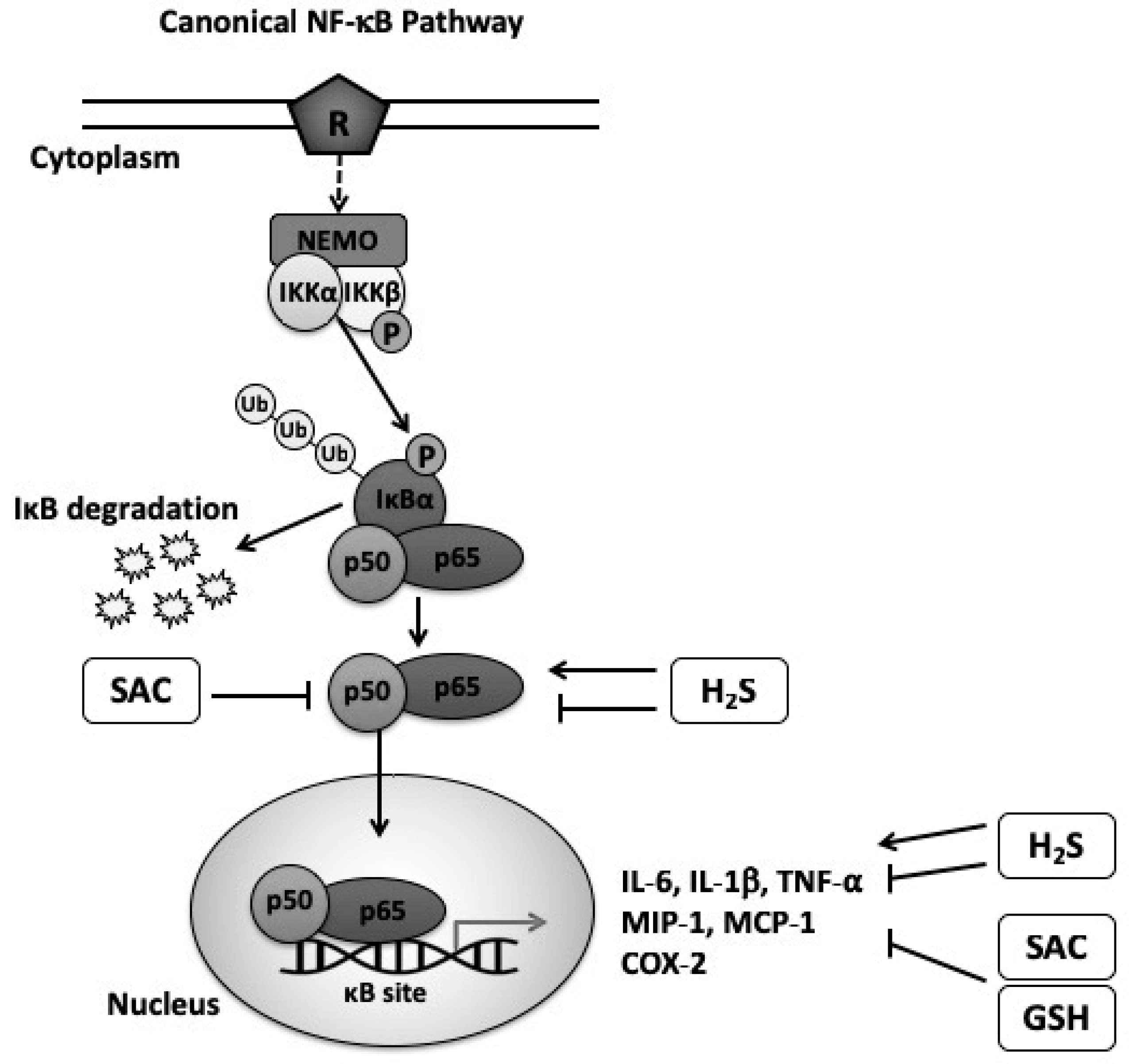
Table 1.
Summary of previous studies on the effect of glutathione in the immune response.
| Model | Treatment | Dose | Immune Response | Reference |
|---|---|---|---|---|
| Cell culture of isolated DC and T cells from study mice | Cysteine | T Cell Proliferation Assay: 50 μM cysteine Metabolic Labeling: 1 μCi/mL [35S] cysteine per 2 × 10⁶ cells | -Redox modeling of Tregs through reduction of GSH synthesis in DC. -Blocking of GSH redistribution from the nucleus to the cytoplasm in Tregs in response to T cell activation. | [96] |
| Cell culture of isolated leukocytes from healthy human blood | GSH-oxidizing agents (diamide and BHP) | 50 nmoL per 106 cells | -Regulation of phagocytosis in PMN through the inhibition of the assembly of microtubules and consequently reduction of H2O2 production in GSH homeostasis. | [97] |
| Cell culture of isolated splenic naive T cells from mice | NAC in GSH depleted cells | 20 mM NAC per 106 cells in the presence of 1 mM BSO | -Inhibition of T cell proliferation and reduction IL-2 production in GSH depleted cells. -Stimulation of T cell proliferation and increase IL-2 production in restored GSH cells. | [98] |
| Cell culture of isolated glomerular mesangial cell from rats | GSH | 3–5 mM GSH per 0.5–1 × 10⁶ cells | -Inhibition of the complement activation cascade and the binding of antibodies to antigens. | [99] |
| Human randomized controlled clinical trial in HIV-infected patients | NAC | ~7 g/day, oral | -Increase in GSH levels in the blood and in CD4 T cells. | [100] |
| Human randomized controlled clinical trial in HIV-infected patients | Liposomal-GSH | 1.26 g/day, oral | -Increase in the levels of total GSH in CD4 T cells, IL-6, IL-12, IL-2, IFN-γ and reduction in the levels of IL-10. | [101] |
| Patients with cirrhosis | GSH precursor (oxathizolidine-4-carboxylic acid) | 210 mg/kg/day, intravenous | -Reduction in the levels of IL-8 and IL-6 from isolated monocytes. | [102] |
Abbreviations: DC (dendritic cells); GSH (glutathione); NAC (N-acetylcysteine); BSO (L-buthionine-sulfoximine); HIV (human immunodeficiency virus); IL (interleukin); IFN-γ (interferon-gamma).
Table 2.
Previous studies on the effect of garlic derivatives in the immune response.
| Model | Treatment | Dose | Immune response | Reference |
|---|---|---|---|---|
| Cell culture of isolated PBMC from healthy individuals | Fresh garlic extract | 0.5–4 μg/mL per 106 cells | -Decrease of IL-17 expression when stimulated with PHA. | [48] |
| Cell culture of 3T3-L1 adipocytes | Alliin | 100 μmol/L (number of cells were not specified) | -Down-regulation of IL-6, MCP-1 genes expression and decrease of IL-6, MCP-1 protein levels. -Up-regulation of genes involved with immune response. | [127] |
| Cell culture of human colorectal cancer SW480 cells | DADS | 2.5–40 μmol/L (number of cells were not specified) | -Reduction of phosphorylation and nuclear translocation of NF-κB/p65 protein in a dose dependent way. | [128] |
| Cell culture of isolated splenocytes from mice | AGE | 0.5-1 v/v% per 0.2–1 × 10⁷ cells | -Increase in NK cell activation and cytotoxic T cells with suppression of tumor cell growth. -Increase of IL-2, TNF-α and IFN-γ levels in spleen cells. | [130] |
| Cell culture of Jurkat T cells | SAC | 0, 0.5, 1, 1.5, or 2 mg/mL of SAC in 106 cells | -Inhibition of NF-κB p50/65 heterodimer activation. | [3] |
| Mice with leukemia | DATS | 1 and 10 mg/kg, oral | -Increase of phagocytosis activity on PBMC. | [123] |
| Mice with implanted breast tumors | Purified protein fraction from garlic extract | 0.01–0.04 mg/mL, inoculation into the lesion | -Increase of T-cell activation and increase of intra-tumor infiltration of lymphocytes in all doses | [124] |
| Mice with alcoholic fatty liver disease | Allicin | 5 mg/kg/day and 20 mg/kg/day, oral | -Reduction of TNF-α, IL-1β and IL-6 levels with 20 mg dose. | [46] |
| Mice | Purified fraction of garlic extract | 10–40 mg/mL, oral | -Increase of NK activity with the 20 mg/mL dose. | [125] |
| Mice with colitis | Alliin | 500 mg/kg, oral | -Suppression of LPS-induced AP-1/NF-κB/STAT-1 activation. | [129] |
| Mice exposed to Aflatoxin B1 | AGE | 20 mg/kg/day, intraperitoneal | -Decrease in IFN-γ and IL-4 levels in isolated splenocytes. -Decrease in the number of Treg in the spleen. | [131] |
| Mice with implanted fibrosarcoma tumor | AGE | 100 mg/kg (containing 0.4 g of garlic/mL), intraperitoneal | -Increase in spleen lymphocyte subpopulation ratio and IFN-γ levels. -Increase in the amount of CD4+/CD8+ ratio. | [132] |
| Rats with induced neuroinflammation | AGE | 125, 250, 500 mg/kg, oral | -Decrease in the density of CD11b-positive microglia immunoreactivity and decrease in up-regulation of IL-1β in hippocampal region at all doses. | [133] |
| Mice with diabetes | SAC | 0.5 g/L, oral by drinking water | -Reduction in renal levels of IL-6 and TNF-α. -Suppression of renal NF-κB activity, mRNA expression and protein production. | [5] |
| Rats with diabetes | SAC | 150 mg/kg/day, oral | -Reduction in hippocampal NF-κB, TLR4 and TNF-α. | [6] |
| Rats with chromium (VI)-induced hepatotoxicity | SAC | 100 mg/kg, oral | -Decrease in the expression of NF-κB, TNF-α and iNOS in the liver. | [136] |
| Human randomized crossover study in healthy individuals | Raw crushed garlic | 5 g, oral | -Increase of NFAM1 gene expression. | [126] |
| Human randomized controlled clinical trial in patients with advanced colon, liver and pancreatic cancer | AGE | 500 mg/day, oral | -Increase in NK cell count and activity. | [64] |
| Human randomized controlled clinical trial in healthy individuals | AGE | 2.56 g/day, oral | -Increase in γδ-T cells and NK cells. -Attenuation of the severity of reported of cold and flu-like symptoms. | [134] |
| Human randomized controlled clinical trial in obese individuals | AGE | 3.6 g/day, oral | -Increase in γδ-T cell population and decrease in NKT cell population, IL-6 and TNF-α levels in the serum. | [135] |
Abbreviations: PBMC (peripheral blood mononuclear cells); MCP-1 (monocyte chemoattractant protein 1); DADS (diallyl disulfide); NF-κB (nuclear factor kappa-light-chain-enhancer of activated B cells); NF-κB/p65 (nuclear factor kappa-light-chain-enhancer of activated B cells/p65 subunit); AGE (aged garlic extract); NK (natural killer); TNF-α (tumor necrosis factor-alpha); IFN-γ (interferon-gamma); SAC (S-allyl cysteine); DATS (diallyl trisulfide); LPS (lipopolysaccharide); AP-1 (activator protein 1); STAT-1 (signal transducer and activator of transcription 1); CD (clutter of differentiation); TLR (toll-like receptor); iNOS (inducible nitric oxide synthase).
Table 3.
Previous studies on the effect of hydrogen sulfide on inflammation.
| Model | Treatment | Dose | Immune response | Reference |
|---|---|---|---|---|
| Cell culture of RAW264.7 cells | FW1256 (H2S donor) | 200 μM per 2 × 10⁵ or 106 cells | -Decrease in TNF-α, IL-6, PGE2, NO levels and expression of IL-1β, COX2, and iNOS. -Inhibition of NF-κB activation. | [14] |
| Cell culture of THP-1 cells | NaHS | 0.01, 0.1, 0.5 or 1 mM per 2 × 10⁶ cells | -Decrease in IL-6 and TNF-α levels in a dose dependent manner. | [141] |
| Cell culture of U937 cells | NaHS | 0.01, 0.1, or 1 mM (number of cells were not specified) | -Increase in mRNA expression and protein levels of TNF-α, IL-1β and IL-6 with 0.1 and 1mM doses. -Up-regulation of CD11b with 0.1 mM dose. -Increase in NF-κB p65 activity with 0.1 and 1mM doses. | [13] |
| Cell culture of human fibroblast-like synoviocytes | NaHS | 0.05–5 mM (number of cells were not specified) | -Increase in mRNA expression of TNF-α, IL-8, IL-1α, IL-1β and COX-2 at 2 mM dose. -Increase in mRNA expression of IL-8 and COX-2 at doses below 0.25 mM. -NaHS did not affect NF-κB activation at none of the doses. | [148] |
| Mice with colitis | ATB-429 (H2S donor) | 33–130 mg/kg, oral | -Reduction of mRNA expression of TNF-α, IFN-γ, IL-1, IL-2, IL-12 p40 and RANTES. | [142] |
| Rats with colitis | NaHS and Lawesson’s reagent (H2S donors) | 30 μmol/kg, intracolonically | -Reduction of mRNA expression of TNF-α with both donors. -Reduction in TNF-α protein levels with NaHS. | [143] |
| Mice with LPS-induced airway inflammation | GYY4137 (H2S donor) and NaHS | GYY4137:0.3 or 1 mg/kg NaHS: 1 or 3 mg/kg, intranasal | -Prevention of neutrophil increase in bronchoalveolar lavage fluid with both donors. -Prevention of IL-1β levels increase with both donors. | [144] |
| Rats with chronic renal failure | NaHS | 100 μmol/kg, intraperitoneal | -Decrease in TNF-α, IL-6, IL-10, and MCP-1 renal levels with both donors. -Decrease in NF-κB-p50, p65, and p-p65 levels with both donors. | [145] |
Abbreviations: COX2 (cyclooxygenase-2); iNOS (inducible nitric oxide synthase); RANTES (regulated on activation, normal T cell expressed and secreted); NaHS (sodium hydrosulfide).
© 2019 by the authors. Licensee MDPI, Basel, Switzerland. This article is an open access article distributed under the terms and conditions of the Creative Commons Attribution (CC BY) license (http://creativecommons.org/licenses/by/4.0/).
Share and Cite
MDPI and ACS Style
Rodrigues, C.; Percival, S.S. Immunomodulatory Effects of Glutathione, Garlic Derivatives, and Hydrogen Sulfide. Nutrients 2019, 11, 295. https://doi.org/10.3390/nu11020295
AMA Style
Rodrigues C, Percival SS. Immunomodulatory Effects of Glutathione, Garlic Derivatives, and Hydrogen Sulfide. Nutrients. 2019; 11(2):295. https://doi.org/10.3390/nu11020295
Chicago/Turabian StyleRodrigues, Camila, and Susan S. Percival. 2019. "Immunomodulatory Effects of Glutathione, Garlic Derivatives, and Hydrogen Sulfide" Nutrients 11, no. 2: 295. https://doi.org/10.3390/nu11020295
Note that from the first issue of 2016, this journal uses article numbers instead of page numbers. See further details here.