
Ag Decoration and SnO2 Coupling Modified Anatase/Rutile Mixed Crystal TiO2 Composite Photocatalyst for Enhancement of Photocatalytic Degradation towards Tetracycline Hydrochloride

 ,
, 
Abstract
:1. Introduction
2. Experimental Section
2.1. Material Preparation
2.2. Characterization
2.3. Photocatalysis Experiment
3. Results and Discussion
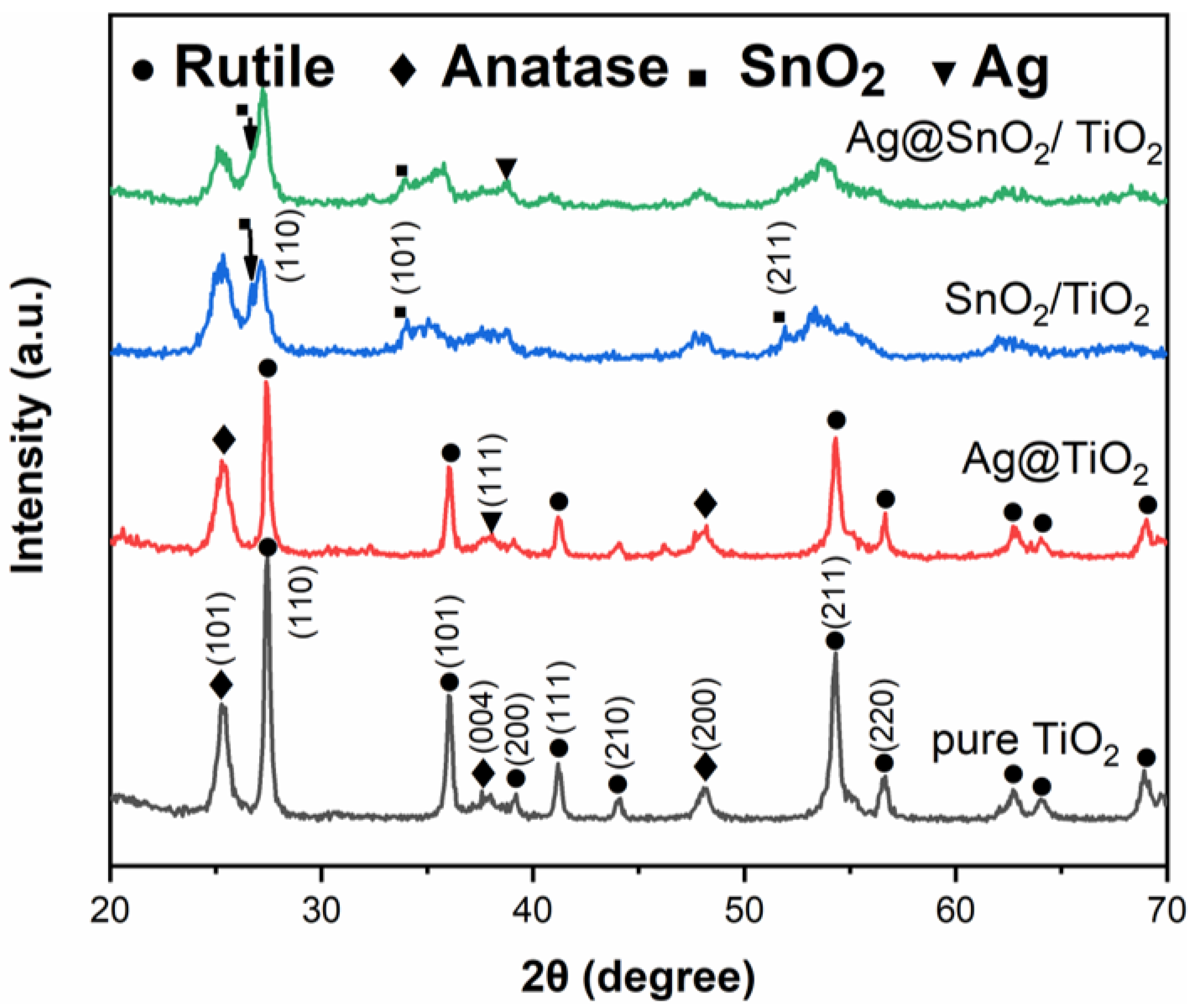
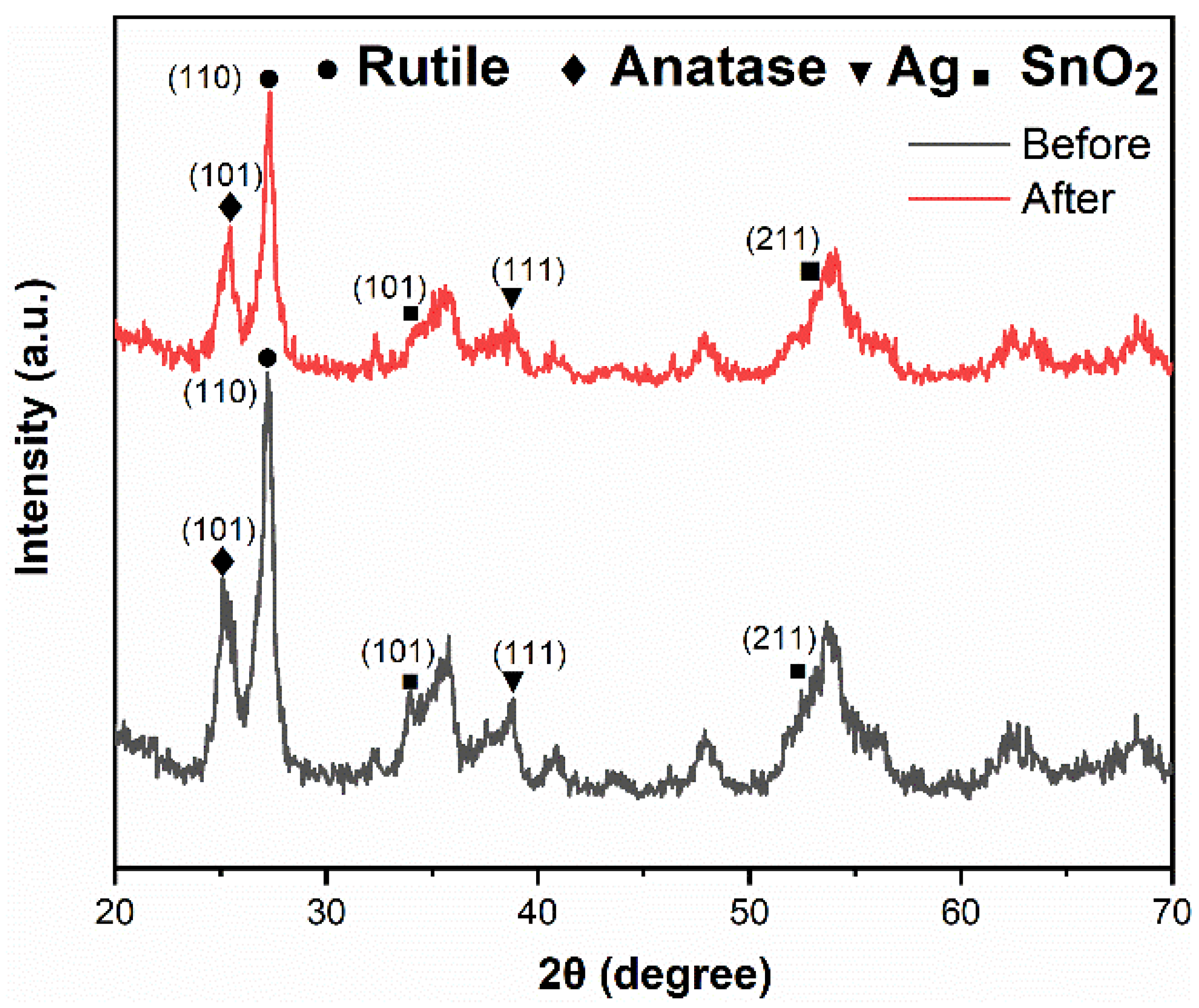
3.1. XRD Analysis
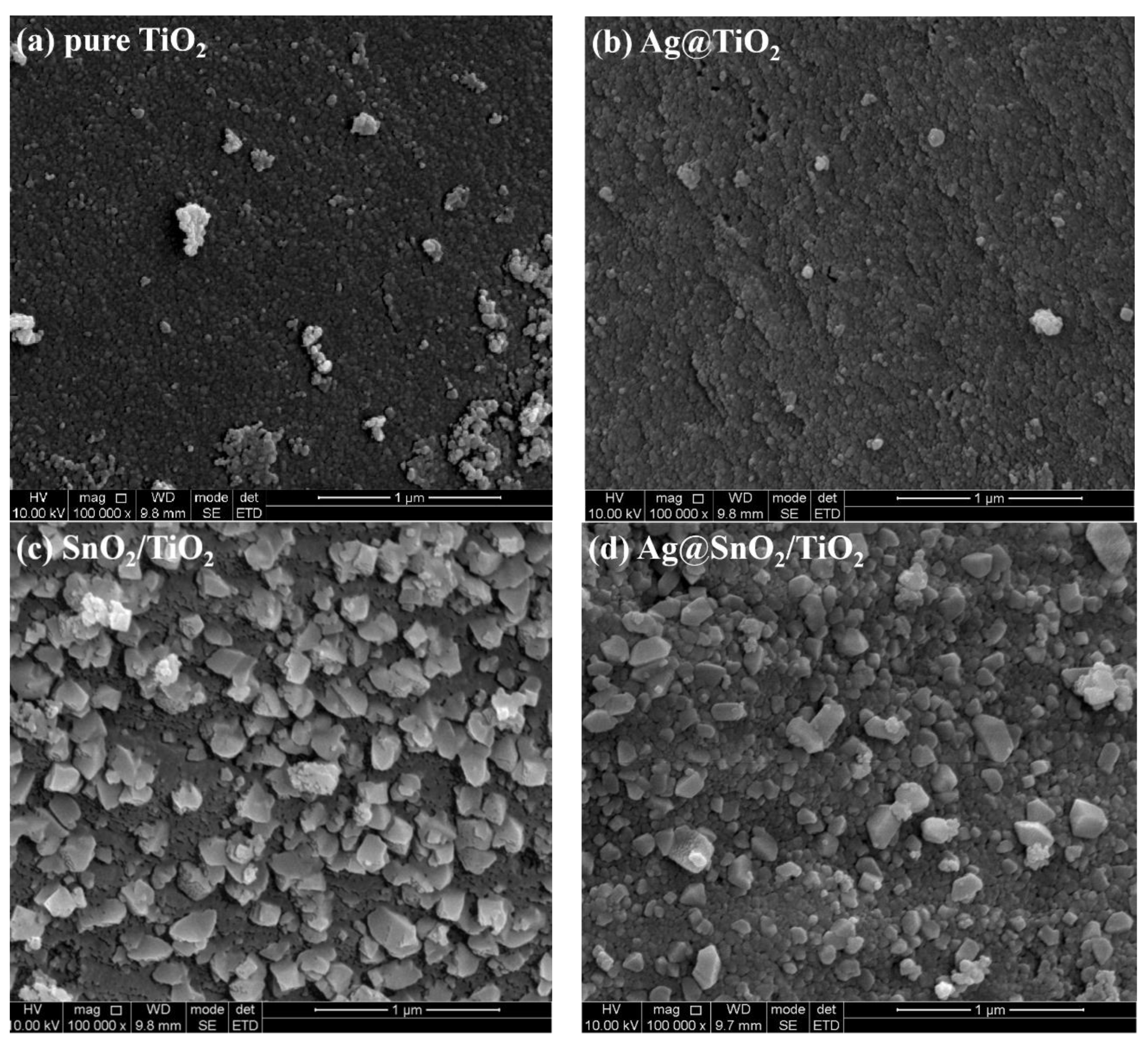
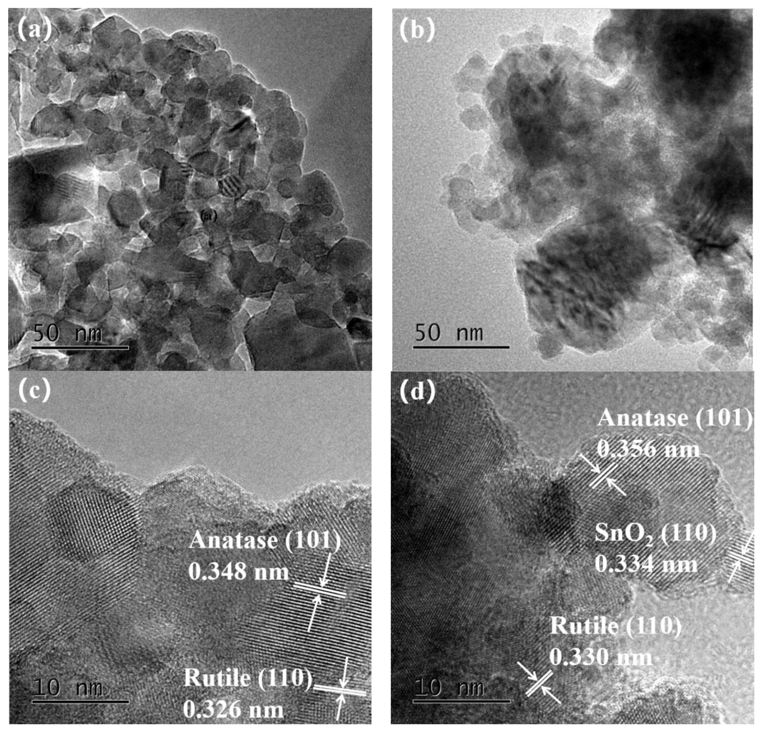
3.2. SEM and TEM Analyses
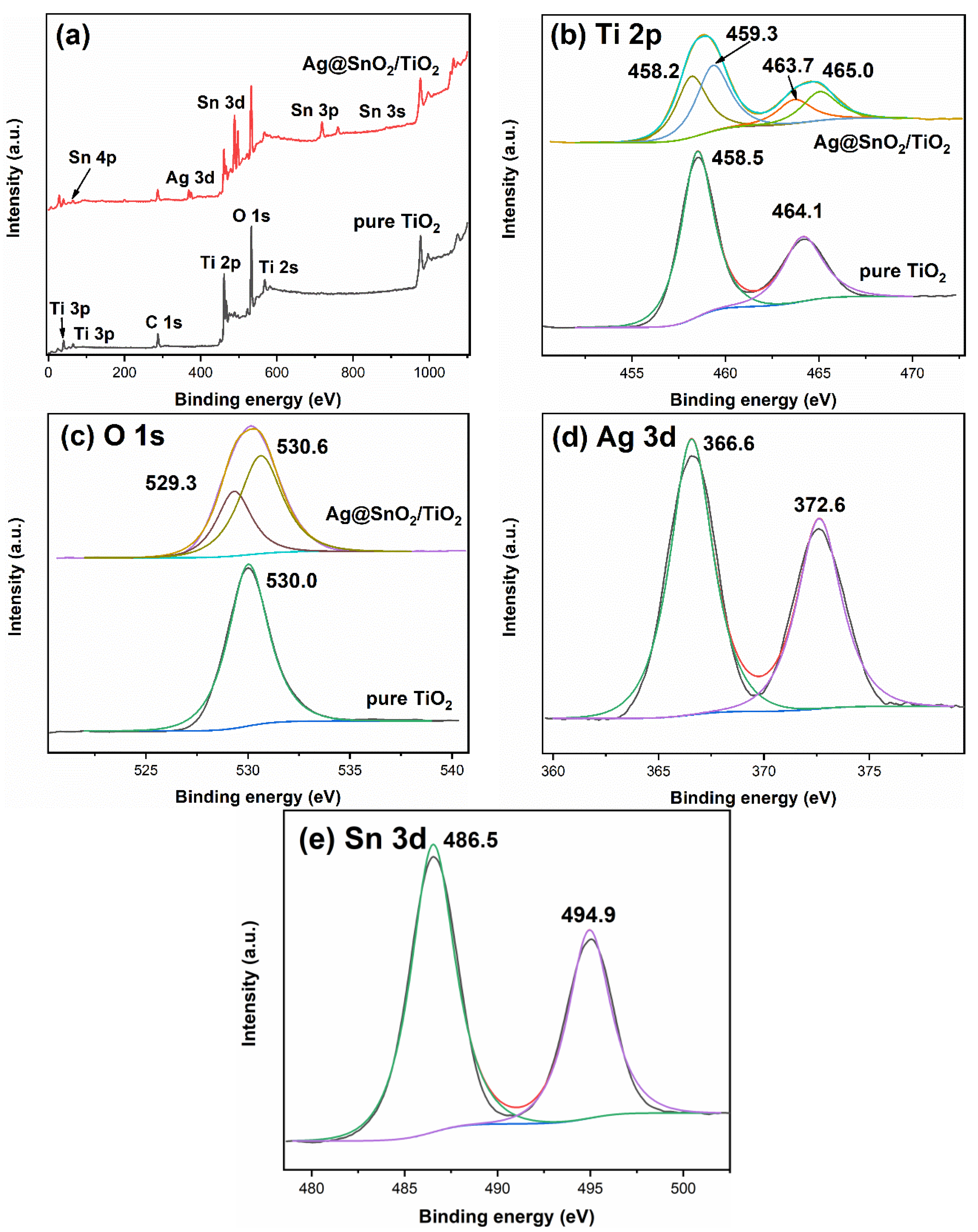
3.3. XPS Analysis
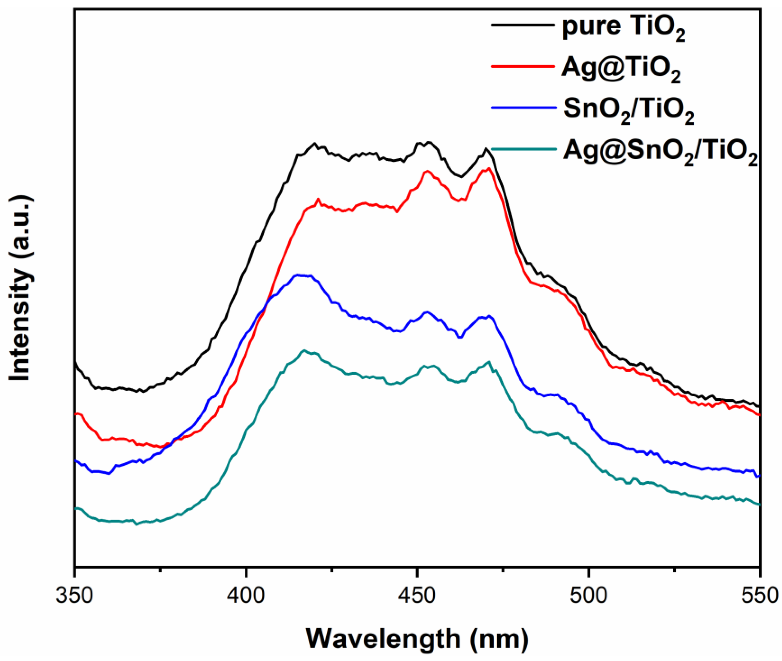
3.4. PL Analysis
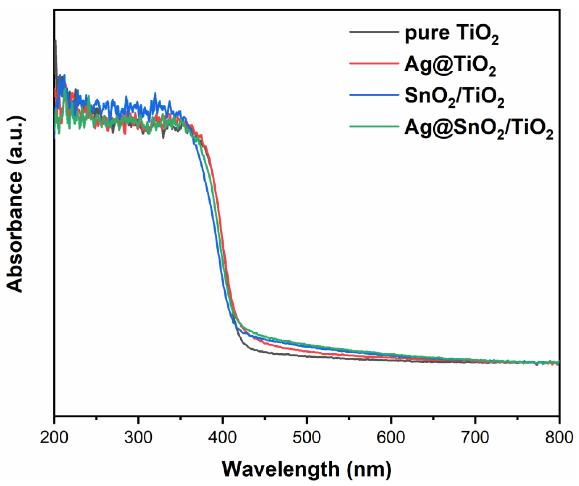
3.5. DRS Analysis
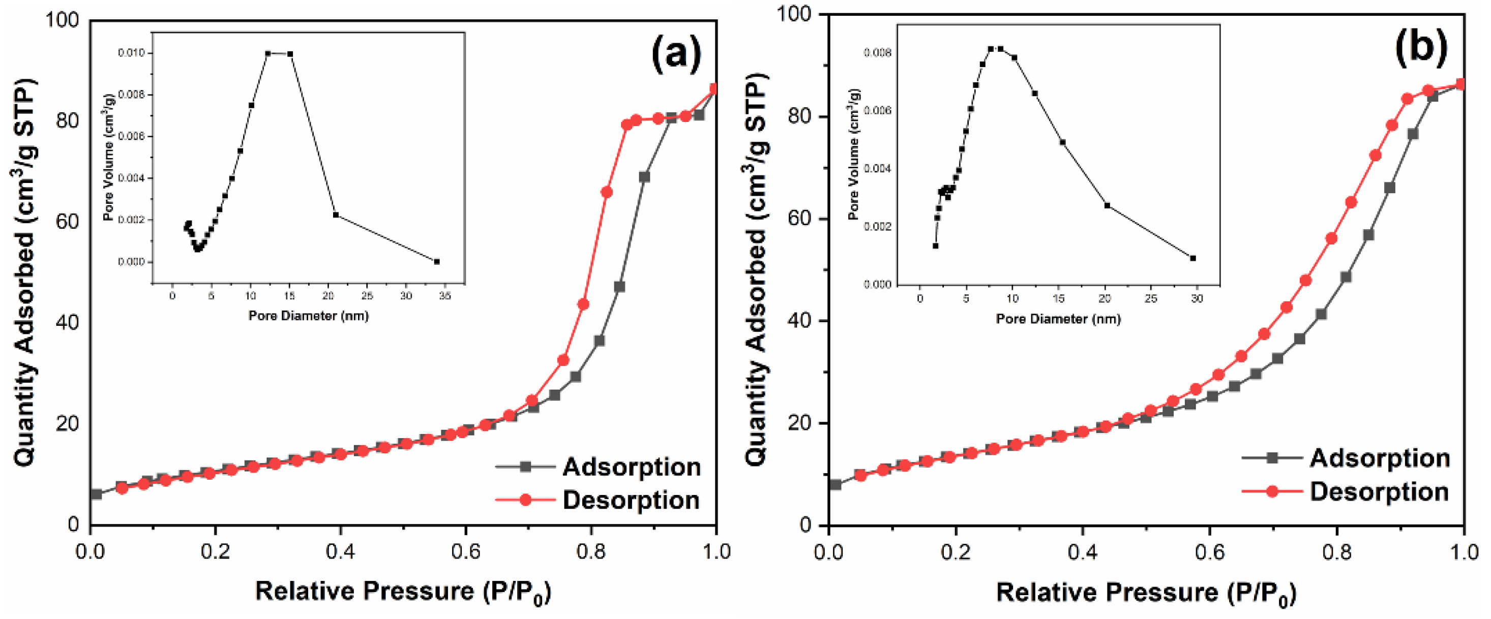
3.6. BET Analysis
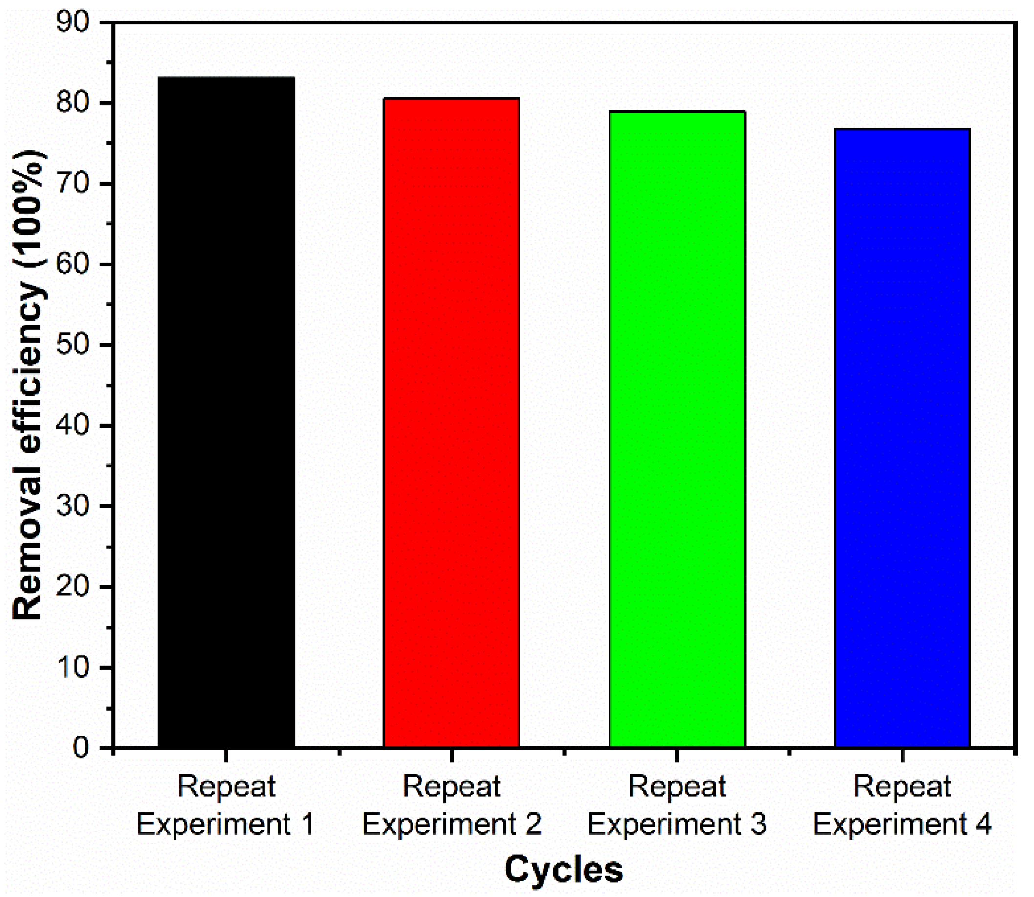
3.7. Photocatalytic Activity
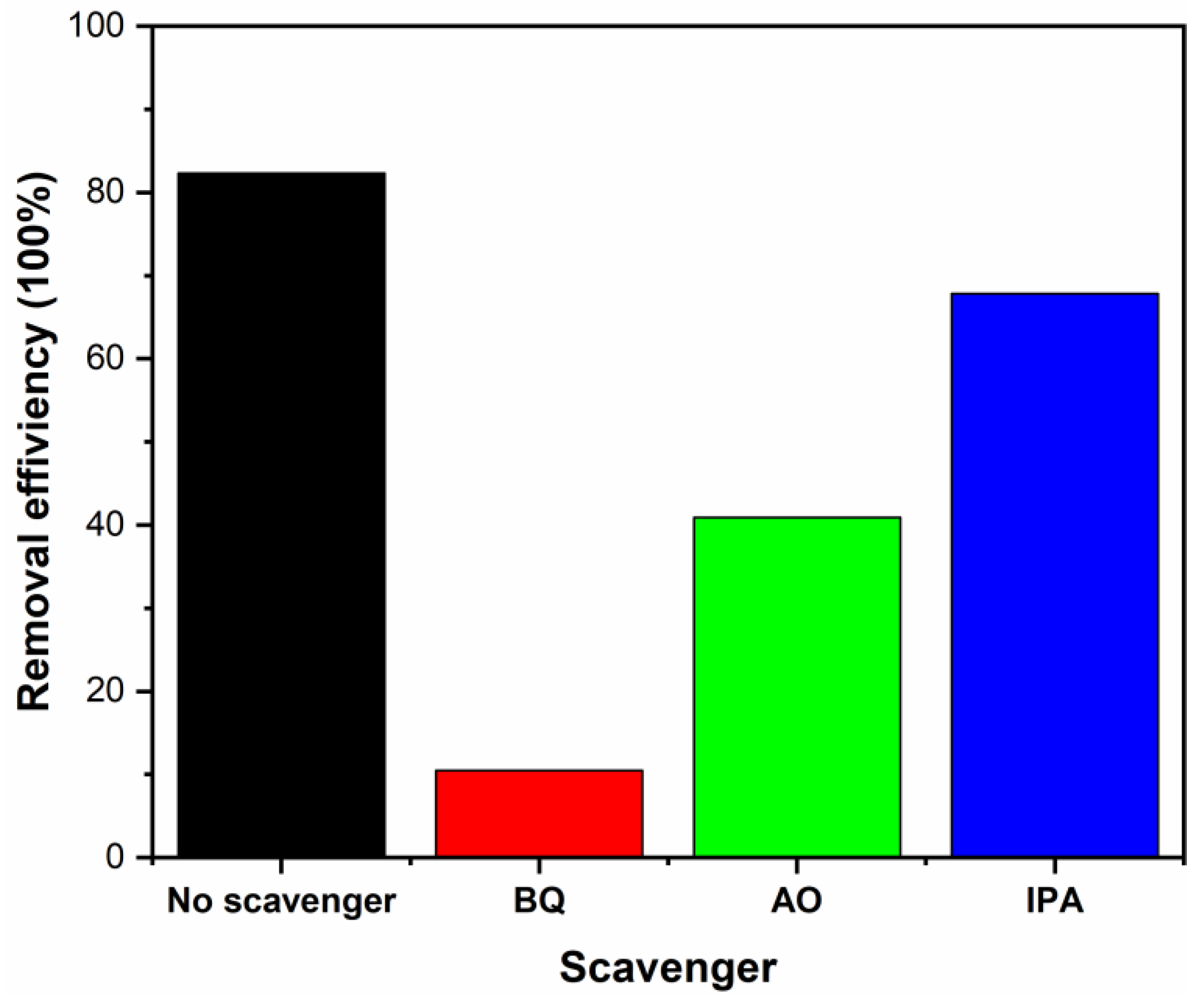
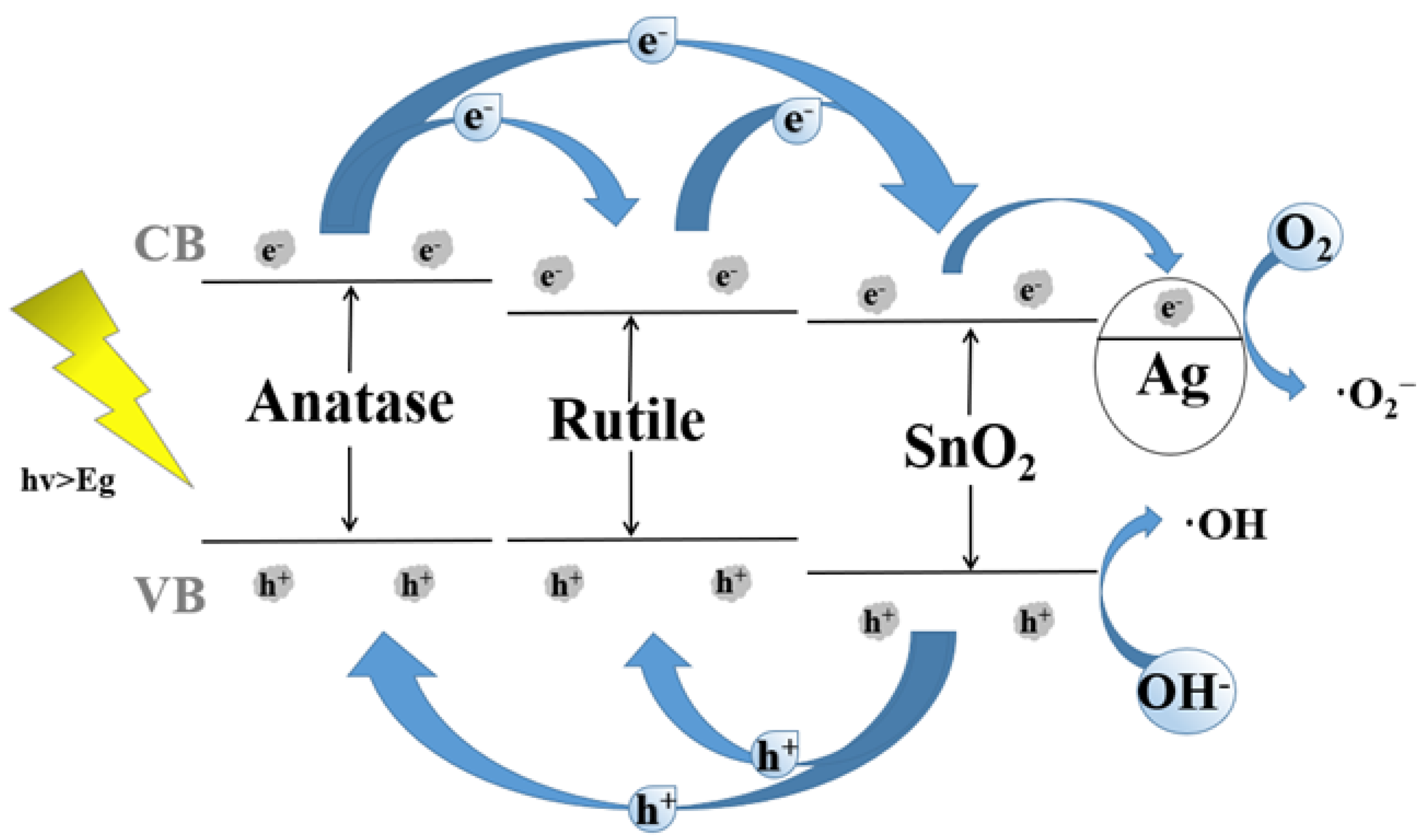
3.8. The Degradation Mechanism
4. Conclusions
Author Contributions
Funding
Institutional Review Board Statement
Informed Consent Statement
Data Availability Statement
Conflicts of Interest
References
- Wang, K.; Li, Y.; Zhang, G.K.; Li, J.; Wu, X.Y. 0D Bi nanodots/2D Bi3NbO7 nanosheets heterojunctions for efficient visible light photocatalytic degradation of antibiotics: Enhanced molecular oxygen activation and mechanism insight. Appl. Catal. B Environ. 2018, 240, 39–49. [Google Scholar] [CrossRef]
- Li, S.J.; Wang, C.C.; Liu, Y.P.; Xue, B.; Jiang, W.; Liu, Y.; Mo, L.Y.; Chen, X.B. Photocatalytic degradation of antibiotics using a novel Ag/Ag2S/Bi2MoO6 plasmonic p-n heterojunction photocatalyst: Mineralization activity, degradation pathways and boosted charge separation mechanism. Chem. Eng. J. 2021, 415, 12899. [Google Scholar] [CrossRef]
- Hajipour, P.; Eslami, A.; Bahrami, A.; Hosseini-Abari, A.; Saber, F.Y.; Mohammadi, R.; Mehr, M.Y. Surface modification of TiO2 nanoparticles with CuO for visible-light antibacterial applications and photocatalytic degradation of antibiotics. Ceram. Int. 2021, 47, 33875–33885. [Google Scholar] [CrossRef]
- Kuppusamy, S.; Kakarla, D.; Venkateswarlu, K.; Megharaj, M.; Yoon, Y.E.; Lee, Y.B. Veterinary antibiotics (VAs) contamination as a global agro-ecological issue: A critical view. Agric. Ecosysy. Environ. 2018, 257, 47–59. [Google Scholar] [CrossRef]
- He, D.; Sun, Y.B.; Xin, L.; Feng, J.W. Aqueous tetracycline degradation by non-thermal plasma combined with nano-TiO2. Chem. Eng. J. 2014, 258, 18–25. [Google Scholar] [CrossRef]
- Li, Y.H.; Liu, L.F.; Yang, F.L. Destruction of tetracycline hydrochloride antibiotics by FeOOH/TiO2 granular activated carbon as expanded cathode in low-cost MBR/MFC coupled system. J. Membr. Sci. 2017, 525, 202–209. [Google Scholar] [CrossRef]
- Wang, W.; Fang, J.J.; Shao, S.F.; Lai, M.; Lu, C.H. Compact and uniform TiO2@g-C3N4 core-shell quantum heterojunction for photocatalytic degradation of tetracycline antibiotics. Appl. Catal. B. 2017, 217, 57–64. [Google Scholar] [CrossRef]
- Shayegan, Z.; Lee, C.S.; Haghighat, F. TiO2 photocatalyst for removal of volatile organic compounds in gas phase–A review. Chem. Eng. J. 2018, 334, 2408–2439. [Google Scholar] [CrossRef] [Green Version]
- Nur, Y.; Lead, J.R.; Baalousha, M. Evaluation of charge and agglomeration behavior of TiO2 nanoparticles in ecotoxicological media. Sci. Total Environ. 2015, 535, 45–53. [Google Scholar] [CrossRef]
- Middepogu, A.; Hou, J.; Gao, X.; Lin, D.H. Effect and mechanism of TiO2 nanoparticles on the photosynthesis of Chlorella pyrenoidosa. Ecotoxicol. Environ. Saf. 2018, 161, 497–506. [Google Scholar] [CrossRef]
- Vinesh, V.; Shaheer, A.R.M.; Neppolian, B. Reduced graphene oxide (rGO) supported electron deficient B-doped TiO2 (Au/B-TiO2/rGO) nanocomposite: An efficient visible light sonophotocatalyst for the degradation of Tetracycline (TC). Ultrason. Sonochem. 2019, 50, 302–310. [Google Scholar] [CrossRef] [PubMed]
- Bahruji, H.; Bowker, M.; Davies, P.R.; Kennedy, J.; Morgan, D.J. The importance of metal reducibility for the photo-reforming of methanol on transition metal-TiO2 photocatalysts and the use of non-precious metals. Int. J. Hydrog. Energy 2015, 40, 1465–1471. [Google Scholar] [CrossRef]
- Tian, F.; Zhu, R.S.; Ouyang, F. Synergistic photocatalytic degradation of pyridine using precious metal supported TiO2 with KBrO3. J. Environ. Sci. 2013, 25, 2299–2305. [Google Scholar] [CrossRef]
- Zhang, Y.; Wang, T.; Zhou, M.; Wang, Y.; Zhang, Z.M. Hydrothermal preparation of Ag-TiO2 nanostructures with exposed {001}/{101} facets for enhancing visible light photocatalytic activity. Ceram. Int. 2017, 43, 3118–3126. [Google Scholar] [CrossRef]
- Li, H.; Shen, L.; Zhang, K.; Sun, B.; Ren, L.; Qiao, P.; Pan, K.; Wang, L.; Zhou, W. Surface plasmon resonance-enhanced solar-driven photocatalytic performance from Ag nanoparticle-decorated self-floating porous black TiO2 foams. Appl. Catal. B Enviorn. 2018, 220, 111–117. [Google Scholar] [CrossRef]
- Stucchi, M.; Bianchi, C.L.; Argirusis, C.; Pifferi, V.; Neppolian, B.; Cerrato, G.; Boffito, D.C. Ultrasound assisted synthesis of Ag-decorated TiO2 active in visible light. Ultrason. Sonochem. 2018, 40, 282–288. [Google Scholar] [CrossRef] [PubMed]
- Ali, T.; Ahmed, A.; Alam, U.; Uddin, I.; Tripathi, P.; Muneer, M. Enhanced photocatalytic and antibacterial activities of Ag-doped TiO2 nanoparticles under visible light. Mater. Chem. Phys. 2018, 212, 325–335. [Google Scholar] [CrossRef]
- Kong, J.H.; Song, C.X.; Zhang, W.; Xing, Y.H.; Wan, M.; Wang, Y.Q. Enhanced visible-light-active photocatalytic performances on Ag nanoparticles sensitized TiO2 nanotube arrays. Superlattice. Microst. 2017, 109, 579–587. [Google Scholar] [CrossRef]
- Odling, G.; Robertson, N. BiVO4-TiO2 Composite Photocatalysts for Dye Degradation Formed Using the SILAR Method. ChemPhysChem 2016, 17, 2872–2880. [Google Scholar] [CrossRef] [Green Version]
- Golestanbagh, M.; Parvini, M.; Pendashteh, A. Preparation, Characterization and Photocatalytic Properties of Visible-Light-Driven CuO/SnO2/TiO2 Photocatalyst. Catal. Lett. 2018, 148, 2162–2178. [Google Scholar] [CrossRef]
- Guo, X.L.; Wan, J.F.; Yu, X.J.; Lin, Y.H. Study on preparation of SnO2-TiO2/Nano-graphite composite anode and electro-catalytic degradation of ceftriaxone sodium. Chemosphere 2016, 164, 421–429. [Google Scholar] [CrossRef]
- Xun, H.T.; Zhang, Z.B.; Yu, A.H.; Yi, J.X. Remarkably enhanced hydrogen sensing of highly-ordered SnO2-decorated TiO2 nanotubes. Sens. Actuators B Chem. 2018, 273, 983–990. [Google Scholar] [CrossRef]
- De Mendonça, V.R.; Waldir, A.; Arenal, R.; Ribeiro, C. A building blocks strategy for preparing photocatalytically active anatase TiO2/rutile SnO2 heterostructures by hydrothermal annealing. J Colloid Interf. Sci. 2017, 505, 454–459. [Google Scholar] [CrossRef] [Green Version]
- Hu, W.Y.; Dong, F.Q.; Zhang, J.; Liu, M.X.; He, H.C.; Yang, D.M.; Deng, H.Q. A high-efficiency photocatalyst, flaky anatase@natural rutile composite using one-step microwave hydrothermal synthesis. Res. Chem. Intermed. 2018, 44, 705–720. [Google Scholar] [CrossRef]
- Kampouri, S.; Ireland, C.P.; Valizadeh, B.; Oveisi, E.; Schouwink, P.; Mensi, M.; Stylianou, K.C. Mixed-phase MOF-derived Titanium Dioxide for Photocatalytic Hydrogen Evolution: The Impact of the Templated Morphology. ACS Appl. Energy Mater. 2018, 1, 6541–6548. [Google Scholar] [CrossRef]
- Scotti, R.; D’Arienzo, M.; Testino, A.; Morazzoni, F. Photocatalytic mineralization of phenol catalyzed by pure and mixed phase hydrothermal titanium dioxide. Appl. Catal. B Environ. 2009, 88, 497–504. [Google Scholar] [CrossRef]
- Lei, Y.C.; Yang, Y.; Zhang, P.L.; Zhou, J.J.; Wu, J.; Li, K.; Wang, W.W.; Chen, L.Y. Controllable One-Step Synthesis of Mixed-Phase TiO2 Nanocrystals with Equivalent Anatase/Rutile Ratio for Enhanced Photocatalytic Performance. Nanomaterials 2021, 11, 1347. [Google Scholar] [CrossRef]
- Fu, W.; Li, G.; Wang, Y.; Zeng, S.; Yan, Z.; Wang, J.; Xin, S.; Zhang, L.; Wu, S.; Zhang, Z. Facile formation of mesoporous structured mixed-phase (anatase/rutile) TiO2 with enhanced visible light photocatalytic activity. Chem. Commun. 2017, 54, 58–61. [Google Scholar] [CrossRef]
- Cheng, H.; Ye, J.; Sun, Y.; Yuan, W.; Tian, J.; Bogale, R.F.; Tian, P.; Ning, G. Template-induced synthesis and superior antibacterial activity of hierarchical Ag/TiO2 composites. RSC Adv. 2015, 5, 80668. [Google Scholar] [CrossRef]
- Khojasteh, F.; Mersagh, M.R.; Hashemipour, H. The influences of Ni, Ag-doped TiO2 and SnO2, Ag-doped SnO2/TiO2 nanocomposites on recombination reduction in dye synthesized solar cells. J. Alloys Compd. 2021, 890, 161709. [Google Scholar] [CrossRef]
- Nakajima, T.; Nakamura, T.; Tsuchiya, T. Crystal-Plane Dependence of Nb-Doped Rutile TiO2 Single Crystals on Photoelectrochemical Water Splitting. Catalysts 2019, 9, 725. [Google Scholar] [CrossRef] [Green Version]
- De, D.S.; Behara, D.K.; Saha, S.; Kumar, A.; Subramaniam, A.; Sivakumar, S.; Pala, R.G.S. Design of iso-material heterostructures of TiO2 via seed mediated growth and arrested phase transitions. Phys. Chem. Chem. Phys. 2020, 22, 25366. [Google Scholar] [CrossRef] [PubMed]
- Yu, Y.; Zhang, Y.; Zou, K.; Chen, G.; Zhang, Y.; Li, H.; Lu, Y.; Zhang, Q.; He, Y.B. High energy density and efficiency in (Pb,La)(Zr,Sn,Ti)O3 antiferroelectric ceramics with high La3+ content and optimized Sn4+ content. Ceram. Int. 2019, 45, 24419–24424. [Google Scholar] [CrossRef]
- Jia, C.; Chen, H.-S.; Yang, P. Construction of hollow waxberry-like rutile-/anatase-TiO2/SnO2 towards enhanced photocatalysis. J. Ind. Eng. Chem. 2018, 58, 278–289. [Google Scholar] [CrossRef]
- Jia, C.; Dong, T.; Li, M.; Wang, P.; Yang, P. Preparation of anatase/rutile TiO2/SnO2 hollow heterostructures for gas sensor. J. Alloys Compd. 2018, 769, 521–531. [Google Scholar] [CrossRef]
- Zhang, Z.; Ma, Y.; Bu, X.; Wu, Q.; Hang, Z.; Dong, Z.; Wu, X. Facile one-step synthesis of TiO2/ Ag/SnO2 ternary heterostructures with enhanced visible light photocatalytic activity. Sci. Rep. 2018, 8, 10532. [Google Scholar] [CrossRef] [PubMed] [Green Version]
- Liu, H.; Fan, H.; Wu, R.; Tian, L.; Yang, X.; Sun, Y. Nitrogen-doped black TiO2 spheres with enhanced visible light photocatalytic performance. SN Appl. Sci. 2019, 1, 487. [Google Scholar] [CrossRef] [Green Version]
- Sheng, J.; Tong, H.; Xu, H.; Tang, C. Preparation and photocatalytic activity of SnO2@TiO2 core-shell composites modified by Ag. Catal. Surv. Asia 2016, 20, 167–172. [Google Scholar] [CrossRef] [Green Version]
- Zhao, L.N.; Jia, Y.H.; You, H.; Wang, S.T.; Fu, L. Photocatalytic performance and application outlook of 3D TiO2/titanium mesh modified by GO-Ag joined-deposition. Catal. Today 2020, 340, 106–114. [Google Scholar] [CrossRef]
- Žerjav, G.; Arshad, M.S.; Djinović, P.; Junkar, I.; Kovač, J.; Zavašnik, J.; Pintar, A. Improved electron-hole separation and migration in anatase TiO2 nanorod/reduced graphene oxide composites and their significance on photocatalytic performance. Nanoscale 2017, 9, 4578–4592. [Google Scholar] [CrossRef] [Green Version]
- Gao, Y.; Fang, P.; Chen, F.; Liu, Y.; Liu, Z.; Wang, D.; Dai, Y. Enhancement of stability of N-doped TiO2 photocatalysts with Ag loading. Appl. Surf. Sci. 2013, 265, 796–801. [Google Scholar] [CrossRef]
- Sun, B.; Chen, Y.; Tao, L.; Zhao, H.; Zhou, G.; Xia, Y.; Wang, H.; Zhao, Y. Nanorods array of SnO2 quantum dots interspersed multiphase TiO2 heterojunctions with highly photocatalytic water splitting and self-rechargeable battery-like applications. ACS Appl. Mater. Inter. 2019, 11, 2071–2081. [Google Scholar] [CrossRef] [PubMed]
- Ye, J.; Xu, J.T.; Tian, D.; Zhao, X.; Wang, Q.; Wang, J.; Li, Y.; Zhao, C.; Liu, Z.; Fu, Y. Efficient photocatalytic reduction of CO2 by a rhenium-doped TiO2-x/SnO2 inverse opal S-scheme heterostructure assisted by the slow-phonon effect. Sep. Purif. Technol. 2021, 277, 119431. [Google Scholar] [CrossRef]
- Jia, T.; Liu, M.; Zheng, C.; Long, F.; Min, Z.; Fu, F.; Yu, D.; Li, J.; Lee, J.H.; Kim, N.H. One-Pot Hydrothermal Synthesis of La-Doped ZnIn2S4 Microspheres with Improved Visible-Light. Nanomaterials 2020, 10, 2026. [Google Scholar] [CrossRef]
- Adyani, S.M.; Ghorbani, M. A comparative study of physicochemical and photocatalytic properties of visible light responsive Fe; Gd and P single and tri-doped TiO2 nanomaterials. J. Rare Earths 2018, 36, 72–85. [Google Scholar] [CrossRef]
- Lei, X.F.; Xue, X.X.; Yang, H. Preparation and characterization of Ag-doped TiO2 nanomaterials and their photocatalytic reduction of Cr (VI) under visible light. Appl. Surf. Sci. 2014, 321, 396–403. [Google Scholar] [CrossRef]
- Yu, J.; Yu, J.C.; Zhao, X.J. The Effect of SiO2 Addition on the Grain Size and Photocatalytic Activity of TiO2 Thin Films. J. Sol-Gel Sci. Technol. 2002, 24, 95–103. [Google Scholar] [CrossRef]
- Chen, Y.; Liu, K.R. Fabrication of Ce/N co-doped TiO2/diatomite granule catalyst and its improved visible-light-driven photoactivity. J. Hazard. Mater. 2017, 324, 139–150. [Google Scholar] [CrossRef]
- Sibu, C.P.; Kumar, S.R.; Mukundan, P.; Warrier, K.G.K. Structural modifications and associated properties of lanthanum oxide doped sol-gel nanosized titanium oxide. Chem. Mater. 2002, 14, 2876–2881. [Google Scholar] [CrossRef]
- Wu, D.; Li, C.; Zhang, D.S.; Wang, L.L.; Zhang, X.; Shi, Z.; Lin, Q. Photocatalytic improvement of Y3+ modified TiO2 prepared by a ball milling method and application in shrimp wastewater treatment. RSC Adv. 2019, 9, 14609. [Google Scholar] [CrossRef] [Green Version]
- Tang, M.; Xia, Y.; Yang, D.; Liu, J.; Zhu, X.; Tang, R. Effects of Hydrothermal Time on Structure and Photocatalytic Property of Titanium Dioxide for Degradation of Rhodamine B and Tetracycline Hydrochloride. Materials 2021, 14, 5674. [Google Scholar] [CrossRef]
- Wang, Y.; Qiao, M.; Lv, J.; Xu, G.; Zheng, Z.; Zhang, X.; Wu, Y. g-C3N4/g-C3N4 isotype heterojunction as an efficient platform for direct photodegradation of antibiotic. Fuller. Nanotub. Carbon Nanostruct. 2018, 26, 210–217. [Google Scholar] [CrossRef]
- Zhu, X.; Zhu, R.; Pei, L.; Liu, H.; Xu, L.; Wang, J.; Feng, W.; Jiao, Y.; Zhang, W. Fabrication, characterization, and photocatalytic activity of anatase/rutile/SnO2 nanocomposites. J. Mater. Sci. 2019, 30, 21210–21218. [Google Scholar] [CrossRef]
- Zhang, Y.; Wang, L.; Yu, S.; Jiang, H.; Yun, Y.; Sun, Y.; Shi, J. Ag-induced synthesis of three dimensionally ordered macroporous anatase/rutile homojunction for solar light-driven Z-scheme photocatalysis. Sol. Energy 2018, 174, 770–779. [Google Scholar] [CrossRef]
- Qin, J.; Wang, J.; Yang, J.; Hu, Y.; Fu, M.; Ye, D. Metal organic framework derivative-TiO2 composite as efficient and durable photocatalyst for the degradation of toluene. Appl. Catal. B Environ. 2020, 267, 118667. [Google Scholar] [CrossRef]
- Prakash, K.; Kumar, P.S.; Pandiaraj, S.; Saravanakumar, K.; Karuthapandian, S. Controllable synthesis of SnO2 photocatalyst with superior photocatalytic activity for the degradation of methylene blue dye solution. J. Exp. Nanosci. 2021, 11, 1–18. [Google Scholar] [CrossRef] [Green Version]
- Wu, D.; Guo, J.; Ge, Z.-H.; Feng, J. Facile Synthesis Bi2Te3 based nanocomposites: Strategies for enhancing charge carrier separation to improve photocatalytic activity. Nanomaterials 2021, 11, 3390. [Google Scholar] [CrossRef]


Publisher’s Note: MDPI stays neutral with regard to jurisdictional claims in published maps and institutional affiliations. |
© 2022 by the authors. Licensee MDPI, Basel, Switzerland. This article is an open access article distributed under the terms and conditions of the Creative Commons Attribution (CC BY) license (https://creativecommons.org/licenses/by/4.0/).
Share and Cite
Tang, M.; Xia, Y.; Yang, D.; Lu, S.; Zhu, X.; Tang, R.; Zhang, W. Ag Decoration and SnO2 Coupling Modified Anatase/Rutile Mixed Crystal TiO2 Composite Photocatalyst for Enhancement of Photocatalytic Degradation towards Tetracycline Hydrochloride. Nanomaterials 2022, 12, 873. https://doi.org/10.3390/nano12050873
Tang M, Xia Y, Yang D, Lu S, Zhu X, Tang R, Zhang W. Ag Decoration and SnO2 Coupling Modified Anatase/Rutile Mixed Crystal TiO2 Composite Photocatalyst for Enhancement of Photocatalytic Degradation towards Tetracycline Hydrochloride. Nanomaterials. 2022; 12(5):873. https://doi.org/10.3390/nano12050873
Chicago/Turabian StyleTang, Mao, Yangwen Xia, Daixiong Yang, Shiji Lu, Xiaodong Zhu, Renyong Tang, and Wanming Zhang. 2022. "Ag Decoration and SnO2 Coupling Modified Anatase/Rutile Mixed Crystal TiO2 Composite Photocatalyst for Enhancement of Photocatalytic Degradation towards Tetracycline Hydrochloride" Nanomaterials 12, no. 5: 873. https://doi.org/10.3390/nano12050873