Review

The State of the Art of Theranostic Nanomaterials for Lung, Breast, and Prostate Cancers

by
Lucas F. Freitas
1,*,
Aryel H. Ferreira
1,2,
Velaphi C. Thipe
1,
Gustavo H. C. Varca
1,*,
Caroline S. A. Lima
1,
Jorge G. S. Batista
1,
Fabiane N. Riello
1,
Kamila Nogueira
1,
Cassia P. C. Cruz
1,
Giovanna O. A. Mendes
1,
Adriana S. Rodrigues
1,
Thayna S. Sousa
1,
Victoria M. Alves
1 and
Ademar B. Lugão
1
1
Nuclear and Energy Research Institute, IPEN-CNEN/SP, Sao Paulo 05508-000, Brazil
2
MackGraphe-Graphene and Nanomaterial Research Center, Mackenzie Presbyterian University, Sao Paulo 01302-907, Brazil
*
Authors to whom correspondence should be addressed.
Nanomaterials 2021, 11(10), 2579; https://doi.org/10.3390/nano11102579
Submission received: 26 July 2021 / Revised: 14 September 2021 / Accepted: 24 September 2021 / Published: 30 September 2021

Abstract

:
The synthesis and engineering of nanomaterials offer more robust systems for the treatment of cancer, with technologies that combine therapy with imaging diagnostic tools in the so-called nanotheranostics. Among the most studied systems, there are quantum dots, liposomes, polymeric nanoparticles, inorganic nanoparticles, magnetic nanoparticles, dendrimers, and gold nanoparticles. Most of the advantages of nanomaterials over the classic anticancer therapies come from their optimal size, which prevents the elimination by the kidneys and enhances their permeation in the tumor due to the abnormal blood vessels present in cancer tissues. Furthermore, the drug delivery and the contrast efficiency for imaging are enhanced, especially due to the increased surface area and the selective accumulation in the desired tissues. This property leads to the reduced drug dose necessary to exert the desired effect and for a longer action within the tumor. Finally, they are made so that there is no degradation into toxic byproducts and have a lower immune response triggering. In this article, we intend to review and discuss the state-of-the-art regarding the use of nanomaterials as therapeutic and diagnostic tools for lung, breast, and prostate cancer, as they are among the most prevalent worldwide.

1. Introduction

The unsolved oncologic challenges of current treatment regimens are hindered by (i) their inability to detect distant micrometastases and prognostic tumor aggressiveness, (ii) non-specific and non-selective delivery with poor biodistribution yielding toxicity, (iii) differentiation between indolent tumors and tumors exhibiting metastatic potential, and (iv) real-time monitoring and predictive treatment response—which affords physicians to adjust dosimetry to prevent overtreatment resulting in harmful side-effects or undertreatment that can lead to incomplete cancer remission. It is paramount to circumvent systemic distribution or off-target accumulation of therapeutics that can be detrimental to patients.
According to recent data, breast cancer is the most common cancer among women and is characterized by multifactorial pathogenesis [1]. There are three main types of breast cancer, classified according to biochemical and molecular characteristics, disease stage, and response to treatment: HER2-positive cancers (human epidermal growth factor receptor 2), the hormone receptor-positive cancers, and the triple-negative cancers (they are negative for HER2 receptors, estrogen receptors, and progesterone receptors). Their incidence depends mostly on ethnicity, age, and life habits, and both the therapeutic approach and the prognosis are dependent on the cancer types as well [2].
Prostate cancer, on the other hand, is the second most prevalent malignancy among men, and the prognosis is worse according to age, ethnicity, daily habits, and the time after diagnosis. The most common biomarker related to prostate cancer is PSA (Prostate-Specific Antigen), which is often elevated in prostate malignancies [3]. It can also be used as a targeting agent, according to recent data and some recent clinical trials. Although current treatment interventions, e.g., enzalutamide and other specific inhibitors or immunotherapeutics, have demonstrated modest progression-free survival increases, researchers keep searching for novel and more efficient approaches for prostate malignancies [4].
Finally, lung cancer is the lead cause of death worldwide due to malignant disease in both sexes. In spite of all the advances obtained throughout the last decades, the overall survival still remains low; therefore, the research in this field stays at a fast pace. The main risk factors for lung malignancies are smoking and air pollution, but it is known that there are genetic factors involved [5].
Advances in the synthesis and engineering of nanomaterials offer more robust and specific systems for the treatment of severe diseases, such as cancer, with technologies that combine therapy with imaging diagnostic tools in the so-called nanotheranostics [6]. The broad and most used definition of nanomaterials is provided by the regulatory organ International Organization for Standardization (ISO), which defines a nanomaterial as any material with external dimensions, internal structure, or surface structure within the nanometric scale (10−9 m), with an average size between 1 and 100 nm [7].
Synthetic nanomaterials are still the most commonly used compared to natural ones, either produced by chemical or physical methods, as those methods provide easy scalability and precise control on size and morphology. Another interesting factor is the possibility of anchoring or conjugating different reagents and chemical compounds with accuracy. One of the main concerns about synthetic nanomaterials, though, is the environmental impact they might have, which can be even higher than their bulk counterparts [8].
Most of the advantages of nanomaterials over the classic anticancer therapies come from their optimal size, which prevents the elimination by the kidneys and enhances their permeation in the tumor due to the abnormal blood vessels present in cancer tissues. Furthermore, the drug delivery and the contrast efficiency for imaging are enhanced, especially due to the increased surface area and the selective accumulation in the desired tissues. This property leads to the reduced drug dose necessary to exert the desired effect and to longer action within the tumor. Finally, they are made so that there is no degradation into toxic byproducts and lower immune response triggering [6].
In this article, we intend to review and discuss the state-of-the-art regarding the use of nanomaterials as therapeutic and diagnostic tools for lung, breast, and prostate cancer, as they are among the most prevalent worldwide.

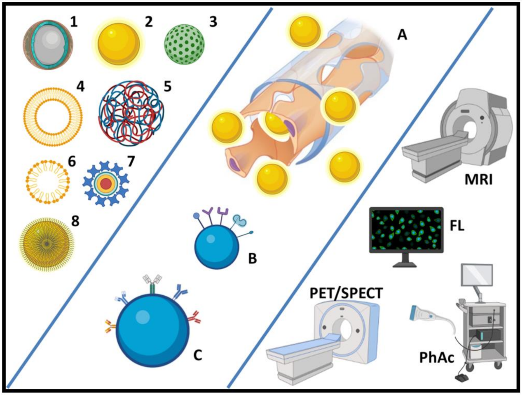
2. Theranostic Nanomaterials

Nanotheranostics (the field of science focused on developing materials and devices that act both as therapeutic and diagnostic agents) represents one of the scientific frontiers involving different multidisciplinary aspects and can contribute to human health advancement. Nanotheranostic systems offer advantages when compared to conventional therapy, and diagnostic, pharmaceutical approaches applied separately [9]. Ideally, nanotheranostic systems should provide the therapeutic and diagnostic agent delivery exclusively to the desired location, minimizing the amount needed to obtain the desired effect and, consequently, adverse effects [10]. This specific targeting is achieved according to the design and materials used in its construction.
In this field, a wide variety of component systems that modulate the speed and direct the drug to be released at a specific site have been the nanotechnology research subject [11]. Among the most studied systems, there are quantum dots, liposomes, polymeric nanoparticles, inorganic nanoparticles, magnetic nanoparticles, dendrimers, and gold nanoparticles (Figure 1) [12].
Quantum dots
Quantum dots (QD) are light-emitting inorganic nanocrystals composed of semiconductor elements from the periodic groups II–VI or III–V [13]. They have unique optical characteristics, absorbing the light energy intensely in the spectral region from the ultraviolet to the near-infrared (NIR) and emitting fluorescence by narrow and symmetrical bands whose position depends on both the size of the nanocrystal and the type of semiconductor used [14]. The core materials are generally composed of CdSe, CdTe, CdS, ZnS, ZnSe, PbS and PbSe, InP, and InGaP, with a core-shell structure of 1 to 10 nm in diameter [15]. The inorganic coating with ZnS and CdS on the particle’s surface improves the photoluminescent quantum efficiency and protects the nucleus from oxidation in biological environments, as they are formed from inorganic elements whose cytotoxicity can be significant even at low concentrations [16,17].
These systems can release toxic ionized metals exposed to oxidation promoted by the environment. However, QD’s degradation and cytotoxicity can be minimized by encapsulating it with a biocompatible system [18]. The use of specific compounds, such as aptamers to functionalize QD can increase their affinity with selected locations in the organism, enabling its selective transport or drug delivery [19]. QDs are considered an alternative to molecular fluorophores, such as fluorescein and rhodamine, as they have better photostability and long-lasting fluorescence time (~10–100 ns), enabling their use for an extended period of time without losing efficiency, even in extremely low concentrations [20].
Polymeric Nanoparticles
Polymeric nanoparticles have a diameter of less than 1 µm and can be prepared from natural or synthetic polymers. They include nanocapsules and nanospheres that differ in composition and structural organization [21]. In general, polymer-based nanotheranostic materials comprise: (i) a polymer component that offers stabilization and biocompatibility, (ii) a therapeutic agent (i.e., small molecule drug, siRNA, etc.), and (iii) an imaging agent (i.e., MRI contrast agent, radionuclide, fluorophore, etc.) [22]. They are considered one of the most promising tools for theranostics applications.
The nanocapsules are constructed with a polymeric shell that surrounds a hydrophobic core. The drug is usually dissolved in the core but can also be present in the nanocapsule’s surface [23]. On the other hand, nanospheres are formed by a polymeric matrix, and a drug can be retained or adsorbed on the matrix. Synthetic polymers, such as poly (lactic-co-glycolic acid) (PLGA), Poly lactic Acid (PLA), and natural polymers, such as albumin, collagen, and chitosan, have been efficiently used in these systems [24].
Polymer-based distribution systems are preferred because they have an inherent versatility in their structures. The potential for modifying their surface (with peptide molecules, proteins, etc.) allows specific targeting of therapeutic and/or diagnostic agents to specific regions, increasing the effectiveness, sensitivity, and specificity of the therapy and diagnosis approach. Furthermore, they can be easily adapted to improve the theranostic conjugate solubility and biocompatibility. [25].
Liposomes
Liposomes are concentric vesicles with one or more lipid bilayers of natural or synthetic phospholipids and cholesterol. [26]. Liposomes are used for drug delivery and as diagnostic agents due to several advantages: they are suitable for transport compounds with different physicochemical characteristics (hydrophilic, hydrophobic, and amphiphilic); they can be functionalized in order to decrease toxicity and adverse effects; they provide protection and a controlled release of the diagnostic agent in the body; they are considered suitable for different routes of administration and a wide variety of physiological conditions; their size can be tuned according to necessity; and, finally, due to their nature, liposomes are generally biocompatible [27]. Despite some disadvantages in using liposomes that might appear in certain conditions, i.e., their high production costs, low stability (phospholipids are easily hydrolyzed and oxidized) associated with a reduced half-life, liposomes are one of the main nanotechnological resources when it comes to encapsulating drugs and active compounds, and many clinical trials are currently using this approach.
Micelles
Micelles, with a size ranging between 5 and 100 nm, are colloidal dispersions of amphiphilic substances, with hydrophilic and hydrophobic regions, which self-organize in an aqueous solution through the establishment of hydrophobic interactions [28]. Micelles have been widely studied as drug carriers, being prepared from various amphiphilic materials, allowing the increase of hydrophobic molecules solubility and the incorporation of multiple functionalities in a single structure [12].
Usually, micelles are created through the self-assembly of copolymers in an aqueous medium that occurs in a concentration-dependent manner [29]. The drug and the diagnostic agents can be attached to the polymer before forming the micelle or become trapped in the micelle’s hydrophobic core. The micellar structure is highly advantageous in drug administration since the hydrophobic core increases the solubility of hydrophobic drugs and the hydrophilic periphery protects them. Micelles are not easily recognized by phagocytes, enabling the drug to remain in the bloodstream until it reaches the therapeutic target. Additionally, they can be composed of biocompatible and biodegradable copolymers and are quickly eliminated from the body by renal excretion, which contributes to their low immunogenicity [30].
Nevertheless, some hindrances must be eliminated in order for micelles to be used in actual therapeutic interventions. For instance, their stability in the bloodstream must be improved so that the micelle concentration in the blood remains sufficiently high, and the encapsulation efficiency might also be improved with some strategies, such as pi-stacking interactions, free-radical polymerization, and entrapment of micelles into other structures, i.e., CaCO3 crystals [28].
Dendrimers
The term dendrimer refers to their characteristic appearance, which are monodispersed macromolecules with a highly branched and regular three-dimensional structure around the nucleus [31]. Two strategies may be used to synthesize them: the dendrimer can grow out of a central nucleus in a process known as a divergent method, or prepared by the convergent method, in which the dendrimer is synthesized from the periphery to the interior, ending in the nucleus [32].
They present some ideal characteristics for a release system: the possibility of structural control of size and shape; biocompatibility and low toxicity; high capacity for incorporating substances in their interior or on the surface; the ability to promote controlled drug release; the different functional structures on its surface are subject to modification for labeling with cell-specific molecules; low immunogenicity; cell adhesion, endocytosis, and appropriate cell trafficking depending on their surface clusters; ability to promote drug isolation at the molecular level during transit to target cells and thereby to protect it from inactivation; and, high solubility in a large number of organic solvents favoring the process and a rapid dissolution [33,34]. The main concern about dendrimers, however, is regarding their cytotoxicity depending on the polymer they are made of. In general, some structural modifications must be made, or more biocompatible polymers must be recruited in order to offer more biologically suited alternatives [35].
Gold-based nanotheranostic agents
The theranostic inorganic nanoparticle application using gold (AuNPs) has been investigated due to the unique combination of their intrinsic optical and thermal properties. The AuNPs have adjustable size, shape, and surface chemistry, with morphology varying from spheres, cubes, rods, clusters to threads, requiring precise shape control, which influences physical properties and affects their use in theranostic applications [36]. Due to the strong interaction between the thiol groups and gold, the modification of the AuNPs’ surface is predominantly driven by the addition of thiolated species. The functionalization of gold nanoparticles can be used to orient and release drugs to specific cell sites or groups [37].
One of the most important properties of AuNPs comes from the interaction with an electromagnetic field, which, at specific frequencies, can induce a resonant and coherent oscillation of the free electrons on the surface of the nanostructures. This oscillation is known as surface plasmon resonance (SPR) [38,39]. Gold nanoparticles can absorb this energy and convert it into heat, which causes tumor cell ablation, mainly due to the destruction of cell membranes [40,41].
Another theranostic approach is the use of radioactive gold nanoparticles (198AuNP and 199AuNP). Both radionuclides are beta (−β) and gamma (γ) emitters, where beta particles are used to destroy the tumor tissue and gamma rays to scintigraphy or single photon emission computed tomography/computed tomography (SPECT/CT) image acquisition [42].
Magnetic Nanoparticles
Magnetic nanoparticles (MNPs) have been considered important nanomaterials in theranostic nanoparticle design, especially iron oxide nanoparticles (IONPs) composed of magnetite (Fe3O4) or hematite (Fe2O3) [43]. They are used for several purposes, such as contrast to magnetic resonance imaging (MRI), drug delivery, controlled/sustained release, and hyperthermia treatment. Some of them are in clinical trials for humans [44].
MNPS in theranostic approaches has important advantages: IONPs are paramagnetic and can be used as imaging agents to diagnose and monitor pathological conditions and release drugs by applying an external magnetic field in target tissue [45]. These systems have simultaneous therapeutic and diagnostic functions in oncology due to their potential for hyperthermia generation (42−45 °C) after an alternating external magnetic field application, with consequent cell death or making tumor tissue more sensitive to the radiation effects and some antineoplastic drugs. Cell death occurs selectively (in the tissues that contain nanoparticles), leading to reduced adverse effects. In addition to the advantages mentioned above, it is essential to highlight that these nanoparticles used are considered safe as they are easily degraded and metabolized in the serum iron pool to form hemoglobin or other metabolic processes [46,47].
The following tables (Table 1 and Table 2) summarize some relevant studies using nanomaterials for breast, lung, and prostate cancer and their synthesis methods, as well as some clinical trials that are currently using nanoparticles for several purposes. Furthermore, Figure 1 summarizes the main nanomaterials as well as their possible applications addressed in this review.

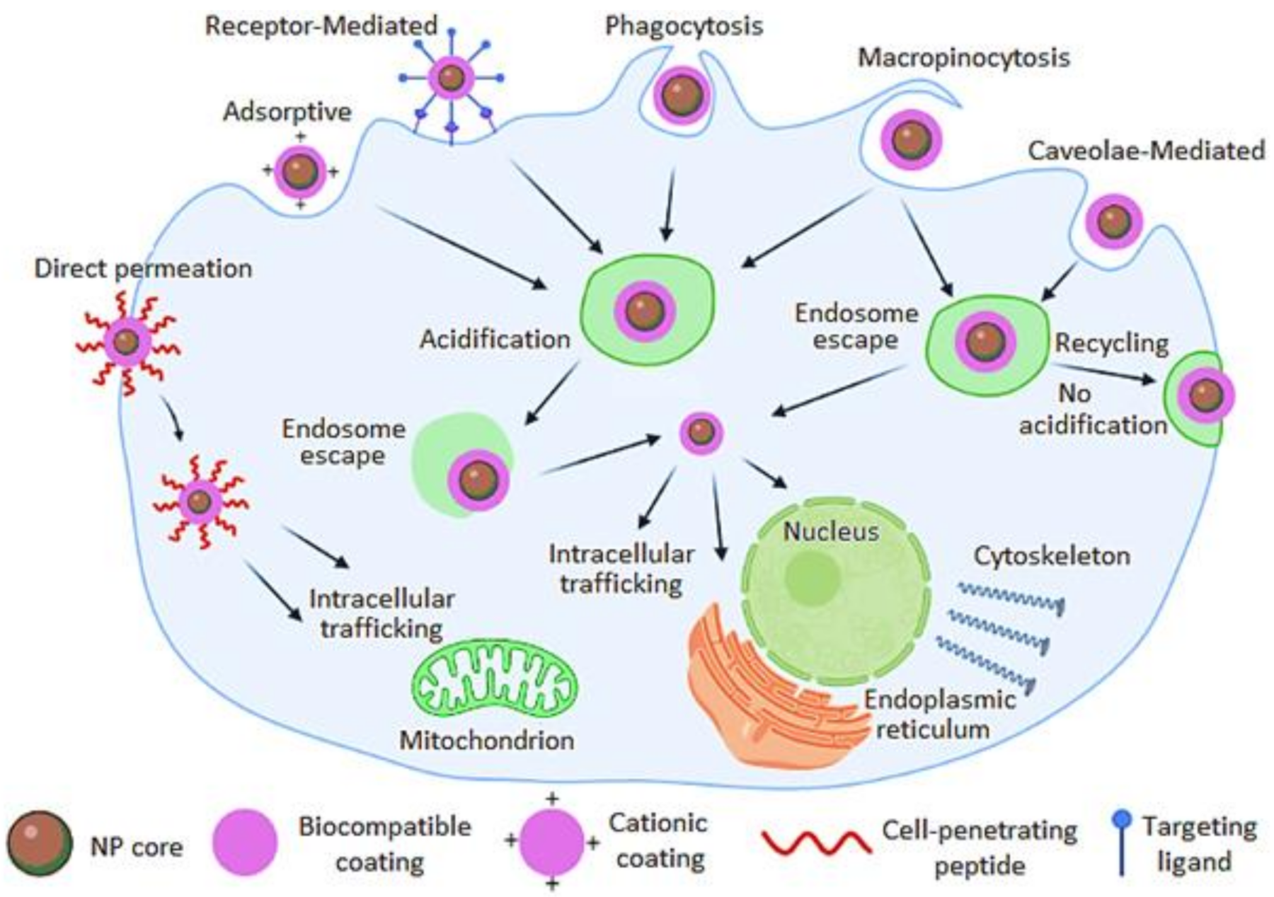
3. Targeting Strategies

Tumor Internationalization and Tumor Microenvironments: Active and Passive Targeting
The delivery of theranostic materials can be facilitated through active and passive targeting. Passive targeting is based on two physiological processes, which include the convection process driven by rhythmic blood circulation pressure responsible for the transport of large molecules through the tumor microvasculature, and diffusion process facilitated by a concentration gradient and mainly responsible for the transport of highly lipophilic and low molecular weight compounds across the cell membrane [75]. On the other hand, active targeting is achieved by specific interaction between the target cell and the carrier by selective cognate binding efficiency to overexpressed receptors in the tumor site, thereby improving cell recognition and uptake [76]. Active targeting with conjugated ligands avoids the destruction of surrounding nearby healthy cells/tissues. However, it is imperative to consider that the carriers must reach the tumor, thereafter, internalize within the tumor cells, and this is achieved by the enhanced permeability and retention (EPR) effect through proper cellular trafficking throughout the body and accumulation in the tumor [75,77].
Most nanotheranostic agents are internalized through endocytosis pathways, which are facilitated by receptor-mediated endocytosis and adsorptive endocytosis (i.e., clathrin-coated pits) [78]. Cellular internalization can also occur via phagocytosis, which is the main uptake mechanism into macrophage cells, while caveolae-mediated endocytosis occurs in non-clathrin-coated plasma membrane present on the surface of some cells. Micropinocytosis is a fluid-phase endocytosis mechanism and other mechanisms that do not involve clathrin or caveolae. Receptor-mediated endocytosis is achieved by attaching a targeting moiety/ligand to the surface of the theranostic agent that recognizes overexpressed receptors on the surface of the tumor cell, as shown in Figure 2. Generally, when the theranostic agent is bound to the cell surface receptor, the theranostic agent is engulfed and once internalized, the theranostic agent is wrapped within an endocytic vesicle [78]. The theranostic must extravasate the endosome, proper cellular-trafficking to the intracellular site of action (cytoplasm, nucleus, mitochondria, Golgi apparatus, or cytoskeleton) to improve the therapeutic efficacy and exhibit real-time monitoring treatment response through imaging [79].
Targeting Moieties/Ligands
Lee and colleagues [80] emphasized the importance of new strategies to discover target antigens and combinatorial targeting of antigens to overcome the heterogeneity and plasticity inherent to solid tumors. The theranostic agent, once extravasated from the blood into the tumor site, it is imperative for the theranostic agent to achieve optimal tumor internalization to confer efficient therapeutic payload without affecting surrounding normal tissues [77]. There are several targeting strategies, according to the different materials used as targeting agents (Figure 3 and Table 3).
Antibodies
Antibodies (mAbs) are proteins with two epitope-binding sites with extraordinary selectivity and specificity to their antigen. The mAbs; such as humanized bevacizumab (Avastin), for VEGF targeting; humanized trastuzumab (Herceptin), which targets HER2; chimeric cetuximab (Erbitux), which targets the human epidermal growth factor receptor (EGFR); chimeric rituximab (Rituxan), for targeting hematopoietic differentiation antigen CD20; and humanized panitumumab (Vectibix), for targeting EGFR, can be conjugated to theranostic nanomaterials.
Aptamers
Produced using systemic evolution of ligands by exponential enrichment (SELEX), aptamers are small synthetic ligands (~15 kD) that have demonstrated superior binding affinity with high specificity when compared with antibodies (~150 kD). Aptamers can be conjugated to FeONPs via peptide bonds between the amine group of the aptamer and the carboxylic group of FeONPs.
Peptides
Peptides have a low molecular size, are highly stable, and exhibit high specificity and low immunogenicity [81]. They can promote selective intracellular delivery of a plethora of therapeutic agents, and they can be designed to target specific cell receptors, avoiding undesired side effects. Furthermore, they present advantages over other targeting agents (i.e., antibodies), such as their small size, which enhances tumor penetration, their higher stability in cyclization or in D-amino acid forms, and easy synthesis protocols for their manufacture and modification. Novel peptide-based targeting agents have been constantly discovered, especially by the phage-display technique. Nevertheless, the precision and the efficiency of these delivery systems can still be further improved [82].
Small molecules
Small molecules are one of the most powerful targeting moieties due to their size, stability, reproducibility, and that they offer high ligand densities as corona on the theranostic nanomaterials. These include glycoproteins, such as gum arabic and folic acid (folate), with the latter exhibiting high binding affinity (Kd = 10−9 M) to folate receptors (α or β) overexpressed in various tumors, such as triple-negative breast, which has limited therapeutic options and poor prognosis [83]. Carbohydrate ligands also make part of small targeting moieties (i.e., dextran, galactose, glucose, mannose and their derivatives) that can selectively target and recognize lectin receptors, all affording for specific targeted delivery [81].
Table 3. Major overexpressed receptors on breast, prostate, and lung cancer as targets for theranostic targeting.
Membrane ReceptorsLigandsCancerRef.
Hormone Receptor-Positive (80%): Estrogen receptor positive (ER+) or progesterone receptor positive (PR+)21-[18F]fluorofuranylnorprogesterone (FFNP)Breast cancerDehdashti et al. [84]
Human epidermal growth factor receptor-2 (HER2) (20%)Herceptin antibody
Gastrin-releasing peptide (GRP) (65–75% and > 90%)Series of Bombesin (BBN) peptide conjugatesBreast, prostate, and lung cancerKübler and Albrecht [85], Baratto et al. [86], Tangthong et al. [87]
Somatostatin (sst2) > 90% (antagonist†)Octreotide, fc[CFwKTC]T(ol)
RC-121 (D- Phe-Cys-Tyr-D-Trp-Lys-Val-Cys-Thr-NH2)
Breast, prostate, and lung cancerChatzisideri et al. [88], Mukherjee et al. [89]
Triple-Negative (10–20%)—BRCA1 and folate receptorsFolateBreast, prostate, and lung cancerMarko et al. [90], Thakur and Kutty [91]
Prostate-specific membrane antigen (PSMA) and androgen receptorPSMA peptide
Monoclonal antibody RM2
Prostate cancerBaratto et al. [86], Cifuentes-Rius et al. [92]
Epidermal growth factor receptors (EGFRs)EGF, EGF-like ligands, TGF-α, and HRGsBreast and prostate cancerMaennling et al. [93]
Lectin-binding glycoproteins (e.g., P-glycoprotein)LectinBreast cancerZhuo et al. [94]
Prostate stem cell antigen (PSCA)PSCA-specific chimeric antigen receptor (CAR)-engineered T cellsProstate cancerLee et al. [80]
Integrin αvβ3Various types of arginine-glycine-aspartic acid (RGD) such as c(RGDyK), c(RGDfK) and (c(RGDf[N-Me]V to target tumor-associated endothelial cellsBreast and prostate cancerChatzisideri et al. [88],
Li et al. [95]
Transferrin receptor and urokinase-type plasminogen activator receptor (uPAR)VitronectinLung cancerMontuori et al. [96]
Targeted Theranostic in Breast Cancer
A study by Lee et al. [97] evaluated HER2-targeted 64Cu-labeled nanoparticle, 64Cu-MM-302 (64Cu-labeled HER2-targeted PEGylated liposomal doxorubicin) in nineteen patients with HER2-positive metastatic breast cancer for optimal doxorubicin delivery. Results revealed that 64Cu-MM-302 tumor accumulation at 24–48 h post-IV infusion varied 35-fold (0.52 to 18.5% ID/kg), including deposition in bone and brain lesions due to the nature of the metastatic tumor associated with more favorable treatment outcomes attributed by increased tumor delivery, was observed with increasing dose (1–13 μg doxorubicin per gram of tumor tissue (μg DOX/g)). Liu et al. [98] developed a superparamagnetic MXene-based theranostic nanoplatform composed of tantalum carbide (Ta4C3) MXenes nanosheets doped with IONPs stabilized with soybean phospholipids (Ta4C3-IONP-SPs) nanotheranostic for targeted PTT of breast tumor in a mice model facilitated by a magnetic-field with MRI and CT imaging capabilities.
Targeted Theranostic in Prostate Cancer
Prostate-specific membrane antigen (PSMA) targeting serves as one of the major promising targets for paradigm-changing practice for improving prostate cancer patient outcomes [89]. Functionalized targeted aptamer-conjugated polymeric nanoparticles (docetaxel-PLGA/PEG-A10 aptamer) have been reported to bind to PSMA [99]. Mangadlao and colleagues [61] developed a theranostic agent composed of PSMA-1 targeted pegylated AuNPs loaded with phthalocyanine-based Pc4 (AuNP-5kPEG-PSMA-1-Pc4) for enhanced PDT. The results showed that AuNP-5kPEG-PSMA-1-Pc4 demonstrated an enhanced binding affinity (IC50 = 0.17 nM), this was found to be attributed by the multivalency of AuNPs, a 12-fold increase binding avidity compared to PSMA-1 alone against PSMA expressing prostate tumors; moreover, it was found that internalization was via clathrin-mediated endocytosis [100].
Additionally, this resulted in the efficient delivery of Pc4 and PDT ablation at 300 J/cm2 with remission 14 days post-treatment. Katti et al. [101] demonstrated the use of Mangiferin conjugated radioactive gold nanoparticles (MGF-198AuNPs), gum arabic 198AuNPs (GA-198AuNPs), and EGCG-198AuNPs for targeted delivery to prostate tumors through glucose-moiety of MGF, which led to efficient endocytosis, EPR effect of the GA, and laminin receptor specificity of EGCG overexpressed on prostate tumor cells, all collectively augmenting tumor uptake and retention; of which, 198Au (βmax = 0.96 MeV; half-life of 2.7 days) β energy emission and half-life contribute for the destruction of tumor cells/tissue. Hosoya et al. [102] developed and evaluated a theranostic targeted heat sensitive-based liposome (HSL)-containing hydrogel-based nanoplatform composed of targeting both breast and prostate tumor with a NIR irradiation facilitating photon-to-heat conversion for triggered drug delivery and multimodal imaging.
Targeted Theranostic in Lung Cancer
Lung cancer is categorized into two major subtypes, which include small cell lung cancer (SCLC) and non-small cell lung cancer (NSCLC), where NSCLC is responsible for ≥ 90% of deaths. Gamal-Eldeen et al. [103] utilized gum arabic conjugated gold nanoparticles (GA-AuNPs) for the targeted delivery of A549 tumor-bearing mice; results showed that GA-AuNPs treated lung tumor-bearing mice followed by laser exposure enhanced the antitumoral efficacy. In another study, Li et al. [104] developed and evaluated ultra-pH-sensitive indocyanine green (ICG)-pegylated polymer nanoparticles (PEG-b-(PR-r-ICG)) for fluorescence imaging-guided targeted delivery via EPR effect to A549 tumor-bearing mice for enhanced PTT.
The results revealed that PEG-b-(PR-r-ICG), when irradiated with 808 nm NIR, produced maximum thermal (Tmax) heat of 51.9–53.1 °C, which completely eliminated the tumor. Zhao et al. [105] used an sgc8c aptamer-functionalized Fe3O4@carbon@doxorubicin NP (Apt-Fe3O4@C@DOX), where the sgc8c aptamer served as an active tumor-targeting ligand by endocytosis, Fe3O4 allowed for MR imaging and PTT with a Tmax of 55.6 °C for the optimal delivery of DOX, as a synergetic enabled nanotheranostic for monitoring of the therapeutic responses of tumor [106]. Moreover, work by Gong and colleagues [107] developed a macrophage-cancer cell hybrid membrane-coated doxorubicin (Dox)-loaded poly (lactic-co-glycolic acid) (PLGA) nanoparticle (DPLGA@[RAW-4T1] NPs) for targeting and treating breast cancer-derived lung metastases. Their results demonstrated that DPLGA@[RAW-4T1] NPs (163 ± 9.61 nm) targeted α4β1 integrin via caveolar- and Na+/H+ exchange (micropinocytosis)-mediated pathways and exhibited 88.9% anti-metastasis efficacy.

4. Mechanisms of Diagnosis for Breast, Lung and Prostate Cancer

The current diagnostic trend consists of non-invasive real-time imaging techniques, which often decrease systemic toxicity and discomfort to patients. Such techniques can even be extrapolated to therapy when prodrugs are administered, as the conversion of the prodrug into the active drug can be followed in real-time with imaging strategies. The use of theranostic nanoparticles as tools for imaging the drug accumulation in tumors and observing the therapeutic effects in real-time has the potential to revolutionize personalized medicine, as it would enable a more accurate prediction of the treatment outcomes and precise adjustments that might be needed to increase the therapeutic success [108].
In this regard, Chen and collaborators developed a complex nanosystem composed of three covalently linked cores (a therapeutic prodrug-activating cytosine deaminase (bCD), poly-L-lysine labeled with the fluorescent dye Cy5.5 as an imaging reporter, and a vector for siRNA delivery and targeting with Indium-111-Dotatate (or [111In]DOTA) for the acquisition of SPECT images). The system was functionalized with a urea-based PSMA-targeting group conjugated with maleimide-PEG-NH2; therefore, the nanoparticles were developed as a theranostic approach for prostate cancer. A total of 48 h after the injections, the authors observed an increased accumulation of the nanoparticles in the prostate tumors; however, more toxicity and immunogenicity studies should be performed before a clinical application could be implemented [109].
Iron nanoparticles present excellent results as contrast agents for magnetic resonance imaging, especially due to their magnetic properties. In this regard, Zhu et al. [110] created superparamagnetic iron oxide nanoparticles coupled with PSMA for targeting the nanoparticles to prostate cancer foci. The authors observed a specific uptake of the nanoparticles in PSMA-expressing cells with a significantly enhanced MRI signal.
Other excellent contrast agents for MRI are superparamagnetic iron oxide nanoparticles (SPIONs). They can be further enhanced and applied for theranostic applications, as demonstrated by Manigandan and collaborators. In their study, a self-assembled amphipathic chitosan micelle complexed with a SPION system was developed for encapsulating doxorubicin in order to treat breast cancer. The targeting strategy was the conjugation with specific anti-integrin monoclonal antibodies since those integrins tend to be overexpressed in tumors, especially metastatic ones. The tumor accumulation could be observed by MRI, and it was shown that the nanomicelle-SPION complex retention into the tumor led to a great therapeutic effect [111].
An excellent non-invasive treatment option for prostate cancers is called magnetic resonance-guided focused ultrasound surgery (MRgFUS), characterized as a non-invasive, real-time monitoring, and three-dimensional imaging approach that increases therapeutic success. Wang and collaborators developed SPIONs to be used in this approach, and the nanomaterials were PEGylated and functionalized with anti-EGFR monoclonal antibodies. The specific delivery could be followed by MRI in lung cancer in vivo, and the authors observed an increased MRI sensitivity when the SPIONs were used; therefore, they could be a useful tool for MRgFUS for a rapidly-spreading cancer, such as lung tumors [112].
Gadolinium, on the other hand, has been commonly applied as a contrast for magnetic resonance imaging due to its paramagnetic properties, its ability to shorten the T1 relaxation time, and the possibility to cross the blood–brain barrier after some kind of damage. Manganese is another metal that can be used for this purpose, and different Mn-based nanomaterials have already been developed and tested, i.e., carbon-based Mn nanoparticles for brain imaging and composite Mn-Au nanoparticles as MRI contrast for stem cell labeling [113].
Dufort and co-workers developed rigid nanoparticles composed of gadolinium-based polysiloxane functionalized with chelating agents, such as DOTA, as theranostic agents for lung cancer. The nanoparticles exhibit optimal imaging properties for magnetic resonance imaging (MRI), SPECT, CT, and fluorescence imaging, either after intravenous or intrapulmonary administration [114], especially because gadolinium-based contrast agents present interesting characteristics for imaging (e.g., high paramagnetic profile due to the configuration of seven unpaired electron spins) [108].
Gadolinium was also used encapsulated in fullerenes, more specifically C82, as a magnetic contrast for MRI, providing an in vivo relaxivity 12 times higher than commercial gadolinium diethylenetriaminepentaacetic acid ([Gd]-DTPA) contrast agent. This relaxivity enhancement is probably due to the electronic interactions between the paramagnetic gadolinium-fullerene cage with water molecules surrounding the fullerenes [115]. One must bear in mind, though, that fullerenes lack water solubility unless some chemical modifications are made to the carbon cage, and they easily aggregate in an aqueous environment, such as plasma. The aggregation degree also influences their overall toxicity. Thus, the use of fullerenes for biomedical applications often requires attention and adjustments [116].
Gold nanoparticles have already been tested as contrast agents for computer tomography (CT) scans and demonstrated promising results [117]. The reasons for considering AuNPs as contrast agents are the significant electron density of gold and its surface plasmon resonance, which are optimal for CT, Raman spectroscopy, photoacoustic image, and multiphoton microscopy [118].
Compared to iodine, a common contrast for CT, gold can be thrice as good due to its superior absorption coefficient relative to all other biological tissues and tumors. By functionalizing AuNPs with targeting agents, e.g., anti-EGFR antibodies, the tumor-specificity and contrast efficiency can be significantly enhanced, as demonstrated by [119] for head and neck squamous cell carcinoma in mice.
Butterworth and colleagues tested potential theranostic AuNPs functionalized with DTPA for prostate cancer radiotherapy and found that the rectal and bladder toxicity could be significantly decreased, and the good CT contrast provided by the nanoparticles might be useful for minimizing the risk of damaging healthy tissues during radiotherapy [118].
Gold was used in the synthesis of mesoporous magnetic gold nanoclusters for drug delivery and chemo-photothermal co-therapy, while the magnetic property allows selective accumulation by extra-magnetic field and enables the use of the device as an MRI contrast agent [120].
A bimodal approach with gold and iron nanoparticles was investigated by Revia and co-workers. FeNPs could be used as contrast agents for MRI, while AuNPs are useful for X-ray computed tomography and photoacoustic imaging. At the same time, the metal nanoparticles could serve as carriers for RNA oligomers as an iRNA-based therapeutic approach; therefore, a complete theranostic system could be created and applied [121].
Another common theranostic approach consists of encapsulating a therapeutic agent in the core of a nanoparticle and functionalizing the surface of this NP with a targeting and signaling agent. Cano-Cortes et al. (2020) used polystyrene nanoparticles to encapsulate doxorubicin as a therapeutic agent, near-infrared cyanine dye (C7), and a targeting peptide (CRGDK sequence), which can be recognized by the neuropilin-1 receptor, overexpressed in triple-negative breast cancer. Polystyrene was used for the in vitro and in vivo tests due to the biostability and compatibility with the synthetic, functionalizing, and encapsulating protocols. The authors observed that the near-infrared dye provided a good fluorescence imaging of the targeted tumor with a high signal-to-noise ratio, and the tumor volume was significantly decreased due to doxorubicin [122].
While paclitaxel is one of the most used chemotherapeutics for several cancers, indocyanine green (ICG) is an FDA-approved optical contrast for diagnostic imaging. A combination of both agents with the aid of nanotechnology could provide a synergistic effect for cancer therapeutics. In this regard, Liu and collaborators took advantage of the red emission of 8 nm albumin-coated gold nanoclusters and the easy drug conjugation enabled by albumin itself and developed BSA-gold nanoclusters functionalized with hyaluronic acid, loading the material with indocyanine green (photothermal ablative agent) as a breast cancer ablative approach. The authors observed a remarkable antitumor effect, and the drug accumulation could be observed as the tumor hyaluronidase degraded the hyaluronic acid from the nanoparticles; therefore, the nanomaterials were considered promising for future anticancer theranostics [123].
Flores and collaborators reported the use of polymeric nanoparticles conjugated with folate groups for prostate cancer targeted theranostics. It was possible to follow the accumulation of the nanoparticles in the tumor tissue due to a fluorescent probe coupled to folate, responsible for targeting PSMA-expressing cells [124].
Regarding lung cancer diagnostics, nuclear imaging techniques offer the advantage of providing high-sensitivity three-dimensional images, compared to other diagnostic tools, such as chest radiography, needle biopsy, sputum cytology, bronchoscopy [125].
Quantum dots are known for their intense fluorescence generation, which could be useful for diagnostic and theranostic purposes; however, the heavy metals in their composition may cause significant toxicity; therefore, other strategies have to be used for a safe imaging protocol. Silica-based Cornell dots (C-dots) are examples in this regard, as they are composed of a silica core with embedded fluorophores and are surrounded by a PEGylated silica shell labeled with monoclonal antibodies that guide the nanoparticles to the target site. The diagnosis, in this case, is obtained by illuminating the tumor site with a near-infrared light source, which will indicate the presence of the dots bound to the tumor. This property can be used in surgical optical guidance as well [108].
Wang et al. developed NIR fluorescent InP/ZnS quantum dots functionalized with amphiphilic copolymer polylactide-b-poly (ethylene glycol) (PLA-PEG) and anti-EGFR monoclonal antibodies, resulting in theranostic micelles for EGFR-overexpressing cancers, especially triple-negative breast cancer. The fluorescence would be used for tracking and diagnosis, while aminoflavone was used as the anticancer drug. The authors observed a significant tumor volume decrease and a reduction in the drug dose needed for inhibiting tumor growth [126].
Biotinylated graphene quantum dots might be an alternative for cancer theranostics, as tumors cells tend to overexpress biotin. Graphene quantum dots, besides the lower toxicity compared to heavy metal quantum dots, exhibit optimal surface tenability properties that allow custom modifications and thus can be designed for a plethora of theranostic applications [127].
Graphene quantum dots are other non-toxic alternatives for theranostic approaches, i.e., molecular imaging, drug delivery, and photothermal therapy. Ko et al. (2017) loaded graphene quantum dots with HER and doxorubicin for a targeted approach against breast cancer. The HER-driven accumulation of the nanomaterial could be followed by the blue light emitted by the quantum dots, and the selective doxorubicin release contributed to significant inhibition of the tumor cells [128].
Another theranostic use for carbon-based dots was reported by Wu and co-workers in 2016, in a study describing carbon dots functionalized with positively-charged polyethyleneimine (PEI) and negatively-charged siRNA molecules as well as folate as the tumor-targeting moiety. The accumulation of the nanomaterials could be followed by bioluminescence (the dots absorb in 360 nm and emit in 400 nm), and the reduction of lung tumor volume in vivo could be clearly observed by the authors [129].
When it comes to radionuclide-based imaging techniques, i.e., single-photon emission computer tomography (SPECT) and positron-emission tomography (PET), nanotechnology can be very useful due to the possibility of fabricating nanomaterials labeled with for most promising theranostic radioactive agents [108].
Lutecium (177Lu) has been consolidated as a therapeutic radionuclide in the clinic, but other beta emitters have been calling the attention of physicians and researchers, e.g., 67Cu, 47Sc, and 161Tb, as they present biologically viable half-lives (2.58, 3.35, and 6.91 days, respectively), and provide theranostic possibilities, with PET and SPECT imaging potential. By combining those radionuclides with targeting ligands, such as PSMA for prostate cancer, in a nanosized carrier, the theranostic potential is considerable [130].
Simple liposomes loaded with doxorubicin, functionalized with PSMA, and radiolabeled with 99mTc were synthesized by Yari and colleagues so that the accumulation and drug delivery by the liposomes could be monitored in real-time by a gamma camera in LNCaP prostate tumor cells. The results indicate that these nanomaterials might be useful for theranostic applications in solid tumors [131].
Theranostic nanodevices offering PSMA-targeting for prostate cancer can be helpful, especially if coupled with beta-, alpha-, or Auger-emitting radioisotopes for therapy and positron-emitters for diagnostics. This approach enhances the prognosis even for chemotherapeutic-resistant and metastatic tumors. 68Ga, for instance, shows superior PET/CT contrast properties than other isotopes, such as 18F, especially regarding the detection of metastases, and it has the advantage of being eluted from a 68Ge/68Ga generator; therefore, it is more available than cyclotron-derived radionuclides [132]. Virgolini and collaborators show the most recurrent isotopes used in association with PSMA for prostate cancer.
Another promising imaging modality is called photoacoustic imaging, consisting of irradiating a specific target with a near-infrared light source in the presence of targeted nanoparticles, which create acoustic pressure waves after absorbing the energy, and those waves can be detected by an ultrasound transducer. In the case of pulsed radiofrequencies being used, the imaging is called thermoacoustic instead. Both techniques have been tested with Prussian blue nanocubes coated with silica, modified with PEG, and loaded with doxorubicin, especially for breast cancer, with good results. These biophotonic technologies can improve diagnostics by enabling simultaneous multiplex imaging with different color emitters according to different targets [108].
Photodynamic therapy (PDT) can be used in theranostic approaches, as demonstrated by Mangadlao (2018). The study describes a nanoparticle system to carry a photosensitizer, Pc4, and target prostate tumors in vivo. The animals showed tumor remission after 14 days of the photodynamic treatment, and the accumulation of the nanomaterial could be observed via the dye fluorescence; therefore, this nanomaterial could be used for surgical guidance and as a theranostic agent [61].
The combination of MRI and near-infrared fluorescence for imaging-guided photodynamic therapy was the approach recently chosen by Wang and collaborators, using cathepsin-B-activatable ultrasmall superparamagnetic iron oxide nanoprobes. These probes were functionalized with a fibronectin-targeting peptide (CREKA) and the fluorescent dye squarain. The authors observed via fluorescence a selective accumulation of the probes in triple-negative breast cancer cells, and the squarain fluorescence could act as a photosensitizer for NIR photodynamic therapy [133].

5. Mechanisms of Treatment for Breast, Lung and Prostate Cancer

The protocol for lung cancer treatment will depend on the malignancy and stage at the time of diagnosis and may involve surgery, chemotherapy, immunotherapy, and radiotherapy. Notwithstanding, most cases are detected at advanced stages because of the difficulty of early-stage diagnosis. In these cases, frequent local tumor invasion or distant metastasis makes them not suitable for surgical intervention. Thus, systemic chemotherapy is the main treatment protocol in the seek for extending survival. The first-line chemotherapy for lung cancer consists of platinum-based drugs, such as cisplatin and carboplatin. Albeit these drugs present dose-limiting side effects, such as nephro- and cardiotoxicity, anemia, intestinal injury, peripheral neuropathy, nausea, and fatigue. Because of their high toxicity, platinum-based drugs are usually combined with other anticancer agents, such as taxanes (paclitaxel, docetaxel) or gemcitabine [134]. That way, it is critical that the development of new and more efficient therapeutics for lung cancer as current treatments present poor response and low survival rates. In this field, hybrid nanoparticles with genes, drugs, and other biomolecules have attracted attention as novel therapeutic possibilities [135].
Concerning breast cancer treatment, the first choice is to avoid mastectomy. However, tumor recurrence, even after radiotherapy, usually leaves no option but surgery. In some cases, hormonal therapy is a good option, but in cases of triple-negative breast cancer—the most complex and aggressive type of breast cancer—it is not suitable due to the absence of expression of estrogen receptors, progesterone receptors, and human epidermal growth factor receptor 2 on tumor cells. Thus, in these cases, systemic chemotherapy with drugs, such as anthracyclins and taxanes is usually the choice. Repeated chemo cycles can kill cancer cells, but often cause damage to healthy cells surrounding the tumor as well [91]. The treatment and early detection facilitated by nanomaterials can elevate the survival rate and quality of life of the patients [136]. Theranostics is a critical factor because it provides dynamic feedback through the treatment process and helps the optimization of the therapeutic operation [137].
Prostate cancer is clinically treated with systemic chemotherapy, surgery, and radiotherapy. Paclitaxel, doxorubicin, and docetaxel are commonly used drugs that, despite being able to prolong survival, their therapeutical efficacy is considered low. Moreover, they can induce several side effects, such as hair loss, nausea, cardiac, liver, and kidney toxicity and cause damage to healthy cells close to the tumors [138].
In this topic, we will briefly talk about the mechanisms of treatment concerning nanomaterials that have been being developed to improve cancer therapy.
Chemotherapy
Chemotherapy is a treatment that has been used for several types of cancer but still holds some concerns because of its lack of specificity and consequent toxicity that leads to several side effects. In that scenario, these anticancer drugs have been conjugated to nanomaterials to increase their efficiency, reducing side effects. Currently, there are some FDA-approved nanoparticle-based chemotherapeutics drugs, such as Abraxane®®, which is based on albumin nanoparticles loaded with paclitaxel, and Doxil®® that consists of PEG nanoparticles containing doxorubicin [139].
Nanoemulsions are an alternative for conventional delivery vehicles, presenting better stability and efficacy for chemotherapy. They can be administered by different routes. The administration of paclitaxel in a nanoemulsion system has demonstrated enhanced release, permeation, and improved cellular uptake in breast cancer cells. Additionally, this type of delivery platform can provide longer retention of the drug in systemic circulation, better oral bioavailability, and safety [140,141]. Another study demonstrated the faster onset action of intravenous administration of a nanoemulsion containing DHA-SBT-1214 (omega-3 fatty acid-conjugated taxoid prodrug) to treat prostate cancer in mice. An 88% decrease in tumor growth was observed, and the nanoemulsion formulation was demonstrated to be much more efficient than Abraxane®® [141,142].
Immunotherapy
Immunotherapy is a technique that stimulates the body’s immune system to kill cancer cells. These therapies may consist of antibodies with the capacity to block suppressive immune-check-points pathways, cellular therapies with dendritic cells or engineered T cells, or even vaccines that will trigger an antigen-specific immune response in cancer [141].
The latest trends in immunotherapy for breast cancer include immune checkpoint antagonists (monoclonal antibodies) that are specific for CTLA-4, PD-1, and PD-L1. Good results have been achieved with commercially available antibodies, i.e., avelumab, atezolizumab, and pembrolizumab, although some are not yet FDA-approved. Trastuzumab is an HER-2-targeting monoclonal antibody that is often used in association with chemotherapy for breast cancer ablation, prolonging the overall survival in advanced disease stages and decreasing the chances of tumor relapse in early stages [142]. Checkpoint antagonists, such as nivolumab (FDA-approved for non-small cell lung cancer), pembrolizumab, and atezolizumab, are also useful for lung cancers. Novel treatment modalities are currently in development, which associate checkpoint inhibitors, such as ipilimumab and radiotherapy, in order to increase the antitumor efficacy of those treatments [143].
As for prostate cancer, apart from the previously mentioned immune checkpoint inhibitors, some other passive therapeutic protocols are in development, i.e., chimeric antigen receptor (CAR)-T cell therapy, radiolabeled monoclonal antibodies against PSMA, and prostate stem cell antigen (PSCA) as an immunotherapeutic agent. Active therapeutic protocols include Sipuleucel-T (Provenge, FDA-approved) immunotherapy targeting prostatic acid phosphatase and some viral vectors carrying modified prostatic antigens and co-stimulatory molecules [144].
Immunotherapy is a great challenge in nanomedicine because it is paramount to guarantee the targeted delivery of immunomodulatory and immunostimulatory molecules to the correct cells; however, it holds the best promises to overcome such targeting issues [137].
Gene Therapy
RNA nanoparticles are known to present low immunogenicity and toxicity, target specificity, therapeutic component, and others very interesting properties. In addition to that, their physicochemical properties allow the delivery of siRNA and microRNA, the imaging via fluorogenic RNA and RNA aptamers, and the module for tumor targeting [145].
In gene therapy, toxic genes are delivered to cancer cells, causing their death. To do that, microRNA, an endogenously expressed non-coding RNA molecule, has been considered a target for many diseases. Further, a vector is required to guarantee the preservation of the gene in the bloodstream [137].
siRNA also holds the property of blocking or silencing the genes responsible for causing cancer. Studies have demonstrated that the presence of cholesterol ligand on the head of the arrow-shaped RNA structure induces the loading of the nanoparticles to the extracellular vesicle, allowing the delivery of siRNA to the tumor cells and blocking their growth. Researchers have reported that the delivery of siRNAs nanoparticles was able to silence prohibitin 1 expression and block the growth of prostate tumor [146,147].
Photodynamic Therapy (PDT)
Photodynamic or photochemotherapy is a treatment that involves a photosensitizing chemical substance (photosensitizer) that will be irradiated with specific wavelengths to induce the production of reactive oxygen species (ROS), for instance, singlet oxygen, peroxide, superoxide, and hydroxyl radicals. These ROS can induce cell death at the place where they are generated, and therefore, can be used as a treatment for some conditions, including cancer.
Specifically, for cancer, there is a different kind of photochemotherapy with two-photon excitation (TPE) that merges the advantages of TPE near-infrared (NIR) photosensitizers and nanotechnology [148]. As a result of the absorption of two relatively low-energy NIR photons, there will be the emission of high-energy photons in the visible spectrum that in turn will sensitize oxygen-producing cytotoxic ROS. The difference between TPE and single-photon-based PDT is the long-wavelength light used that makes it possible to reach deeper tissues, and therefore, tumors that are located deeper. Several lines of evidence show that the TPE technique may also be used for theranostics [108].
Photothermal Therapy (PTT)
Photothermal therapy presents similar principles to PDT, with the application of nanoheaters/heating agents to achieve and accumulate on the tumor site via EPR or active targeting in order to provide the localized temperature increase. This process destroys proteins and DNA/RNA molecules, inducing membrane rupture or necrosis and, thus, cell death. However, in PTT, the presence of oxygen is not necessary to kill the tumor cells. Moreover, plasmonic PTT (PPTT) has been gaining attention lately regarding the use of gold nanoparticles to be irradiated with infrared or near-infrared light, causing excitation of its conduction electrons at the surface (because of the surface plasmon resonance—SPR). When that happens, these electrons produce localized heat waves that can kill cancer cells.
Studies with nanotheranostics materials for tumor targeting, photon-to-heat conversion, and drug delivery enabling drug-controlled release for prostate and breast cancer have been described in the literature [102,107].
Therapies with radionuclides
Radionuclides applied to target therapy have been gaining attention as an option for cancer treatment for presenting the advantage of delivering high radiation doses to the tumor without compromising healthy tissues in the neighborhood.
For instance, PSMA-targeting ligands labeled with radionuclides have been used in clinical studies and indicating encouraging results for prostate cancer. The radiation used for this therapy may be of β-particles, α-particles, and Auger electrons depending on the radionuclide chosen. β-particles are emitted by radionuclides, such as yttrium-90, lutetium-177, iodine-131, and terbium-161. They have a low linear energy transfer (LET) of 0.2 Kev/µm, but enough to cause some damage to DNA, such as base chemical modifications and protein crosslinks. These particles can travel through 1–10 mm of tissue and, thus, may cause damage to surrounding cells. On the other side, α-particles have a short range in tissue, from 50–100 µm. This way, the 4He nucleus is suitable for small tumors, micro-metastases, and individual neoplastic cells.
However, α-particles can kill more cells with less radiation due to their capacity to induce lethal DNA double-strand breaks. Finally, Auger electrons have extremely low energy and can travel through only some nanometers. Nonetheless, radionuclides that emit Auger electrons also release γ-rays, X-rays, β-particles, and internal conversion electrons, resulting in several energy deposition distances [148].
In neutron capture therapy (NCT), radiation is generated inside the tumor site by a nuclear reaction. Usually, boron atoms are bombarded with thermal neutrons to produce α-particles—10B(n,α). Gd has also been considered for this type of therapy; however, the toxicity of Gd3+ has been a concern. Radioimmunotherapy (RIT) is a treatment based on the use of monoclonal antibodies (mAbs), or fractions of mAbs, labeled with radioactive isotopes (α, β, ou Auger-electrons emitters) to irradiate the tumor. This technique has been used for years, and the number of studies concerning nanoparticles for RIT is increasing [108].
Magnetic Therapy
The most common type of magnetic therapy uses an alternating-current magnetic field to generate hyperthermia, inducing tumor cells to apoptosis. The heat comes from the Brownian and Neel relaxation processes, and the smaller the nanoparticles used, the more the Neel relaxation processes will take over the Brownian, heating the tissue and causing cell death. There are some situations that a direct-current magnetic field is used; in that case, magnetocytolysis happens, causing cellular disruption. In this kind of therapy, magnetic nanoparticles may also be used as contrast enhancers for MRI and, therefore, work as a theranostics platform [108,149].
Iron oxide particles have been evaluated and used in magnetic resonance technology-based biomedical applications, such as multifunctional theranostic complexes, due to their magnetic properties, combining tumor targeting, imaging, and nanotherapy for personalized cancer treatment. For breast cancer, there are experiments on blocking the IL4-α receptor (IL4Rα) using PEGylated superparamagnetic iron oxide nanoparticles (SPIONs) to inhibit breast cancer cell proliferation. By blocking this receptor, there was a notable decrease in cell viability and apoptosis in 4T1 cells. Additionality, a merged treatment using SPION-IL4Rα-doxorubicin caused a significant growth in apoptosis, cell death, and oxidative stress when compared to SPION-IL4Rα or doxorubicin alone [108].
Another promising theranostic for breast cancer is pH-sensitive poly (β-thiopropionate) nanoparticles with super magnetic core and folic acid (FA) conjugation (FA-doxorubicin-iron oxide nanoparticles [FA-DOX@IONPs]) for the delivery of an antineoplastic drug, DOX, for the treatment of folate receptor (FR)-overexpressed breast cancer. Aside from their imaging function, the nanoparticles can release in response to pH 5, namely the acidic environment of the tumors. It was shown that FA-DOX@IONPs cause cellular apoptosis and have the strongest cytotoxicity against breast cancer cells when compared to free DOX or non-FR targeted nanoparticles (DOX@IONPs). Furthermore, FA-DOX@IONPs with magnetic field treatment suppressed the growth of in vivo tumors in mice much better than either treatment alone. Furthermore, the nanoparticles exerted no toxicity against other healthy organs [150].

6. Future Directions

The development of nanomaterials for theranostic purposes must take into consideration some difficulties that may arise regarding conjugation chemistries, synthesis, and encapsulation protocols. Theranostic materials require good compatibility among the nanoparticle core, the targeting agent, the diagnostic moiety, and the active drug [122]. The ideal nanomaterial for theranostic purposes should have a synthesis protocol with the fewest steps possible, affordable and realistic costs, high reproducibility and ease to scale-up, and should provide diagnostic and therapeutic efficiency. Targeting agents with imaging properties may improve the sensitivity and accuracy of both treatment and diagnostic outcomes, providing a real-time follow-up that is a trend in the medical field, especially in cancer treatments. In this regard, activatable prodrugs may also be applied for real-time monitoring of cancer theranostics, thus helping in the decision-making process regarding the most effective treatment approach for each patient [151].
A plethora of imaging techniques can be applied for theranostic purposes, i.e., fluorescence imaging, scintillography, tomography (PET or SPECT), magnetic resonance, ultrasound, Upconversion imaging. More than one technique may even be combined in order to increase the efficiency of the nanomaterials. Therefore, the nanomaterials for theranostic purposes must be synthesized with all those features as an ultimate goal in order to enhance the therapeutic outcomes whenever they are used.

Author Contributions

L.F.F., A.H.F., V.C.T. conceived and designed the work, as well as contributed to writing, reviewing and data analysis; C.S.A.L., J.G.S.B., F.N.R., K.N., C.P.C.C., G.O.A.M., A.S.R., T.S.S. and V.M.A. contributed to literature search, writing and data analysis; G.H.C.V. and A.B.L. contributed to writing, reviewing and data analysis. All authors have read and agreed to the published version of the manuscript.

Funding

The authors would like to thank Fundação de Amparo à Pesquisa do Estado de São Paulo (FAPESP) for the financial support given to the researchers Lucas F. de Freitas (Process 2018/15598-2) and Caroline S.A. de Lima (Process 2019/01315-1), as well as Conselho Nacional de Desenvolvimento Científico e Tecnológico (CNPq) for the scholarship program supporting the undergraduate students involved in this manuscript, and the International Atomic Energy Agency (IAEA) coordinated Research Project: Nanosized Delivery Systems for Radiopharmaceuticals (CRP code: F22064) for financial support.

Data Availability Statement

No new data were created or analyzed in this study. Data sharing is not applicable to this article.

Conflicts of Interest

The authors declare no conflict of interest.

References

  1. Zendehdel, M.; Niakan, B.; Keshtkar, A.; Rafiei, E.; Salamat, F. Subtypes of Benign Breast Disease as a Risk Factor for Breast Cancer: A Systematic Review and Meta-Analysis Protocol. Iran. J. Med. Sci. 2018, 43, 1–8. [Google Scholar] [PubMed]
  2. Momenimovahed, Z.; Salehiniya, H. Epidemiological characteristics of and risk factors for breast cancer in the world. Breast Cancer Targets Ther. 2019, 11, 151–164. [Google Scholar] [CrossRef] [Green Version]
  3. Rawla, P. Epidemiology of Prostate Cancer. World J. Oncol. 2019, 10, 63–89. [Google Scholar] [CrossRef] [Green Version]
  4. Sartor, O.; de Bono, J.S. Metastatic Prostate Cancer. N. Engl. J. Med. 2018, 378, 645–657. [Google Scholar] [CrossRef] [PubMed]
  5. Bade, B.C.; Dela Cruz, C.S. Lung Cancer 2020: Epidemiology, Etiology, and Prevention. Clin. Chest Med. 2020, 41, 1–24. [Google Scholar] [CrossRef]
  6. Medavenkata, S.P.; Akshatha, H.S. Nano Theranostics—A Breakthrough in Cancer Diagnosis and Treatment and Regulations of Nano Technology Products. Int. J. Pharm. Sci. Res. 2018, 9, 3136–3149. [Google Scholar] [CrossRef]
  7. ISO. Nanotechnologies—Plain Language Explanation of Selected Terms from the ISO/IEC 80004 Series. ISO/TR 18401:2017(en). 2017. Available online: https://www.iso.org/obp/ui/#iso:std:iso:tr:18401:ed-1:v1:en (accessed on 3 September 2021).
  8. Wagner, S.; Gondikas, A.; Neubauer, E.; Hofmann, T.; von der Kammer, F. Spot the Difference: Engineered and Natural Nanoparticles in the Environment-Release, Behavior, and Fate. Angew. Chemie Int. Ed. 2014, 53, 12398–12419. [Google Scholar] [CrossRef]
  9. Madamsetty, V.S.; Mukherjee, A.; Mukherjee, S. Recent trends of the bio-inspired nanoparticles in cancer theranostics. Front. Pharmacol. 2019, 10, 1264. [Google Scholar] [CrossRef]
  10. Siafaka, P.I.; Okur, N.Ü.; Karantas, I.D.; Okur, M.E.; Gündoğdu, E.A. Current update on nanoplatforms as therapeutic and diagnostic tools: A review for the materials used as nanotheranostics and imaging modalities. Asian J. Pharm. Sci. 2021, 16, 24–46. [Google Scholar] [CrossRef]
  11. Zahin, N.; Anwar, R.; Tewari, D.; Kabir, M.T.; Sajid, A.; Mathew, B.; Uddin, M.S.; Aleya, L.; Abdel-Daim, M.M. Nanoparticles and its biomedical applications in health and diseases: Special focus on drug delivery. Environ. Sci. Pollut. Res. 2020, 27, 19151–19168. [Google Scholar] [CrossRef]
  12. Patra, J.K.; Das, G.; Fraceto, L.F.; Campos, E.V.R.; Rodriguez-Torres, M.D.P.; Acosta-Torres, L.S.; Diaz-Torres, L.A.; Grillo, R.; Swamy, M.K.; Sharma, S.; et al. Nano based drug delivery systems: Recent developments and future prospects 10 Technology 1007 Nanotechnology 03 Chemical Sciences 0306 Physical Chemistry (incl. Structural) 03 Chemical Sciences 0303 Macromolecular and Materials Chemistry 11 Medical and He. J. Nanobiotechnol. 2018, 16, 71. [Google Scholar] [CrossRef] [Green Version]
  13. Popescu, R.C.; Fufă, M.O.M.; Andronescu, E.; Grumezescu, A.M. Specifically targeted imaging using functionalized nanoparticles. In Nanobiomaterials in Medical Imaging: Applications of Nanobiomaterials; Elsevier Inc.: Amsterdam, The Netherlands, 2016; pp. 1–44. ISBN 9780323417389. [Google Scholar]
  14. Zayed, D.G.; Abdelhamid, A.S.; Freag, M.S.; Elzoghby, A.O. Hybrid quantum dot-based theranostic nanomedicines for tumor-Targeted drug delivery and cancer imaging. Nanomedicine 2019, 14, 225–228. [Google Scholar] [CrossRef] [Green Version]
  15. Hu, M.Z.; Zhu, T. Semiconductor Nanocrystal Quantum Dot Synthesis Approaches Towards Large-Scale Industrial Production for Energy Applications. Nanoscale Res. Lett. 2015, 10, 1–15. [Google Scholar] [CrossRef] [PubMed] [Green Version]
  16. Reiss, P.; Protière, M.; Li, L. Core/shell semiconductor nanocrystals. Small 2009, 5, 154–168. [Google Scholar] [CrossRef]
  17. Kalinowska, D.; Grabowska-Jadach, I.; Drozd, M.; Pietrzak, M. Comparative studies of biological activity of cadmium-based quantum dots with different surface modifications. Appl. Nanosci. 2018, 8, 309–321. [Google Scholar] [CrossRef] [Green Version]
  18. Zhu, C.; Chen, Z.; Gao, S.; Goh, B.L.; Bin Samsudin, I.; Lwe, K.W.; Wu, Y.; Wu, C.; Su, X. Recent advances in non-toxic quantum dots and their biomedical applications. Prog. Nat. Sci. Mater. Int. 2019, 29, 628–640. [Google Scholar] [CrossRef]
  19. Ahar, M.J. A Review on Aptamer-Conjugated Quantum Dot Nanosystems for Cancer Imaging and Theranostic. J. Nanomed. Res. 2017, 5, 00117. [Google Scholar] [CrossRef] [Green Version]
  20. Zhao, M.X.; Zeng, E.Z. Application of functional quantum dot nanoparticles as fluorescence probes in cell labeling and tumor diagnostic imaging. Nanoscale Res. Lett. 2015, 10, 1–9. [Google Scholar] [CrossRef] [PubMed] [Green Version]
  21. Karlsson, J.; Vaughan, H.J.; Green, J.J. Biodegradable Polymeric Nanoparticles for Therapeutic Cancer Treatments. Annu. Rev. Chem. Biomol. Eng. 2018, 9, 105–127. [Google Scholar] [CrossRef]
  22. Luk, B.T.; Zhang, L. Current advances in polymer-based nanotheranostics for cancer treatment and diagnosis. ACS Appl. Mater. Interfaces 2014, 6, 21859–21873. [Google Scholar] [CrossRef] [Green Version]
  23. Deng, S.; Gigliobianco, M.R.; Censi, R.; Di Martino, P. Polymeric nanocapsules as nanotechnological alternative for drug delivery system: Current status, challenges and opportunities. Nanomaterials 2020, 10, 847. [Google Scholar] [CrossRef]
  24. Emami, F.; Mostafavi Yazdi, S.J.; Na, D.H. Poly(lactic acid)/poly(lactic-co-glycolic acid) particulate carriers for pulmonary drug delivery. J. Pharm. Investig. 2019, 49, 427–442. [Google Scholar] [CrossRef] [Green Version]
  25. Ogay, V.; Mun, E.A.; Kudaibergen, G.; Baidarbekov, M.; Kassymbek, K.; Zharkinbekov, Z.; Saparov, A. Progress and prospects of polymer-based drug delivery systems for bone tissue regeneration. Polymers 2020, 12, 2881. [Google Scholar] [CrossRef]
  26. Bulbake, U.; Doppalapudi, S.; Kommineni, N.; Khan, W. Liposomal formulations in clinical use: An updated review. Pharmaceutics 2017, 9, 12. [Google Scholar] [CrossRef]
  27. Beltrán-Gracia, E.; López-Camacho, A.; Higuera-Ciapara, I.; Velázquez-Fernández, J.B.; Vallejo-Cardona, A.A. Nanomedicine review: Clinical developments in liposomal applications. Cancer Nanotechnol. 2019, 10, 1–40. [Google Scholar] [CrossRef]
  28. Hanafy, N.A.N.; El-Kemary, M.; Leporatti, S. Micelles structure development as a strategy to improve smart cancer therapy. Cancers 2018, 10, 238. [Google Scholar] [CrossRef] [PubMed] [Green Version]
  29. Atanase, L.I.; Riess, G. Self-assembly of block and graft copolymers in organic solvents: An overview of recent advances. Polymers 2018, 10, 62. [Google Scholar] [CrossRef] [Green Version]
  30. Lu, Y.; Park, K. Polymeric micelles and alternative nanonized delivery vehicles for poorly soluble drugs. Int. J. Pharm. 2013, 453, 198–214. [Google Scholar] [CrossRef] [Green Version]
  31. Munavalli, B.B.; Naik, S.R.; Torvi, A.I.; Kariduraganavar, M.Y. Kariduraganavar, “Dendrimers”; Springer: Cham, Switzerland, 2019; pp. 289–345. [Google Scholar]
  32. Sandoval-Yañez, C.; Rodriguez, C.C. Dendrimers: Amazing platforms for bioactive molecule delivery systems. Materials 2020, 13, 570. [Google Scholar] [CrossRef] [Green Version]
  33. Najafi, F.; Salami-Kalajahi, M.; Roghani-Mamaqani, H. A review on synthesis and applications of dendrimers. J. Iran. Chem. Soc. 2021, 18, 503–517. [Google Scholar] [CrossRef]
  34. Mandal, A.K. Dendrimers in targeted drug delivery applications: A review of diseases and cancer. Int. J. Polym. Mater. Polym. Biomater. 2021, 70, 287–297. [Google Scholar] [CrossRef]
  35. Yan, X.; Yang, Y.; Sun, Y. Dendrimer Applications for Cancer Therapies. J. Phys. Conf. Ser. 2021, 1948, 012205. [Google Scholar] [CrossRef]
  36. Li, W.; Cao, Z.; Liu, R.; Liu, L.; Li, H.; Li, X.; Chen, Y.; Lu, C.; Liu, Y. AuNPs as an important inorganic nanoparticle applied in drug carrier systems. Artif. Cells Nanomed. Biotechnol. 2019, 47, 4222–4233. [Google Scholar] [CrossRef] [Green Version]
  37. Tepale, N.; Fernández-Escamilla, V.V.A.; Carreon-Alvarez, C.; González-Coronel, V.J.; Luna-Flores, A.; Carreon-Alvarez, A.; Aguilar, J. Nanoengineering of gold nanoparticles: Green synthesis, characterization, and applications. Crystals 2019, 9, 612. [Google Scholar] [CrossRef] [Green Version]
  38. Liu, J.; He, H.; Xiao, D.; Yin, S.; Ji, W.; Jiang, S.; Luo, D.; Wang, B.; Liu, Y. Recent advances of plasmonic nanoparticles and their applications. Materials 2018, 11, 1833. [Google Scholar] [CrossRef] [Green Version]
  39. Yu, H.; Peng, Y.; Yang, Y.; Li, Z.Y. Plasmon-enhanced light–matter interactions and applications. NPJ Comput. Mater. 2019, 5, 1–14. [Google Scholar] [CrossRef]
  40. Bai, X.; Wang, Y.; Song, Z.; Feng, Y.; Chen, Y.; Zhang, D.; Feng, L. The basic properties of gold nanoparticles and their applications in tumor diagnosis and treatment. Int. J. Mol. Sci. 2020, 21, 2480. [Google Scholar] [CrossRef] [Green Version]
  41. Ashikbayeva, Z.; Tosi, D.; Balmassov, D.; Schena, E.; Saccomandi, P.; Inglezakis, V. Application of nanoparticles and nanomaterials in thermal ablation therapy of cancer. Nanomaterials 2019, 9, 1195. [Google Scholar] [CrossRef] [PubMed] [Green Version]
  42. Xu, H.; Jiang, S.; Wang, J.; Li, X.; Wu, T.; Xu, P.; Santos-Oliveira, R.; Zhang, A. Radioactive Gold Nanoparticle in Two Forms (19879Au GNPs and 99mTc-GNPs) for Lung Cancer Antiproliferative Induction and Intralesional Imaging: A Proof of Concept. Anticancer Agents Med. Chem. 2020, 20, 1648–1653. [Google Scholar] [CrossRef]
  43. Ganapathe, L.S.; Mohamed, M.A.; Mohamad Yunus, R.; Berhanuddin, D.D. Magnetite (Fe3O4) Nanoparticles in Biomedical Application: From Synthesis to Surface Functionalisation. Magnetochemistry 2020, 6, 68. [Google Scholar] [CrossRef]
  44. Mukherjee, S.; Liang, L.; Veiseh, O. Recent advancements of magnetic nanomaterials in cancer therapy. Pharmaceutics 2020, 12, 147. [Google Scholar] [CrossRef] [PubMed] [Green Version]
  45. Ferreira, M.; Sousa, J.; Pais, A.; Vitorino, C. The role of magnetic nanoparticles in cancer nanotheranostics. Materials 2020, 13, 266. [Google Scholar] [CrossRef] [PubMed] [Green Version]
  46. Yang, S.-J.; Huang, C.-H.; Wang, C.-H.; Shieh, M.-J.; Chen, K.-C. The Synergistic Effect of Hyperthermia and Chemotherapy in Magnetite Nanomedicine-Based Lung Cancer Treatment. Int. J. Nanomed. 2020, 15, 10331–10347. [Google Scholar] [CrossRef] [PubMed]
  47. Liu, X.; Zhang, Y.; Wang, Y.; Zhu, W.; Li, G.; Ma, X.; Zhang, Y.; Chen, S.; Tiwari, S.; Shi, K.; et al. Comprehensive understanding of magnetic hyperthermia for improving antitumor therapeutic efficacy. Theranostics 2020, 10, 3793–3815. [Google Scholar] [CrossRef] [PubMed]
  48. Bae, S.; Ma, K.; Kim, T.H.; Lee, E.S.; Oh, K.T.; Park, E.S.; Lee, K.C.; Youn, Y.S. Doxorubicin-loaded human serum albumin nanoparticles surface-modified with TNF-related apoptosis-inducing ligand and transferrin for targeting multiple tumor types. Biomaterials 2012, 33, 1536–1546. [Google Scholar] [CrossRef]
  49. Blanco, E.; Sangai, T.; Hsiao, A.; Ferrati, S.; Bai, L.; Liu, X.; Meric-Bernstam, F.; Ferrari, M. Multistage delivery of chemotherapeutic nanoparticles for breast cancer treatment. Cancer Lett. 2013, 334, 245–252. [Google Scholar] [CrossRef]
  50. Peixoto, R.C.A.; Miranda-Vilela, A.L.; de Souza Filho, J.; Carneiro, M.L.B.; Oliveira, R.G.S.; da Silva, M.O.; de Souza, A.R.; Báo, S.N. Antitumor effect of free rhodium (II) citrate and rhodium (II) citrate-loaded maghemite nanoparticles on mice bearing breast cancer: A systemic toxicity assay. Tumor Biol. 2015, 36, 3325–3336. [Google Scholar] [CrossRef] [Green Version]
  51. Kossatz, S.; Grandke, J.; Couleaud, P.; Latorre, A.; Aires, A.; Crosbie-Staunton, K.; Ludwig, R.; Dähring, H.; Ettelt, V.; Lazaro-Carrillo, A.; et al. Efficient treatment of breast cancer xenografts with multifunctionalized iron oxide nanoparticles combining magnetic hyperthermia and anti-cancer drug delivery. Breast Cancer Res. 2015, 17, 1–17. [Google Scholar] [CrossRef] [Green Version]
  52. Ahamed, M.; Khan, M.A.M.; Akhtar, M.J.; Alhadlaq, H.A.; Alshamsan, A. Role of Zn doping in oxidative stress mediated cytotoxicity of TiO2 nanoparticles in human breast cancer MCF-7 cells. Sci. Rep. 2016, 6, 30196. [Google Scholar] [CrossRef] [Green Version]
  53. Minafra, L.; Porcino, N.; Bravatà, V.; Gaglio, D.; Bonanomi, M.; Amore, E.; Cammarata, F.P.; Russo, G.; Militello, C.; Savoca, G.; et al. Radiosensitizing effect of curcumin-loaded lipid nanoparticles in breast cancer cells. Sci. Rep. 2019, 9, 11134. [Google Scholar] [CrossRef] [Green Version]
  54. Kundu, M.; Sadhukhan, P.; Ghosh, N.; Chatterjee, S.; Manna, P.; Das, J.; Sil, P.C. pH-responsive and targeted delivery of curcumin via phenylboronic acid-functionalized ZnO nanoparticles for breast cancer therapy. J. Adv. Res. 2019, 18, 161–172. [Google Scholar] [CrossRef] [PubMed]
  55. Zheng, G.; Zheng, M.; Yang, B.; Fu, H.; Li, Y. Improving breast cancer therapy using doxorubicin loaded solid lipid nanoparticles: Synthesis of a novel arginine-glycine-aspartic tripeptide conjugated, pH sensitive lipid and evaluation of the nanomedicine in vitro and in vivo. Biomed. Pharmacother. 2019, 116, 109006. [Google Scholar] [CrossRef] [PubMed]
  56. Gnanasekar, S.; Balakrishnan, D.; Seetharaman, P.; Arivalagan, P.; Chandrasekaran, R.; Sivaperumal, S. Chrysin-Anchored Silver and Gold Nanoparticle-Reduced Graphene Oxide Composites for Breast Cancer Therapy. ACS Appl. Nano Mater. 2020, 3, 4574–4585. [Google Scholar] [CrossRef]
  57. Cristofolini, T.; Dalmina, M.; Sierra, J.A.; Silva, A.H.; Pasa, A.A.; Pittella, F.; Creczynski-Pasa, T.B. Multifunctional hybrid nanoparticles as magnetic delivery systems for siRNA targeting the HER2 gene in breast cancer cells. Mater. Sci. Eng. C 2020, 109, 110555. [Google Scholar] [CrossRef] [PubMed]
  58. Mohan Yallapu, M.; Ray Dobberpuhl, M.; Michele Maher, D.; Jaggi, M.; Chand Chauhan, S. Design of Curcumin loaded Cellulose Nanoparticles for Prostate Cancer. Curr. Drug Metab. 2011, 13, 120–128. [Google Scholar] [CrossRef] [PubMed] [Green Version]
  59. Wang, H.; Zhang, Y.; Yu, H.; Wu, D.; Ma, H.; Li, H.; Du, B.; Wei, Q. Label-free electrochemical immunosensor for prostate-specific antigen based on silver hybridized mesoporous silica nanoparticles. Anal. Biochem. 2013, 434, 123–127. [Google Scholar] [CrossRef] [PubMed]
  60. Yeh, C.Y.; Hsiao, J.K.; Wang, Y.P.; Lan, C.H.; Wu, H.C. Peptide-conjugated nanoparticles for targeted imaging and therapy of prostate cancer. Biomaterials 2016, 99, 1–15. [Google Scholar] [CrossRef] [PubMed]
  61. Mangadlao, J.D.; Wang, X.; McCleese, C.; Escamilla, M.; Ramamurthy, G.; Wang, Z.; Govande, M.; Basilion, J.P.; Burda, C. Prostate-Specific Membrane Antigen Targeted Gold Nanoparticles for Theranostics of Prostate Cancer. ACS Nano 2018, 12, 3714–3725. [Google Scholar] [CrossRef]
  62. Gu, F.; Hu, C.; Xia, Q.; Gong, C.; Gao, S.; Chen, Z. Aptamer-conjugated multi-walled carbon nanotubes as a new targeted ultrasound contrast agent for the diagnosis of prostate cancer. J. Nanopart. Res. 2018, 20, 1–10. [Google Scholar] [CrossRef] [Green Version]
  63. Islam, M.A.; Xu, Y.; Tao, W.; Ubellacker, J.M.; Lim, M.; Aum, D.; Lee, G.Y.; Zhou, K.; Zope, H.; Yu, M.; et al. Restoration of tumour-growth suppression in vivo via systemic nanoparticle-mediated delivery of PTEN mRNA. Nat. Biomed. Eng. 2018, 2, 850–864. [Google Scholar] [CrossRef]
  64. Luo, D.; Wang, X.; Zeng, S.; Ramamurthy, G.; Burda, C.; Basilion, J.P. Targeted Gold Nanocluster-Enhanced Radiotherapy of Prostate Cancer. Small 2019, 15, 1–8. [Google Scholar] [CrossRef]
  65. Tunki, L.; Jangid, A.K.; Pooja, D.; Bhargava, S.K.; Sistla, R.; Kulhari, H. Serotonin-Functionalized Vit-E Nanomicelles for Targeting of Irinotecan to Prostate Cancer Cells. ACS Appl. Bio Mater. 2020, 3, 5093–5102. [Google Scholar] [CrossRef]
  66. Emanet Ciofani, M.; Şen, Ö.; Çulha, M. Hexagonal Boron Nitride Nanoparticles for Prostate Cancer Treatment. ACS Appl. Nano Mater. 2020, 3, 2364–2372. [Google Scholar] [CrossRef]
  67. Kim, S.Y.; Naskar, D.; Kundu, S.C.; Bishop, D.P.; Doble, P.A.; Boddy, A.V.; Chan, H.K.; Wall, I.B.; Chrzanowski, W. Formulation of Biologically-Inspired Silk-Based Drug Carriers for Pulmonary Delivery Targeted for Lung Cancer. Sci. Rep. 2015, 5, 11878. [Google Scholar] [CrossRef] [PubMed]
  68. Choi, S.H.; Byeon, H.J.; Choi, J.S.; Thao, L.; Kim, I.; Lee, E.S.; Shin, B.S.; Lee, K.C.; Youn, Y.S. Inhalable self-assembled albumin nanoparticles for treating drug-resistant lung cancer. J. Control. Release 2015, 197, 199–207. [Google Scholar] [CrossRef]
  69. Wu, D.; Wang, C.; Yang, J.; Wang, H.; Han, H.; Zhang, A.; Yang, Y.; Li, Q. Improving the Intracellular Drug Concentration in Lung Cancer Treatment through the Codelivery of Doxorubicin and miR-519c Mediated by Porous PLGA Microparticle. Mol. Pharm. 2016, 13, 3925–3933. [Google Scholar] [CrossRef]
  70. Ramalingam, V.; Varunkumar, K.; Ravikumar, V.; Rajaram, R. Target delivery of doxorubicin tethered with PVP stabilized gold nanoparticles for effective treatment of lung cancer. Sci. Rep. 2018, 8, 3815. [Google Scholar] [CrossRef]
  71. Menon, J.U.; Kuriakose, A.; Iyer, R.; Hernandez, E.; Gandee, L.; Zhang, S.; Takahashi, M.; Zhang, Z.; Saha, D.; Nguyen, K.T. Dual-Drug Containing Core-Shell Nanoparticles for Lung Cancer Therapy. Sci. Rep. 2017, 7, 13249. [Google Scholar] [CrossRef] [Green Version]
  72. Amreddy, N.; Babu, A.; Panneerselvam, J.; Srivastava, A.; Muralidharan, R.; Chen, A.; Zhao, Y.D.; Munshi, A.; Ramesh, R. Chemo-biologic combinatorial drug delivery using folate receptor-targeted dendrimer nanoparticles for lung cancer treatment. Nanomed. Nanotechnol. Biol. Med. 2018, 14, 373–384. [Google Scholar] [CrossRef]
  73. Zheng, L.; Wei, J.; Lv, X.; Bi, Y.; Wu, P.; Zhang, Z.; Wang, P.; Liu, R.; Jiang, J.; Cong, H.; et al. Detection and differentiation of influenza viruses with glycan-functionalized gold nanoparticles. Biosens. Bioelectron. 2017, 91, 46–52. [Google Scholar] [CrossRef]
  74. Li, S.; Zhang, R.; Wang, D.; Feng, L.; Cui, K. Synthesis of hollow maghemite (<gamma>-Fe2O3) particles for magnetic field and pH-responsive drug delivery and lung cancer treatment. Ceram. Int. 2021, 47, 7457–7464. [Google Scholar] [CrossRef]
  75. Morales-Cruz, M.; Delgado, Y.; Castillo, B.; Figueroa, C.M.; Molina, A.M.; Torres, A.; Milián, M.; Griebenow, K. Smart targeting to improve cancer therapeutics. Drug Des. Dev. Ther. 2019, 13, 3753–3772. [Google Scholar] [CrossRef] [Green Version]
  76. Fallis, A. Cancer Nanotechnology Plan 2015. J. Chem. Inf. Model. 2013, 53, 1689–1699. [Google Scholar]
  77. Kanapathipillai, M.; Brock, A.; Ingber, D.E. Nanoparticle targeting of anti-cancer drugs that alter intracellular signaling or influence the tumor microenvironment. Adv. Drug Deliv. Rev. 2014, 79, 107–118. [Google Scholar] [CrossRef] [PubMed]
  78. Saadat, M.; Zahednezhad, F.; Zakeri-Milani, P.; Heidari, H.R.; Shahbazi-Mojarrad, J.; Valizadeh, H. Drug targeting strategies based on charge dependent uptake of nanoparticles into cancer cells. J. Pharm. Pharm. Sci. 2019, 22, 191–220. [Google Scholar] [CrossRef] [PubMed] [Green Version]
  79. Kievit, F.M.; Zhang, M. Cancer nanotheranostics: Improving imaging and therapy by targeted delivery across biological barriers. Adv. Mater. 2011, 23, 1–31. [Google Scholar] [CrossRef] [PubMed] [Green Version]
  80. Lee, J.K.; Bangayan, N.J.; Chai, T.; Smith, B.A.; Pariva, T.E.; Yun, S. Systemic surfaceome profiling identifies target antigens for immune-based therapy in subtypes of advanced prostate cancer. Proc. Natl. Acad. Sci. USA 2018, 115, E4473–E4482. [Google Scholar] [CrossRef] [Green Version]
  81. Reda, A.; Hosseiny, S.; El-Sherbiny, I.M. Next-generation nanotheranostics targeting cancer stem cells. Nanomedicine 2019, 14, 2487–2514. [Google Scholar] [CrossRef] [PubMed]
  82. Mousavizadeh, A.; Jabbari, A.; Akrami, M.; Bardania, H. Cell targeting peptides as smart ligands for targeting of therapeutic or diagnostic agents: A systematic review. Colloids Surf. B Biointerfaces 2017, 158, 507–517. [Google Scholar] [CrossRef]
  83. Loukanov, A.; Nikolova, S.; Filipov, C.; Nakabayashi, S. Nanomaterials for cancer medication: From individual nanoparticles toward nanomachines and nanorobots. Pharmacia 2019, 66, 1–10. [Google Scholar] [CrossRef] [Green Version]
  84. Dehdashti, F.; Wu, N.; Ma, C.X.; Naughton, M.J.; Katzenellenbogen, J.A.; Siegel, B.A. Association of PET-based estradiol-challenge test for breast cancer progesterone receptors with response to endocrine therapy. Nat. Commun. 2021, 12, 733. [Google Scholar] [CrossRef]
  85. Kübler, E.; Albrecht, H. Large set data mining reveals overexpressed GPCRs in prostate and breast cancer: Potential for active targeting with engineered anti-cancer nanomedicines. Oncotarget 2018, 9, 24882–24897. [Google Scholar] [CrossRef] [Green Version]
  86. Baratto, L.; Duan, H.; Helmut, M.; Iagaru, A. Imaging the Distribution of Gastrin-Releasing Peptide Receptors in Cancer. J. Nucl. Med. 2020, 61, 792–798. [Google Scholar] [CrossRef] [PubMed]
  87. Tangthong, T.; Piroonpan, T.; Thipe, V.C.; Khoobchandani, M.; Katti, K.; Katti, K.V.; Pasanphan, W. Water-Soluble Chitosan Conjugated DOTA-Bombesin Peptide Capped Gold Nanoparticles as a Targeted Therapeutic Agent for Prostate Cancer. Nanotechnol. Sci. Appl. 2021, 1, 69–89. [Google Scholar] [CrossRef] [PubMed]
  88. Chatzisideri, T.; Leonidis, G.; Sarli, V. Cancer-targeted delivery systems based on peptides. Future Med. Chem. 2018, 10, 2201–2226. [Google Scholar] [CrossRef] [PubMed]
  89. Mukherjee, A.; Paul, M.; Mukherjee, S. Recent Progress in the Theranostics Application of Nanomedicine in Lung Cancer. Cancers 2019, 11, 597. [Google Scholar] [CrossRef] [PubMed] [Green Version]
  90. Marko, A.J.; Borah, B.M.; Siters, K.E.; Missert, J.R.; Gupta, A.; Pera, P.; Isaac-Lam, M.F.; Pandey, R.K. Targeted Nanoparticles for Fluorescence Imaging of Folate Receptor Positive Tumors. Biomolecules 2020, 10, 1651. [Google Scholar] [CrossRef] [PubMed]
  91. Thakur, V.; Kutty, R.V. Recent advances in nanotheranostics for triple negative breast cancer treatment. J. Exp. Clin. Cancer Res. 2019, 38, 430. [Google Scholar] [CrossRef] [PubMed] [Green Version]
  92. Cifuentes-Rius, A.; Butler, L.M.; Voelcker, N.H. Precision nanomedicines for prostate cancer. Nanomedicine 2018, 13, 803–807. [Google Scholar] [CrossRef] [Green Version]
  93. Maennling, A.E.; Tur, M.K.; Niebert, M.; Klockenbring, T.; Zeppernick, F.; Gattenlöhner, S.; Meinhold-Heerlein, I.; Hussain, A.F. Molecular Targeting Therapy against EGFR Family in Breast Cancer: Progress and Future Potentials. Cancers 2019, 11, 1826. [Google Scholar] [CrossRef] [Green Version]
  94. Zhou, S.-M.; Cheng, L.; Guo, S.-J.; Wang, Y.; Czajkowsky, D.M.; Gao, H.; Hu, X.-F.; Tao, S.-C. Lectin RCA-I specifically binds to metastasis-associated cell surface glycans in triple-negative breast cancer. Breast Cancer Res. 2015, 17, 36. [Google Scholar] [CrossRef] [Green Version]
  95. Li, L.; Wang, J.; Kong, H.; Zeng, Y.; Liu, G. Functional biomimetic nanoparticles for drug delivery and theranostic applications in cancer treatment. Sci. Technol. Adv. Mater. 2018, 19, 771–790. [Google Scholar] [CrossRef] [Green Version]
  96. Montuori, N.; Pesapane, A.; Rossi, F.W.; Giudice, V.; De Paulis, A.; Selleri, C.; Ragno, P. Urokinase type plasminogen activator receptor (uPAR) as a new therapeutic target in cancer. Transl. Med. UniSa 2016, 15, 15–21. [Google Scholar]
  97. Lee, H.; Shields, A.F.; Siegel, B.A.; Miller, K.D.; Krop, I.; Ma, C.X.; LoRusso, P.M.; Munster, P.N.; Campbell, K.; Gaddy, D.F.; et al. (64)Cu-MM-302 Positron Emission Tomography Quantifies Variability of Enhanced Permeability and Retention of Nanoparticles in Relation to Treatment Response in Patients with Metastatic Breast Cancer. Clin. Cancer Res. 2017, 23, 4190–4202. [Google Scholar] [CrossRef] [Green Version]
  98. Liu, Z.; Lin, H.; Zhao, M.; Dai, C.; Zhang, S.; Peng, W.; Chen, Y. 2D Superparamagnetic Tantalum Carbide Composite MXenes for Efficient Breast-Cancer Theranostics. Theranostics 2018, 8, 1648. [Google Scholar] [CrossRef]
  99. Fu, Z.; Xiang, J. Aptamer-Functionalized Nanoparticles in Targeted Delivery and Cancer Therapy. Int. J. Mol. Sci. 2020, 21, 9123. [Google Scholar] [CrossRef] [PubMed]
  100. Navya, P.N.; Kaphle, A.; Srinivas, S.P.; Bhargava, S.K.; Rotello, V.M.; Daima, H.K. Current trends and challenges in cancer management and therapy using designer nanomaterials. Nano Converg. 2019, 6, 1–30. [Google Scholar] [CrossRef] [Green Version]
  101. Katti, K.V.; Khoobchandani, M.; Thipe, V.C.; Al-Yasiri, A.Y.; Katti, K.K.; Loyalka, S.K.; Sakr, T.M.; Lugão, A.B. Prostate tumor therapy advances in nuclear medicine: Green nanotechnology toward the design of tumor specific radioactive gold nanoparticles. J. Radioanal. Nucl. Chem. 2018, 318, 1737–1747. [Google Scholar] [CrossRef]
  102. Hosoya, H.; Dobroff, A.S.; Driessen, W.H.P.; Cristini, V.; Brinker, L.M. Integrated nanotechnology platform for tumor-targeted multimodal imaging and therapeutic cargo release. Proc. Natl. Acad. Sci. USA 2016, 113, 1877–1882. [Google Scholar] [CrossRef] [PubMed] [Green Version]
  103. Gamal-Eldeen, A.M.; Moustafa, D.; El-Daly, S.M.; Abo-Zeid, M.A.M.; Saleh, S.; Khoobchandani, M.; Katti, K.; Shukla, R.; Katti, K.V. Gum Arabic-encapsulated gold nanoparticles for a non-invasive photothermal ablation of lung tumor in mice. Biomed. Pharmacother. 2017, 89, 1045–1054. [Google Scholar] [CrossRef]
  104. Li, Z.; Yin, Q.; Chen, B.; Wang, Z.; Yan, Y.; Qi, T.; Chen, W.; Zhang, Q.; Wang, Y. Ultra-pH-sensitive indocyanine green-conjugated nanoprobes for fluorescence imaging-guided photothermal cancer therapy. Nanomed. Nanotechnol. Biol. Med. 2019, 17, 287–296. [Google Scholar] [CrossRef]
  105. Zhao, C.; Song, X.; Jin, W.; Wu, F.; Zhang, Q. Analytica Chimica Acta Image-guided cancer therapy using aptamer-functionalized cross-linked magnetic-responsive Fe3O4 @ carbon nanoparticles. Anal. Chim. Acta 2019, 1056, 108–116. [Google Scholar] [CrossRef]
  106. Tariq, H.; Bokhari, S.A.I. Surface-functionalised hybrid nanoparticles for targeted treatment of cancer. IET Nanobiotechnol. 2020, 14, 537–547. [Google Scholar] [CrossRef]
  107. Gong, C.; Yu, X.; You, B.; Wu, Y.; Wang, R.; Han, L.; Wang, Y.; Gao, S.; Yuan, Y. Macrophage-cancer hybrid membrane-coated nanoparticles for targeting lung metastasis in breast cancer therapy. J. Nanobiotechnol. 2020, 18, 1–17. [Google Scholar] [CrossRef] [PubMed]
  108. Elgqvist, J. Nanoparticles as theranostic vehicles in experimental and clinical applications-focus on prostate and breast cancer. Int. J. Mol. Sci. 2017, 18, 1102. [Google Scholar] [CrossRef] [Green Version]
  109. Chen, Z.; Penet, M.-F.; Nimmagadda, S.; Li, C.; Banerjee, S.R.; Winnard, P.T.; Artemov, D.; Glunde, K.; Pomper, M.G.; Bhujwalla, Z.M. PSMA-Targeted Theranostic Nanoplex for Prostate Cancer Therapy. ACS Nano 2012, 6, 7752–7762. [Google Scholar] [CrossRef] [PubMed] [Green Version]
  110. Zhu, Y.; Sun, Y.; Chen, Y.; Liu, W.; Jiang, J.; Guan, W.; Zhang, Z.; Duan, Y. In Vivo Molecular MRI Imaging of Prostate Cancer by Targeting PSMA with Polypeptide-Labeled Superparamagnetic Iron Oxide Nanoparticles. Int. J. Mol. Sci. 2015, 16, 9573–9587. [Google Scholar] [CrossRef] [Green Version]
  111. Manigandan, A.; Handi, V.; Sundaramoorthy, N.S.; Dhandapani, R.; Radhakrishnan, J.; Sethuraman, S.; Subramanian, A. Responsive Nanomicellar Theranostic Cages for Metastatic Breast Cancer. Bioconjug. Chem. 2018, 29, 275–286. [Google Scholar] [CrossRef] [PubMed]
  112. Wang, Z.; Qiao, R.; Tang, N.; Lu, Z.; Wang, H.; Zhang, Z.; Xue, X.; Huang, Z.; Zhang, S.; Zhang, G.; et al. Active targeting theranostic iron oxide nanoparticles for MRI and magnetic resonance-guided focused ultrasound ablation of lung cancer. Biomaterials 2017, 127, 25–35. [Google Scholar] [CrossRef] [Green Version]
  113. Dufort, S.; Bianchi, A.; Henry, M.; Lux, F.; Le Duc, G.; Josserand, V.; Louis, C.; Perriat, P.; Crémillieux, Y.; Tillement, O.; et al. Nebulized Gadolinium-Based Nanoparticles: A Theranostic Approach for Lung Tumor Imaging and Radiosensitization. Small 2015, 11, 215–221. [Google Scholar] [CrossRef] [PubMed]
  114. Ma, X.; Zhao, Y.; Liang, X. Theranostic Nanoparticles Engineered for Clinic and Pharmaceutics. Acc. Chem. Res. 2011, 44, 1114–1122. [Google Scholar] [CrossRef]
  115. Kyzyma, O.A.; Avdeev, M.V.; Bolshakova, O.I.; Melentev, P.; Sarantseva, S.V.; Ivankov, O.I.; Korobov, M.V.; Mikheev, I.V.; Tropin, T.V.; Kubovcikova, M.; et al. State of aggregation and toxicity of aqueous fullerene solutions. Appl. Surf. Sci. 2019, 483, 69–75. [Google Scholar] [CrossRef]
  116. Barabadi, H.; Vahidi, H.; Mahjoub, M.A.; Kosar, Z.; Damavandi Kamali, K.; Ponmurugan, K.; Hosseini, O.; Rashedi, M.; Saravanan, M. Emerging Antineoplastic Gold Nanomaterials for Cervical Cancer Therapeutics: A Systematic Review. J. Clust. Sci. 2020, 31, 1173–1184. [Google Scholar] [CrossRef]
  117. Butterworth, K.T.; Nicol, J.R.; Ghita, M.; Rosa, S.; Chaudhary, P.; McGarry, C.K.; McCarthy, H.O.; Jimenez-Sanchez, G.; Bazzi, R.; Roux, S.; et al. Preclinical evaluation of gold-DTDTPA nanoparticles as theranostic agents in prostate cancer radiotherapy. Nanomedicine 2016, 11, 2035–2047. [Google Scholar] [CrossRef] [Green Version]
  118. Reuveni, T.; Motiei, M.; Romman, Z.; Popovtzer, A.; Popovtzer, R. Targeted gold nanoparticles enable molecular CT imaging of cancer: An in vivo study. Int. J. Nanomed. 2011, 6, 2859. [Google Scholar] [CrossRef] [Green Version]
  119. Peng, J.; Qi, T.; Liao, J.; Chu, B.; Yang, Q.; Qu, Y.; Li, W.; Li, H.; Luo, F.; Qian, Z. Mesoporous Magnetic Gold “Nanoclusters” as Theranostic Carrier for Chemo-Photothermal Co-therapy of Breast Cancer. Theranostics 2014, 4, 678–692. [Google Scholar] [CrossRef] [PubMed] [Green Version]
  120. Revia, R.A.; Stephen, Z.R.; Zhang, M. Theranostic Nanoparticles for RNA-Based Cancer Treatment. Acc. Chem. Res. 2019, 52, 1496–1506. [Google Scholar] [CrossRef]
  121. Cano-Cortes, M.V.; Navarro-Marchal, S.A.; Ruiz-Blas, M.P.; Diaz-Mochon, J.J.; Marchal, J.A.; Sanchez-Martin, R.M. A versatile theranostic nanodevice based on an orthogonal bioconjugation strategy for efficient targeted treatment and monitoring of triple negative breast cancer. Nanomed. Nanotechnol. Biol. Med. 2020, 24, 102120. [Google Scholar] [CrossRef]
  122. Liu, R.; Hu, C.; Yang, Y.; Zhang, J.; Gao, H. Theranostic nanoparticles with tumor-specific enzyme-triggered size reduction and drug release to perform photothermal therapy for breast cancer treatment. Acta Pharm. Sin. B 2019, 9, 410–420. [Google Scholar] [CrossRef]
  123. Flores, O.; Santra, S.; Kaittanis, C.; Bassiouni, R.; Khaled, A.S.; Khaled, A.R.; Grimm, J.; Perez, J.M. PSMA-Targeted Theranostic Nanocarrier for Prostate Cancer. Theranostics 2017, 7, 2477–2494. [Google Scholar] [CrossRef]
  124. Karpuz, M.; Silindir-Gunay, M.; Kursunel, M.A.; Esendagli, G.; Dogan, A.; Ozer, A.Y. Design and in vitro evaluation of folate-targeted, co-drug encapsulated theranostic liposomes for non-small cell lung cancer. J. Drug Deliv. Sci. Technol. 2020, 57, 101707. [Google Scholar] [CrossRef]
  125. Wang, Y.; Wang, Y.; Chen, G.; Li, Y.; Xu, W.; Gong, S. Quantum-Dot-Based Theranostic Micelles Conjugated with an Anti-EGFR Nanobody for Triple-Negative Breast Cancer Therapy. ACS Appl. Mater. Interfaces 2017, 9, 30297–30305. [Google Scholar] [CrossRef]
  126. Tade, R.S.; Patil, P.O. Theranostic Prospects of Graphene Quantum Dots in Breast Cancer. ACS Biomater. Sci. Eng. 2020, 6, 5987–6008. [Google Scholar] [CrossRef]
  127. Ko, N.R.; Nafiujjaman, M.; Lee, J.S.; Lim, H.-N.; Lee, Y.-k.; Kwon, I.K. Graphene quantum dot-based theranostic agents for active targeting of breast cancer. RSC Adv. 2017, 7, 11420–11427. [Google Scholar] [CrossRef] [Green Version]
  128. Wu, Y.-F.; Wu, H.-C.; Kuan, C.-H.; Lin, C.-J.; Wang, L.-W.; Chang, C.-W.; Wang, T.-W. Multi-functionalized carbon dots as theranostic nanoagent for gene delivery in lung cancer therapy. Sci. Rep. 2016, 6, 21170. [Google Scholar] [CrossRef] [PubMed]
  129. Lütje, S.; Slavik, R.; Fendler, W.; Herrmann, K.; Eiber, M. PSMA ligands in prostate cancer—Probe optimization and theranostic applications. Methods 2017, 130, 42–50. [Google Scholar] [CrossRef] [PubMed]
  130. Yari, H.; Nkepang, G.; Awasthi, V. Surface Modification of Liposomes by a Lipopolymer Targeting Prostate Specific Membrane Antigen for Theranostic Delivery in Prostate Cancer. Materials 2019, 12, 756. [Google Scholar] [CrossRef] [Green Version]
  131. Virgolini, I.; Decristoforo, C.; Haug, A.; Fanti, S.; Uprimny, C. Current status of theranostics in prostate cancer. Eur. J. Nucl. Med. Mol. Imaging 2018, 45, 471–495. [Google Scholar] [CrossRef] [PubMed] [Green Version]
  132. Wang, Y.; Jiang, L.; Zhang, Y.; Lu, Y.; Li, J.; Wang, H.; Yao, D.; Wang, D. Fibronectin-Targeting and Cathepsin B-Activatable Theranostic Nanoprobe for MR/Fluorescence Imaging and Enhanced Photodynamic Therapy for Triple Negative Breast Cancer. ACS Appl. Mater. Interfaces 2020, 12, 33564–33574. [Google Scholar] [CrossRef]
  133. Hussain, S. Nanomedicine for treatment of lung cancer. Adv. Exp. Med. Biol. 2016, 890, 137–147. [Google Scholar] [CrossRef]
  134. Mottaghitalab, F.; Farokhi, M.; Fatahi, Y.; Atyabi, F.; Dinarvand, R. New insights into designing hybrid nanoparticles for lung cancer: Diagnosis and treatment. J. Control. Release 2019, 295, 250–267. [Google Scholar] [CrossRef]
  135. Mohammadpour, Z.; Majidzadeh-A, K. Applications of Two-Dimensional Nanomaterials in Breast Cancer Theranostics. ACS Biomater. Sci. Eng. 2020, 6, 1852–1873. [Google Scholar] [CrossRef]
  136. Gao, T.; Bi, A.; Yang, S.; Liu, Y.; Kong, X.; Zeng, W. Applications of Nanoparticles Probes for Prostate Cancer Imaging and Therapy. In Advances in Experimental Medicine and Biology; Schatten, H., Ed.; Springer: Cham, Switzerland, 2018; Volume 1126, p. E3. ISBN 9783319992860. [Google Scholar]
  137. Golombek, S.K.; May, J.N.; Theek, B.; Appold, L.; Drude, N.; Kiessling, F.; Lammers, T. Tumor targeting via EPR: Strategies to enhance patient responses. Adv. Drug Deliv. Rev. 2018, 130, 17–38. [Google Scholar] [CrossRef]
  138. Gorain, B.; Choudhury, H.; Nair, A.B.; Dubey, S.K.; Kesharwani, P. Theranostic application of nanoemulsions in chemotherapy. Drug Discov. Today 2020, 25, 1174–1188. [Google Scholar] [CrossRef]
  139. Ahmad, G.; El Sadda, R.; Botchkina, G.; Ojima, I.; Egan, J.; Amiji, M. Nanoemulsion formulation of a novel taxoid DHA-SBT-1214 inhibits prostate cancer stem cell-induced tumor growth. Cancer Lett. 2018, 406, 71–80. [Google Scholar] [CrossRef]
  140. Scheetz, L.; Park, K.S.; Li, Q.; Lowenstein, P.R.; Maria, G.; Schwendeman, A.; Moon, J.J. Engineering Patient-Specific Cancer Immunotherapies. Nature Biom Eng. 2020, 3, 768–782. [Google Scholar] [CrossRef]
  141. Emens, L.A. Breast cancer immunotherapy: Facts and hopes. Clin. Cancer Res. 2018, 24, 511–520. [Google Scholar] [CrossRef] [PubMed] [Green Version]
  142. Doroshow, D.B.; Sanmamed, M.F.; Hastings, K.; Politi, K.; Rimm, D.L.; Chen, L.; Melero, I.; Schalper, K.A.; Herbst, R.S. Immunotherapy in non-small cell lung cancer: Facts and hopes. Clin. Cancer Res. 2019, 25, 4592–4602. [Google Scholar] [CrossRef] [PubMed] [Green Version]
  143. Cha, H.R.; Lee, J.H.; Ponnazhagan, S. Revisiting immunotherapy: A focus on prostate cancer. Cancer Res. 2020, 80, 1615–1623. [Google Scholar] [CrossRef] [PubMed] [Green Version]
  144. Tan, Y.Y.; Yap, P.K.; Xin Lim, G.L.; Mehta, M.; Chan, Y.; Ng, S.W.; Kapoor, D.N.; Negi, P.; Anand, K.; Singh, S.K.; et al. Perspectives and advancements in the design of nanomaterials for targeted cancer theranostics. Chem. Biol. Interact. 2020, 329, 109221. [Google Scholar] [CrossRef] [PubMed]
  145. Xu, X.; Wu, J.; Liu, Y.; Saw, P.E.; Tao, W.; Yu, M.; Zope, H.; Si, M.; Victorious, A.; Rasmussen, J.; et al. Multifunctional Envelope-Type siRNA Delivery Nanoparticle Platform for Prostate Cancer Therapy. ACS Nano 2017, 11, 2618–2627. [Google Scholar] [CrossRef] [Green Version]
  146. Liu, Y.; Bhattarai, P.; Dai, Z.; Chen, X. Photothermal therapy and photoacoustic imaging: Via nanotheranostics in fighting cancer. Chem. Soc. Rev. 2019, 48, 2053–2108. [Google Scholar] [CrossRef] [PubMed]
  147. Czerwińska, M.; Bilewicz, A.; Kruszewski, M.; Wegierek-Ciuk, A.; Lankoff, A. Targeted radionuclide therapy of prostate cancer-from basic research to clinical perspectives. Molecules 2020, 25, 1743. [Google Scholar] [CrossRef] [Green Version]
  148. Shen, Y.; Shuhendler, A.J.; Ye, D.; Xu, J.J.; Chen, H.Y. Two-photon excitation nanoparticles for photodynamic therapy. Chem. Soc. Rev. 2016, 45, 6725–6741. [Google Scholar] [CrossRef] [PubMed]
  149. Zhu, L.; Zhou, Z.; Mao, H.; Yang, L. Magnetic nanoparticles for precision oncology: Theranostic magnetic iron oxide nanoparticles for image-guided and targeted cancer therapy. Nanomedicine 2017, 12, 73–87. [Google Scholar] [CrossRef] [PubMed] [Green Version]
  150. Pan, C.; Liu, Y.; Zhou, M.; Wang, W.; Shi, M.; Xing, M.; Liao, W. Theranostic pH-sensitive nanoparticles for highly efficient targeted delivery of doxorubicin for breast tumor treatment. Int. J. Nanomed. 2018, 13, 1119–1137. [Google Scholar] [CrossRef] [Green Version]
  151. Chen, C.; Wang, C.; Wang, Y.; Liao, W.; Chen, Y.; Kuo, C.; Kuo, H.; Hung, C. Effects of Low-Level Laser Therapy on M1-Related Cytokine Expression in Monocytes via Histone Modification. Mediat. Inflamm. 2014, 2014, 1–13. [Google Scholar] [CrossRef]
Figure 1. Most common nanoparticles for theranostic purposes in breast, lung, and prostate cancer: (1) core-shell NPs, (2) metal NPs for Magnetic Resonance Imaging (MRI) contrast, (3) mesoporous NPs, (4) liposomes, (5) polymer nanocapsules, (6) micelles, (7) quantum dots, (8) functionalized NPs; strategies for specific targeting: (A) enhanced permeation and retention effect, (B) functionalization with specific ligands, (C) functionalization with monoclonal antibodies; and main diagnostic applications of NPs for those cancers: (MRI) Magnetic Resonance Imaging, (FL) Fluorescence Imaging, (PET/SPECT) Positron-Emission Tomography/Single Photon-Emission Computer Tomography, (PhAc) Photoacoustic Imaging.
Nanomaterials 11 02579 g001
Figure 2. Passive and active targeting of theranostic nanomaterials for cellular internalization in tumors via direct permeation and various endocytosis mechanisms.
Nanomaterials 11 02579 g002
Figure 3. Overall theranostic architecture for theranostic delivery.
Nanomaterials 11 02579 g003
Table 1. Relevant studies using nanomaterials for breast, lung and prostate cancer and their synthesis methods.
Synthesis Protocols
NanopartilcesType of NanoPreparation MethodApplicationsType of CANCERSourceYear
Human Serum AlbuminOrganicDesolvation technique for preparation of TRAIL/transferrin/doxorubicin HSA nanoparticles (TRAIL/Tf/DoxHSA-NPs).Drug deliveryHCT 116, MCF-7/ADR and CAPAN-1 cell linesBae, S. et al. [48]2012
Paclitaxel (PTX)-(PEG-PCL) polymer micellesOrganicDiscoidal porous silicon particles were fabricated by modification of protocols that combined electrochemical etching and photolithography. A solvent evaporation procedure was used to fabricate PTX micelles. Chemotherapeutic; drug deliveryBreast cancer, MCF-7 and MDA-MB-468Blanco, E. et al. [49]2013
Maghemite NPs coated with rhodium (II) citrateMetallicMaghemite nanoparticles were synthesized by alkaline co-precipitation of Fe2+ and Fe3+ ions. Then Magh-Rh2Cit was prepared using 5 mL of the colloidal dispersion with 1 mL of Rh2Cit and stirred for 24 h.Drug deliveryBearing 4T1 breast carcinomaPeixoto, R. et al. [50]2015
SPIONs (MF66) (MF66-N6LDOX)Organic/MetallicThe magnetic nanoparticles were produced by means of the co-precipitation technique and coated with oleic acid and dispersed in toluene, and a solution of DMSA in dimethyl sulfoxide (DMSO) was added to perform a ligand exchange from oleic acid to DMSA. Drug deliveryBreast adenocarcinoma (MDA-MB-231).Kossatz, S. et al. [51]2015
Zn-doped TiO2 nanoparticlesMetallicTitanium (IV) isopropoxide Ti [OCH (CH3)2]4 and zinc nitrate [Zn (NO3)2. 6 H2O] were prepared in ethanol and transformed into a gel prior to doping with Zn. Cancer therapyBreast cancer MCF-7 cellsAhamed et al. [52]2016
Curcumin-loaded solid nanoparticlesOrganicEmpty and curcumin-loaded solid lipid nanoparticles (SLNs) were prepared by using an ethanolic precipitation technique.Cancer therapyBreast cancer (MCF7 and MDA-MB-231)Minafra, L. et al. [53]2019
Zinc OxideMetallicI. Synthesis of amine-functionalized zinc oxide nanoparticles (ZnO NPs); II. Tagging of 3-carboxybenzeneboronic acid (PBA) to ZnO NPs; III. Loading of curcumin to ZnO-PBA NPs.Drug deliveryBreast cancerKundu, M. et al. [54]2019
Arginine-glycine-aspartic (RGD) tripeptide modifiedOrganicArginine-glycine-aspartic (RGD) tripeptide modified is encapsulated in pH-sensitive solid lipid nanoparticles (SLNs). RGD-HZ-GMS was applied to encapsulate doxorubicin (DOX) to construct a RGD-modified, DOX-loaded SLNs (RGD-DOX-SLNs).Drug deliveryBreast cancer (MCF-7 and MCF7/ADR)Zheng, G. et al. [55]2019
Chrysin-Anchored Silver and Gold Nanoparticle-Reduced Graphene OxideMetallic/Non-metallicAnticancer flavone chrysin (5,7-dihydroxyflavone ChR) was employed to fabricate silver (AgNPs), and gold nanoparticles (AuNPs) hybridized with reduced graphene oxide (rGO) nanocomposites (ChR@Ag-rGONCs and ChR@Au-rGONCs)Cancer therapyBreast cancer (MDA-MD-468 and MDA-MD-231)Gnanasekar, S. et al. [56]2020
SPIONsMetallicMultifunctional hybrid nanoparticles composed of iron oxide, coated with caffeic acid, and stabilized by layers of calcium phosphate and PEG-polyanion block copolymer for incorporation of siRNA that was used in magnetic delivery systems for siRNA the HER2 Gene in the case of breast cancer.Cancer therapy Breast cancer cell HER2-positive line HCC1954Cristofolin, T. et al. [57]2020
Curcumin-loaded Cellulose NanoparticlesOrganicCellulose curcumin (cellulose-CUR) nanoformulation was prepared in an aqueous solution in the presence of acetone with overnight stirring. Cancer therapyProstate cancer (C4-2, LNCaP, DU-145; PC-3)Yallapu, M. et al. [58]2012
Mesoporous silica nanoparticlesOrganic/Non-metallicThe MSNs (10 mg) with AgNO3 and BSA were prepared by an electron-deposition method.Prostate cancer theranostic Prostate cancer PSA detectionWang, H. et al. [59]2013
Curcumin-loaded PLGA/PVA/PLL nanoparticlesOrganicThe curcumin-loaded organic PLGA/PVA/PLL nanoparticles were prepared by nano-precipitation technique. Cancer therapyProstate cancer (PC-3; DU-145)Yallapu, M. et al. [58]2014
Peptide-conjugated SPIONsOrganic/MetallicMaleimide-functionalized QDs were conjugated with targeting peptide SP204-GGGC in an aqueous solution.Cancer theranosticPC-3 human prostate carcinomaYeh, C. et al. [60]2016
Antigen-targeted gold nanoparticlesOrganic/MetallicPc4 loading was performed by adding 40-fold excess of Pc4 to AuNP-5kPEG-PSMA-1 solution in chloroform. Cancer therapy Prostate cancer, cell lines PC-3flu and PSMA-positive PC-3pipMangadlao, J. et al. [61]2018
Multi-walled carbon nanotubes with PEG and anti-PSMA aptamer.Organic/Non-metallicTo stabilize MWCNTs in a solution, PEG-coated MWCNTs were prepared, given the highly hydrophobic surface of MWCNTs. Then 50 nM AntiPSMA aptamer with 5′ modification of amino group was added to the solution and stirred for 24 h at room temperature. Cancer theranosticPC-3 cells overexpressing PSMAGu, F. et al. [62]2018
Systemic nanoparticle-mediated delivery of PTEN mRNAOrganicThe prepare the hybrid mRNA NPs, the cationic lipid-like compound G0-C14 and poly(lactic-coglycolic acid) (PLGA) polymer coated with a lipid–PEG shell44 were used. Enhanced green fluorescent protein (EGFP) mRNA was used as a model mRNA in the presence of EGFP mRNA NP coated with ceramide–PEG.Drug delivery Prostate cancer; PCA cells DU145 and LNCaPIslam, M.A. et al. [63]2018
Gold nanoclusters
as radiosensitizing agents
Organic/MetallicTo generate PSMA-targeted Au25 NCs, the ligand CY-PSMA- was combined at pH 12 with Au3+ ions resulting in the formation of Au25 NCs.Cancer treatmentPC-3pip and PC3flu Prostate cancer Luo, D. et al. [64]2019
Serotonin conjugated IIrinotecan loaded
nanomicelles
OrganicBriefly, 10 mg of TPGS and 2 mg of IRI were dissolved in 1 mL of methanol and added to 5 mL of phosphate buffer pH 4.5 under magnetic stirring and kept for solvent evaporation. For the preparation of ligand (serotonin) conjugated, targeted nanomicelles, plain TPGS was replaced with TPGS-ST conjugate and the rest of the procedure was the same as above.Cancer chemotherapyPC-3 human prostate cancer cellsTunki, L. et al. [65]2020
Hexagonal boron nitride nanoparticlesNon-metallichBNs were synthesized using BA as a boron source and ammonia as a nitrogen source. The synthesis was carried out in a high-temperature furnace.Cancer therapyProstate cancer (DU145 and PC3)Ciofani, M.E. et al. [66]2020
Solid Lipid Curmcumin NanopartcilesOrganicSLN-Curcumin(2:1), were self-assembled and combined in an O/W environment.Cancer therapy Non-small-cell lung cancer cell linesWang, W. et al. 2012
Silk FibroinOrganicTwo methods were used to formulate silk-based particles: spray drying and spray-freeze-drying. Cisplatin was incorporated at concentrations of 0.05% (w/v) into the silk formulations. In order to produce crosslinked silk formulations, genipin was added to the silk solutions at 0.05% (w/v) prior to the incorporation of cisplatin. Drug carrier, targeted deliveryLung cancer cells line A549Kim, S. et al. [67]2015
Doxorubicin-conjugated HSA nanoparticles coated with TRAILOrganicThiolated doxorubicin was conjugated with sulfo-SMCC-modified HAS in aqueous media. Dox I-NP (40 mg as I) was then suspended in 0.1 mL of TRAIL solution (1 mg/mL) and sonicated in an ice bath.Cancer therapy Lung cancer; H226 cell-induced metastatic tumorsChoi, S. et al. [68]2015
Co-delivery of Doxorubicin and miR-519c Mediated by Porous PLGA MicroparticleOrganicThe organic microparticles were prepared through the water-oil-water emulsion solvent evaporation method. Drug deliveryHuman lung; adenocarcinoma cell line A549Wu, D. et al. [69]2015
Target delivery of doxorubicin tethered with PVP stabilized gold nanoparticlesMetallic/OrganicThe synthesis of AuNPs a standard reduction of HauCl4 in NaBH4 as a reducing and CTAB as the capping agent. AuNPs were added to PVP and conjugated with doxorubicin.Target deliveryHuman lung adenocarcinoma cells (A549), human large-cell lung carcinoma cells (H460)Ramalingam, V. et al. [70]2018
MDNP containing a poly(N-isopropylacrylamide)-carboxymethyl chitosan shell and (PLGA)OrganicThe PLGA core was prepared by a standard emulsion method, as previously mentioned. Briefy, 4.5 mg NU7441, 20 mg SPIO, and 90 mg PLGA (L/G ratio: 50:50, inherent viscosity: 0.15–0.25 dL/g) in 5 mL dichloromethane solution was added dropwise to 5% (w/v) PVA (MW: 13,000–23,000) solution and sonicated for 10 min at 50 W. Following overnight stirring, the solution was centrifuged at 15,000 rpm for 30 min, washed, and lyophilized to obtain the PLGA NPs.Cancer therapy Lung cancer cells lines A549 and H460Menon, J. et al. [71]2016
Folic acid (FA)-conjugated polyamidoamine dendrimer (Den)-based nanoparticle (NP) system for co-delivery of siRNA OrganicPolyethyleneimine (PEI) was covalently conjugated to fourth-generation Poly (amidoamine) dendrimer (Den) through a biofunctionalized PEG crosslinker molecule. CDDP encapsulation into Den-PEI nanoparticles was carried out via hydrolysis. The FA-PEG-NHS was conjugated to Den-PEI-CDDP (Den-PEI-CDDP-FA) through amide covalent linkage. The siRNA was encapsulated via electrostatic interaction in Den-PEI-CDDP and DenPEI-CDDP-FA nanoparticles by mixing the nanoparticles with siRNA.Drug delivery receptor targetedNon-small-cell lung cancer (H1299 and A549) Amreddy, N. et al. [72]2017
Gold nanoparticles synthesized from Magnolia officinalisOrganicMagnolia officinalis leaves were dilapidated to make the aqueous extract. A digestive budding method was used to separate gold nanoparticles from polyscattering nanoparticles using the digestive budding agents.Cancer therapyLung cancer cells line A549Zheng, Y. et al. [73]2017
Synthesis of hollow maghemite (<gamma>-Fe2O3)MetallicThe synthesis of hollow maghemite (γ-Fe2O3) particles was modified from spray pyrolysis. The particles at the exit of the furnace were collected with a permanent (Nd-Fe-B) magnet, followed by washing with DI water and ethanol and drying at 50 °C for 6 h.Cancer therapyLung cancer cells line A549Li, S. et al. [74]2019
Table 2. Clinical trials using nanomaterials for several applications.
NanoparticleApplicationIdentifier
Hafnium oxide (HfO2) nanoparticle activated by radiotherapyLocoregional recurrent (LRR) or recurrent and metastatic (R/M) head and neck squamous cell carcinoma (HNSCC) and lung and liver metastases from any primary cancer eligible for anti-PD-1 therapyNCT03589339
Iron NPs. Magnetic responsive for Thermo-ablationProstate CancerNCT02033447
Superparamagnetic iron oxide nanoparticles (SPIONs) with spinning magnetic fieldOsteosarcomaNCT04316091
Magnetic nanoparticles with cultured human corneal endothelial cellsCorneal edemaNCT04894110
Carbon nanoparticlesLymph node tracer in rectal cancerNCT03550001
NCT04482803
NCT04759820
Liposomes containing RNA for patient-specific tumor-associated antigens and p53 RNATriple-negative breast cancerNCT02316457
Nab-paclitaxel pegylated liposomal doxorubicin (PLD)Triple-negative breast cancer or ovarian cancerNCT03719326
Lipid nanoparticle encapsulating mRNAs encoding human OX40L, IL-23, and IL-36γRelapsed/refractory solid tumor malignancies or lymphomaNCT03739931
Nab-paclitaxel/rituximab-coated nanoparticle AR160Non-Hodgkin lymphomaNCT03003546
Nab-paclitaxel-pegylated liposomal doxorubicin hydrochloride lAdvanced solid tumors (spread to other places in the body)NCT03907475
Lipid nanoparticle carrying mRNACOVID-19 vaccineNCT04813796
NCT04860258
NCT04838847
NCT04674189
NCT04652102
NCT04515147
NCT04449276
NCT04848467
Lipid nanoparticle carrying mRNARespiratory syncytial virus vaccineNCT04528719
Lipid nanoparticle carrying mRNARabies vaccineNCT03713086
Lipid nanoparticle carrying mRNACytomegalovirus vaccineNCT04232280
Lipid nanoparticle carrying mRNACombined human metapneumovirus and parainfluenza virus type 3 vaccineNCT04144348
Lipid nanoparticle carrying mRNAAdvanced solid tumor malignanciesNCT03323398
Lipid nanoparticle carrying mRNAAdvanced solid tumor malignanciesNCT03739931
NCT02872025
Lipid nanoparticle carrying mRNAPersonalized cancer vaccineNCT03313778
NCT03897881
Lipid nanoparticle carrying mRNAKRAS vaccineNCT03948763
Lipid nanoparticle carrying mRNAPersonalized cancer vaccineNCT03313778
NCT03897881
Lipid nanoparticle carrying mRNAAdvanced solid tumorsNCT03946800
Lipid nanoparticle carrying mRNACOVID-19 vaccineNCT04821674
Size- and charge-based RNA-lipoplex nanoparticles for targeting dendritic cellsMetastatic melanoma vaccineNCT04526899
Size- and charge-based RNA-lipoplex nanoparticles for targeting dendritic cellsProstate cancer vaccineNCT04382898
Size- and charge-based RNA-lipoplex nanoparticles for targeting dendritic cellsHead and neck cancer vaccineNCT04534205
mRNA-lipoplex nanoparticlesOvarian cancerNCT04163094
Size- and charge-based RNA-lipoplex nanoparticles for targeting dendritic cellsColorectal cancer, melanoma, lung cancer, bladder cancerNCT04486378
NCT03815058
NCT03289962
Liver-targeting lipid nanoparticleMultiple solid tumorsNCT04710043
NCT04455620
NCT04710043
Size- and charge-based RNA-lipoplex nanoparticles for targeting dendritic cellsSolid tumorNCT04503278
Lipid-enabled and unlocked nucleomonomer agent mRNA (LUNAR®®)COVID-19 vaccineNCT04728347
NCT04668339
NCT04480957
Lipid-enabled and unlocked nucleomonomer agent mRNA (LUNAR®®)Ornithine transcarbamylase deficiencyNCT04442347
LiposomeAdvanced lymphoid malignanciesNCT04072458
Army liposomal formulation (adjuvant)COVID-19 vaccineNCT04784767
Lipid-Inorganic Nanoparticle (LION™); 15-nm superparamagnetic iron oxideCOVID-19 vaccine (repRNA)NCT04844268
Lipid nanoparticlesTransthyretin amyloidosisNCT04601051
Large surface area microparticles (nanoparticulates)Urothelial carcinomaNCT03636256
NCT04060628
Large surface area microparticles (nanoparticulates)Pancreatic adenocarcinoma, lung cancerNCT04314895
NCT03077685
NCT03756311
Poly(lactic-co-glycolic acid) (PLGA) nanoparticleEsophageal Squamous Cell Carcinoma-1 positive cancersNCT04751786
Self-assembling protein nanoparticle immunogensCOVID-19 vaccineNCT04742738
NCT04750343
Recombinant hemagglutinin protein nanoparticle with saponin-based Matrix-M adjuvantInfluenza vaccineNCT04120194
Recombinant spike protein nanoparticle with saponin-based Matrix-M1 adjuvantCOVID-19 vaccineNCT04611802
NCT04368988
NCT04533399
NCT04583995
Publisher’s Note: MDPI stays neutral with regard to jurisdictional claims in published maps and institutional affiliations.

