Review

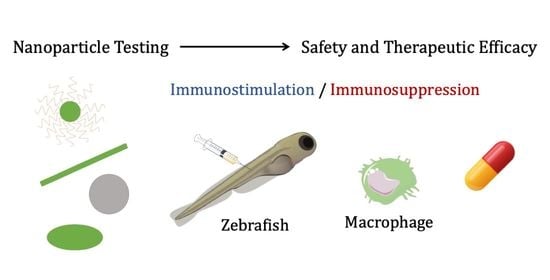
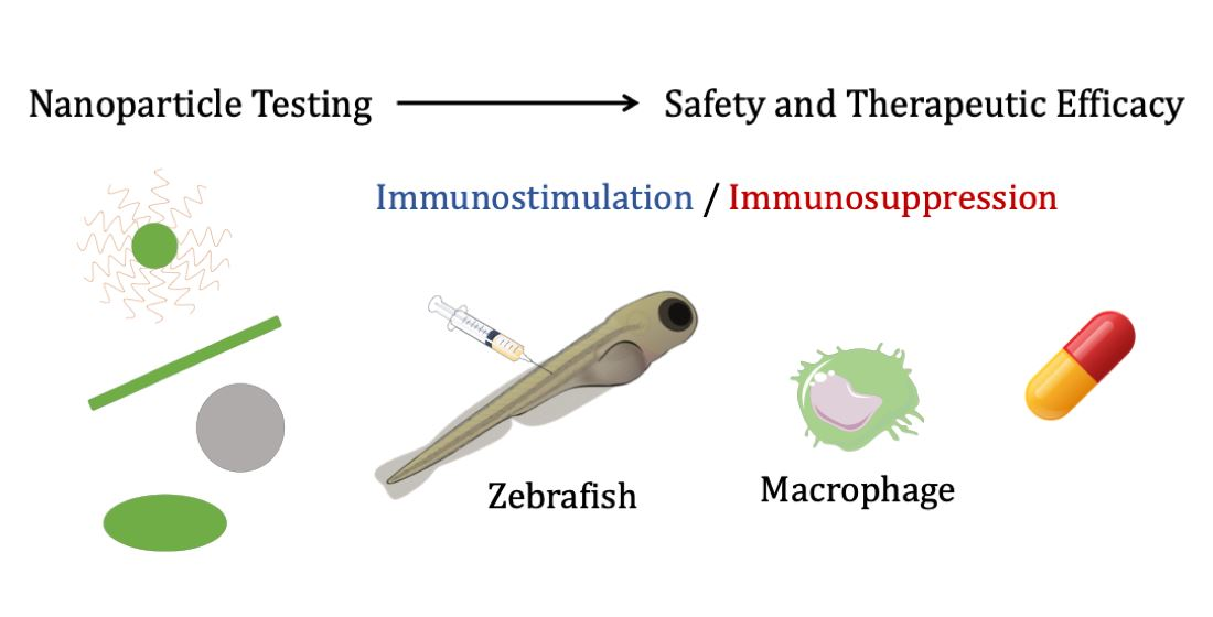
Zebrafish Models for the Safety and Therapeutic Testing of Nanoparticles with a Focus on Macrophages

by
Alba Pensado-López
1,2,
Juan Fernández-Rey
1,2,
Pedro Reimunde
3,4,
José Crecente-Campo
2,
Laura Sánchez
1,*,† and
Fernando Torres Andón
2,*,†
1
Department of Zoology, Genetics and Physical Anthropology, Campus de Lugo, Universidade de Santiago de Compostela, 27002 Lugo, Spain
2
Center for Research in Molecular Medicine & Chronic Diseases (CIMUS), Campus Vida, Universidade de Santiago de Compostela, 15706 Santiago de Compostela, Spain
3
Department of Physiotherapy, Medicine and Biomedical Sciences, Universidade da Coruña, Campus de Oza, 15006 A Coruña, Spain
4
Department of Neurosurgery, Hospital Universitario Lucus Augusti, 27003 Lugo, Spain
*
Authors to whom correspondence should be addressed.
These authors have contributed equally to this work.
Nanomaterials 2021, 11(7), 1784; https://doi.org/10.3390/nano11071784
Submission received: 11 June 2021 / Revised: 5 July 2021 / Accepted: 7 July 2021 / Published: 9 July 2021

Abstract

:
New nanoparticles and biomaterials are increasingly being used in biomedical research for drug delivery, diagnostic applications, or vaccines, and they are also present in numerous commercial products, in the environment and workplaces. Thus, the evaluation of the safety and possible therapeutic application of these nanomaterials has become of foremost importance for the proper progress of nanotechnology. Due to economical and ethical issues, in vitro and in vivo methods are encouraged for the testing of new compounds and/or nanoparticles, however in vivo models are still needed. In this scenario, zebrafish (Danio rerio) has demonstrated potential for toxicological and pharmacological screenings. Zebrafish presents an innate immune system, from early developmental stages, with conserved macrophage phenotypes and functions with respect to humans. This fact, combined with the transparency of zebrafish, the availability of models with fluorescently labelled macrophages, as well as a broad variety of disease models offers great possibilities for the testing of new nanoparticles. Thus, with a particular focus on macrophage–nanoparticle interaction in vivo, here, we review the studies using zebrafish for toxicological and biodistribution testing of nanoparticles, and also the possibilities for their preclinical evaluation in various diseases, including cancer and autoimmune, neuroinflammatory, and infectious diseases.

Graphical Abstract

1. Introduction

Recent advances in nanotechnology offer the possibility to engineer a wide variety of new nanoparticles and biomaterials with potential application in medicine, but also with commercial interest, providing solutions for numerous sectors in society (i.e., electricity, cosmetics, food packaging, etc.) [1,2]. There is considerable evidence that nano- and microscale materials have unique biological interactions when compared with molecules or bulk materials [3]. Thus, safety assessment results are, of course, of paramount importance [4]. In the case of medical applications, nanomaterials are designed for drug delivery, imaging, diagnosis, sensing, and/or therapeutic purposes. Thus, the benefit/risk for the use of nanotechnologies in this case acquires a different dimension similar to the pharmacological/toxicological profile of other drugs [5]. For these studies, in silico and in vitro methods, including cell- and organ-based assays, are encouraged. However, animal tests are still needed [4,6].
In this context, the zebrafish (Danio rerio) has become a well-established model for the toxicological and pharmacological screening of new drugs [7] and nanomaterials [8]. Rapid embryo development, small size and transparency, genetic and physiological conservation, ethical and economic advantages have made zebrafish stand out from all other in vivo models [9]. Furthermore, the innate branch of the immune system, including macrophage functions, is well-conserved between humans and the zebrafish, presenting a powerful model for the study of immunotoxicity and also diseases causing and/or caused by inflammatory disorders (i.e., cancer, autoimmune diseases or infectious diseases) [10,11]. Several zebrafish models with fluorescently labeled macrophages have been developed and optimized for studies with a particular focus on the role of macrophages in the toxicological or therapeutic effects of new compounds and nanomaterials. Thus, it is our purpose in this review to collect the information available related to the safety and biomedical testing of new nanomaterials using zebrafish models, with a particular focus on their interaction with macrophages, starting with toxicological and/or biodistribution experimentation, followed by preclinical testing of nanoparticles for therapeutic purposes.

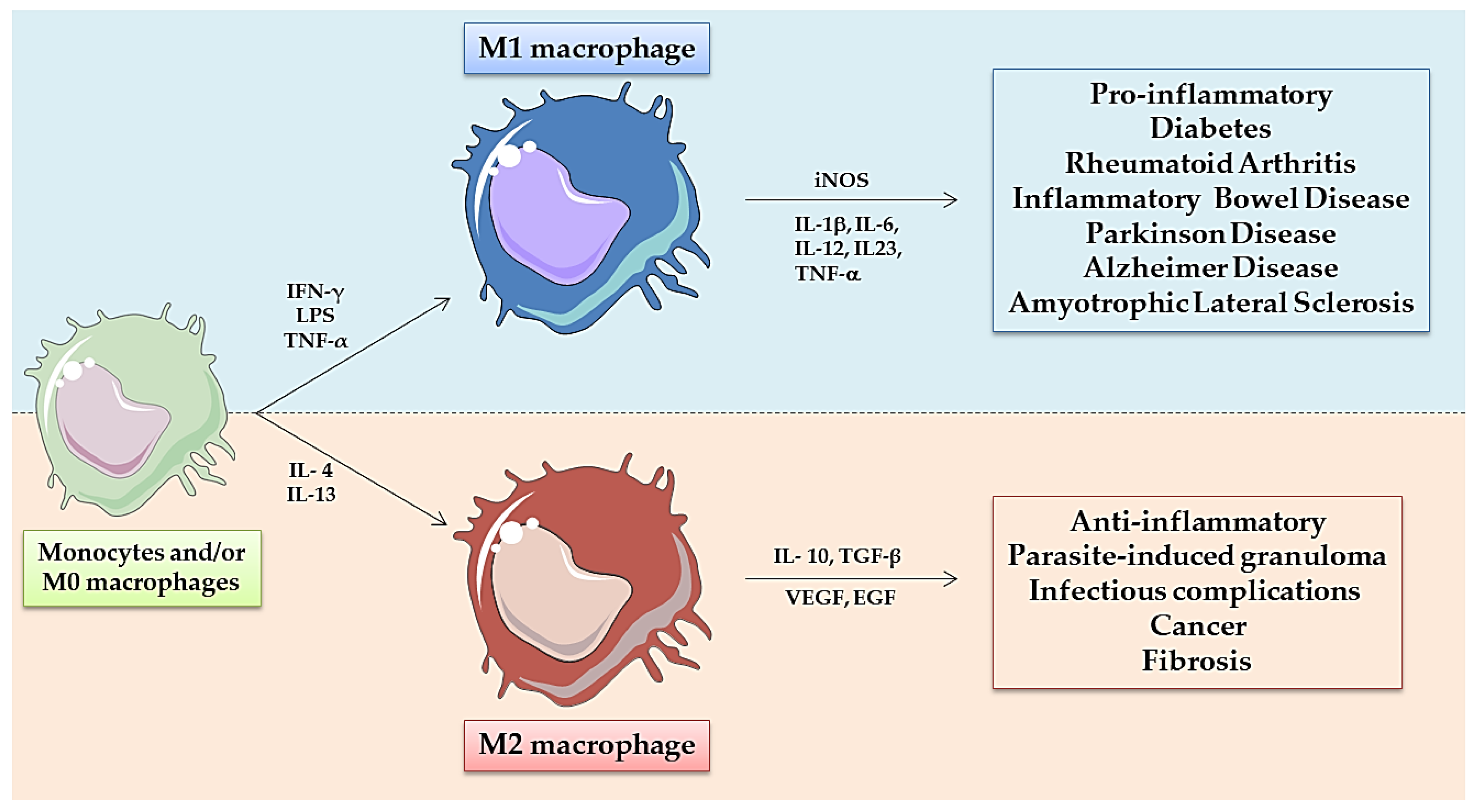
2. Zebrafish Innate Immune System: Role of Macrophages

The zebrafish immune system is able to develop both innate and adaptive responses. Despite differences in anatomical sites and time points with respect to mammals. Key cell types, molecular pathways, genetic programs, and transcription factors are highly conserved [12]. Whereas the innate immune system components are already present in early zebrafish embryo stages, the development of adaptive responses does not occur until 2–4 weeks post-fertilization (wpf), and complete immunocompetence is not achieved until 4–6 wpf [13]. Zebrafish innate immune system is composed of similar types of cells than mammals, being macrophages the first leukocytes in development and key players in directing the host immune response [14]. The origin of primitive macrophages in zebrafish occurs at 14–15 h post-fertilization (hpf). These cells migrate to the yolk sac, differentiate, and enter the circulation from 25 hpf [15,16]. The caudal hematopoietic tissue (CHT) at two days post-fertilization (dpf), equivalent to fetal liver or placenta in mammals, acts as a transient hematopoietic site and is a source of embryonic macrophages and neutrophils [17,18]. The thymus produces mature T-cells and the kidney, functional ortholog of mammalian bone marrow, produces myeloid, erythroid, thromboid, and lymphoid cells throughout adulthood [19,20].
Macrophages are phagocytic cells from the innate immune system which can display a wide variety of functions due to their plasticity, versatility and continuous adaptation and response to specific stimuli [21]. In mammals, macrophages have been classically classified according to their polarization extremes observed in the initiation or in the resolution of inflammatory processes [22]. And these cells have been respectively denominated as M1, classically activated or pro-inflammatory macrophages, versus the M2, alternatively activated or wound-healing macrophages [23,24] (Figure 1). These populations have been characterized in terms of gene expression, the pattern of surface molecules, and the production of biological mediators and metabolites [25,26]. An imbalance between M1 and M2 macrophages has been found in pathological tissues and correlated with a worse prognosis of the disease (i.e., high numbers of M1-like macrophages in arthritis or infiltration of M2-like macrophages in solid tumors) [21,27]. Furthermore, taking into account this knowledge, the reprogramming of M1-like macrophages towards M2-like anti-inflammatory effectors appears to be a reasonable strategy for the treatment of some autoimmune diseases [23], while the stimulation of M2-like immunosuppressive macrophages towards M1-like pro-inflammatory-anti-tumor effector cells is a promising approach for the treatment of cancer [21,25,28,29,30,31].
In 2015, Nguyen-Chi et al. provided a seminal report on the polarization of macrophages in zebrafish, showing a great similarity of the M1-like and M2-like phenotypes with respect to mammals [32]. They used the Tg(mpeg1:mCherryF) transgenic zebrafish line, which enables to track macrophages [33,34], and generated the Tg (tnfa:eGFP-F) line to label M1-macrophages expressing tumor necrosis factor-α (TNF-α). Following fin wounding-induced inflammation or Escherichia coli inoculation in zebrafish larvae, they observed a recruitment of macrophages to the wound after amputation or to the muscle after bacteria-inoculation. To characterize the M1/M2 polarization, they mated both lines to generate the Tg(tnfa:eGFP-F/mpeg1:mCherryF), sorted mCherry+/eGFP+ and mCherry+ cells during the early and late phases of inflammation, and compared their RNA expression patterns, observing high levels of tnfb, il1b and il6 pro-inflammatory markers of M1-like macrophages in double-labeled cells, while high levels of tgfb1, ccr2 and cxcr4b anti-inflammatory markers were found in the M2-like macrophages only labeled with mCherry [24,32]. Similar results were obtained by Sanderson et al. using the Tg(irg1:EGFP)/Tg(mpeg1:mCherry) transgenic line, in which irg1 specifically labeled M1-like macrophages upon LPS inoculation or M2-like macrophages upon injection of human metastatic breast cancer cells [35]. Other zebrafish-macrophage-fluorescent reporter lines, such as Tg(mpeg1:eGFP)gl22 [33] or Tg(mpeg1:mCherry)UMSF001 [36], have enabled to track the precise behavior and physiology of these cells in vivo, during inflammatory, cancer or infectious processes, thus providing a highly valuable tool to study their role in the context of these pathologies, and also for the screening of new macrophage-targeted therapeutic approaches, including nanoparticles.

3. Toxicological and Biodistribution Evaluation of Nanoparticles Using Zebrafish: Focus on Macrophages

3.1. Toxicological Studies

The zebrafish is commonly used as an in vivo model for the toxicological evaluation of new compounds and nanomaterials, due to its reduced cost, ease of husbandry, and high fecundity rates [37,38]. Zebrafish and mammals present concordance in toxicological assays ranging from 64% to 100% [39]. Due to its short-life cycle, zebrafish can be used to evaluate intergenerational toxicity by exposing a generation (F0) to a compound and studying the effects on the next generations (F1, F2, etc.) [40,41]. Reproductive impairment can also be studied by assessing sperm motility, egg depositions, and steroid hormone levels [42,43].
The usual methods of exposure to new compounds or nanomaterials are microinjection and immersion [44,45]. A widely used harmonized approach is the OECD Fish Embryo Acute Toxicity (FET) Test [46]. The OECD guidelines present immersion exposure as the preferred method and propose that tests should be performed within the first 120 hpf (5 dpf) limit [47,48]. Nishimura et al. reviewed the methodology used in several zebrafish-based developmental toxicity tests and indicated that 5 hpf is the preferred time to start the exposure to a new sample, because at this point embryos are in the late blastula stage and dechorionation can be safely performed [49]. Variations of the FET test have served other researchers to develop assays destined to screen large libraries of compounds or to evaluate organ-specific drug toxicities. For example, Cornet et al. took advantage of the brief zebrafish development to combine in one test the study of cardio-, neuro-, and hepato-toxicity effects in the same individual, enabling recompilation of organ-specific toxicity data for 24 compounds [50]. In a different study, 91 compounds, including pesticides, drugs, and flame retardants, were screened for teratological and behavioral effects, by immersing dechorionated zebrafish embryos in solutions of the different chemicals [51]. In combination with in silico and in vitro assays, zebrafish models were used for the screening of compounds with anti-inflammatory activity from a library of more than 1200 chemicals [52].
Nanotoxicological studies are commonly divided into two fields: environmental health and nanomedicine safety [8]. In both fields, similar types of in vitro and in vivo assays are performed, which in the case of zebrafish models take into account endpoints such as death, hatching rate, developmental malformations, behavioral changes and gene-profiling to assess toxicity [8,53,54]. The main factors influencing nanotoxicological effects, such as composition, particle size, particle shape and surface charge [54], have been investigated in zebrafish [55,56,57]. Zeng et al. studied the effects of Ag nanoparticles (NPs) on zebrafish and found a decreased activity of enzymes implicated in oxidative stress, such as superoxide dismutase, hydrogen peroxide, and malondiadehyde [55]. Another study evaluated how the size of AgNPs could affect toxicity, revealing a higher sensitivity of zebrafish to 20 nm AgNPs versus 100 nm AgNPs [56]. To assess the effects of shape, Abramenko et al. followed OECD guidelines and observed that Ag nanoplates induce higher toxicity than spherical AgNPs [57]. The toxicity of AgNPs on neural development was also evaluated through the study of gfap and ngn1 genetic profiling [58]. The safety of other inorganic based NPs (Au, Mg, Si, Zn) has been studied in zebrafish following similar methodologies [59,60,61,62,63]. Synergistic toxicity of methylmercury, SiNPs and AuNPs including surfactants have also been evaluated in zebrafish, proving it as a useful model to explore the toxicological effects of different compounds in the same organism [64,65]. A more comprehensive toxicological methodology, using zebrafish, and focused on 21 endpoints, was used for the screening of several NPs, leading to the conclusion that surface charge is a major determinant for NP-toxicity [66].
Numerous organic-based nanomaterials have been also tested in zebrafish [67,68,69,70]. The toxicity of polyamidoamine and polypropylenimin dendrimers was tested in zebrafish following the FET method, and with a parallel test in dechorionated embryos, showing for both dendrimers a lower toxicity in chorionated embryos [67]. In a different study, nanographene oxide with an external layer of polyethilenglycol was microinjected in zebrafish to evaluate its toxicity profile and effect on angiogenesis [68]. In another study, Teijeiro-Valiño et al. examined the toxicity of polymeric nanocapsules with an outer shell of hyaluronic acid and protamine [69].
The interaction of NPs with macrophages was investigated using zebrafish models, having important implications in the biocompatibility-toxicity, as well as in the biodistribution of the NPs (reviewed in the next section) [70,71,72,73,74]. On one hand, NPs may trigger direct cell toxicity through different molecular mechanisms [75], on the other hand numerous NPs can also trigger inflammatory responses when they are recognized as foreign agents by immune cells (i.e., macrophages) [5]. In response to NPs, the immune cells commonly produce soluble mediators, such as cytokines, chemokines, and complement factors, which result in the recruitment of more cells and development of acute or chronic inflammatory responses [76]. We should also remember that, in some cases, the inflammatory cells are capable of the biodegradation of NPs, e.g., through oxidative stress or enzymatic degradation (i.e., myeloperoxidase or eosinophil peroxidase) [77]. The balance of these responses is of foremost importance for immunotoxicological studies. As an example, graphene oxide immunotoxicity was evaluated by measuring glutathione, malondialdehyde, superoxide dismutase, catalase, and genetic profiling in adult zebrafish. TNF-α, IL-1β and IL-6 expression levels were significantly increased in a dose-dependent manner [74]. Similarly, it has been reported that several types of metallic NPs, such as gold NPs (AuNPs), silver NPs (AgNPs), or zinc oxide NPS (ZnO-NPs), may induce oxidative stress and disrupt signaling pathways related to innate immune responses [78,79,80]. Other studies involving drugs, nanocarriers, and macrophages practiced their experiments on zebrafish [81,82]. For example, poly(lactic-co-glycolic acid) NPs (PLGA-NPs) loaded with thioridazine were evaluated for toxicity and their therapeutic efficacy was tested in murine macrophages, human macrophages and zebrafish models [81]. These immunotoxicological studies, using zebrafish as in vivo model, demonstrate the relevance of safety assessment for nanomaterials with potential application in a variety of industrial sectors (i.e., cosmetics, paints, food package, etc.), and they also provide pharmacological/toxicological information of interest for their application with medical purposes.

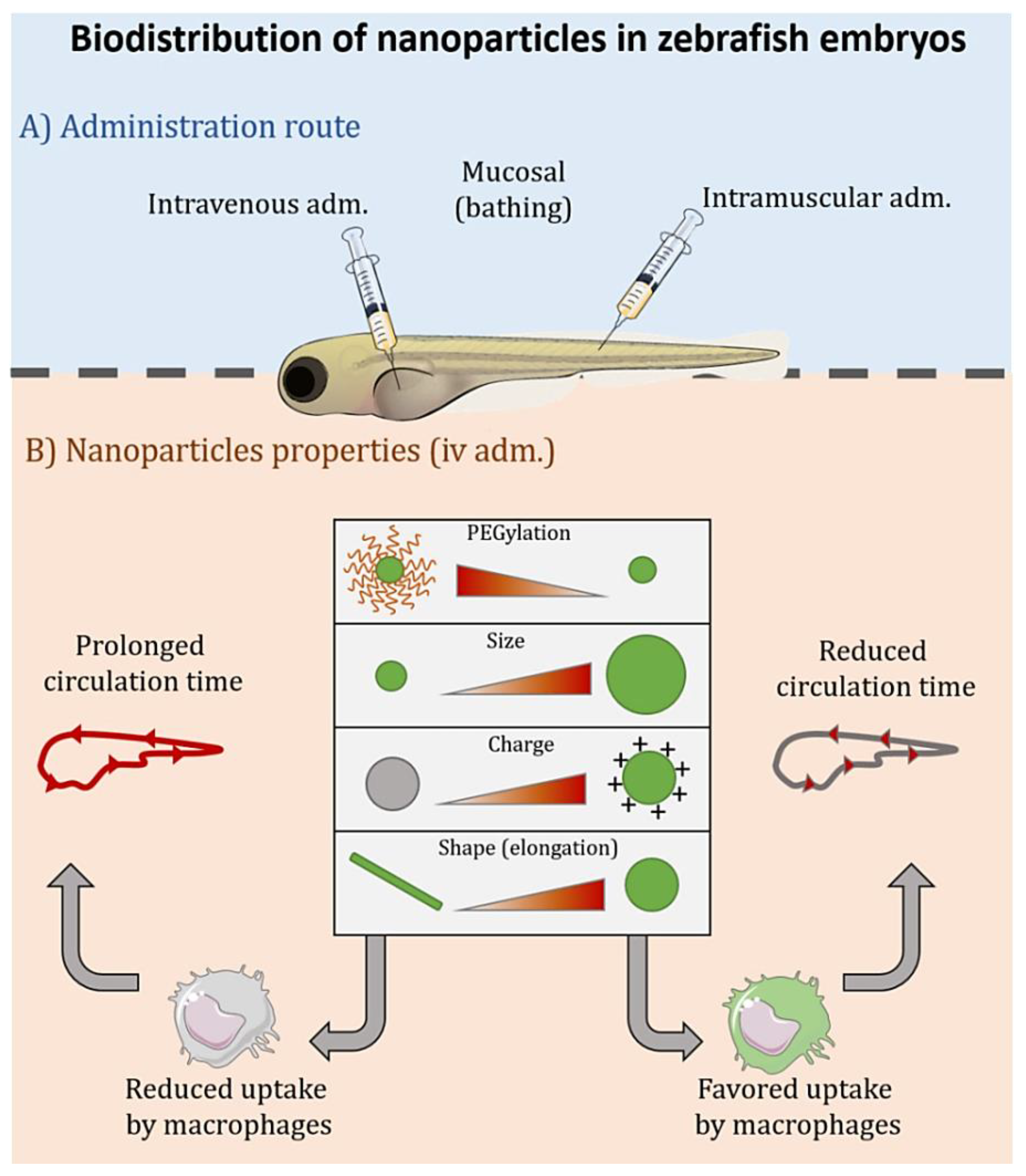
3.2. Biodistribution

Nanotechnology is commonly used to improve the biodistribution/pharmacokinetics of drugs. Thus, biodistribution moves from being determined by the drug’s physicochemical characteristics to be dictated by the NP’s-features. Besides, by tuning the physicochemical properties of the NPs, a preferential accumulation in specific organs and/or cell populations can be achieved [83]. Taking this into consideration, zebrafish is gaining importance in the nanomedicine field as a simple and reliable model to screen biodistribution of drugs and/or NPs. The transparency of the embryos [84] and the availability of transgenic lines offer the possibility to study in biodistribution studies the role of the interaction of NPs at the cellular level, for example, by using macrophage-labelled embryos [85]. In this context, different scenarios can happen depending on the aimed cellular and/or molecular target: macrophages can be the final target for the nanomedicine, such as in the case of autoimmune or infectious diseases, and nano-vaccines [86], or they can be a cellular population to be avoided, due to the undesirable clearance of the therapeutic entity mediated by these phagocytic cells, such as in the case of drugs targeted towards cancer cells or others [72]. Of note, in the context of cancer both approaches, targeting and/or avoiding macrophages, could be intended to improve the efficacy of certain drugs, directed towards macrophages (i.e., immunotherapy) or to cancer cells (i.e., chemotherapy), respectively [87].
The overall biodistribution of a NP is governed by the combined effect of the administration route, the dose and the nanostructure’s composition and properties (Figure 2). As previously described for the toxicity, the size, shape, charge and flexibility are also critical parameters that determine the final fate of the NPs [83,88]. The circulation time of NPs administered intravenously (i.v.) depends, mainly, on their interaction with immune and/or endothelial cells [89]. In fact, the majority of the NPs i.v. administered are cleared in the liver, by macrophages or by sinusoidal cells [85,90]. Part of these NPs are also captured by circulating monocytes/macrophages, and/or other immune cells, such as neutrophils, before reaching their target tissue, as it was recently shown for gelatine nanospheres [86] and liposomes [91]. In zebrafish, liposomes of 60 nm showed a decreased macrophage uptake as compared to larger 120 nm liposomes, which were more accumulated in the spleen [91]. Apart from immune cells, uptake by endothelial cells can also be responsible for the limited circulation time of certain NPs. For example, polystyrene (PS) NPs of 1000 nm were found adhered to the endothelium some minutes after the injection. On the contrary, with a lower adherence to the endothelium, 200 nm PS NPs showed a more prolonged circulation time. Our group evaluated the diffusion of NCs of different sizes and surface charges in zebrafish embryos [70]. These NPs are versatile nanosystems with applications in different fields, such as cancer, vaccination, and ocular diseases [92,93,94,95]. Both positively and negatively charged NCs of 70 nm spread faster and at a higher extent than medium size NCs (200 nm) after i.v. injection in zebrafish. Chitosan NCs (positively charged) were found to be attached to the endothelial cells of the blood vessels at a higher degree than inulin NCs (negatively charged). As a whole, zebrafish injected with the 70 nm chitosan NCs showed the highest intensity, due to the fluorescent-NCs, in the circulatory system, and also higher accumulation in other tissues, such as the brain and visceral organs. All NCs showed certain degree of co-localization with GFP-labelled macrophages. However, the positively charged NCs showed the highest accumulation in these cells. In relation to their particle size, 200 nm NCs interacted with macrophages from early time points (0.5 h), whereas 70 nm NCs presented less interaction at 0.5 h, but their accumulation in these cells increased with time. From this and other studies, it can be concluded that larger particles are commonly attached or captured by macrophages and/or endothelial cells, thus showing shorter circulation times.
Particle shape and flexibility have also an impact on NPs biodistribution. Generally elongated and rod-shaped NPs present a prolonged time in circulation compared with spherical particles, especially if they are flexible [96]. This effect was observed in different animal models (i.e., mice or pigs) [97], and also zebrafish [98]. In this case, the non-spherical NPs were poorly taken up by macrophages because of hydrodynamic shearing. However, other authors found that spherical NPs present prolonged circulation time versus NPs with fibrillar morphologies upon i.v. administration in zebrafish [99]. Overall, it is important to highlight that the final biodistribution and toxicological behaviour of a NP is orchestrated by the interplay between its different physicochemical properties (i.e., size, charge, and shape), and focusing only in one parameter can be misleading.
The composition of the NP’s surface and/or its functionalization with specific molecules is also a key factor affecting its biodistribution. For instance, liposomes presented substantially higher circulation time after i.v. administration in zebrafish than PS NPs of similar size (nm) [72]. The different circulation patterns can be explained by the high affinity of PS NPs for the endothelium, while liposomes showed a slow macrophage-mediated clearance. With respect to decoration of NPs surface, polyethylene glycol (PEG) and other molecules have been used to avoid macrophage interaction, while ligands for specific receptors on macrophages-surface or other cells have been investigated. Functionalization with PEG chains is commonly implemented to “mask” a therapeutic agent from the immune system, reducing immunogenicity and antigenicity. PEGylated nanosystems show longer circulation times after i.v. administration than non-PEGylated ones, by reducing their interaction with opsonins and making NPs “invisible” to the immune system [72,89]. As an example, liposomes commonly cleared from the circulation by macrophages, when PEGylated, increased their circulation time, and their elimination by macrophages, although still occurring, is delayed [89]. In the case of PEGylated PS NPs, the increase in their circulation time has been attributed not only to their reduced capture by macrophages, but also to their reduced interaction with the endothelial cells [72]. The degree of PEGylation and its molecular weight also impacts on NPs biodistribution, showing decreased clearance by macrophages for liposomes decorated with a higher density of PEGs or increased molecular weight [91]. Other polymers with “shielding behaviour” have been used, such as polysarcosine or poly(N,N-dimethylacrylamide), showing an increase in NP-circulation time in zebrafish models [89].
Zebrafish models have also been used to study NP-biodistribution through other routes of administration. Upon intramuscular (i.m.) injection in zebrafish, NPs generally only spread along the muscle tissue close to the injection site, at least for short times [86]. As time goes by, the NPs are commonly internalized by macrophages to be cleared [86]. In our previously mentioned work, after i.m. injection, the 70 nm NCs disseminated further than 200 nm NCs, spreading through all the myomere and limited by myosepta [70]. Positively charged NCs (chitosan based) recruited more macrophages than negatively charged (inulin based) NCs, following a similar pattern to the i.v. route. To study the mucosal administration of new therapies using zebrafish, NPs can be simply incubated in water. Verrier and colleagues found that 200 nm PLA-NPs were able to cross the epithelial barrier of different mucosae (nasal, gills, gut, and skin) and be accumulated in antigen presenting cells, such as dendritic cells, macrophages and B cells in the gills and skin [100]. Interestingly, the NPs were able to enter the bloodstream through the gills, and to then reach internal organs, such as the liver and kidney. Our group showed the importance of surface composition in the ability of NCs to diffuse through the chorion before the zebrafish hatching [69]. The presence of a PEGylated surfactant is supposed to favour the diffusion of these NCs through the thick chorion barrier. Using hatched embryos, we demonstrated that hyaluronic acid NCs are not able to cross the epidermis. However, protamine-hyaluronic acid NCs were internalized and reached the yolk sac, the stomach, the esophagus and the olfactory pit. In this case, protamine, a well-known cell penetrating peptide, could be important to facilitate the internalization and transport through the zebrafish skin epithelium [69]. The particle size also influences de biodistribution by the mucosal routes. For instance, after incubation with zebrafish in water, coumarin nanocrystals of 70 nm showed better permeability across the chorion, blood brain barrier, blood retinal barrier and gastrointestinal barrier than their counterparts of 200 nm. Besides, the smaller nanocrystals accumulated at a higher extent in internal organs via lipid raft-mediated endocytosis [101]. The importance of the particle shape was demonstrated by Vijver and his group [102]. After waterborne exposure of zebrafish embryos to gold NPs of different shapes, the macrophage’s abundance was higher for urchin-shaped NPs compared to the spherical ones.

4. Preclinical Testing of Nanoparticles Using Zebrafish Models of Disease: Relevance of Macrophages

4.1. Cancer

Due to its unique features, zebrafish tumor models, mainly induced by transgenesis or xenotransplantation, are increasingly being used for cancer research and discovery of new antitumoral drugs [103]. Xenografts are routinely performed by implantation of human or murine cancer cells into different anatomical sites of zebrafish embryos (yolk sac, duct of Cuvier or perivitelline space) to obtain heterotopic or eventually orthotopic in vivo tumor models. The injection of labeled cancer cells in “transparent zebrafish” allows to track their survival, progression, migration, and interaction with the host microenvironment [104,105]. For the transgenic models, genetically modified zebrafish lines, mainly based on the expression of human oncogenes driven by tissue-specific or ubiquitous promoters (e.g., BRAFV600E, HRASG12V or KRASG12V) have been used [106,107,108]. While the transgenic models are commonly preferred for the mechanistic understanding of tumor development and/or interaction of cancer cells with the tumor microenvironment (TME), xenografts are more used for drug screenings. As the generation of stable transgenic zebrafish lines requires several months, it is technically challenging and less cost-effective when compared to the simple injection of tumor cells in xenografts [109]. In addition, xenograft models have been recently used to understand the metabolic and/or stem cell properties of cancer cells and they also offer the possibility to study patient-derived cancer cells in vivo [110,111].
The study of macrophages in tumors is nowadays a very active field of research. Tumors are complex tissues, comprising a heterogeneous population of cells plus the extracellular matrix they produce, which constitute the TME [112,113,114,115]. The cellular fraction is composed of malignant cancer cells, as well as endothelial cells, cancer-associated fibroblasts, and tumor-associated macrophages (TAMs), with the latter being the latest the most abundant cell type [116]. In human tumors, TAMs originate mostly from circulating precursor monocytes, but resident macrophages can be originally present in the tissue, later developing in a tumor [117]. TAM infiltration in tumor tissues has been shown to support tumor growth, angiogenesis, invasion and metastasis, and their high density in tumors has been correlated with tumor progression and resistance to therapies [118]. The secretion of CSF-1, CCL2 and VEGF, by cancer cells, induce the recruitment of macrophages towards the TME, and the Th2 cytokines IL-4, IL-13, IL-10 and TGF-β, metabolic signals (i.e., lactic acid and hypoxia) produced also by Treg and TAMs are key drivers of immunosuppression [116]. With the aim to better mimic human tumors, including the TME, zebrafish xenografts are being continuously improved, using reporter lines for analysis of vasculature [119], neutrophils [120] and/or macrophages [32,33,35]. Orthotopic xenotransplantation, consisting in the injection of tumor cells in the equivalent anatomical site, and the use of patient-derived xenografts (PDX), allows the preservation of tumor cells’ original phenotype, and represents a further step to recapitulate the TME and tumor cell-host interactions [121]. The knowledge and practice acquired using these models will not only be useful for a better understanding of tumors, but also determinant for the development and evaluation of new antitumoral therapies [122], including nanotechnological strategies.
An enormous variety of nano-oncologicals (nanostructures for the treatment of cancer) have been developed and evaluated with the main purpose to improve the delivery of pharmacological molecules to the TME and reduce off-target effects [123,124,125]. NPs have been designed to improve the efficacy of classical chemotherapies, immunomodulatory drugs, but also delicate molecules such as nucleic acids, and even new cellular therapies [126,127]. Main therapeutic strategies to target and impact on TAMs include: (i) inhibition of TAM recruitment to the tumor, (ii) direct killing of TAMs, (iii) re-education of TAM from their M2-like protumoral phenotype into a M1-like antitumoral phenotype [87]; offering promising opportunities to switch the tumor-promoting immune suppressive microenvironment, characteristic of tumors rich in macrophages, to one that kills tumor cells, is anti-angiogenic and promotes adaptive immune responses [87,128].
In this context, zebrafish models offer the possibility to test the antitumoral effect of new nano-oncologicals, not only at the level of tumor growth in vivo [109], but also at the cellular level within the tumor (i.e., cancer cells or TAMs in vivo). Relevant examples of nano-oncologicals tested in zebrafish are reviewed below and summarized in Table 1.
Nadar and colleagues investigated the ability of drug-loaded hydroxyapatite NPs to release active therapeutics in vivo [129]. To this end, they used a zebrafish xenograft model with MDA-MB-231 breast cancer cells to test the antitumoral efficacy of kiteplatin-pyrophosphate-loaded hydroxyapatite NPs (PtPP-HA). Two days after the co-injection of cancer cells and the platinum-loaded HA NPs into 48 hpf zebrafish blood circulation, they found a significant decrease in survival of breast cancer cells [129]. Different types of metal oxide-NPs, not loaded with additional pharmacological molecules, have also been tested for antitumoral efficacy. ZnO-NPs were evaluated using a gingival squamous cell carcinoma xenograft model, showing antitumoral activity via induction of reactive oxygen species (ROS) and reduction of anti-oxidative enzymes with consequent oxidative damage to cells and tissues [130,138,139]. DiL-labeled Ca9-22 cells were implanted into the yolk sac of 48 hpf Tg(fli1:EGFP) embryos and fluorescence monitoring showed no effect on survival but dose-dependent inhibition of tumor growth [130]. The physicochemical properties of nanomaterials have also been exploited for photodynamic therapy (PDT) and tested in zebrafish. For instance, Jimenez et al. developed, and tested in zebrafish tumor xenografts, porous porphyrin-based organosilica NPs (PMOsPOR-NH2) [131]. At 2 dpi, treated-embryos were subjected to two-photon-excited photodynamic therapy (TPE-PDT) and a complete extinction of GFP-cancer cells was observed. Additionally, to analyze the efficiency of gene delivery, the same NPs were complexed with anti-GFP siRNA and co-injected together with GFP mRNA at one-cell stage, leading to a reduction in GFP expression a few hours later, thus also revealing the potential use of these NPs for gene therapy. Similarly, small-seized non-porous porphyrin-based bridged silsesquioxane NPs (PORBSNs) functionalized with PEG and mannose (20–30 nm) were developed for i.v. injection and tested in zebrafish with MDA-MB-231-GFP cells subjected to TPE-PDT, showing decreased tumor growth after irradiation [132]. Moreover, with targeting purposes, Peng et al. synthesized fluorescent cellulose acetate NPs functionalized with folate groups for their preferential accumulation in epithelial cancer cells overexpressing folic acid receptors [140]. These NPs can be tuned within the entire UV-VIS-NIR spectrum and were capable to target tumors in vivo. To test the combinatorial antitumoral efficacy of chemotherapy and radiotherapy using NPs, Wu et al. synthesized G4.5 polyamidoamine (PAMAM) dendrimers conjugated with l-cysteine (GC), acting the later as radiosensitizer, and further loaded with doxorubicin (PAMAM-GC/DOX) [133]. Cervical carcinoma HeLa cancer cells labeled with carboxyfluorescein succinimidyl ester (CFSE) were injected into the yolk sac at 2 dpf. Embryos were immersed in PAMAM-GC/DOX and further exposed to γ-radiation at 3 dpf. Synergistic antitumoral effect for the combination chemotherapy and radiotherapy with NPs was confirmed, versus only radiation or free DOX with or without irradiation. Likewise, PAMAM dendrimers, functionalized with PEG using a hypoxia-induced sensitive linker, were loaded with DOX and hypoxia-inducible factor 1a siRNA (PAMAM-DOX-siHIF) [134]. The antitumor activity of free DOX, PAMAM+DOX, and PAMAM-DOX+si-HIF was tested upon implantation of MCF-7-CM-DiL breast cancer cells into the perivitelline space of 48 hpf Tg(fli1:EGFP) embryos, and intracardiac injection of free DOX, PAP+DOX and PAP-DOX+si-HIF at 1 dpi, showing the best results for the “triple-therapeutic combination” (PAMAM-DOX+si-HIF). Despite these promising results in zebrafish, we still foresee some limitations for the translation of some results to the clinic, in part due to the differences in zebrafish anatomy versus mammals, because of the need for deeper penetration of radiotherapy in hidden tumors. However, a positive experience was recently reported by Costa et al., showing the utility of zebrafish to distinguish radiosensitive from radioresistant tumors using colorectal cancer cell lines and patient biopsies, and clinical response was correlated with induction of apoptosis in zebrafish [141].
Several redox- and pH-responsive-NPs have been developed, to favor the control delivery of drugs in the reducing and acidic TME, and several zebrafish models were optimized for the testing of these NPs. Transgenic mifepristone-inducible liver tumor zebrafish line expressing the enhanced green fluorescence protein (EGFP)-Krasv12 oncogene [142], as a model of hepatocellular carcinoma with elevated glutathione and liver acidity, was used [143,144]. While embryos treated with free DOX died at 2–3 dpi, lower toxicity and sustained regression of tumor size was observed for the DOX-loaded-NPs, demonstrating improved drug release to liver tumor cells and lower systemic toxicity [145]. A pH-sensitive hydrazone-linked DOX nanogel (NanogelDOX) was i.v. injected in zebrafish, previously implanted with B6 mouse melanoma cells into the neural tube at 48 hpf, showing significant reduction in tumor growth versus no effect for free DOX. [135]. Redox-responsive silica-gold nanocomposites functionalized with transferrin and loaded with DOX (Tf-DOX-ReSi-Au) were injected in a zebrafish colorectal cancer model, showing positive antitumoral activity without the typical DOX-related cardiotoxic adverse effects [136]. Others have tested the capacity of fluorescent-NPs to detect cancer cells in vivo, using similar zebrafish tumor models [146,147].
To understand the interaction between immune and tumor cells, several zebrafish xenograft models have been developed. These models have allowed to study the role of immune cells in tumor vascularization and invasion [148], the dynamic interaction between immune and cancer cells [149], or the positive correlation between the number of immune cells recruited to the tumor site and the degree of angiogenesis [150,151]. Póvoa et al. showed distinct engraftment profiles from the same patient at different stages of tumor progression in colorectal zebrafish xenograft models and explored the innate immune contribution to this process [152]. While cells derived from the primary tumor were able to recruit macrophages and neutrophils, thus being rapidly cleared (regressors), those cancer cells derived from a lymph node metastasis polarized macrophages towards a M2-like protumoral phenotype, engrafting very efficiently (progressors). Interestingly, mixing both types of cells resulted in decreased regressors clearance, reduced numbers of innate cells and increased M2-like polarization. Furthermore, depletion of macrophages resulted in a significant increase in the engraftment of regressors. These results provide the first experimental evidence of therapeutic manipulation of macrophages in zebrafish tumor models [152]. Detailed studies, using NPs for reprogramming TAMs into antitumoral M1-like macrophages, have still not been performed using zebrafish models of cancer. Nevertheless, a few investigations have explored the circulation time of NPs, their accumulation at the tumor site and their specific uptake by macrophages in tumors using these models. Evensen et al. were the first to image the accumulation of NPs to human tumor-like structures in a zebrafish xenograft model [72]. Comparing labeled polystyrene NPs and liposomes, with or without PEGylation, upon injection into the posterior cardinal vein of 2 dpf embryos, they noted that PEG-liposomes displayed the longest circulation time due to their lower affinity to the endothelium, the lowest macrophage uptake, and the highest survival rate. Using xenografts, with melanoma and kidney cancer cells, the PEG-liposomes showed a specific and rapid accumulation into the tumor and outside the vasculature after only 2–5 h post- injection (hpi) [72]. Kocere and colleagues demonstrated the selective accumulation of Cy5-labelled poly(ethylene glycol)-block-poly(2-(diisopropyl amino) ethyl methacrylate) (PEG-PDPA) NPs in TAMs using a melanoma xenograft model [137]. By injection of B6 mouse melanoma cells (labeled with GFP or RFP) in the neural tube of 3 dpf transgenic embryos ((Tg(fli1a:EGFP, Tg(mpeg1:mcherry), Tg(mpx:GFP)) [33,119,153], they were able to observe tumor growth, angiogenesis, and accumulation of macrophages, but not neutrophils, within the tumor at 10 dpf. Additionally, xenografts in 3 dpf Tg(mpeg1:GAL4/UAS:NTR-mCherry) embryos, which express a nitroreductase in macrophages in presence of metronidazole causing their selective apoptosis, revealed a slightly increased tumor growth when macrophages were absent. The injection of PEG-PDPA NPs, showed selective accumulation of NPs and increased number of macrophages at the tumor site. Moreover, a small fraction of NPs was internalized by cancer cells and TAMs. These DOX-loaded-PEG-PDPA NPs were i.v. injected in the melanoma model at 1 dpi, showing reduced toxicity, decreased proliferation, and increased apoptosis of cancer cells six days after treatment [137].
As a whole, these studies provide a consistent knowledge and experience for the development of several types of tumors in zebrafish and the testing of nano-oncologicals with different features. Zebrafish models provide excellent opportunities for genetic modifications and for in vivo evaluation/tracking of innate immune cells, being these key aspects for the testing of NPs. A major limitation for the use of zebrafish embryos to test antitumoral therapies is their lack of adaptive immune system. This challenge has partially been addressed through the suppression of the adult immune system, either by γ-irradiation, dexamethasone treatment [154,155,156] or more recently using adult immunocompromised strains [157,158,159], followed by the injection of human or murine immune cells (i.e., T cells). These adult zebrafish xenografts enable a closer resemblance of cell–tumor microenvironment interactions, a longer tumor engraftment and a clinically-relevant dose response [160,161]. Remarkably, Yan et al. created an optically clear, homozygous mutants (prkdc−/−, il2rga−/−) which lacks T, B, and natural killer (NK) cells. These animals survived at 37 °C (optimal for mammal cells), robustly engrafted a variety of human tumor cells and subsequently responded to drug treatments. Importantly, similar histological and molecular features in both fish and mouse xenografts were confirmed and pharmacokinetics of antitumoral treatments, such as olaparib and temozolomide, were comparable to that found in both mouse preclinical models and humans. Nevertheless, this adult immunocompromised strain must be improved, as several cancer cell types failed to engraft into the model and pre-treating fish with clodronate liposomes to deplete macrophages was required [159]. Additional disadvantages of zebrafish models are that some strains do not breed, develop gill inflammation, and likely autoimmunity [158], or they could be quite prone to infection and require specialized food and antibiotic treatment [162], thus raising the cost of maintenance. Despite these challenges, the results demonstrate that zebrafish tumor models are important tools with high potential to improve the translation of nano-oncologicals towards the clinic.

4.2. Autoimmune Diseases

4.2.1. Inflammatory Bowel Disease

Inflammatory bowel diseases (IBDs) refer to chronic inflammatory disorders of the gastrointestinal tract which comprise both Crohn’s disease and ulcerative colitis. Although their etiology is not clear, they are thought to be a result of host genetic susceptibility and environmental factors (e.g., diet) [163,164], which lead to altered interactions between gut microbiota and the intestinal immune system [165]. Intestinal homeostasis is partly maintained by resident macrophages with enhanced phagocytic and bactericidal activity and decreased production of pro-inflammatory cytokines [166]. Nevertheless, when gut dysbiosis and further disruption of normal mucosal immunity occur, monocytes are continuously recruited to become inflammatory macrophages, they participate in the inflammatory response and contribute to chronic intestine inflammation [167]. In this context, zebrafish models are useful to study the relationship between immune system and inflammation. For instance, Coronado et al. treated fish with a previously established inflammatory diet [168] and found a strong increase in the number of neutrophils, macrophages and T helper cells recruited to the gut [169]. Looking into the genetic susceptibility, Kaya et al. were able to validate the implication of GPR35-expressing macrophages in intestinal immune homeostasis and inflammation by generating a zebrafish mutant line [170]. Other researchers have shown the potential of zebrafish for the screening of drugs to treat IBDs [171,172,173]. With regard to NPs, to date, we only found one study where the administration of copper NPs to zebrafish resulted in intestinal developmental defects, through ER stress and ROS generation, showing similar alterations to IBD patients (Table 2) [174]. Thus, these studies provide the starting point for the study of IBD pathology and testing of NPs which might offer new solutions to patients suffering these intestinal disorders.

4.2.2. Type I Diabetes Mellitus

Type 1 diabetes mellitus (T1DM) is an autoimmune disease caused by immune-mediated progressive destruction of the pancreatic β-cells, driven by the interaction of multiple environmental and genetic factors. The pathogenesis of T1DM is characterized by the infiltration of islet antigen-specific T cells and pro-inflammatory APCs associated with impairment of Foxp3+ Tregs. The destruction of β-cells leads to the loss of ability to produce insulin and in turn, to chronic hyperglycemia [175,176]. T1DM treatment is mainly based on lifelong insulin replacement therapy and several nanoparticles have been designed to improve its administration [177,178]. Others have explored the inhibition of the destructive autoimmune response against insulin-producing β-cells, for example by regulating T-cell autoreactivity as a therapeutic approach [179,180]. In zebrafish models, to mimic T1DM, the destruction of β-cells has been achieved by either surgery [181], chemical destruction [182], or genetic ablation [183], followed by their subsequent regeneration ability allowing to study the mechanisms of β-cells regeneration and also the testing of antidiabetic drugs [184,185]. A number of chemical screens to induce β-cell generation in zebrafish have been reported [186,187,188] and the antidiabetic effect of different bioactive molecules and NPs has been tested (Table 2).
Silver nanoparticles loaded with Eysenhardtia polystachya (EP/AgNPs), with a spherical shape and diameter of 5–21 nm, were tested on glucose-induced diabetic adult zebrafish and the results confirmed the effectiveness of NPs in ameliorating hyperglycemia [189]. The utility of quercetin NPs (NQs) in ameliorating diabetic retinopathy, a common complication derived from diabetes was shown by Wang et al. [190]. Chemically induced diabetes and diabetic retinopathy were established in adult zebrafish, and further treatment with NQs led to a reduction in glucose blood levels as wells as to the improvement of different morphological, behavioral, and biochemical parameters linked to diabetic retinopathy [190]. Others have evaluated the biocompatibility/toxicological profile of different types of NPs with potential antidiabetic activity, such as peptide-major histocompatibility complexes-NPs or curcumin encapsulated in polycaprolactone-grafted oligocarrageenan nanomicelles [191,192]. These studies provide a solid basis for the further use of zebrafish models to screen new antidiabetic nanotechnological approaches.

4.2.3. Rheumatoid Arthritis

Rheumatoid arthritis (RA) is a chronic inflammatory and autoimmune disease characterized by synovial and joint swelling, pain, and bone destruction [193]. The pathogenesis of RA is a multistep process, initially starting outside the joints by the aberrant activation of antigen-presenting cells, triggered by genetic or environmental causes, which leads to the activation of a pro-inflammatory cascade, production of autoantibodies as well as altered T-cell and B-cell cross-activation [194,195]. These events lead to monocyte recruitment to the diseased tissue and activation and polarization of macrophages towards a M1-like pro-inflammatory phenotype, boosting the inflammatory cascade [196]. Current RA-treatments include nonsteroidal anti-inflammatory drugs (NSAIDs), corticosteroids, disease-modifying antirheumatic drugs, and different natural substances to reduce joint inflammation [197]. With the aim to mimic human disease, improve its understanding and test new therapies, several animal models have been developed [198,199], and zebrafish models could be also implemented as a useful tool. Zebrafish have served as a model to evaluate the anti-inflammatory properties of different synthetic and natural compounds, although no reports on nanomaterials are available yet. Clerodendrum cyrtophyllum Turcz is a commonly used plant in Vietnam for treating RA. Ngyuyen et al. confirmed the anti-inflammatory properties of ethanol extracts of this plant in a copper-induced inflammation zebrafish model, via downregulation of inflammation mediators and pro-inflammatory cytokines (e.g., cox-2 or il-1β) [200]. Similarly, Jiang et al. demonstrated anti-inflammatory effects of isothiocyanate prodrugs in a zebrafish neutrophilic inflammation model, as the number of migrating neutrophils in treated zebrafish was smaller than in the control group [201]. Wang et al. demonstrated the inhibition of cyclooxygenase, as the anti-inflammatory mechanisms of action of Gentiana dahurica roots, in a zebrafish model of induced production of cyclooxygenases 1 and 2 [202]. Genetic approaches using zebrafish were applied to study the role of c5orf30, whose variants have been associated with RA. Upon tail transection in the c5orf30 knockdown fish an increased recruitment of macrophages to the wound site was observed, confirming the anti-inflammatory role of c5orf30, with implications in RA [203]. With reference to early diagnosis, Feng et al. designed and synthesized fluorescent probes for the quantitative detection of hypochlorous acid, a biomarker of RA, and confirmed their efficiency in an LPS-induced inflammatory model of adult zebrafish [204]. Additionally, zebrafish has served as a platform to evaluate the toxicity of new treatments with potential application in RA [205,206]. Although mammalian models of RA are needed before the clinical translation of new therapeutic approaches, these zebrafish models provide a valuable tool for the initial screening of innovative nanotechnological approaches to treat RA.

4.2.4. Neuroinflammatory and Neurodegenerative Diseases

Several zebrafish models have been established and optimized for the understanding of neuroinflammatory or neurodegenerative diseases [207,208]. In parallel, different types of NPs have been engineered for the treatment and/or diagnosis of neuro-related disorders [209,210]. Interestingly, zebrafish models have also been used to study the neurotoxic or neuroprotective effects of a wide variety of NPs and biomaterials [207]. Below, we provide some examples of investigations using zebrafish models and NPs to improve drug targeting and/or efficacy in the context of neuroinflammatory and neurodegenerative diseases (Table 2).
Neuroprotective effects of NPs in Parkinson’s disease (PD) using zebrafish models have been observed, commonly mediated by the antioxidant and/or neuro-antiinflammatory activity of these NPs. Bacopa monnieri platinum NPs (BmE-PtNPs) demonstrated the same activity of Complex I, as that of oxidizing NADH to NAD(+), suggesting that BmE-PtNPs could be a potential medicinal substance for oxidative stress mediated disease with suppressed mitochondrial complex I as it happens in PD. Hence, in 1-methyl-4-phenyl-1,2,3,6-tetrahydropyridine (MPTP)-induced experimental Parkinsonism in zebrafish model, BmE-PtNPs pretreatment significantly reversed the toxic effects of MPTP by increasing the levels of dopamine, its metabolites, GSH, and activities of GPx, catalase, SOD and complex I, and reducing levels of MDA along with enhanced locomotor activity [211]. Schisantherin A (SA) is a promising anti-Parkinsonism Chinese herbal medicine and SA nanocrystals (SA-NC) were used to reverse the MPTP-induced dopaminergic neuronal loss and locomotion deficiency in zebrafish. This strong neuroprotective effect of SA-NC may be partially mediated by the activation of the protein kinase B (Akt)/glycogen synthase kinase-3β (Gsk3β) pathway [212]. On the other hand, it has been shown that, after exposure of different concentrations of titanium dioxide NPs (TiO2 NPs) to zebrafish embryos from fertilization to 96 hpf, the hatching time of zebrafish was decreased accompanied by an increase in malformation rate, while no significant increases in mortality relative to controls were observed [213]; moreover, accumulation of TiO2 NPs was found in the brain of zebrafish larvae, resulting in loss of dopaminergic neurons, ROS generation and cell death in hypothalamus. Meanwhile, q-PCR analysis showed that TiO2 NPs exposure increased the pink1, parkin, α-syn, and uchl1 gene expressions, which are related with the formation of Lewy bodies [213]. Data from zebrafish behavioral phenotype revealed observable effects of silica nanoparticles (SiNPs) on disturbing light/dark preference, dampening exploratory behavior and inhibiting memory capability; furthermore, the relationship between neurotoxic symptom and the transcriptional alteration of autophagy- and parkinsonism-related genes was showed [62]. Similarly, another study showed that 15-nm silica SiNPs produced significant changes in advanced cognitive neurobehavioral patterns (color preference) and caused PD-like behavior compared with 50-nm SiNPs. Analyses at the tissue, cell and molecular levels corroborated the behavioral observations [214]. Both studies demonstrated that nanosilica acted on the retina and dopaminergic neurons to change color preference and to cause PD-like behavior [62,214]. Puerarin has emerged as a promising herb-derived anti-Parkinsonism compound and puerarin nanocristals (PU-NCs) demonstrated no obvious toxic effects on zebrafish, as evidenced by the unaltered morphology, hatching, survival rate, body length, and heart rate; fluorescence resonance energy transfer (FRET) imaging revealed that intact nanocrystals were found in the intestine and brain of adult zebrafish [215,216]. Moreover, other NPs with alleged neuroprotective effects for treating PD, such as polymeric NPs of Ginkgolide B, have shown correct bioavailability and cerebral accumulation in zebrafish models [217].
Similarly, as with PD, some authors have shown neuroprotective and neuroregenerative effects of different NPs in Alzheimer’s disease (AD) zebrafish models. Thereby, it has been shown that casein coated-gold nanoparticles (βCas AuNPs) in systemic circulation translocate across the blood brain barrier (BBB) of zebrafish larvae, sequester intracerebral Aβ42 and its elicited toxicity in a nonspecific chaperone-like manner. This was evidenced by behavioral pathology, ROS, and neuronal dysfunction biomarkers assays, complemented by brain histology and inductively coupled plasma-mass spectroscopy. The capacity of βCas AuNPs in recovering the mobility and cognitive function of adult zebrafish exposed to Aβ was demonstrated [218]. Other study evaluated the role of solid lipid NPs of quercetin (SLN-Q), a flavonoid with multiple pharmacological actions like vascular integrity and regulatory action on the BBB, using pentylenetetrazole (PTZ) induced cognitive impairment of Danio rerio species [219]. The intraperitoneal pretreatment of SLN-Q showed an attenuating effect in PTZ induced neurocognitive impairments, along with amelioration of biochemical changes (acetylcholinesterase activity, lipid peroxidation, and reduced glutathione levels), showing differences with fish treated with donepezil. Some authors have demonstrated with confocal image analyses that amphiphilic yellow-emissive carbon dots (Y-CDs) crossed the BBB of five-day old wild-type zebrafish, most probably by passive diffusion due to the amphiphilicity of Y-CDs; furthermore, Y-CDs were internalized by the cells, inhibiting the overexpression of human amyloid precursor protein (APP) and β-amyloid (Aβ) which is a major factor responsible for AD pathology [220].
Amyotrophic Lateral Sclerosis (ALS), a fatal neurodegenerative disease affecting the upper and lower motor neurons in the motor cortex and spinal cord, could be ameliorated by reducing the levels of superoxide dismutase I (SOD1). Thus, calcium phosphate lipid coated nanoparticles (CaP-lipid NPs) were developed and tested in zebrafish for the delivery of SOD1 antisense oligonucleotides (ASO) with success, and their preferential accumulation in the brain, blood stream, and spinal cord was observed [221].
In addition, the toxic profile of several NPs with potential interest for neurological diseases was also evaluated using zebrafish models. As examples, Carbamazepine or Tacrine were co-administered with PAMAM dendrimers and neurotoxicity, cardiotoxicity, or hepatotoxicity were evaluated in zebrafish larvae [222,223]. These reports provide satisfactory experience in the study of neuro-related disorders using zebrafish models and a good basis for their use as a screening platform to support new nanotechnologies for the treatment of these diseases.

4.3. Infectious Diseases

Zebrafish models have been largely used to study infectious diseases, taking advantage of its transparency, possibilities to study both innate and adaptive immunity, and feasibility for the highly controlled administration of pathogens, commonly through microinjection. Prospective treatments are mostly administered by microinjection too, although other routes such as intubation have been described [224]. Most of the studies reviewed in this section have been performed using zebrafish in the first month of life, which does not present a completely developed adaptive immune system but allows for the separate study of macrophages and neutrophils during pathogenic infection [225,226]. Transgenic lines that attach fluorescent proteins to robust macrophages markers, such as mpeg1 or csf1ra, have been used [227]. As an example, Palha et al. infected Tg(fnφ1:mCherry) zebrafish larvae with two Chikungunya virus strains, one of which expressed GFP, allowing to follow infection progress and its quantification by flow cytometry. This work, using zebrafish, demonstrated similarities with the process in mammals and a critical role of the IFN response to control the infection. Furthermore, the differential role of macrophages and neutrophils was investigated using a transgenic metronidazole-inducible cell ablation system to deplete macrophages. Neutrophil depletion was studied in csf3r knockdown larvae that were highly susceptible to Chikungunya virus, exhibiting a high increase of virus transcripts and mortality [228]. In another study, selective depletion of macrophages by incubation in metronidazole reduced the virulence of infection caused by the Burkholderia cepacia complex, revealing the important role of these cells in the infectious process [229]. The easiness for monitoring macrophages in zebrafish allowed extensive studies of tuberculosis and facilitated the observation of granulomas caused by Mycobacterium marinum [230,231,232]. Clay et al. took advantage of the transparency of the zebrafish to report a dichotomous role of macrophages in early M. marinum infection. First, they performed dual fluorescent antibody detection of L-plastin and myeloperoxidase to confirm that only macrophages and not neutrophils phagocytose bacteria. Most bacteria were found in macrophages (L-plastin positive but MPO-negative). Second, they assessed whether macrophages upregulate inflammatory cytokines as a response to M. marinum. They injected separately the bacteria and similar-sized fluorescent beads into the hindbrain, demonstrating that macrophages migrated to the area in response to M. marinum but not in response to the beads. Macrophage-defective zebrafish embryos were created by the injection of morpholinos against the pu.1 gene to evaluate the role of macrophages on mycobacterial growth. Finally, the Tg(fli1:EGFP) transgenic line and pu.1 morphants were used to study bacteria dissemination through the vascular system and the role of macrophages. By injecting red fluorescent bacteria into the bloodstream, they found that control embryos had a higher number of extravascular bacteria than pu.1 morphants and bacteria injected in the hindbrain could only disseminate out of this space in the control zebrafish with macrophages [231]. Similarly, Davis and Ramakrishnan exploited zebrafish transparency to assess the role of macrophages in tuberculous infection through three-dimensional differential interference contrast microscopy (3D DIC) and fluorescence in vivo microscopy. Embryos were infected with wild type and attenuated M. marinum, lacking the ESX-1/RD1 secretion system locus, throughout their experiments. Upon injection of the bacteria into the hindbrain ventricle and daily monitoring, infected macrophages recruited uninfected macrophages in a RD1-dependent manner. Further, the combination of 3D DIC with time-lapse microscopy showed that uninfected macrophages become infected quicker when they are recruited by RD1-competent bacteria. Zebrafish transparency also enabled close observation of the macrophage’s morphology both in WT and RD1 defective bacteria, which proved to be different. Finally, after proving that macrophages become infected when phagocyting dead infected macrophages, granuloma dissemination initiated by macrophages from primary granulomas was also observed. To assess the migration of infected macrophages, they used a bacteria strain that constitutively expresses the Kaede photoactivable protein [232]. Other pathogens relevant for humans (Candida albicans, Herpes simplex, Pseudomonas aeruginosa, Staphylococcus aureus, Streptococcus pneumonia) [233,234,235,236] and fish (Vibrio anguillarum, Aeromonas salmonicida, Yersinia ruckeria, etc.) [237,238,239] have also been studied using zebrafish models [240,241]. In relation with the study of tuberculosis, Oksanen et al. demonstrated that zebrafish is a useful model for preclinical DNA vaccine development. They vaccinated zebrafish with a combination of plasmids encoding for Ag85B, CFP-10, and ESAT-6, well known mycobacterial antigens, through intramuscular microinjection. Three weeks after immunisation, zebrafish were challenged with a high dose of M. marinum (20,500 CFU) by microinjection. Vaccinated fish showed increased survival and the analysis of the bacterial load by qPCR revealed that unvaccinated zebrafish had a higher load than the vaccinated group [242]. Taken together these studies are proof of the versatility of zebrafish to study pathogen infection and dissemination in vivo. Genetic-wise, besides an already existing repertoire of transgenic lines, Clay et al. proved D. rerio can be tailored to the needs of the researchers for the study of macrophages by the knockdown of key regulator genes [231].
Several nanotechnology-based-approaches have been developed for vaccination, prophylactic and therapeutic purposes, in the context of infectious diseases, and tested in zebrafish models (Table 3). With prophylactic purposes, Torrealba et al. generated inclusion bodies (IBs) containing TNFα or CCL4 [243]. The resulting nanostructures (IBs) showed good stability under different pH conditions (2.5 and 8). While IBTNFα were cylindrical with a diameter between 380–900 nm and an average length of 1134.6 ± 196.6 nm, the IBCCL4 showed spherical shape with a diameter between 220–850 nm. Both IBs were added to the cell culture of ATTC® CRL-2643 (zebrafish liver cells) and RT-HKM (trout macrophages) to evaluate their uptake, showing positive results for both cell types. Further, treatment of RT-HKM with IBTNFα stimulated the expression of pro-inflammatory cytokines. In vivo, zebrafish were immunised with IBTNFα, IBCCL4, or IBiRFP-H6 (an IB containing a control protein) by microinjection and later challenged with P. aeruginosa. Zebrafish injected intraperitoneally with both types of IBs exhibited reduced mortality after a challenge with P. aeruginosa. Similar inclusion bodies were also used to encapsulate proteins of pancreatic necrosis virus (IPNV), haemorrhagic septicaemia virus (VHSV) and viral nervous necrosis virus (VNNV) for the development of oral prophylactics [224]. IPNV-IBs were barrel shaped and porous with an average width and length of 607 and 734 nm, respectively. VHSV-IBs were round with a width and length of 488 and 608 nm. VNNV-IBs presented an irregular shape with spherical protrusions and a mean diameter of 422 nm. Gene expression analysis of trout macrophages stimulated in vitro with IPNV-IBs and VHSV-IBs showed upregulation of pro-inflammatory markers: vig1, gig2, stat1b, mx, irf7, and ccl4. In vivo uptake of the fluorescently labelled IBs was evaluated by intubating zebrafish adults with the IBs solutions and later analysing gut cells by flow cytometry. Uptake of VHSV-IBs or VNNV-IBs was observed by all fish, while IPNV-IBs were only taken up by 75% of fish. In another study, various recombinant vaccines based on glycoprotein G of viral haemorrhagic septicaemia virus encapsulated in NPs were developed. NPs were produced by complexing poly(I:C) with chitosan, size ranged from 100 to 550 nm with an average diameter of 368 ± 1.3 nm and an average surface charge of +36.2 mV. Fish vaccinated with NPs containing poly(I:C) showed lower mortality rates than non-vaccinated fish or fish vaccinated with NPs without poly(I:C) [244]. Chitosan was also used as a polymer to coat Piscirickettsia salmonis membrane nanovesicles for immunisation. The chitosan-coated NPs showed an average diameter of 182.2 ± 4.3 nm and a Z-potential of 31.2 ± 1.8 mV. Immunisation was successful and the upregulation of immune related genes (IL-8, IL-1β, IL-10, and IL-6) was reported by analysis of kidney samples [245]. In essence, these articles provide evidence for the use of adult zebrafish as a valid model for immunological studies and as an economic platform for the testing of nano-based approaches designed to improve fish survival, which is of particular interest in aquaculture.
In the search for anti-bacterial compounds to deal with antibiotic resistance, Díez-Martínez et al. assayed the anti-bacterial effect of Auranofin, an FDA-approved drug for the treatment of rheumatoid arthritis, free and encapsulated in nanocapsules of poly(lactic-co-glycolic acid) (PLGA-NPs) in vivo using a zebrafish infection model of Streptococcus pneumoniae. PLGA-NPs loaded with Auranofin were spherical with a diameter of 60 nm and a negative surface charge (−30mV). Then, 48 hpf zebrafish embryos were infected by immersion with the pathogen and later treated with free auranofin or auranofin-PLGA-NPs, again by immersion. Encapsulated auranofin was more efficient at rescuing infected embryos in a dose dependent manner than the free drug. Moreover, when compared with encapsulated ampicillin, auranofin PLGA-NPs were still more efficient at increasing survival rates of infected fish [246]. As a cheap, high-throughput model organism, zebrafish could greatly contribute to the initial development of new approaches in the fight against antimicrobial resistance.
Taken together, these findings demonstrate that zebrafish models are very useful for the study of infectious diseases and for the testing of new therapeutic approaches, including NPs. As described above, the evaluation of macrophage and/or neutrophil behavior in this context is of foremost interest, and zebrafish models offer an appropriate environment to study the role of these innate immune cells along the course of the disease and in response to treatment.

5. Conclusions

In the last years, the use of zebrafish models in biomedical research has increased substantially to study the cellular and/or molecular basis of human diseases, and for the faster and more economic testing of new compounds, drugs, biomaterials, and nanoparticles. In Figure 3, we can clearly observe the rising number of studies published each year, including zebrafish or nanomaterials, related to toxicity, biodistribution, macrophages, cancer, infectious diseases, or other autoimmune disorders. Manuscripts with a focus on toxicity, macrophages, and cancer are the most frequent, and their number has been consistently increasing in the last decade. Only a few nanomaterials have still been tested in zebrafish models, but the number of this type of studies is also clearly increasing.
In addition to economic and ethical issues, zebrafish models provide excellent opportunities for genetic modifications and for in vivo evaluation/tracking of innate immune cells, such as macrophages. Such unique features make zebrafish amenable to a multitude of methodologies and the establishment of disease models which have already proven viable to study the interaction of macrophages with nanoparticles. These technical advances have been used for a precise toxicological and/or biodistribution testing of nanomaterials with safety or medical purposes. Following a similar trend, we foresee an increase in the testing of nanotechnological approaches for the treatment of cancer, infectious disease, or other autoimmune disorders, using zebrafish models of disease, and providing further information about the role of macrophages in the initiation, progression, and remission of the disease over the course of the treatment. Ultimately, we expect that these studies will contribute to the safe use of nanotechnologies and to their translation towards the clinic, providing new solutions for patients.

Author Contributions

J.F.-R., P.R. and J.C.-C. have contributed to writing and elaboration of Figure 2 and Figure 3, A.P.-L. to writing and elaboration of Figure 1, the tables, review and editing. L.S. and F.T.A. to the writing, review, and editing. All authors have read and agreed to the published version of the manuscript.

Funding

A.P.-L. is supported by the Xunta de Galicia Pre-doctoral Fellowship (ED481A-2018/095); F.T.A. is recipient of a grant by the AECC (“Asociación Española Contra el Cáncer”, Spain). J.F.-R. was supported by a scholarship awarded by “Fundación Barrié”.

Acknowledgments

The images used for the different figures were reproduced from Servier Medical Art under a Creative Commons Attribution 3.0 Unported License (https://creativecommons.org/licenses/by/3.0).

Conflicts of Interest

The authors declare no conflict of interest. The authors certify that they have no affiliations with or involvement in any organization or entity with any financial interest or non-financial interest in the subject matter or materials discussed in this manuscript.

Abbreviations

ADAlzheimer’s disease
AgNPsSilver NPs
ALSAmyotrophic Lateral Sclerosis
APCsAntigen-presenting cells
APPHuman amyloid precursor protein
ASO-CaP-lipid NPsSOD1 antisense oligonucleotide-calcium phosphate lipid coated NPs
AuNPsGold NPs
β-amyloid
BBBBlood-brain-barrier
BmE-PtNPsBacopa monnieri platinum NPs
CFSECarboxyfluorescein succinimidyl ester
CHTCaudal hematopoietic tissue
cox-2Cyclooxygenase-2
CuNPsCopper nanoparticles
DOXDoxorubicin
dpfDays post-fertilization
dpidays post-injection
EGFEpithelial growth factor
EP/AgNPsEysenhardtia polystachya-loaded silver NPs
EREndoplasmic reticulum
FETFish Embryo Acute Toxicity
GFP Green fluorescent protein
GSHGlutathione
GSH-PxGlutathione peroxidase
hpfHours post-fertilization
hpiHours post-injection
HSCsHematopoietic stem cells
i.m.Intramuscular
i.v.Intravenously
IBDInflammatory bowel disease
IBsInclusion bodies
IFN-γInterferon-γ
ILInterleukin
IPNVPancreatic necrosis virus
LPSLipopolysaccharide
MDAMalondialdehyde
MPTP1-methyl-4-phenyl-1,2,3,6-tetrahydropyridine
mRNA Messenger RNA
MVsMembrane nanovesicles
NCsNanocapsules
NKNatural killer
NPsNanoparticles
NQsQuercetin NPs
NSAIDsNonsteroidal anti-inflammatory drugs
PAMAMPolyamidoamine
PDParkinson’s disease
PDTPhotodynamic therapy
PDXPatient-derived xenografts
PEGPolyethylene glycol
PEG-PCL NPsPoly(ethylene glycol)-co-poly(ε-caprolactone) NPs
PEG-PDPA NPsPoly(ethylene glycol)-block-poly(2-(diisopropyl amino) ethyl methacrylate) NPs
PLA NPsPoly(Lactic Acid) NPs
PLGA-NPsPoly(lactic-co-glycolic acid) NPs
pMHC-NPsPeptide-major histocompatibility complexes NPs
PMOsPOR-NH2Porous porphyrin-based organosilica NPs
PORBSNsNon-porous porphyrin-based bridged silsesquioxane NPs
PSPolystyrene
PtPP-HAKiteplatin-pyrophosphate-loaded hydroxyapatite NPs
PTZPentylenetetrazole
PU-NCsPuerarin nanocristals
RARheumatoid arthritis
RFPRed fluorescent protein
ROSReactive oxygen species
SA-NCsSchisantherin nanocrystals
siHIFHypoxia-inducible factor 1a siRNA
SiNPsSilica NPs
SLN-QSolid lipid NPs of quercetin
SODSuperoxide dismutase
STZStreptozotocin
T1DMType 1 diabetes mellitus
TAMsTumor-associated macrophages
Tf-DOX-ReSi-AuRedox-responsive silica-gold NPs functionalized with transferrin- DOX
TGF-βTransforming growth factor-β
TiO2 NPsTitanium dioxide NPs
TMETumor microenvironment
TNF-αTumor necrosis factor-α
TPE-PDTTwo-photon-excited photodynamic therapy
VEGFVascular endothelial growth factor
VHSVViral hemorrhagic septicemia virus
VNNVViral nervous necrosis virus
wpfWeeks post-fertilization
Y-CDsAmphiphilic yellow-emissive carbon dots
ZnO-NPsZinc oxide NPs
βCas AuNPsCasein coated-gold NPs

References

  1. Pelaz, B.; Alexiou, C.; Alvarez-Puebla, R.A.; Alves, F.; Andrews, A.M.; Ashraf, S.; Balogh, L.P.; Ballerini, L.; Bestetti, A.; Brendel, C.; et al. Diverse Applications of Nanomedicine. ACS Nano 2017, 11, 2313–2381. [Google Scholar] [CrossRef] [Green Version]
  2. Ramsden, J. Nanotechnology: An Introduction; William Andrew: Norwich, NY, USA, 2016. [Google Scholar]
  3. Faria, M.; Björnmalm, M.; Thurecht, K.J.; Kent, S.J.; Parton, R.G.; Kavallaris, M.; Johnston, A.P.R.; Gooding, J.J.; Corrie, S.R.; Boyd, B.J.; et al. Minimum information reporting in bio–nano experimental literature. Nat. Nanotechnol. 2018, 13, 777–785. [Google Scholar] [CrossRef] [PubMed]
  4. Fadeel, B.; Farcal, L.; Hardy, B.; Vázquez-Campos, S.; Hristozov, D.; Marcomini, A.; Lynch, I.; Valsami-Jones, E.; Alenius, H.; Savolainen, K. Advanced tools for the safety assessment of nanomaterials. Nat. Nanotechnol. 2018, 13, 537–543. [Google Scholar] [CrossRef] [PubMed]
  5. Andón, F.T.; Fadeel, B. Nanotoxicology: Towards safety by design. In Nano-Oncologicals: New Targeting and Delivery Approaches; Alonso, M.J., Garcia-Fuentes, M., Eds.; Springer International Publishing: Cham, Germany, 2014; pp. 391–424. [Google Scholar] [CrossRef]
  6. Wang, H.; Brown, P.C.; Chow, E.C.Y.; Ewart, L.; Ferguson, S.S.; Fitzpatrick, S.; Freedman, B.S.; Guo, G.L.; Hedrich, W.; Heyward, S.; et al. 3D Cell Culture Models: Drug Pharmacokinetics, Safety Assessment, and Regulatory Consideration. Clin. Transl. Sci. 2021. [Google Scholar] [CrossRef]
  7. Fazio, M.; Ablain, J.; Chuan, Y.; Langenau, D.M.; Zon, L.I. Zebrafish patient avatars in cancer biology and precision cancer therapy. Nat. Rev. Cancer 2020, 20, 263–273. [Google Scholar] [CrossRef] [PubMed]
  8. Jia, H.R.; Zhu, Y.X.; Duan, Q.Y.; Chen, Z.; Wu, F.G. Nanomaterials meet zebrafish: Toxicity evaluation and drug delivery applications. J. Control. Release 2019, 311, 301–318. [Google Scholar] [CrossRef]
  9. Zon, L.I. Zebrafish: A new model for human disease. Genome Res. 1999, 9, 99–100. [Google Scholar]
  10. Xie, Y.; Meijer, A.H.; Schaaf, M.J.M. Modeling inflammation in zebrafish for the development of anti-inflammatory drugs. Front. Cell Dev. Biol. 2021, 8. [Google Scholar] [CrossRef]
  11. Astin, J.; Keerthisinghe, P.; Du, L.; Sanderson, L.; Crosier, K.; Crosier, P.; Hall, C. Innate immune cells and bacterial infection in zebrafish. In Methods in Cell Biology; Elsevier: Amsterdam, The Netherlands, 2017; Volume 138, pp. 31–60. [Google Scholar]
  12. Renshaw, S.A.; Trede, N.S. A model 450 million years in the making: Zebrafish and vertebrate immunity. Dis. Models Mech. 2012, 5, 38–47. [Google Scholar] [CrossRef] [PubMed] [Green Version]
  13. Lam, S.; Chua, H.; Gong, Z.; Lam, T.; Sin, Y. Development and maturation of the immune system in zebrafish, Danio rerio: A gene expression profiling, in situ hybridization and immunological study. Dev. Comp. Immunol. 2004, 28, 9–28. [Google Scholar] [CrossRef]
  14. de Jong, J.L.; Zon, L.I. Use of the zebrafish system to study primitive and definitive hematopoiesis. Annu. Rev. Genet. 2005, 39, 481–501. [Google Scholar] [CrossRef] [PubMed] [Green Version]
  15. Paik, E.J.; Zon, L.I. Hematopoietic development in the zebrafish. Int. J. Dev. Biol. 2010, 54, 1127–1137. [Google Scholar] [CrossRef] [PubMed]
  16. Herbomel, P.; Thisse, B.; Thisse, C. Ontogeny and behaviour of early macrophages in the zebrafish embryo. Development 1999, 126, 3735–3745. [Google Scholar] [CrossRef] [PubMed]
  17. Gore, A.V.; Pillay, L.M.; Venero Galanternik, M.; Weinstein, B.M. The zebrafish: A fintastic model for hematopoietic development and disease. Wiley Interdiscip. Rev. Dev. Biol. 2018, 7, e312. [Google Scholar] [CrossRef] [PubMed]
  18. Murayama, E.; Kissa, K.; Zapata, A.; Mordelet, E.; Briolat, V.; Lin, H.F.; Handin, R.I.; Herbomel, P. Tracing hematopoietic precursor migration to successive hematopoietic organs during zebrafish development. Immunity 2006, 25, 963–975. [Google Scholar] [CrossRef] [Green Version]
  19. Liang, D.; Jia, W.; Li, J.; Li, K.; Zhao, Q. Retinoic acid signaling plays a restrictive role in zebrafish primitive myelopoiesis. PLoS ONE 2012, 7, e30865. [Google Scholar] [CrossRef]
  20. Chen, A.T.; Zon, L.I. Zebrafish blood stem cells. J. Cell. Biochem. 2009, 108, 35–42. [Google Scholar] [CrossRef]
  21. Locati, M.; Curtale, G.; Mantovani, A. Diversity, Mechanisms, and Significance of Macrophage Plasticity. Annu. Rev. Pathol. 2020, 15, 123–147. [Google Scholar] [CrossRef] [Green Version]
  22. Varol, C.; Mildner, A.; Jung, S. Macrophages: Development and Tissue Specialization. Annu. Rev. Immunol. 2015, 33, 643–675. [Google Scholar] [CrossRef]
  23. Funes, S.C.; Rios, M.; Escobar-Vera, J.; Kalergis, A.M. Implications of macrophage polarization in autoimmunity. Immunology 2018, 154, 186–195. [Google Scholar] [CrossRef] [Green Version]
  24. Mantovani, A.; Sozzani, S.; Locati, M.; Allavena, P.; Sica, A. Macrophage polarization: Tumor-associated macrophages as a paradigm for polarized M2 mononuclear phagocytes. Trends Immunol. 2002, 23, 549–555. [Google Scholar] [CrossRef]
  25. Murray, P.J. Macrophage Polarization. Annu. Rev. Physiol. 2017, 79, 541–566. [Google Scholar] [CrossRef]
  26. Bronte, V.; Brandau, S.; Chen, S.H.; Colombo, M.P.; Frey, A.B.; Greten, T.F.; Mandruzzato, S.; Murray, P.J.; Ochoa, A.; Ostrand-Rosenberg, S.; et al. Recommendations for myeloid-derived suppressor cell nomenclature and characterization standards. Nat. Commun. 2016, 7, 12150. [Google Scholar] [CrossRef] [Green Version]
  27. Sica, A.; Erreni, M.; Allavena, P.; Porta, C. Macrophage polarization in pathology. Cell. Mol. Life Sci. CMLS 2015, 72, 4111–4126. [Google Scholar] [CrossRef]
  28. Zhang, X.; Mosser, D.M. Macrophage activation by endogenous danger signals. J. Pathol. 2008, 214, 161–178. [Google Scholar] [CrossRef]
  29. Mosser, D.M.; Edwards, J.P. Exploring the full spectrum of macrophage activation. Nat. Rev. Immunol. 2008, 8, 958–969. [Google Scholar] [CrossRef] [PubMed]
  30. Martinez, F.O.; Gordon, S. The M1 and M2 paradigm of macrophage activation: Time for reassessment. F1000Prime Rep. 2014, 6, 13. [Google Scholar] [CrossRef] [PubMed] [Green Version]
  31. Allavena, P.; Anfray, C.; Ummarino, A.; Andón, F.T. Therapeutic manipulation of tumor-associated macrophages: Facts and hopes from a clinical and translational perspective. Clin. Cancer Res. 2021, 27, 3291–3297. [Google Scholar] [CrossRef] [PubMed]
  32. Nguyen-Chi, M.; Laplace-Builhe, B.; Travnickova, J.; Luz-Crawford, P.; Tejedor, G.; Phan, Q.T.; Duroux-Richard, I.; Levraud, J.P.; Kissa, K.; Lutfalla, G.; et al. Identification of polarized macrophage subsets in zebrafish. eLife 2015, 4, e07288. [Google Scholar] [CrossRef] [PubMed]
  33. Ellett, F.; Pase, L.; Hayman, J.W.; Andrianopoulos, A.; Lieschke, G.J. mpeg1 promoter transgenes direct macrophage-lineage expression in zebrafish. Blood 2011, 117, e49–e56. [Google Scholar] [CrossRef] [Green Version]
  34. Nguyen-Chi, M.; Phan, Q.T.; Gonzalez, C.; Dubremetz, J.F.; Levraud, J.P.; Lutfalla, G. Transient infection of the zebrafish notochord with E. coli induces chronic inflammation. Dis. Models Mech. 2014, 7, 871–882. [Google Scholar] [CrossRef] [PubMed] [Green Version]
  35. Sanderson, L.E.; Chien, A.T.; Astin, J.W.; Crosier, K.E.; Crosier, P.S.; Hall, C.J. An inducible transgene reports activation of macrophages in live zebrafish larvae. Dev. Comp. Immunol. 2015, 53, 63–69. [Google Scholar] [CrossRef]
  36. Bernut, A.; Herrmann, J.L.; Kissa, K.; Dubremetz, J.F.; Gaillard, J.L.; Lutfalla, G.; Kremer, L. Mycobacterium abscessus cording prevents phagocytosis and promotes abscess formation. Proc. Natl. Acad. Sci. USA 2014, 111, E943–E952. [Google Scholar] [CrossRef] [Green Version]
  37. Johnston, H.J.; Verdon, R.; Gillies, S.; Brown, D.M.; Fernandes, T.F.; Henry, T.B.; Rossi, A.G.; Tran, L.; Tucker, C.; Tyler, C.R.; et al. Adoption of in vitro systems and zebrafish embryos as alternative models for reducing rodent use in assessments of immunological and oxidative stress responses to nanomaterials. Crit. Rev. Toxicol. 2018, 48, 252–271. [Google Scholar] [CrossRef]
  38. Brannen, K.C.; Chapin, R.E.; Jacobs, A.C.; Green, M.L. Alternative models of developmental and reproductive toxicity in pharmaceutical risk assessment and the 3Rs. ILAR J. 2016, 57, 144–156. [Google Scholar] [CrossRef] [Green Version]
  39. Sipes, N.S.; Padilla, S.; Knudsen, T.B. Zebrafish: As an integrative model for twenty-first century toxicity testing. Birth Defects Res. Part C Embryo Today Rev. 2011, 93, 256–267. [Google Scholar] [CrossRef]
  40. Cheng, H.; Yan, W.; Wu, Q.; Liu, C.; Gong, X.; Hung, T.C.; Li, G. Parental exposure to microcystin-LR induced thyroid endocrine disruption in zebrafish offspring, a transgenerational toxicity. Environ. Pollut. 2017, 230, 981–988. [Google Scholar] [CrossRef] [PubMed]
  41. Han, Z.; Li, Y.; Zhang, S.; Song, N.; Xu, H.; Dang, Y.; Liu, C.; Giesy, J.P.; Yu, H. Prenatal transfer of decabromodiphenyl ether (BDE-209) results in disruption of the thyroid system and developmental toxicity in zebrafish offspring. Aquat. Toxicol. (Amst. Neth.) 2017, 190, 46–52. [Google Scholar] [CrossRef] [PubMed]
  42. Newman, T.A.C.; Carleton, C.R.; Leeke, B.; Hampton, M.B.; Horsfield, J.A. Embryonic oxidative stress results in reproductive impairment for adult zebrafish. Redox Biol. 2015, 6, 648–655. [Google Scholar] [CrossRef] [PubMed] [Green Version]
  43. Cao, F.; Zhu, L.; Li, H.; Yu, S.; Wang, C.; Qiu, L. Reproductive toxicity of azoxystrobin to adult zebrafish (Danio rerio). Environ. Pollut. 2016, 219, 1109–1121. [Google Scholar] [CrossRef] [PubMed]
  44. Kim, J.Y.; Kim, S.J.; Bae, M.A.; Kim, J.R.; Cho, K.H. Cadmium exposure exacerbates severe hyperlipidemia and fatty liver changes in zebrafish via impairment of high-density lipoproteins functionality. Toxicol. Vitr. 2018, 47, 249–258. [Google Scholar] [CrossRef]
  45. Qin, X.; Laroche, F.F.J.; Peerzade, S.; Lam, A.; Sokolov, I.; Feng, H. In Vivo Targeting of Xenografted Human Cancer Cells with Functionalized Fluorescent Silica Nanoparticles in Zebrafish. J. Vis. Exp. 2020, 159, e61187. [Google Scholar] [CrossRef]
  46. Rothenbücher, T.S.P.; Ledin, J.; Gibbs, D.; Engqvist, H.; Persson, C.; Hulsart-Billström, G. Zebrafish embryo as a replacement model for initial biocompatibility studies of biomaterials and drug delivery systems. Acta Biomater. 2019, 100, 235–243. [Google Scholar] [CrossRef]
  47. No, O.T. 236: Fish embryo acute toxicity (FET) test. OECD Guidel. Test. Chem. Sect. 2013, 2, 1–22. [Google Scholar]
  48. OJEU. Directive 2010/63/EU of the European Parliament and of the Council on the protection of animals used for scientific purposes. Off. J. Eur. Union 2010, 276, 33–79. [Google Scholar]
  49. Nishimura, Y.; Inoue, A.; Sasagawa, S.; Koiwa, J.; Kawaguchi, K.; Kawase, R.; Maruyama, T.; Kim, S.; Tanaka, T. Using zebrafish in systems toxicology for developmental toxicity testing. Congenit. Anom. 2016, 56, 18–27. [Google Scholar] [CrossRef] [PubMed]
  50. Cornet, C.; Calzolari, S.; Miñana-Prieto, R.; Dyballa, S.; van Doornmalen, E.; Rutjes, H.; Savy, T.; D’Amico, D.; Terriente, J. ZeGlobalTox: An Innovative Approach to Address Organ Drug Toxicity Using Zebrafish. Int. J. Mol. Sci. 2017, 18, 864. [Google Scholar] [CrossRef] [Green Version]
  51. Dach, K.; Yaghoobi, B.; Schmuck, M.R.; Carty, D.R.; Morales, K.M.; Lein, P.J. Teratological and Behavioral Screening of the National Toxicology Program 91-Compound Library in Zebrafish (Danio rerio). Toxicol. Sci. 2019, 167, 77–91. [Google Scholar] [CrossRef] [PubMed]
  52. Marrero-Ponce, Y.; Siverio-Mota, D.; Gálvez-Llompart, M.; Recio, M.C.; Giner, R.M.; García-Domènech, R.; Torrens, F.; Arán, V.J.; Cordero-Maldonado, M.L.; Esguera, C.V.; et al. Discovery of novel anti-inflammatory drug-like compounds by aligning in silico and in vivo screening: The nitroindazolinone chemotype. Eur. J. Med. Chem. 2011, 46, 5736–5753. [Google Scholar] [CrossRef] [PubMed]
  53. Chakraborty, C.; Sharma, A.R.; Sharma, G.; Lee, S.S. Zebrafish: A complete animal model to enumerate the nanoparticle toxicity. J. Nanobiotechnol. 2016, 14, 65. [Google Scholar] [CrossRef] [PubMed] [Green Version]
  54. Bai, C.; Tang, M. Toxicological study of metal and metal oxide nanoparticles in zebrafish. J. Appl. Toxicol. JAT 2020, 40, 37–63. [Google Scholar] [CrossRef] [Green Version]
  55. Zeng, J.; Xu, P.; Chen, G.; Zeng, G.; Chen, A.; Hu, L.; Huang, Z.; He, K.; Guo, Z.; Liu, W.; et al. Effects of silver nanoparticles with different dosing regimens and exposure media on artificial ecosystem. J. Environ. Sci. (China) 2019, 75, 181–192. [Google Scholar] [CrossRef] [PubMed]
  56. Liu, H.; Wang, X.; Wu, Y.; Hou, J.; Zhang, S.; Zhou, N.; Wang, X. Toxicity responses of different organs of zebrafish (Danio rerio) to silver nanoparticles with different particle sizes and surface coatings. Environ. Pollut. 2019, 246, 414–422. [Google Scholar] [CrossRef] [PubMed]
  57. Abramenko, N.B.; Demidova, T.B.; Abkhalimov, E.V.; Ershov, B.G.; Krysanov, E.Y.; Kustov, L.M. Ecotoxicity of different-shaped silver nanoparticles: Case of zebrafish embryos. J. Hazard. Mater. 2018, 347, 89–94. [Google Scholar] [CrossRef] [PubMed]
  58. Xin, Q.; Rotchell, J.M.; Cheng, J.; Yi, J.; Zhang, Q. Silver nanoparticles affect the neural development of zebrafish embryos. J. Appl. Toxicol. JAT 2015, 35, 1481–1492. [Google Scholar] [CrossRef] [PubMed]
  59. Brundo, M.V.; Pecoraro, R.; Marino, F.; Salvaggio, A.; Tibullo, D.; Saccone, S.; Bramanti, V.; Buccheri, M.A.; Impellizzeri, G.; Scuderi, V.; et al. Toxicity Evaluation of New Engineered Nanomaterials in Zebrafish. Front. Physiol. 2016, 7, 130. [Google Scholar] [CrossRef]
  60. Ghobadian, M.; Nabiuni, M.; Parivar, K.; Fathi, M.; Pazooki, J. Toxic effects of magnesium oxide nanoparticles on early developmental and larval stages of zebrafish (Danio rerio). Ecotoxicol. Environ. Saf. 2015, 122, 260–267. [Google Scholar] [CrossRef]
  61. Girigoswami, K.; Viswanathan, M.; Murugesan, R.; Girigoswami, A. Studies on polymer-coated zinc oxide nanoparticles: UV-blocking efficacy and in vivo toxicity. Mater. Sci. Eng. C Mater. Biol. Appl. 2015, 56, 501–510. [Google Scholar] [CrossRef]
  62. Li, X.; Ji, X.; Wang, R.; Zhao, J.; Dang, J.; Gao, Y.; Jin, M. Zebrafish behavioral phenomics employed for characterizing behavioral neurotoxicity caused by silica nanoparticles. Chemosphere 2020, 240, 124937. [Google Scholar] [CrossRef]
  63. Vranic, S.; Shimada, Y.; Ichihara, S.; Kimata, M.; Wu, W.; Tanaka, T.; Boland, S.; Tran, L.; Ichihara, G. Toxicological Evaluation of SiO2 Nanoparticles by Zebrafish Embryo Toxicity Test. Int. J. Mol. Sci. 2019, 20, 882. [Google Scholar] [CrossRef] [Green Version]
  64. Duan, J.; Hu, H.; Li, Q.; Jiang, L.; Zou, Y.; Wang, Y.; Sun, Z. Combined toxicity of silica nanoparticles and methylmercury on cardiovascular system in zebrafish (Danio rerio) embryos. Environ. Toxicol. Pharmacol. 2016, 44, 120–127. [Google Scholar] [CrossRef] [PubMed]
  65. Ginzburg, A.L.; Truong, L.; Tanguay, R.L.; Hutchison, J.E. Synergistic Toxicity Produced by Mixtures of Biocompatible Gold Nanoparticles and Widely Used Surfactants. ACS Nano 2018, 12, 5312–5322. [Google Scholar] [CrossRef] [PubMed]
  66. Harper, B.; Thomas, D.; Chikkagoudar, S.; Baker, N.; Tang, K.; Heredia-Langner, A.; Lins, R.; Harper, S. Comparative hazard analysis and toxicological modeling of diverse nanomaterials using the embryonic zebrafish (EZ) metric of toxicity. J. Nanoparticle Res. 2015, 17, 250. [Google Scholar] [CrossRef] [PubMed] [Green Version]
  67. Bodewein, L.; Schmelter, F.; Di Fiore, S.; Hollert, H.; Fischer, R.; Fenske, M. Differences in toxicity of anionic and cationic PAMAM and PPI dendrimers in zebrafish embryos and cancer cell lines. Toxicol. Appl. Pharmacol. 2016, 305, 83–92. [Google Scholar] [CrossRef]
  68. Jeong, J.; Cho, H.J.; Choi, M.; Lee, W.S.; Chung, B.H.; Lee, J.S. In vivo toxicity assessment of angiogenesis and the live distribution of nano-graphene oxide and its PEGylated derivatives using the developing zebrafish embryo. Carbon 2015, 93, 431–440. [Google Scholar] [CrossRef]
  69. Teijeiro-Valiño, C.; Yebra-Pimentel, E.; Guerra-Varela, J.; Csaba, N.; Alonso, M.J.; Sánchez, L. Assessment of the permeability and toxicity of polymeric nanocapsules using the zebrafish model. Nanomed. (Lond. Engl.) 2017, 12, 2069–2082. [Google Scholar] [CrossRef]
  70. Crecente-Campo, J.; Guerra-Varela, J.; Peleteiro, M.; Gutiérrez-Lovera, C.; Fernández-Mariño, I.; Diéguez-Docampo, A.; González-Fernández, Á.; Sánchez, L.; Alonso, M.J. The size and composition of polymeric nanocapsules dictate their interaction with macrophages and biodistribution in zebrafish. J. Control. Release 2019, 308, 98–108. [Google Scholar] [CrossRef]
  71. Chang, H.; Yhee, J.Y.; Jang, G.H.; You, D.G.; Ryu, J.H.; Choi, Y.; Na, J.H.; Park, J.H.; Lee, K.H.; Choi, K.; et al. Predicting the in vivo accumulation of nanoparticles in tumor based on in vitro macrophage uptake and circulation in zebrafish. J. Control. Release 2016, 244, 205–213. [Google Scholar] [CrossRef]
  72. Evensen, L.; Johansen, P.L.; Koster, G.; Zhu, K.; Herfindal, L.; Speth, M.; Fenaroli, F.; Hildahl, J.; Bagherifam, S.; Tulotta, C.; et al. Zebrafish as a model system for characterization of nanoparticles against cancer. Nanoscale 2016, 8, 862–877. [Google Scholar] [CrossRef] [Green Version]
  73. Li, H.; Cao, F.; Zhao, F.; Yang, Y.; Teng, M.; Wang, C.; Qiu, L. Developmental toxicity, oxidative stress and immunotoxicity induced by three strobilurins (pyraclostrobin, trifloxystrobin and picoxystrobin) in zebrafish embryos. Chemosphere 2018, 207, 781–790. [Google Scholar] [CrossRef] [PubMed]
  74. Chen, M.; Yin, J.; Liang, Y.; Yuan, S.; Wang, F.; Song, M.; Wang, H. Oxidative stress and immunotoxicity induced by graphene oxide in zebrafish. Aquat. Toxicol. (Amst. Neth.) 2016, 174, 54–60. [Google Scholar] [CrossRef]
  75. Andón, F.T.; Fadeel, B. Programmed Cell Death: Molecular Mechanisms and Implications for Safety Assessment of Nanomaterials. Acc. Chem. Res. 2013, 46, 733–742. [Google Scholar] [CrossRef] [PubMed]
  76. Bhattacharya, K.; Andón, F.T.; El-Sayed, R.; Fadeel, B. Mechanisms of carbon nanotube-induced toxicity: Focus on pulmonary inflammation. Adv. Drug Deliv. Rev. 2013, 65, 2087–2097. [Google Scholar] [CrossRef] [PubMed]
  77. Andón, F.T.; Kapralov, A.A.; Yanamala, N.; Feng, W.; Baygan, A.; Chambers, B.J.; Hultenby, K.; Ye, F.; Toprak, M.S.; Brandner, B.D.; et al. Biodegradation of Single-Walled Carbon Nanotubes by Eosinophil Peroxidase. Small 2013, 9, 2721–2729. [Google Scholar] [CrossRef]
  78. Truong, L.; Tilton, S.C.; Zaikova, T.; Richman, E.; Waters, K.M.; Hutchison, J.E.; Tanguay, R.L. Surface functionalities of gold nanoparticles impact embryonic gene expression responses. Nanotoxicology 2013, 7, 192–201. [Google Scholar] [CrossRef] [PubMed] [Green Version]
  79. Krishnaraj, C.; Harper, S.L.; Yun, S.I. In Vivo toxicological assessment of biologically synthesized silver nanoparticles in adult Zebrafish (Danio rerio). J. Hazard. Mater. 2016, 301, 480–491. [Google Scholar] [CrossRef] [Green Version]
  80. Brun, N.R.; Lenz, M.; Wehrli, B.; Fent, K. Comparative effects of zinc oxide nanoparticles and dissolved zinc on zebrafish embryos and eleuthero-embryos: Importance of zinc ions. Sci. Total Environ. 2014, 476-477, 657–666. [Google Scholar] [CrossRef]
  81. Vibe, C.B.; Fenaroli, F.; Pires, D.; Wilson, S.R.; Bogoeva, V.; Kalluru, R.; Speth, M.; Anes, E.; Griffiths, G.; Hildahl, J. Thioridazine in PLGA nanoparticles reduces toxicity and improves rifampicin therapy against mycobacterial infection in zebrafish. Nanotoxicology 2016, 10, 680–688. [Google Scholar] [CrossRef]
  82. Velikova, N.; Mas, N.; Miguel-Romero, L.; Polo, L.; Stolte, E.; Zaccaria, E.; Cao, R.; Taverne, N.; Murguía, J.R.; Martinez-Manez, R.; et al. Broadening the antibacterial spectrum of histidine kinase autophosphorylation inhibitors via the use of ε-poly-L-lysine capped mesoporous silica-based nanoparticles. Nanomedicine 2017, 13, 569–581. [Google Scholar] [CrossRef]
  83. Blanco, E.; Shen, H.; Ferrari, M. Principles of nanoparticle design for overcoming biological barriers to drug delivery. Nat. Biotechnol. 2015, 33, 941–951. [Google Scholar] [CrossRef]
  84. Lee, K.Y.; Jang, G.H.; Byun, C.H.; Jeun, M.; Searson, P.C.; Lee, K.H. Zebrafish models for functional and toxicological screening of nanoscale drug delivery systems: Promoting preclinical applications. Biosci. Rep. 2017, 37. [Google Scholar] [CrossRef]
  85. Shwartz, A.; Goessling, W.; Yin, C. Macrophages in Zebrafish Models of Liver Diseases. Front. Immunol. 2019, 10, 2840. [Google Scholar] [CrossRef]
  86. Zhang, X.; Song, J.; Klymov, A.; Zhang, Y.; de Boer, L.; Jansen, J.A.; van den Beucken, J.J.; Yang, F.; Zaat, S.A.; Leeuwenburgh, S.C. Monitoring local delivery of vancomycin from gelatin nanospheres in zebrafish larvae. Int. J. Nanomed. 2018, 13, 5377–5394. [Google Scholar] [CrossRef] [Green Version]
  87. Andón, F.T.; Digifico, E.; Maeda, A.; Erreni, M.; Mantovani, A.; Alonso, M.J.; Allavena, P. Targeting tumor associated macrophages: The new challenge for nanomedicine. Semin. Immunol. 2017, 34, 103–113. [Google Scholar] [CrossRef]
  88. Duan, X.; Li, Y. Physicochemical characteristics of nanoparticles affect circulation, biodistribution, cellular internalization, and trafficking. Small 2013, 9, 1521–1532. [Google Scholar] [CrossRef]
  89. Dal, N.K.; Kocere, A.; Wohlmann, J.; Van Herck, S.; Bauer, T.A.; Resseguier, J.; Bagherifam, S.; Hyldmo, H.; Barz, M.; De Geest, B.G.; et al. Zebrafish Embryos Allow Prediction of Nanoparticle Circulation Times in Mice and Facilitate Quantification of Nanoparticle-Cell Interactions. Small 2020, 16, e1906719. [Google Scholar] [CrossRef] [PubMed]
  90. Campbell, F.; Bos, F.L.; Sieber, S.; Arias-Alpizar, G.; Koch, B.E.; Huwyler, J.; Kros, A.; Bussmann, J. Directing Nanoparticle Biodistribution through Evasion and Exploitation of Stab2-Dependent Nanoparticle Uptake. ACS Nano 2018, 12, 2138–2150. [Google Scholar] [CrossRef] [PubMed]
  91. Sieber, S.; Grossen, P.; Uhl, P.; Detampel, P.; Mier, W.; Witzigmann, D.; Huwyler, J. Zebrafish as a predictive screening model to assess macrophage clearance of liposomes in vivo. Nanomedicine 2019, 17, 82–93. [Google Scholar] [CrossRef] [PubMed]
  92. Teijeiro-Valiño, C.; Novoa-Carballal, R.; Borrajo, E.; Vidal, A.; Alonso-Nocelo, M.; de la Fuente Freire, M.; Lopez-Casas, P.P.; Hidalgo, M.; Csaba, N.; Alonso, M.J. A multifunctional drug nanocarrier for efficient anticancer therapy. J. Control. Release 2019, 294, 154–164. [Google Scholar] [CrossRef] [PubMed]
  93. Crecente-Campo, J.; Lorenzo-Abalde, S.; Mora, A.; Marzoa, J.; Csaba, N.; Blanco, J.; González-Fernández, Á.; Alonso, M.J. Bilayer polymeric nanocapsules: A formulation approach for a thermostable and adjuvanted E. coli antigen vaccine. J. Control. Release 2018, 286, 20–32. [Google Scholar] [CrossRef]
  94. Vicente, S.; Peleteiro, M.; Gonzalez-Aramundiz, J.V.; Díaz-Freitas, B.; Martínez-Pulgarín, S.; Neissa, J.I.; Escribano, J.M.; Sanchez, A.; González-Fernández, A.; Alonso, M.J. Highly versatile immunostimulating nanocapsules for specific immune potentiation. Nanomed. (Lond. Engl.) 2014, 9, 2273–2289. [Google Scholar] [CrossRef]
  95. de la Fuente, M.; Raviña, M.; Paolicelli, P.; Sanchez, A.; Seijo, B.; Alonso, M.J. Chitosan-based nanostructures: A delivery platform for ocular therapeutics. Adv. Drug Deliv. Rev. 2010, 62, 100–117. [Google Scholar] [CrossRef]
  96. Truong, N.P.; Whittaker, M.R.; Mak, C.W.; Davis, T.P. The importance of nanoparticle shape in cancer drug delivery. Expert Opin. Drug Deliv. 2015, 12, 129–142. [Google Scholar] [CrossRef]
  97. Wibroe, P.P.; Anselmo, A.C.; Nilsson, P.H.; Sarode, A.; Gupta, V.; Urbanics, R.; Szebeni, J.; Hunter, A.C.; Mitragotri, S.; Mollnes, T.E.; et al. Bypassing adverse injection reactions to nanoparticles through shape modification and attachment to erythrocytes. Nat. Nanotechnol. 2017, 12, 589–594. [Google Scholar] [CrossRef] [PubMed]
  98. Sangabathuni, S.; Murthy, R.V.; Chaudhary, P.M.; Subramani, B.; Toraskar, S.; Kikkeri, R. Mapping the Glyco-Gold Nanoparticles of Different Shapes Toxicity, Biodistribution and Sequestration in Adult Zebrafish. Sci. Rep. 2017, 7, 4239. [Google Scholar] [CrossRef] [Green Version]
  99. Saez Talens, V.; Arias-Alpizar, G.; Makurat, D.M.M.; Davis, J.; Bussmann, J.; Kros, A.; Kieltyka, R.E. Stab2-Mediated Clearance of Supramolecular Polymer Nanoparticles in Zebrafish Embryos. Biomacromolecules 2020, 21, 1060–1068. [Google Scholar] [CrossRef] [PubMed]
  100. Rességuier, J.; Delaune, E.; Coolen, A.L.; Levraud, J.P.; Boudinot, P.; Le Guellec, D.; Verrier, B. Specific and Efficient Uptake of Surfactant-Free Poly(Lactic Acid) Nanovaccine Vehicles by Mucosal Dendritic Cells in Adult Zebrafish after Bath Immersion. Front. Immunol. 2017, 8, 190. [Google Scholar] [CrossRef] [PubMed] [Green Version]
  101. Li, Y.; Miao, X.; Chen, T.; Yi, X.; Wang, R.; Zhao, H.; Lee, S.M.; Wang, X.; Zheng, Y. Zebrafish as a visual and dynamic model to study the transport of nanosized drug delivery systems across the biological barriers. Colloids Surfaces B Biointerfaces 2017, 156, 227–235. [Google Scholar] [CrossRef] [PubMed]
  102. van Pomeren, M.; Peijnenburg, W.; Vlieg, R.C.; van Noort, S.J.T.; Vijver, M.G. The biodistribution and immuno-responses of differently shaped non-modified gold particles in zebrafish embryos. Nanotoxicology 2019, 13, 558–571. [Google Scholar] [CrossRef] [PubMed] [Green Version]
  103. Brown, J.M.; Recht, L.; Strober, S. The promise of targeting macrophages in cancer therapy. Clin. Cancer Res. 2017, 23, 3241–3250. [Google Scholar] [CrossRef] [Green Version]
  104. Nicoli, S.; Ribatti, D.; Cotelli, F.; Presta, M. Mammalian tumor xenografts induce neovascularization in zebrafish embryos. Cancer Res. 2007, 67, 2927–2931. [Google Scholar] [CrossRef] [Green Version]
  105. Nicoli, S.; Presta, M. The zebrafish/tumor xenograft angiogenesis assay. Nat. Protoc. 2007, 2, 2918. [Google Scholar] [CrossRef] [PubMed]
  106. Patton, E.E.; Widlund, H.R.; Kutok, J.L.; Kopani, K.R.; Amatruda, J.F.; Murphey, R.D.; Berghmans, S.; Mayhall, E.A.; Traver, D.; Fletcher, C.D.; et al. BRAF mutations are sufficient to promote nevi formation and cooperate with p53 in the genesis of melanoma. Curr. Biol. 2005, 15, 249–254. [Google Scholar] [CrossRef] [PubMed] [Green Version]
  107. Michailidou, C.; Jones, M.; Walker, P.; Kamarashev, J.; Kelly, A.; Hurlstone, A.F. Dissecting the roles of Raf- and PI3K-signalling pathways in melanoma formation and progression in a zebrafish model. Dis Model. Mech. 2009, 2, 399–411. [Google Scholar] [CrossRef] [Green Version]
  108. Park, S.W.; Davison, J.M.; Rhee, J.; Hruban, R.H.; Maitra, A.; Leach, S.D. Oncogenic KRAS induces progenitor cell expansion and malignant transformation in zebrafish exocrine pancreas. Gastroenterology 2008, 134, 2080–2090. [Google Scholar] [CrossRef] [Green Version]
  109. Sieber, S.; Grossen, P.; Bussmann, J.; Campbell, F.; Kros, A.; Witzigmann, D.; Huwyler, J. Zebrafish as a preclinical in vivo screening model for nanomedicines. Adv. Drug Deliv. Rev. 2019, 151, 152–168. [Google Scholar] [CrossRef] [PubMed]
  110. Valle, S.; Alcalá, S.; Martin-Hijano, L.; Cabezas-Sáinz, P.; Navarro, D.; Muñoz, E.R.; Yuste, L.; Tiwary, K.; Walter, K.; Ruiz-Cañas, L.; et al. Exploiting oxidative phosphorylation to promote the stem and immunoevasive properties of pancreatic cancer stem cells. Nat. Commun. 2020, 11, 5265. [Google Scholar] [CrossRef] [PubMed]
  111. Pudelko, L.; Edwards, S.; Balan, M.; Nyqvist, D.; Al-Saadi, J.; Dittmer, J.; Almlöf, I.; Helleday, T.; Bräutigam, L. An orthotopic glioblastoma animal model suitable for high-throughput screenings. Neuro-oncology 2018, 20, 1475–1484. [Google Scholar] [CrossRef] [PubMed]
  112. Quail, D.F.; Joyce, J.A. Microenvironmental regulation of tumor progression and metastasis. Nat. Med. 2013, 19, 1423–1437. [Google Scholar] [CrossRef] [PubMed]
  113. Mantovani, A.; Allavena, P.; Sica, A.; Balkwill, F. Cancer-related inflammation. Nature 2008, 454, 436–444. [Google Scholar] [CrossRef]
  114. Hanahan, D.; Weinberg, R.A. Hallmarks of cancer: The next generation. Cell 2011, 144, 646–674. [Google Scholar] [CrossRef] [Green Version]
  115. Sounni, N.E.; Noel, A. Targeting the tumor microenvironment for cancer therapy. Clin. Chem. 2013, 59, 85–93. [Google Scholar] [CrossRef] [Green Version]
  116. Mantovani, A.; Marchesi, F.; Malesci, A.; Laghi, L.; Allavena, P. Tumour-associated macrophages as treatment targets in oncology. Nat. Rev. Clin. Oncol. 2017, 14, 399–416. [Google Scholar] [CrossRef]
  117. Movahedi, K.; Van Ginderachter, J.A. The Ontogeny and Microenvironmental Regulation of Tumor-Associated Macrophages. Antioxid. Redox Signal. 2016, 25, 775–791. [Google Scholar] [CrossRef] [PubMed]
  118. Anfray, C.; Ummarino, A.; Andón, F.T.; Allavena, P. Current Strategies to Target Tumor-Associated-Macrophages to Improve Anti-Tumor Immune Responses. Cells 2019, 9, 46. [Google Scholar] [CrossRef] [PubMed] [Green Version]
  119. Lawson, N.D.; Weinstein, B.M. In Vivo Imaging of Embryonic Vascular Development Using Transgenic Zebrafish. Dev. Biol. 2002, 248, 307–318. [Google Scholar] [CrossRef] [PubMed] [Green Version]
  120. Renshaw, S.; Loynes, C.; Trushell, D.; Elworthy, S.; Ingham, P.; Whyte, M. A transgenic zebrafish model of neutrophilic inflammation. Blood 2007, 108, 3976–3978. [Google Scholar] [CrossRef]
  121. Cabezas-Sáinz, P.; Pensado-López, A.; Sáinz, B., Jr.; Sánchez, L. Modeling Cancer Using Zebrafish Xenografts: Drawbacks for Mimicking the Human Microenvironment. Cells 2020, 9, 1978. [Google Scholar] [CrossRef]
  122. Baxendale, S.; van Eeden, F.; Wilkinson, R. The power of zebrafish in personalised medicine. In Personalised Medicine; Springer: Berlin/Heidelberg, Germany, 2017; pp. 179–197. [Google Scholar]
  123. Germain, M.; Caputo, F.; Metcalfe, S.; Tosi, G.; Spring, K.; Åslund, A.K.O.; Pottier, A.; Schiffelers, R.; Ceccaldi, A.; Schmid, R. Delivering the power of nanomedicine to patients today. J. Control. Release 2020, 326, 164–171. [Google Scholar] [CrossRef]
  124. Shi, J.; Kantoff, P.W.; Wooster, R.; Farokhzad, O.C. Cancer nanomedicine: Progress, challenges and opportunities. Nat. Rev. Cancer 2017, 17, 20–37. [Google Scholar] [CrossRef]
  125. Alonso, M.; Garcia-Fuentes, M. Nano-Oncologicals: New Targeting and Delivery Approaches; Springer: Berlin/Heidelberg, Germany, 2014. [Google Scholar] [CrossRef]
  126. Goldberg, M.S. Improving cancer immunotherapy through nanotechnology. Nat. Rev. Cancer 2019, 19, 587–602. [Google Scholar] [CrossRef]
  127. Lächelt, U.; Wagner, E. Nucleic acid therapeutics using polyplexes: A journey of 50 years (and beyond). Chem. Rev. 2015, 115, 11043–11078. [Google Scholar] [CrossRef]
  128. Hu, G.; Guo, M.; Xu, J.; Wu, F.; Fan, J.; Huang, Q.; Yang, G.; Lv, Z.; Wang, X.; Jin, Y. Nanoparticles Targeting Macrophages as Potential Clinical Therapeutic Agents Against Cancer and Inflammation. Front. Immunol. 2019, 10, 1998. [Google Scholar] [CrossRef]
  129. Nadar, R.A.; Asokan, N.; Degli Esposti, L.; Curci, A.; Barbanente, A.; Schlatt, L.; Karst, U.; Iafisco, M.; Margiotta, N.; Brand, M.; et al. Preclinical evaluation of platinum-loaded hydroxyapatite nanoparticles in an embryonic zebrafish xenograft model. Nanoscale 2020, 12, 13582–13594. [Google Scholar] [CrossRef] [PubMed]
  130. Wang, S.W.; Lee, C.H.; Lin, M.S.; Chi, C.W.; Chen, Y.J.; Wang, G.S.; Liao, K.W.; Chiu, L.P.; Wu, S.H.; Huang, D.M.; et al. ZnO Nanoparticles Induced Caspase-Dependent Apoptosis in Gingival Squamous Cell Carcinoma through Mitochondrial Dysfunction and p70S6K Signaling Pathway. Int. J. Mol. Sci. 2020, 21, 1612. [Google Scholar] [CrossRef] [Green Version]
  131. Mauriello Jimenez, C.; Aggad, D.; Croissant, J.G.; Tresfield, K.; Laurencin, D.; Berthomieu, D.; Cubedo, N.; Rossel, M.; Alsaiari, S.; Anjum, D.H. Porous Porphyrin-Based Organosilica Nanoparticles for NIR Two-Photon Photodynamic Therapy and Gene Delivery in Zebrafish. Adv. Funct. Mater. 2018, 28, 1800235. [Google Scholar] [CrossRef]
  132. Dib, S.; Aggad, D.; Mauriello Jimenez, C.; Lakrafi, A.; Hery, G.; Nguyen, C.; Durand, D.; Morère, A.; El Cheikh, K.; Sol, V.; et al. Porphyrin-based bridged silsesquioxane nanoparticles for targeted two-photon photodynamic therapy of zebrafish xenografted with human tumor. Cancer Rep. 2019, 2, e1186. [Google Scholar] [CrossRef] [PubMed]
  133. Wu, S.Y.; Chou, H.Y.; Yuh, C.H.; Mekuria, S.L.; Kao, Y.C.; Tsai, H.C. Radiation-Sensitive Dendrimer-Based Drug Delivery System. Adv. Sci. 2018, 5, 1700339. [Google Scholar] [CrossRef] [PubMed]
  134. Xie, Z.; Guo, W.; Guo, N.; Huangfu, M.; Liu, H.; Lin, M.; Xu, W.; Chen, J.; Wang, T.; Wei, Q.; et al. Targeting tumor hypoxia with stimulus-responsive nanocarriers in overcoming drug resistance and monitoring anticancer efficacy. Acta Biomater. 2018, 71, 351–362. [Google Scholar] [CrossRef]
  135. Van Driessche, A.; Kocere, A.; Everaert, H.; Nuhn, L.; Van Herck, S.; Griffiths, G.; Fenaroli, F.; De Geest, B.G. pH-Sensitive Hydrazone-Linked Doxorubicin Nanogels via Polymeric-Activated Ester Scaffolds: Synthesis, Assembly, and In Vitro and In Vivo Evaluation in Tumor-Bearing Zebrafish. Chem. Mater. 2018, 30, 8587–8596. [Google Scholar] [CrossRef] [Green Version]
  136. Tu, W.M.; Huang, X.C.; Chen, Y.L.; Luo, Y.L.; Liau, I.; Hsu, H.Y. Longitudinal and quantitative assessment platform for concurrent analysis of anti-tumor efficacy and cardiotoxicity of nano-formulated medication in vivo. Anal. Chim. Acta 2020, 1095, 129–137. [Google Scholar] [CrossRef] [PubMed]
  137. Kocere, A.; Resseguier, J.; Wohlmann, J.; Skjeldal, F.M.; Khan, S.; Speth, M.; Dal, N.J.K.; Ng, M.Y.W.; Alonso-Rodriguez, N.; Scarpa, E.; et al. Real-time imaging of polymersome nanoparticles in zebrafish embryos engrafted with melanoma cancer cells: Localization, toxicity and treatment analysis. EBioMedicine 2020, 58, 102902. [Google Scholar] [CrossRef] [PubMed]
  138. Yu, C.I.; Chen, C.Y.; Liu, W.; Chang, P.C.; Huang, C.W.; Han, K.F.; Lin, I.P.; Lin, M.Y.; Lee, C.H. Sandensolide Induces Oxidative Stress-Mediated Apoptosis in Oral Cancer Cells and in Zebrafish Xenograft Model. Mar. Drugs 2018, 16, 387. [Google Scholar] [CrossRef] [PubMed] [Green Version]
  139. Carmody, R.J.; Cotter, T.G. Signalling apoptosis: A radical approach. Redox Rep. 2001, 6, 77–90. [Google Scholar] [CrossRef]
  140. Peng, B.; Almeqdadi, M.; Laroche, F.; Palantavida, S.; Dokukin, M.; Roper, J.; Yilmaz, O.H.; Feng, H.; Sokolov, I. Ultrabright fluorescent cellulose acetate nanoparticles for imaging tumors through systemic and topical applications. Mater. Today 2019, 23, 16–25. [Google Scholar] [CrossRef]
  141. Costa, B.; Ferreira, S.; Póvoa, V.; Cardoso, M.J.; Vieira, S.; Stroom, J.; Fidalgo, P.; Rio-Tinto, R.; Figueiredo, N.; Parés, O.; et al. Developments in zebrafish avatars as radiotherapy sensitivity reporters-towards personalized medicine. EBioMedicine 2020, 51, 102578. [Google Scholar] [CrossRef] [Green Version]
  142. Nguyen, A.T.; Emelyanov, A.; Koh, C.H.V.; Spitsbergen, J.M.; Parinov, S.; Gong, Z. An inducible kras(V12) transgenic zebrafish model for liver tumorigenesis and chemical drug screening. Dis. Models Mech. 2012, 5, 63–72. [Google Scholar] [CrossRef] [Green Version]
  143. Huang, Z.Z.; Chen, C.; Zeng, Z.; Yang, H.; Oh, J.; Chen, L.; Lu, S.C. Mechanism and significance of increased glutathione level in human hepatocellular carcinoma and liver regeneration. FASEB J. 2001, 15, 19–21. [Google Scholar] [CrossRef]
  144. Volk, T.; Jähde, E.; Fortmeyer, H.P.; Glüsenkamp, K.H.; Rajewsky, M.F. pH in human tumour xenografts: Effect of intravenous administration of glucose. Br. J. Cancer 1993, 68, 492–500. [Google Scholar] [CrossRef]
  145. Ang, C.Y.; Tan, S.Y.; Teh, C.; Lee, J.M.; Wong, M.F.E.; Qu, Q.; Poh, L.Q.; Li, M.; Zhang, Y.; Korzh, V.; et al. Redox and pH Dual Responsive Polymer Based Nanoparticles for In Vivo Drug Delivery. Small 2017, 13, 1602379. [Google Scholar] [CrossRef]
  146. Palantavida, S.; Guz, N.V.; Woodworth, C.D.; Sokolov, I. Ultrabright fluorescent mesoporous silica nanoparticles for prescreening of cervical cancer. Nanomedicine 2013, 9, 1255–1262. [Google Scholar] [CrossRef] [Green Version]
  147. Peerzade, S.; Qin, X.; Laroche, F.J.F.; Palantavida, S.; Dokukin, M.; Peng, B.; Feng, H.; Sokolov, I. Ultrabright fluorescent silica nanoparticles for in vivo targeting of xenografted human tumors and cancer cells in zebrafish. Nanoscale 2019, 11, 22316–22327. [Google Scholar] [CrossRef] [PubMed]
  148. He, S.; Lamers, G.E.; Beenakker, J.W.M.; Cui, C.; Ghotra, V.P.; Danen, E.H.; Meijer, A.H.; Spaink, H.P.; Snaar-Jagalska, B.E. Neutrophil-mediated experimental metastasis is enhanced by VEGFR inhibition in a zebrafish xenograft model. J. Pathol. 2012, 227, 431–445. [Google Scholar] [CrossRef] [Green Version]
  149. Roh-Johnson, M.; Shah, A.N.; Stonick, J.A.; Poudel, K.R.; Kargl, J.; Yang, G.H.; di Martino, J.; Hernandez, R.E.; Gast, C.E.; Zarour, L.R.; et al. Macrophage-Dependent Cytoplasmic Transfer during Melanoma Invasion In Vivo. Dev. Cell 2017, 43, 549–562.e6. [Google Scholar] [CrossRef] [PubMed]
  150. Britto, D.D.; Wyroba, B.; Chen, W.; Lockwood, R.A.; Tran, K.B.; Shepherd, P.R.; Hall, C.J.; Crosier, K.E.; Crosier, P.S.; Astin, J.W. Macrophages enhance Vegfa-driven angiogenesis in an embryonic zebrafish tumour xenograft model. Dis. Models Mech. 2018, 11, dmm035998. [Google Scholar] [CrossRef] [PubMed] [Green Version]
  151. Seoane, S.; Martinez-Ordoñez, A.; Eiro, N.; Cabezas-Sainz, P.; Garcia-Caballero, L.; Gonzalez, L.O.; Macia, M.; Sanchez, L.; Vizoso, F.; Perez-Fernandez, R. POU1F1 transcription factor promotes breast cancer metastasis via recruitment and polarization of macrophages. J. Pathol. 2019, 249, 381–394. [Google Scholar] [CrossRef] [PubMed]
  152. Póvoa, V.; Rebelo de Almeida, C.; Maia-Gil, M.; Sobral, D.; Domingues, M.; Martinez-Lopez, M.; de Almeida Fuzeta, M.; Silva, C.; Grosso, A.R.; Fior, R. Innate immune evasion revealed in a colorectal zebrafish xenograft model. Nat. Commun. 2021, 12, 1156. [Google Scholar] [CrossRef]
  153. Lieschke, G.J.; Oates, A.C.; Crowhurst, M.O.; Ward, A.C.; Layton, J.E. Morphologic and functional characterization of granulocytes and macrophages in embryonic and adult zebrafish. Blood 2001, 98, 3087–3096. [Google Scholar] [CrossRef]
  154. Stoletov, K.; Montel, V.; Lester, R.D.; Gonias, S.L.; Klemke, R. High-resolution imaging of the dynamic tumor cell vascular interface in transparent zebrafish. Proc. Natl. Acad. Sci. USA 2007, 104, 17406–17411. [Google Scholar] [CrossRef] [Green Version]
  155. Heilmann, S.; Ratnakumar, K.; Langdon, E.M.; Kansler, E.R.; Kim, I.S.; Campbell, N.R.; Perry, E.B.; McMahon, A.J.; Kaufman, C.K.; van Rooijen, E.; et al. A Quantitative System for Studying Metastasis Using Transparent Zebrafish. Cancer Res. 2015, 75, 4272. [Google Scholar] [CrossRef] [Green Version]
  156. Traver, D.; Winzeler, A.; Stern, H.M.; Mayhall, E.A.; Langenau, D.M.; Kutok, J.L.; Look, A.T.; Zon, L.I. Effects of lethal irradiation in zebrafish and rescue by hematopoietic cell transplantation. Blood 2004, 104, 1298–1305. [Google Scholar] [CrossRef] [PubMed] [Green Version]
  157. Tang, Q.; Abdelfattah, N.S.; Blackburn, J.S.; Moore, J.C.; Martinez, S.A.; Moore, F.E.; Lobbardi, R.; Tenente, I.M.; Ignatius, M.S.; Berman, J.N. Optimized cell transplantation using adult rag2 mutant zebrafish. Nat. Methods 2014, 11, 821–824. [Google Scholar] [CrossRef] [PubMed] [Green Version]
  158. Moore, J.C.; Tang, Q.; Yordán, N.T.; Moore, F.E.; Garcia, E.G.; Lobbardi, R.; Ramakrishnan, A.; Marvin, D.L.; Anselmo, A.; Sadreyev, R.I.; et al. Single-cell imaging of normal and malignant cell engraftment into optically clear prkdc-null SCID zebrafish. J. Exp. Med. 2016, 213, 2575–2589. [Google Scholar] [CrossRef] [PubMed]
  159. Yan, C.; Brunson, D.C.; Tang, Q.; Do, D.; Iftimia, N.A.; Moore, J.C.; Hayes, M.N.; Welker, A.M.; Garcia, E.G.; Dubash, T.D.; et al. Visualizing Engrafted Human Cancer and Therapy Responses in Immunodeficient Zebrafish. Cell 2019, 177, 1903–1914.e14. [Google Scholar] [CrossRef] [PubMed]
  160. Yan, C.; Yang, Q.; Do, D.; Brunson, D.C.; Langenau, D.M. Adult immune compromised zebrafish for xenograft cell transplantation studies. EBioMedicine 2019, 47, 24–26. [Google Scholar] [CrossRef] [Green Version]
  161. Xiao, J.; Glasgow, E.; Agarwal, S. Zebrafish Xenografts for Drug Discovery and Personalized Medicine. Trends Cancer 2020, 6, 569–579. [Google Scholar] [CrossRef]
  162. Yan, C.; Do, D.; Yang, Q.; Brunson, D.C.; Rawls, J.F.; Langenau, D.M. Single-cell imaging of human cancer xenografts using adult immunodeficient zebrafish. Nat. Protoc. 2020, 15, 3105–3128. [Google Scholar] [CrossRef]
  163. Wawrzyniak, M.; Scharl, M. Genetics and epigenetics of inflammatory bowel disease. Swiss Med. Wkly. 2018, 148, w14671. [Google Scholar] [CrossRef]
  164. Uhlig, H.H.; Powrie, F. Translating Immunology into Therapeutic Concepts for Inflammatory Bowel Disease. Annu. Rev. Immunol. 2018, 36, 755–781. [Google Scholar] [CrossRef]
  165. Yoo, J.Y.; Groer, M.; Dutra, S.V.O.; Sarkar, A.; McSkimming, D.I. Gut Microbiota and Immune System Interactions. Microorganisms 2020, 8, 1587. [Google Scholar] [CrossRef]
  166. Smythies, L.E.; Sellers, M.; Clements, R.H.; Mosteller-Barnum, M.; Meng, G.; Benjamin, W.H.; Orenstein, J.M.; Smith, P.D. Human intestinal macrophages display profound inflammatory anergy despite avid phagocytic and bacteriocidal activity. J. Clin. Investig. 2005, 115, 66–75. [Google Scholar] [CrossRef] [PubMed] [Green Version]
  167. Wang, J.; Chen, W.D.; Wang, Y.D. The Relationship Between Gut Microbiota and Inflammatory Diseases: The Role of Macrophages. Front. Microbiol. 2020, 11, 1065. [Google Scholar] [CrossRef]
  168. Hedrera, M.I.; Galdames, J.A.; Jimenez-Reyes, M.F.; Reyes, A.E.; Avendaño-Herrera, R.; Romero, J.; Feijóo, C.G. Soybean meal induces intestinal inflammation in zebrafish larvae. PLoS ONE 2013, 8, e69983. [Google Scholar] [CrossRef] [Green Version]
  169. Coronado, M.; Solis, C.J.; Hernandez, P.P.; Feijóo, C.G. Soybean Meal-Induced Intestinal Inflammation in Zebrafish Is T Cell-Dependent and Has a Th17 Cytokine Profile. Front. Immunol. 2019, 10, 610. [Google Scholar] [CrossRef]
  170. Kaya, B.; Doñas, C.; Wuggenig, P.; Diaz, O.E.; Morales, R.A.; Melhem, H.; Hernández, P.P.; Kaymak, T.; Das, S.; Hruz, P.; et al. Lysophosphatidic Acid-Mediated GPR35 Signaling in CX3CR1+ Macrophages Regulates Intestinal Homeostasis. Cell Rep. 2020, 32, 107979. [Google Scholar] [CrossRef] [PubMed]
  171. Jardine, S.; Anderson, S.; Babcock, S.; Leung, G.; Pan, J.; Dhingani, N.; Warner, N.; Guo, C.; Siddiqui, I.; Kotlarz, D.; et al. Drug Screen Identifies Leflunomide for Treatment of Inflammatory Bowel Disease Caused by TTC7A Deficiency. Gastroenterology 2020, 158, 1000–1015. [Google Scholar] [CrossRef] [PubMed]
  172. Sheng, Y.; Li, H.; Liu, M.; Xie, B.; Wei, W.; Wu, J.; Meng, F.; Wang, H.Y.; Chen, S. A Manganese-Superoxide Dismutase From Thermus thermophilus HB27 Suppresses Inflammatory Responses and Alleviates Experimentally Induced Colitis. Inflamm. Bowel Dis. 2019, 25, 1644–1655. [Google Scholar] [CrossRef]
  173. Ge, H.; Tang, H.; Liang, Y.; Wu, J.; Yang, Q.; Zeng, L.; Ma, Z. Rhein attenuates inflammation through inhibition of NF-κB and NALP3 inflammasome in vivo and in vitro. Drug Des. Dev. Ther. 2017, 11, 1663–1671. [Google Scholar] [CrossRef] [PubMed] [Green Version]
  174. Zhao, G.; Zhang, T.; Sun, H.; Liu, J.X. Copper nanoparticles induce zebrafish intestinal defects via endoplasmic reticulum and oxidative stress. Metallomics 2020, 12, 12–22. [Google Scholar] [CrossRef]
  175. Eizirik, D.L.; Pasquali, L.; Cnop, M. Pancreatic β-cells in type 1 and type 2 diabetes mellitus: Different pathways to failure. Nat. Rev. Endocrinol. 2020, 16, 349–362. [Google Scholar] [CrossRef]
  176. Cerna, M. Epigenetic Regulation in Etiology of Type 1 Diabetes Mellitus. Int. J. Mol. Sci. 2019, 21, 36. [Google Scholar] [CrossRef] [PubMed] [Green Version]
  177. Hu, Q.; Luo, Y. Recent advances of polysaccharide-based nanoparticles for oral insulin delivery. Int. J. Biol. Macromol. 2018, 120, 775–782. [Google Scholar] [CrossRef]
  178. Wong, C.Y.; Al-Salami, H.; Dass, C.R. Potential of insulin nanoparticle formulations for oral delivery and diabetes treatment. J. Control. Release 2017, 264, 247–275. [Google Scholar] [CrossRef] [PubMed]
  179. Xu, X.; Bian, L.; Shen, M.; Li, X.; Zhu, J.; Chen, S.; Xiao, L.; Zhang, Q.; Chen, H.; Xu, K.; et al. Multipeptide-coupled nanoparticles induce tolerance in ‘humanised’ HLA-transgenic mice and inhibit diabetogenic CD8+ T cell responses in type 1 diabetes. Diabetologia 2017, 60, 2418–2431. [Google Scholar] [CrossRef] [PubMed]
  180. Prasad, S.; Neef, T.; Xu, D.; Podojil, J.R.; Getts, D.R.; Shea, L.D.; Miller, S.D. Tolerogenic Ag-PLG nanoparticles induce tregs to suppress activated diabetogenic CD4 and CD8 T cells. J. Autoimmun. 2018, 89, 112–124. [Google Scholar] [CrossRef] [PubMed]
  181. Delaspre, F.; Beer, R.L.; Rovira, M.; Huang, W.; Wang, G.; Gee, S.; Vitery, M.d.C.; Wheelan, S.J.; Parsons, M.J. Centroacinar Cells Are Progenitors That Contribute to Endocrine Pancreas Regeneration. Diabetes 2015, 64, 3499–3509. [Google Scholar] [CrossRef] [Green Version]
  182. Castañeda, R.; Rodriguez, I.; Nam, Y.H.; Hong, B.N.; Kang, T.H. Trigonelline promotes auditory function through nerve growth factor signaling on diabetic animal models. Phytomedicine 2017, 36, 128–136. [Google Scholar] [CrossRef] [PubMed]
  183. Ye, L.; Robertson, M.A.; Hesselson, D.; Stainier, D.Y.R.; Anderson, R.M. Glucagon is essential for alpha cell transdifferentiation and beta cell neogenesis. Dev. (Camb. Engl.) 2015, 142, 1407–1417. [Google Scholar] [CrossRef] [PubMed] [Green Version]
  184. Zang, L.; Maddison, L.A.; Chen, W. Zebrafish as a Model for Obesity and Diabetes. Front. Cell Dev. Biol. 2018, 6, 91. [Google Scholar] [CrossRef] [Green Version]
  185. Yang, B.; Covington, B.A.; Chen, W. In vivo generation and regeneration of β cells in zebrafish. Cell Regen. 2020, 9, 9. [Google Scholar] [CrossRef]
  186. Lu, J.; Liu, K.-C.; Schulz, N.; Karampelias, C.; Charbord, J.; Hilding, A.; Rautio, L.; Bertolino, P.; Östenson, C.-G.; Brismar, K.; et al. IGFBP1 increases β-cell regeneration by promoting α- to β-cell transdifferentiation. EMBO J. 2016, 35, 2026–2044. [Google Scholar] [CrossRef] [PubMed]
  187. Liu, K.C.; Leuckx, G.; Sakano, D.; Seymour, P.A.; Mattsson, C.L.; Rautio, L.; Staels, W.; Verdonck, Y.; Serup, P.; Kume, S.; et al. Inhibition of Cdk5 Promotes β-Cell Differentiation From Ductal Progenitors. Diabetes 2018, 67, 58–70. [Google Scholar] [CrossRef] [PubMed] [Green Version]
  188. Wang, G.; Rajpurohit, S.K.; Delaspre, F.; Walker, S.L.; White, D.T.; Ceasrine, A.; Kuruvilla, R.; Li, R.J.; Shim, J.S.; Liu, J.O.; et al. First quantitative high-throughput screen in zebrafish identifies novel pathways for increasing pancreatic β-cell mass. eLife 2015, 4, e08261. [Google Scholar] [CrossRef]
  189. Garcia Campoy, A.H.; Perez Gutierrez, R.M.; Manriquez-Alvirde, G.; Muñiz Ramirez, A. Protection of silver nanoparticles using Eysenhardtia polystachya in peroxide-induced pancreatic β-Cell damage and their antidiabetic properties in zebrafish. Int. J. Nanomed. 2018, 13, 2601–2612. [Google Scholar] [CrossRef] [PubMed] [Green Version]
  190. Wang, S.; Du, S.; Wang, W.; Zhang, F. Therapeutic investigation of quercetin nanomedicine in a zebrafish model of diabetic retinopathy. Biomed. Pharmacother. 2020, 130, 110573. [Google Scholar] [CrossRef]
  191. Singha, S.; Shao, K.; Yang, Y.; Clemente-Casares, X.; Solé, P.; Clemente, A.; Blanco, J.; Dai, Q.; Song, F.; Liu, S.W.; et al. Peptide–MHC-based nanomedicines for autoimmunity function as T-cell receptor microclustering devices. Nat. Nanotechnol. 2017, 12, 701–710. [Google Scholar] [CrossRef]
  192. Youssouf, L.; Bhaw-Luximon, A.; Diotel, N.; Catan, A.; Giraud, P.; Gimié, F.; Koshel, D.; Casale, S.; Bénard, S.; Meneyrol, V.; et al. Enhanced effects of curcumin encapsulated in polycaprolactone-grafted oligocarrageenan nanomicelles, a novel nanoparticle drug delivery system. Carbohydr. Polym. 2019, 217, 35–45. [Google Scholar] [CrossRef]
  193. Tateiwa, D.; Yoshikawa, H.; Kaito, T. Cartilage and Bone Destruction in Arthritis: Pathogenesis and Treatment Strategy: A Literature Review. Cells 2019, 8, 818. [Google Scholar] [CrossRef] [Green Version]
  194. Anfray, C.; Mainini, F.; Andón, F.T. Chapter 11—Nanoparticles for immunotherapy. In Frontiers of Nanoscience; Parak, W.J., Feliu, N., Eds.; Elsevier: Amsterdam, The Netherlands, 2020; Volume 16, pp. 265–306. [Google Scholar]
  195. Gambari, L.; Grassi, F.; Roseti, L.; Grigolo, B.; Desando, G. Learning from Monocyte-Macrophage Fusion and Multinucleation: Potential Therapeutic Targets for Osteoporosis and Rheumatoid Arthritis. Int. J. Mol. Sci. 2020, 21, 6001. [Google Scholar] [CrossRef]
  196. Yang, X.; Chang, Y.; Wei, W. Emerging role of targeting macrophages in rheumatoid arthritis: Focus on polarization, metabolism and apoptosis. Cell Prolif. 2020, 53, e12854. [Google Scholar] [CrossRef]
  197. Li, P.; Zheng, Y.; Chen, X. Drugs for Autoimmune Inflammatory Diseases: From Small Molecule Compounds to Anti-TNF Biologics. Front. Pharmacol. 2017, 8, 460. [Google Scholar] [CrossRef] [PubMed]
  198. Yang, Y.; Guo, L.; Wang, Z.; Liu, P.; Liu, X.; Ding, J.; Zhou, W. Targeted silver nanoparticles for rheumatoid arthritis therapy via macrophage apoptosis and Re-polarization. Biomaterials 2020, 264, 120390. [Google Scholar] [CrossRef] [PubMed]
  199. Wu, H.; Su, S.; Wu, Y.; Wu, Y.; Zhang, Z.; Chen, Q. Nanoparticle-facilitated delivery of BAFF-R siRNA for B cell intervention and rheumatoid arthritis therapy. Int. Immunopharmacol. 2020, 88, 106933. [Google Scholar] [CrossRef] [PubMed]
  200. Nguyen, T.H.; Le, H.D.; Kim, T.N.T.; The, H.P.; Nguyen, T.M.; Cornet, V.; Lambert, J.; Kestemont, P. Anti-Inflammatory and Antioxidant Properties of the Ethanol Extract of Clerodendrum Cyrtophyllum Turcz in Copper Sulfate-Induced Inflammation in Zebrafish. Antioxidants 2020, 9, 192. [Google Scholar] [CrossRef] [PubMed] [Green Version]
  201. Jiang, Y.; Li, H.Y.; Li, X.H.; Lu, J.; Zhang, Q.; Bai, C.G.; Chen, Y. Therapeutic effects of isothiocyanate prodrugs on rheumatoid arthritis. Bioorganic Med. Chem. Lett. 2018, 28, 737–741. [Google Scholar] [CrossRef] [PubMed]
  202. Wang, Y.M.; Xu, M.; Wang, D.; Yang, C.R.; Zeng, Y.; Zhang, Y.J. Anti-inflammatory compounds of “Qin-Jiao”, the roots of Gentiana dahurica (Gentianaceae). J. Ethnopharmacol. 2013, 147, 341–348. [Google Scholar] [CrossRef]
  203. Dorris, E.R.; Tazzyman, S.J.; Moylett, J.; Ramamoorthi, N.; Hackney, J.; Townsend, M.; Muthana, M.; Lewis, M.J.; Pitzalis, C.; Wilson, A.G. The Autoimmune Susceptibility Gene C5orf30 Regulates Macrophage-Mediated Resolution of Inflammation. J. Immunol. (Baltim. Md.) 2019, 202, 1069–1078. [Google Scholar] [CrossRef]
  204. Feng, H.; Zhang, Z.; Meng, Q.; Jia, H.; Wang, Y.; Zhang, R. Rapid Response Fluorescence Probe Enabled In Vivo Diagnosis and Assessing Treatment Response of Hypochlorous Acid-Mediated Rheumatoid Arthritis. Adv. Sci. (Weinh. Baden-Wurtt. Ger.) 2018, 5, 1800397. [Google Scholar] [CrossRef]
  205. Sunke, R.; Bankala, R.; Thirupataiah, B.; Ramarao, E.V.V.S.; Kumar, J.S.; Doss, H.M.; Medishetti, R.; Kulkarni, P.; Kapavarapu, R.K.; Rasool, M.; et al. InCl3 mediated heteroarylation of indoles and their derivatization via CH activation strategy: Discovery of 2-(1H-indol-3-yl)-quinoxaline derivatives as a new class of PDE4B selective inhibitors for arthritis and/or multiple sclerosis. Eur. J. Med. Chem. 2019, 174, 198–215. [Google Scholar] [CrossRef]
  206. Gao, X.Y.; Li, K.; Jiang, L.L.; He, M.F.; Pu, C.H.; Kang, D.; Xie, J. Developmental toxicity of auranofin in zebrafish embryos. J. Appl. Toxicol. JAT 2017, 37, 602–610. [Google Scholar] [CrossRef]
  207. Stella, S.L., Jr.; Geathers, J.S.; Weber, S.R.; Grillo, M.A.; Barber, A.J.; Sundstrom, J.M.; Grillo, S.L. Neurodegeneration, Neuroprotection and Regeneration in the Zebrafish Retina. Cells 2021, 10, 633. [Google Scholar] [CrossRef]
  208. Lee, J.G.; Cho, H.J.; Jeong, Y.M.; Lee, J.S. Genetic Approaches Using Zebrafish to Study the Microbiota-Gut-Brain Axis in Neurological Disorders. Cells 2021, 10, 566. [Google Scholar] [CrossRef] [PubMed]
  209. Zhang, W.; Mehta, A.; Tong, Z.; Esser, L.; Voelcker, N.H. Development of Polymeric Nanoparticles for Blood-Brain Barrier Transfer-Strategies and Challenges. Adv. Sci. (Weinh. Baden-Wurtt. Ger.) 2021, 8, 2003937. [Google Scholar] [CrossRef]
  210. Zorkina, Y.; Abramova, O.; Ushakova, V.; Morozova, A.; Zubkov, E.; Valikhov, M.; Melnikov, P.; Majouga, A.; Chekhonin, V. Nano Carrier Drug Delivery Systems for the Treatment of Neuropsychiatric Disorders: Advantages and Limitations. Molecules 2020, 25, 5294. [Google Scholar] [CrossRef]
  211. Nellore, J.; Pauline, C.; Amarnath, K. Bacopa monnieri Phytochemicals Mediated Synthesis of Platinum Nanoparticles and Its Neurorescue Effect on 1-Methyl 4-Phenyl 1,2,3,6 Tetrahydropyridine-Induced Experimental Parkinsonism in Zebrafish. J. Neurodegener. Dis. 2013, 2013, 972391. [Google Scholar] [CrossRef] [PubMed] [Green Version]
  212. Chen, T.; Li, C.; Li, Y.; Yi, X.; Lee, S.M.; Zheng, Y. Oral Delivery of a Nanocrystal Formulation of Schisantherin A with Improved Bioavailability and Brain Delivery for the Treatment of Parkinson’s Disease. Mol. Pharm. 2016, 13, 3864–3875. [Google Scholar] [CrossRef]
  213. Hu, Q.; Guo, F.; Zhao, F.; Fu, Z. Effects of titanium dioxide nanoparticles exposure on parkinsonism in zebrafish larvae and PC12. Chemosphere 2017, 173, 373–379. [Google Scholar] [CrossRef] [PubMed]
  214. Li, X.; Liu, B.; Li, X.L.; Li, Y.X.; Sun, M.Z.; Chen, D.Y.; Zhao, X.; Feng, X.Z. SiO2 nanoparticles change colour preference and cause Parkinson’s-like behaviour in zebrafish. Sci. Rep. 2014, 4, 3810. [Google Scholar] [CrossRef] [Green Version]
  215. Chen, T.; Liu, W.; Xiong, S.; Li, D.; Fang, S.; Wu, Z.; Wang, Q.; Chen, X. Nanoparticles Mediating the Sustained Puerarin Release Facilitate Improved Brain Delivery to Treat Parkinson’s Disease. ACS Appl. Mater. Interfaces 2019, 11, 45276–45289. [Google Scholar] [CrossRef]
  216. Xiong, S.; Liu, W.; Li, D.; Chen, X.; Liu, F.; Yuan, D.; Pan, H.; Wang, Q.; Fang, S.; Chen, T. Oral Delivery of Puerarin Nanocrystals To Improve Brain Accumulation and Anti-Parkinsonian Efficacy. Mol. Pharm. 2019, 16, 1444–1455. [Google Scholar] [CrossRef]
  217. Zhao, Y.; Xiong, S.; Liu, P.; Liu, W.; Wang, Q.; Liu, Y.; Tan, H.; Chen, X.; Shi, X.; Wang, Q.; et al. Polymeric Nanoparticles-Based Brain Delivery with Improved Therapeutic Efficacy of Ginkgolide B in Parkinson’s Disease. Int. J. Nanomed. 2020, 15, 10453–10467. [Google Scholar] [CrossRef] [PubMed]
  218. Javed, I.; Peng, G.; Xing, Y.; Yu, T.; Zhao, M.; Kakinen, A.; Faridi, A.; Parish, C.L.; Ding, F.; Davis, T.P.; et al. Inhibition of amyloid beta toxicity in zebrafish with a chaperone-gold nanoparticle dual strategy. Nat. Commun. 2019, 10, 3780. [Google Scholar] [CrossRef] [PubMed]
  219. Rishitha, N.; Muthuraman, A. Therapeutic evaluation of solid lipid nanoparticle of quercetin in pentylenetetrazole induced cognitive impairment of zebrafish. Life Sci. 2018, 199, 80–87. [Google Scholar] [CrossRef] [PubMed]
  220. Zhou, Y.; Liyanage, P.Y.; Devadoss, D.; Rios Guevara, L.R.; Cheng, L.; Graham, R.M.; Chand, H.S.; Al-Youbi, A.O.; Bashammakh, A.S.; El-Shahawi, M.S.; et al. Nontoxic amphiphilic carbon dots as promising drug nanocarriers across the blood-brain barrier and inhibitors of β-amyloid. Nanoscale 2019, 11, 22387–22397. [Google Scholar] [CrossRef] [PubMed]
  221. Chen, L.; Watson, C.; Morsch, M.; Cole, N.J.; Chung, R.S.; Saunders, D.N.; Yerbury, J.J.; Vine, K.L. Improving the Delivery of SOD1 Antisense Oligonucleotides to Motor Neurons Using Calcium Phosphate-Lipid Nanoparticles. Front. Neurosci. 2017, 11, 476. [Google Scholar] [CrossRef] [PubMed]
  222. Igartúa, D.E.; Martinez, C.S.; Temprana, C.F.; Alonso, S.D.V.; Prieto, M.J. PAMAM dendrimers as a carbamazepine delivery system for neurodegenerative diseases: A biophysical and nanotoxicological characterization. Int. J. Pharm. 2018, 544, 191–202. [Google Scholar] [CrossRef] [PubMed] [Green Version]
  223. Igartúa, D.E.; Martinez, C.S.; Del, V.A.S.; Prieto, M.J. Combined Therapy for Alzheimer’s Disease: Tacrine and PAMAM Dendrimers Co-Administration Reduces the Side Effects of the Drug without Modifying its Activity. AAPS PharmSciTech 2020, 21, 110. [Google Scholar] [CrossRef] [PubMed]
  224. Thwaite, R.; Ji, J.; Torrealba, D.; Coll, J.; Sabés, M.; Villaverde, A.; Roher, N. Protein Nanoparticles Made of Recombinant Viral Antigens: A Promising Biomaterial for Oral Delivery of Fish Prophylactics. Front. Immunol. 2018, 9, 1652. [Google Scholar] [CrossRef]
  225. Sullivan, C.; Kim, C.H. Zebrafish as a model for infectious disease and immune function. Fish. Shellfish. Immunol. 2008, 25, 341–350. [Google Scholar] [CrossRef]
  226. Levraud, J.P.; Palha, N.; Langevin, C.; Boudinot, P. Through the looking glass: Witnessing host-virus interplay in zebrafish. Trends Microbiol. 2014, 22, 490–497. [Google Scholar] [CrossRef]
  227. Torraca, V.; Masud, S.; Spaink, H.P.; Meijer, A.H. Macrophage-pathogen interactions in infectious diseases: New therapeutic insights from the zebrafish host model. Dis. Model. Mech. 2014, 7, 785–797. [Google Scholar] [CrossRef] [Green Version]
  228. Palha, N.; Guivel-Benhassine, F.; Briolat, V.; Lutfalla, G.; Sourisseau, M.; Ellett, F.; Wang, C.H.; Lieschke, G.J.; Herbomel, P.; Schwartz, O.; et al. Real-time whole-body visualization of Chikungunya Virus infection and host interferon response in zebrafish. PLoS Pathog. 2013, 9, e1003619. [Google Scholar] [CrossRef] [PubMed] [Green Version]
  229. Mesureur, J.; Feliciano, J.R.; Wagner, N.; Gomes, M.C.; Zhang, L.; Blanco-Gonzalez, M.; van der Vaart, M.; O’Callaghan, D.; Meijer, A.H.; Vergunst, A.C. Macrophages, but not neutrophils, are critical for proliferation of Burkholderia cenocepacia and ensuing host-damaging inflammation. PLoS Pathog. 2017, 13, e1006437. [Google Scholar] [CrossRef] [PubMed]
  230. Bouz, G.; Al Hasawi, N. The zebrafish model of tuberculosis-no lungs needed. Crit. Rev. Microbiol. 2018, 44, 779–792. [Google Scholar] [CrossRef]
  231. Clay, H.; Davis, J.M.; Beery, D.; Huttenlocher, A.; Lyons, S.E.; Ramakrishnan, L. Dichotomous role of the macrophage in early Mycobacterium marinum infection of the zebrafish. Cell Host Microbe 2007, 2, 29–39. [Google Scholar] [CrossRef] [Green Version]
  232. Davis, J.M.; Ramakrishnan, L. The role of the granuloma in expansion and dissemination of early tuberculous infection. Cell 2009, 136, 37–49. [Google Scholar] [CrossRef] [Green Version]
  233. Chao, C.C.; Hsu, P.C.; Jen, C.F.; Chen, I.H.; Wang, C.H.; Chan, H.C.; Tsai, P.W.; Tung, K.C.; Wang, C.H.; Lan, C.Y.; et al. Zebrafish as a model host for Candida albicans infection. Infect. Immun. 2010, 78, 2512–2521. [Google Scholar] [CrossRef] [PubMed] [Green Version]
  234. Burgos, J.S.; Ripoll-Gomez, J.; Alfaro, J.M.; Sastre, I.; Valdivieso, F. Zebrafish as a new model for herpes simplex virus type 1 infection. Zebrafish 2008, 5, 323–333. [Google Scholar] [CrossRef] [PubMed]
  235. Phennicie, R.T.; Sullivan, M.J.; Singer, J.T.; Yoder, J.A.; Kim, C.H. Specific resistance to Pseudomonas aeruginosa infection in zebrafish is mediated by the cystic fibrosis transmembrane conductance regulator. Infect. Immun. 2010, 78, 4542–4550. [Google Scholar] [CrossRef] [PubMed] [Green Version]
  236. Rounioja, S.; Saralahti, A.; Rantala, L.; Parikka, M.; Henriques-Normark, B.; Silvennoinen, O.; Rämet, M. Defense of zebrafish embryos against Streptococcus pneumoniae infection is dependent on the phagocytic activity of leukocytes. Dev. Comp. Immunol. 2012, 36, 342–348. [Google Scholar] [CrossRef]
  237. Schmidt, J.G.; Korbut, R.; Ohtani, M.; Jørgensen, L.V.G. Zebrafish (Danio rerio) as a model to visualize infection dynamics of Vibrio anguillarum following intraperitoneal injection and bath exposure. Fish Shellfish. Immunol. 2017, 67, 692–697. [Google Scholar] [CrossRef]
  238. Lin, B.; Chen, S.; Cao, Z.; Lin, Y.; Mo, D.; Zhang, H.; Gu, J.; Dong, M.; Liu, Z.; Xu, A. Acute phase response in zebrafish upon Aeromonas salmonicida and Staphylococcus aureus infection: Striking similarities and obvious differences with mammals. Mol. Immunol. 2007, 44, 295–301. [Google Scholar] [CrossRef]
  239. Korbut, R.; Mehrdana, F.; Kania, P.W.; Larsen, M.H.; Frees, D.; Dalsgaard, I.; Jørgensen, L. Antigen Uptake during Different Life Stages of Zebrafish (Danio rerio) Using a GFP-Tagged Yersinia ruckeri. PLoS ONE 2016, 11, e0158968. [Google Scholar] [CrossRef] [PubMed] [Green Version]
  240. Lohi, O.; Parikka, M.; Rämet, M. The zebrafish as a model for paediatric diseases. Acta Paediatr. (Oslo Nor.) 2013, 102, 104–110. [Google Scholar] [CrossRef] [PubMed]
  241. Jørgensen, L.V.G. Zebrafish as a Model for Fish Diseases in Aquaculture. Pathogens 2020, 9, 609. [Google Scholar] [CrossRef]
  242. Oksanen, K.E.; Halfpenny, N.J.; Sherwood, E.; Harjula, S.K.; Hammarén, M.M.; Ahava, M.J.; Pajula, E.T.; Lahtinen, M.J.; Parikka, M.; Rämet, M. An adult zebrafish model for preclinical tuberculosis vaccine development. Vaccine 2013, 31, 5202–5209. [Google Scholar] [CrossRef]
  243. Torrealba, D.; Parra, D.; Seras-Franzoso, J.; Vallejos-Vidal, E.; Yero, D.; Gibert, I.; Villaverde, A.; Garcia-Fruitós, E.; Roher, N. Nanostructured recombinant cytokines: A highly stable alternative to short-lived prophylactics. Biomaterials 2016, 107, 102–114. [Google Scholar] [CrossRef] [PubMed] [Green Version]
  244. Kavaliauskis, A.; Arnemo, M.; Speth, M.; Lagos, L.; Rishovd, A.L.; Estepa, A.; Griffiths, G.; Gjøen, T. Protective effect of a recombinant VHSV-G vaccine using poly(I:C) loaded nanoparticles as an adjuvant in zebrafish (Danio rerio) infection model. Dev. Comp. Immunol. 2016, 61, 248–257. [Google Scholar] [CrossRef]
  245. Tandberg, J.; Lagos, L.; Ropstad, E.; Smistad, G.; Hiorth, M.; Winther-Larsen, H.C. The Use of Chitosan-Coated Membrane Vesicles for Immunization against Salmonid Rickettsial Septicemia in an Adult Zebrafish Model. Zebrafish 2018, 15, 372–381. [Google Scholar] [CrossRef] [PubMed]
  246. Díez-Martínez, R.; García-Fernández, E.; Manzano, M.; Martínez, Á.; Domenech, M.; Vallet-Regí, M.; García, P. Auranofin-loaded nanoparticles as a new therapeutic tool to fight streptococcal infections. Sci. Rep. 2016, 6, 19525. [Google Scholar] [CrossRef] [Green Version]
Figure 1. Macrophages originate from monocytes or tissue-resident macrophages. In response to different microenvironmental stimuli macrophages polarize towards an M1-like or M2-like phenotype, and the excessive accumulation of macrophages with a particular phenotype has been correlated with a poor prognosis in some diseases (on the right). In pathological tissues, these macrophages frequently contribute to the development and progression of the disease, thus their reprogramming towards an opposite polarization status has been recognized as an important therapeutic strategy.
Nanomaterials 11 01784 g001
Figure 2. Biodistribution of nanoparticles in zebrafish is related to their interaction with macrophages. The biodistribution of nanoparticles in zebrafish embryos is mainly dictated by (A) the administration route and (B) the physicochemical properties of the nanoparticles. Among the characteristics that most impact the circulation time after intravenous administration are the degree of PEGylation, the particle size, the surface charge and the shape of the particles. By tuning these properties, the uptake by macrophages can be minimized and the circulation time can be prolonged.
Nanomaterials 11 01784 g002
Figure 3. Number of publications each year in Pubmed for indicated terms from 2010 to 2020: (a) Nanomaterials and macrophages/Zebrafish and macrophages; (b) Nanomaterials and toxicity/Zebrafish and toxicity; (c) Nanomaterials and biodistributrion/Zebrafish and biodistribution; (d) Nanomaterials and cancer/Zebrafish and cancer; (e) Nanomaterials and autoimmunity/Zebrafish and autoimmunity; (f) Nanomaterials and infectious diseases/Zebrafish and infectious diseases.
Nanomaterials 11 01784 g003
Table 1. Evaluation of nanoparticle-based approaches for cancer treatment using xenograft zebrafish models.
NanosystemDrugMechanismTumor Type/
Cell Line
Zebrafish StageInjection SiteNPs DeliveryRemarkable ResultsReference
PtPP-HAKiteplatin pyrophosphateApoptosis through DNA platinationBreast cancer/
MDA-MB-231-GFP
48 hpfDuct of CuvierCo-injection with cellsDecrease in breast cancer cells survival[129]
Zinc oxide NPs-Apoptosis and ROS inductionGingival squamous cell carcinoma/
Ca9-22-DiL
48 hpfYolk sacImmersion/
48 hpf
Dose-dependent antitumoral activity[130]
PMOsPOR-NH2/TPE-PDT-Porphyrin photosensitivity and ROS productionBreast Cancer/
MDA-MB-231-GFP
30 hpfDuct of CuvierPre-treatment of cancer cellsComplete extinction of cancer cells[131]
PORBSNs/
TPE-PDT
-Porphyrin photosensitivity and ROS productionBreast cancer/
MDA-MB-231-GFP
24-30 hpfPerivitelline spaceIntravenously/
4 dpi
Decrease of the tumor area[132]
PAMAM-GC/DOX/γ-radiationDOXGC radiosensitivity increases DOX releaseUterine cervical carcinoma/
HeLa- CSFE
48 hpfYolk sacImmersion/
1 dpi
Synergistic antitumoral effect for the combination of GC/DOX and radiotherapy [133]
PAMAM-DOX-siHIFDOX/siHIFNPs responsiveness to hypoxia and increased drug releaseBreast cancer/
MCF-7-CM-DiL
48 hpfPerivitelline spaceIntracardiac injection/1 dpiFeasibility of the cooperative strategy for in vivo applications[134]
NanogelDOXDOXHydrazone sensitivity to pH accelerate drug releaseMelanoma/
B6-RFP or GPF
48 hpfNeural tubeIntravenously/
1 dpi
Selective accumulation of the NPs in the tumor and reduction in tumor growth[135]
Tf-DOX-ReSi-AuDOXEnhanced tumor targeting by interaction between Tf and Tf receptor Colorectal cancer/
HCT116-GFP
48 hpfYolk sacRetro-orbital injection/1 dpiAntitumoral activity without DOX-related cardiotoxic effects[136]
PEG liposomes--Melanoma/
Melmet 5-dsRed
Kidney/HEK293-mCherry
48 hpfDuct of CuvierIntravenously/
2 dpi
NPs accumulation in human tumor structure, low macrophage uptake and high survival rate[72]
PEG-PDPA-DOXDOXPolymersomes release the drug only at low pHMelanoma/
B6-RFP or GPF
72 hpfNeural tubeIntravenously/
1 dpi
Selective accumulation of NPs in the tumor area, increased cancer cell apoptosis and reduced proliferation[137]
dpi: days post-injection; DOX: doxorubicin; PAMAM-DOX-siHIF: G4.5 polyamidoamine dendrimers loaded with DOX and hypoxia-inducible factor 1a siRNA; PAMAM-GC/DOX: G4.5 polyamidoamine dendrimers with l-cysteine and loaded with DOX; PEG-PDPA-DOX: Poly(ethylene glycol)-block-poly(2-(diisopropyl amino) ethyl methacrylate) NPs loaded with DOX; PMOsPOR-NH2: porous porphyrin-based organosilica NPs; PORBSNs: non-porous porphyrin-based bridged silsesquioxane NPs; PtPP-HA: kiteplatin-pyrophosphate-loaded hydroxyapatite NPs; Tf-DOX-ReSi-Au: Redox-responsive silica-gold nanocomposites functionalized with transferrin and loaded with DOX; TPE-PDT: two-photon-excited photodynamic therapy.
Table 2. Summary of nanoparticles tested in zebrafish models of autoimmune diseases: inflammatory bowel disease, type I diabetes mellitus, parkinson’s disease, alzheimer’s disease, amyotrophic lateral sclerosis.
DiseaseDisease InductionZebrafish StageNanosystemNPs DeliveryRemarkable ResultsReference
Inflammatory Bowel DiseaseCopper NPs- induced intestinal developmental defectsFrom 0 hpfCopper NPsImmersionCuNPs cause intestinal developmental defects via inducing ER stress and ROS generation, which corresponds with elevated serum copper levels in IBD patients[174]
Type 1
diabetes mellitus
Glucose-induced diabetic zebrafishAdultEP/AgNPsImmersionHyperglycemia amelioration[189]
Type 1
diabetes mellitus
STZ- induced diabetic retinopathyAdultQuercetin NPsIntraperitoneal injectionReduction of glycemia and improvement of morphological, behavioral and biochemical parameters linked to retinopathy[190]
Type 1
diabetes mellitus
-From 4 hpfpMHC-NPsImmersionNeither off-target toxicity, nor morphological abnormalities[191]
Parkinson’s DiseaseMPTP-induced parkinsonismAdultBmE-PtNPsIntraperitoneal injectionSignificant reversion of toxic effects of MPTP by increasing the levels of dopamine, GSH, GPx, catalase, SOD and complex I, and reducing levels of MDA[211]
Parkinson’s DiseaseMPTP-induced parkinsonismFrom 72 hpfSchisantherin
nanocrystals
ImmersionReversed dopaminergic neuronal loss and locomotion deficiency by the activation of the Akt/Gsk3β pathway[212]
Parkinson’s Disease-From 0 hpfTitanium dioxide NPsImmersionLoss of dopaminergic neurons, ROS generation and cell death in hypothalamus. Increased Lewy bodies-related markers.[213]
Parkinson’s Disease-AdultSilica NPsImmersionChanges in dopaminergic neurons with disturbed light/dark preference, dampened exploratory behavior, inhibited memory capability and PD-like behavior[62,214]
Parkinson’s Disease-From 6 hpfPuerarin NanocristalsImmersionPromising anti-Parkinsonism NCs. Unaltered morphology, hatching, survival rate, body length and heart rate[215,216]
Parkinson’s Disease-From 120 hpfGinkgolide B-PEG-PCL NPsImmersionCorrect bioavailability and cerebral accumulation in zebrafish models[217]
Alzheimer’s DiseaseAβ- induced toxicityAdultCasein coated-gold NPsRetro-orbital injectionInhibition of Aβ toxicity and recover of the
mobility and cognitive function
[218]
Alzheimer’s DiseasePTZ- induced cognitive impairmentAdultSolid lipid NPs of QuercetinIntraperitoneal injectionAttenuation of PTZ-induced neurocognitive
impairments and amelioration of biochemical changes
[219]
Amyotrophic
Lateral
Sclerosis
-From 96 hpfASO- CaP-lipid NPsBrain, spinal cord, intravenous and retro-orbital injectionSuccessful delivery and preferential accumulation in brain, bloodstream and spinal cord[221]
APP: Human amyloid precursor protein; ASO-CaP-lipid NPs: SOD1 antisense oligonucleotide-calcium phosphate lipid coated NPs; Aβ: β-amyloid; BmE-PtNPs: Bacopa monnieri platinum NPs; EP/AgNPs: Eysenhardtia polystachya-loaded silver NPs; ER: Endoplasmic reticulum; GSH: Glutathione; GSH-Px: Glutathione peroxidase; MDA: Malondialdehyde; MPTP: 1-methyl-4-phenyl-1,2,3,6-tetrahydropyridine; STZ: Streptozotocin; PEG-PCL NPs: Poly(ethylene glycol)-co-poly(ε-caprolactone); pMHC-NPs: Peptide-major histocompatibility complexes NPs; PTZ: Pentylenetetrazole; SOD: Superoxide dismutase.
Table 3. Summary of nanoparticles tested in zebrafish models of infectious diseases.
Disease InductionZebrafish StageNanosystemNPs DeliveryImmune Cells BehaviorRemarkable ResultsReference
Pseudomonas aeruginosa infectionAdultNanostructured
cytokines
(IBTNFα, IBCCL4)
Intraperitoneal injectionInteraction of IBs with immune cellsProphylactic potential in vivo[243]
-AdultIPNV, VHSV and VNNV-encapsulated IBsOral
intubation
-Successful NPs uptake in gut cells after oral
administration
[224]
VHSV infectionAdultViral glycoprotein G
encapsulated in
chitosan-poly(I:C) NPs
Intraperitoneal injectionUpregulation of antiviral cytokinesSignificant protection against VHSV through
induction of anti-viral state
[244]
Piscirickettsia salmonis infectionAdultChitosan-coated MVs from P. salmonisIntraperitoneal injectionUpregulation of immune related genesSuccessful immunisation. Potential use of
chitosan-coated MVs for vaccination
[245]
Streptococcus pneumonia infectionFrom 48 hpfAuranofin-PLGA-NPsImmersion-Auranofin-NPs capability of decreasing the
bacterial population compared to free drug
[246]
IBs: Inclusion bodies; IPNV: Pancreatic necrosis virus; VHSV: Viral hemorrhagic septicemia virus; VNNV: Viral nervous necrosis virus; MVs: Membrane nanovesicles; PLGA-NPs: Poly(lactic-co-glycolic acid) NPs; LPS: Lipopolysaccharide.
Publisher’s Note: MDPI stays neutral with regard to jurisdictional claims in published maps and institutional affiliations.

