Preliminary Assays towards Melanoma Cells Using Phototherapy with Gold-Based Nanomaterials

 ,
,  , and
, and 
Abstract
:1. Introduction
2. Materials and Methods
2.1. Materials
2.2. Methods
2.2.1. Preparation of Gold Nanoparticles (GNPs)
2.2.2. Size, Polydispersity Index and Zeta Potential Measurements
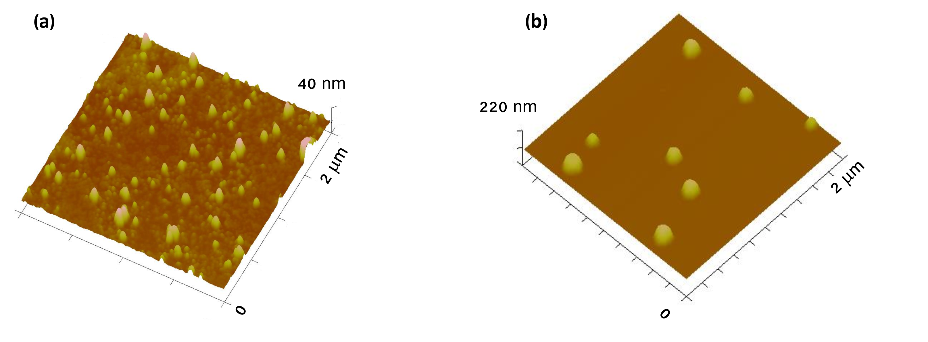
2.2.3. Atomic Force Microscopy (AFM)
2.2.4. Absorbance Spectrum
2.2.5. Cytotoxicity on the Saccharomyces cerevisiae Model
2.2.6. In Vitro Assays on HaCat and B16F10 Cell Lines
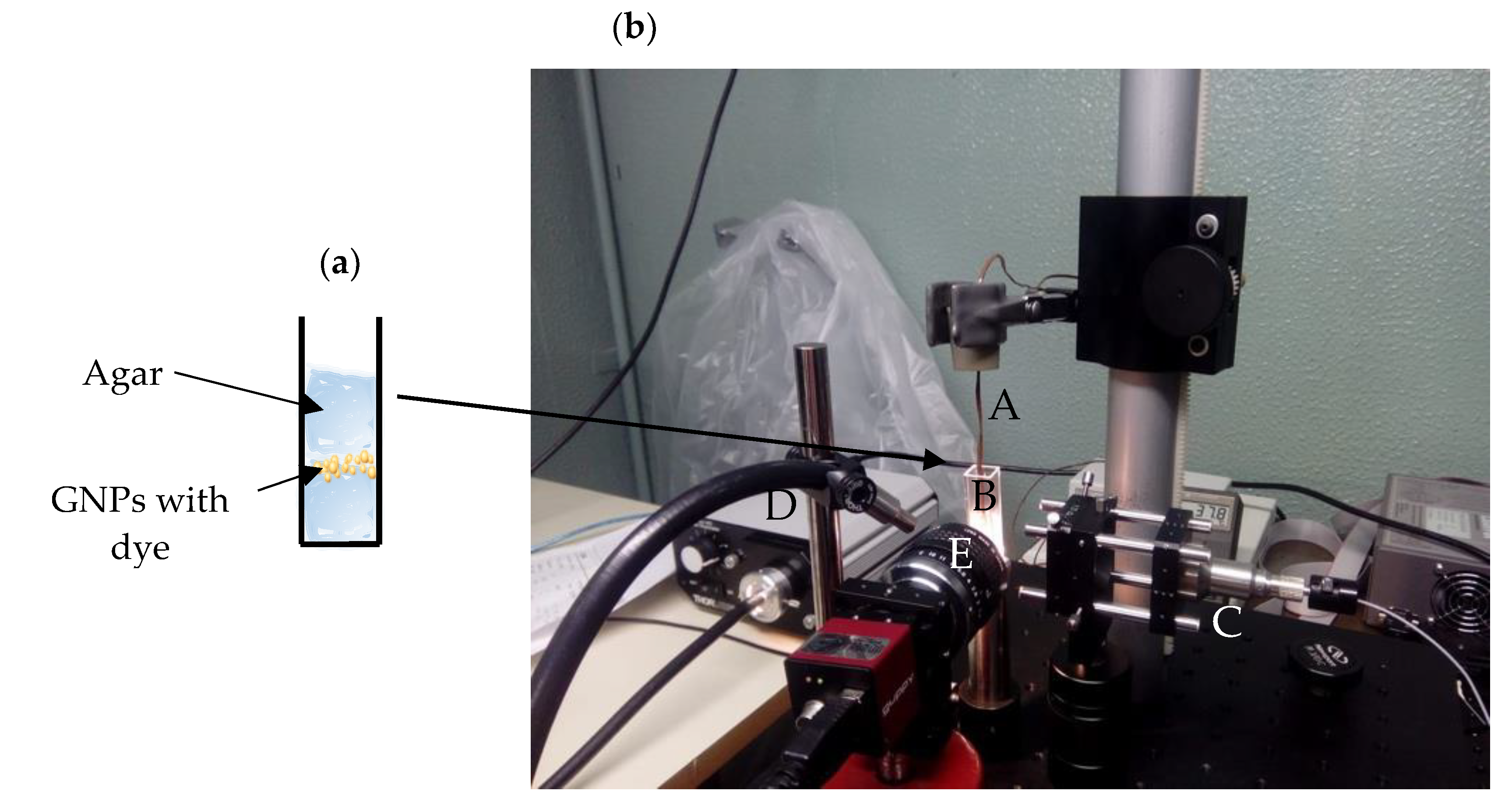
2.2.7. In Vitro Thermal Activation Using the Phantom Model
2.2.8. Statistical Analysis
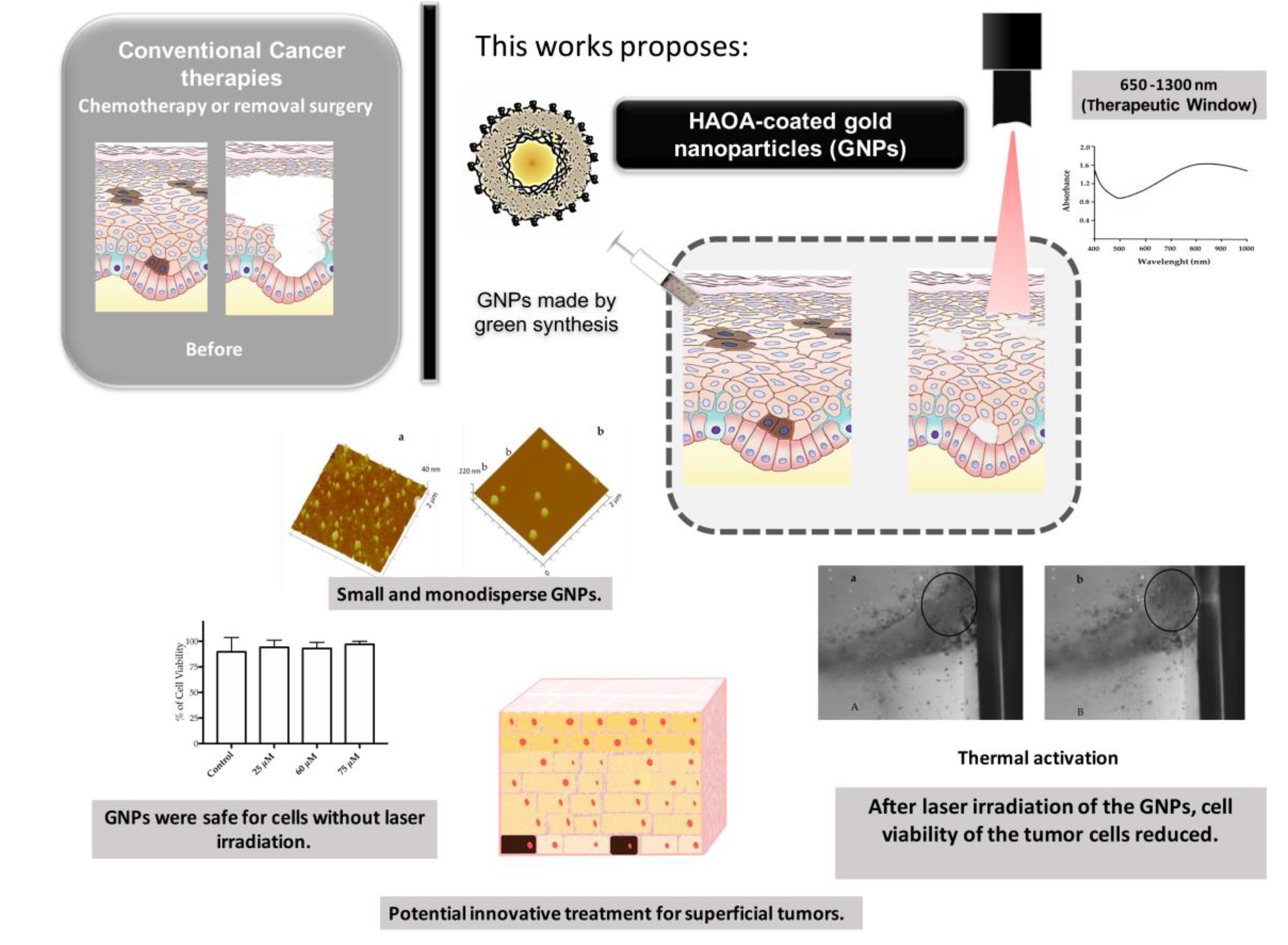
3. Results
3.1. Physical and Optical Properties of GNPs
3.1.1. Size, Polydispersity Index and Zeta Potential Measurements
3.1.2. Atomic Force Microscopy (AFM)
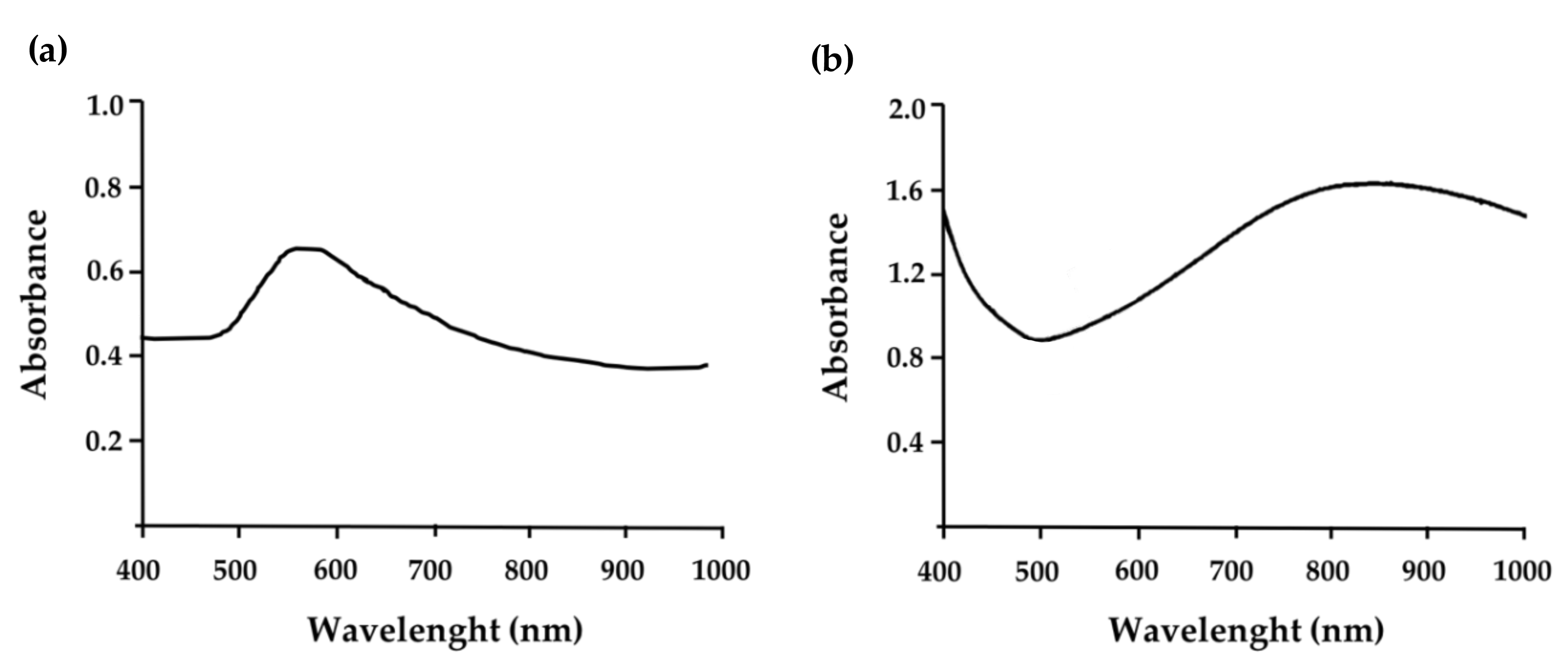
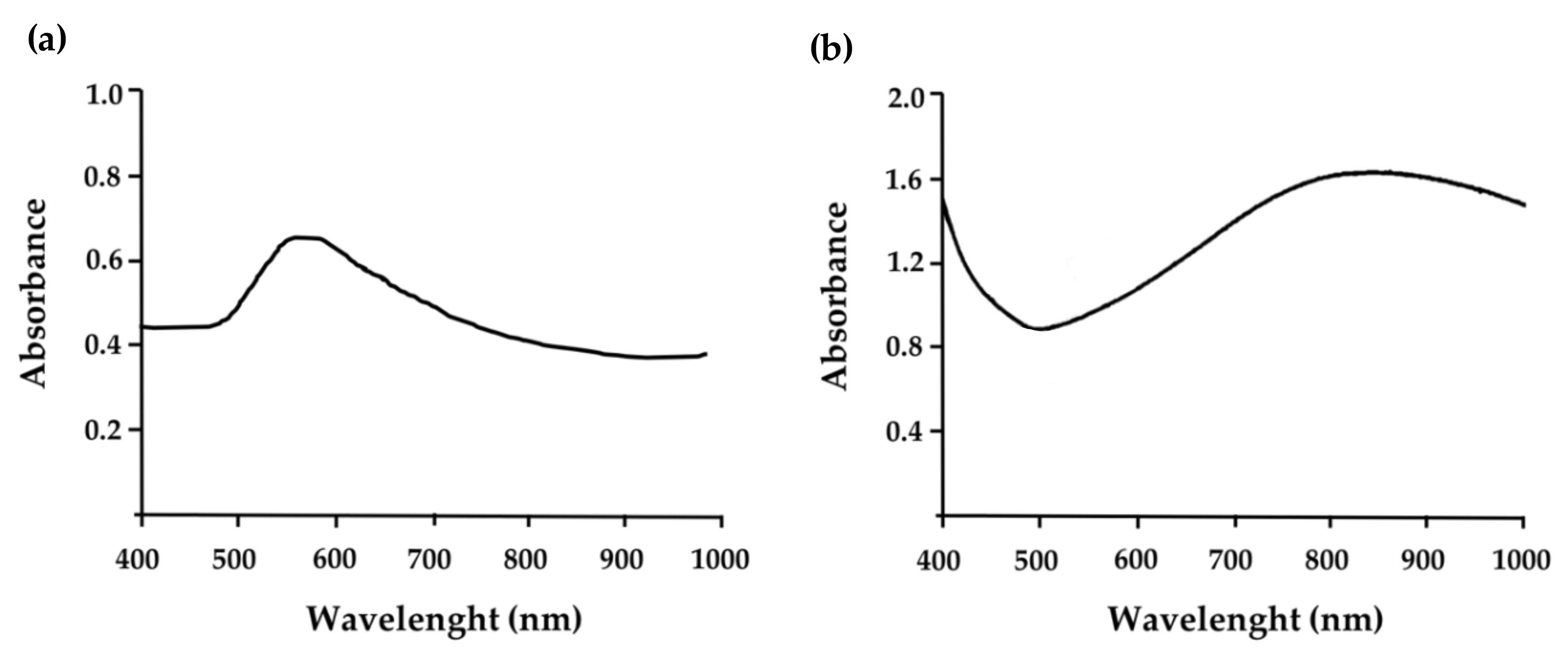
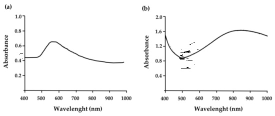
3.1.3. Absorbance Spectrum
3.2. Cytotoxicity Studies of GNPs
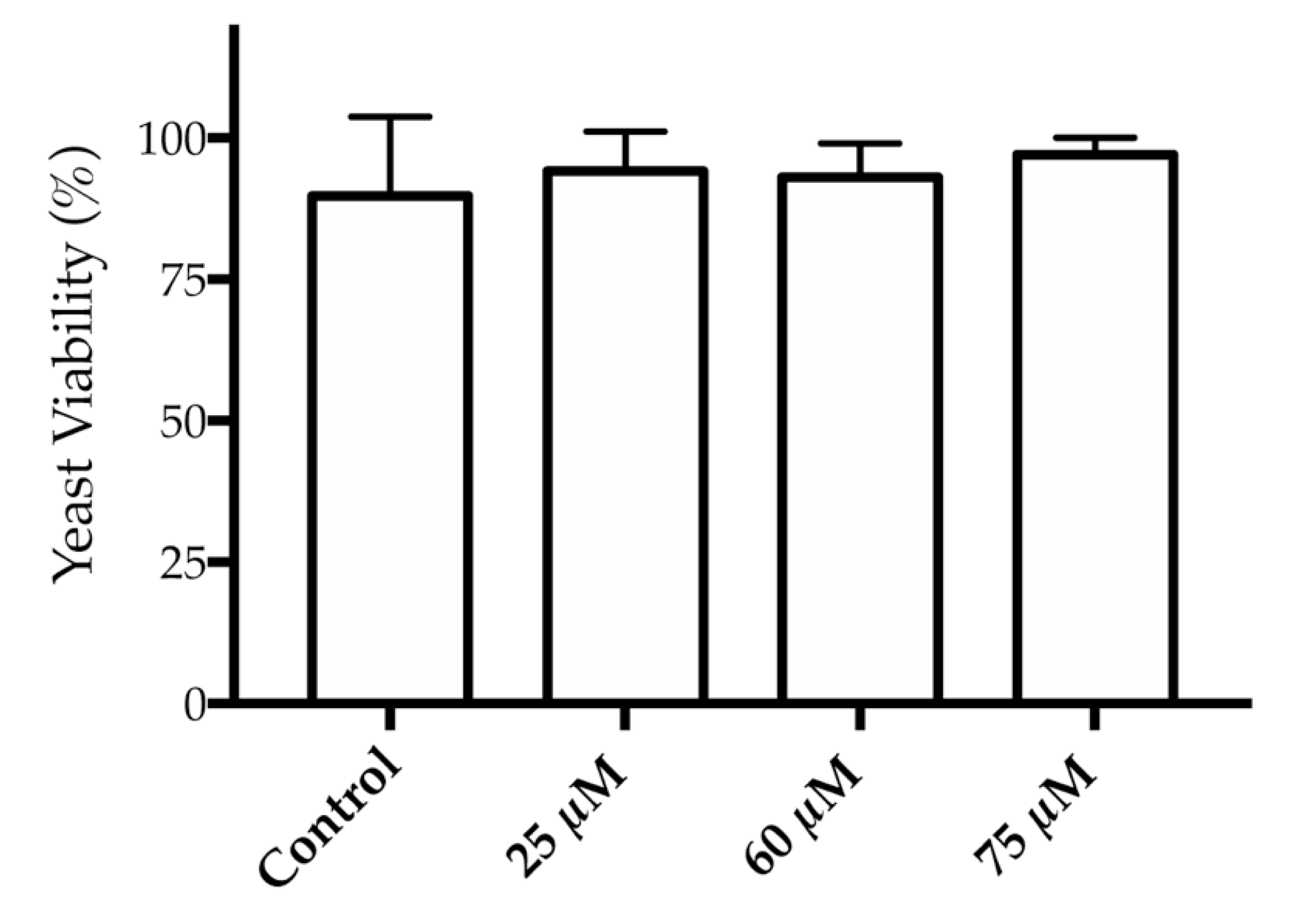
3.2.1. Evaluation on a Saccharomyces cerevisiae Model
3.2.2. Evaluation on HaCat and B16F10 Cell Lines
Evaluation on HaCat and B16F10 Cell Lines after GNPs’ Activation with Laser
3.3. Thermal Activation
4. Discussion
5. Conclusions
Supplementary Materials
Author Contributions
Funding
Acknowledgments
Conflicts of Interest
References
- López-Gómez, M.; Malmierca, E.; Górgolas, M.; Casado, E. Cancer in developing countries: The next most preventable pandemic. The global problem of cancer. Crit. Rev. Oncol. Hematol. 2013, 88, 117–122. [Google Scholar] [CrossRef] [PubMed]
- Centre International de Recherche sur le Cancer. Rapport Biennal 2016/2017; Centre International de Recherche sur le Cancer: Lyon, France, 2018. [Google Scholar]
- Wark, P.A.; Peto, J. Cancer Epidemiology. Int. Encycl. Public Heal. 2008, 1, 416–424. [Google Scholar]
- Siegel, R.L.; Miller, K.D.; Jemal, A. Cancer statistics, 2020. CA Cancer J. Clin. 2020, 70, 7–30. [Google Scholar] [CrossRef] [PubMed]
- National Comprehensive Cancer Network (NCCN). NCCN Guidelines for Patients-Melanoma; National Comprehensive Cancer Network (NCCN): Fort Washington, PA, USA, 2018. [Google Scholar]
- Marks, J.G.; Miller, J.J. Structure and Function of the Skin. In Lookingbill and Marks’ Principles of Dermatology; Elsevier Health Sciences: Amsterdam, The Netherlands, 2018; ISBN 9780323430401. [Google Scholar]
- Martínez, B.M.-A.; Martín, F.J.V.; Poveda, M.D.; Villaverde, R.M. Melanoma. Medicine 2017, 12, 1980–1989. [Google Scholar]
- American Cancer Society American Cancer Society. Facts & Figures 2019. Am. Cancer Soc. 2019, 76. Available online: https://www.cancer.org/content/dam/cancer-org/research/cancer-facts-and-statistics/annual-cancer-facts-and-figures/2019/cancer-facts-and-figures-2019.pdf (accessed on 20 July 2020).
- Quintanilla-Dieck, M.J.; Bichakjian, C.K. Management of Early-Stage Melanoma. Facial Plast. Surg. Clin. N. Am. 2019, 27, 35–42. [Google Scholar] [CrossRef]
- Kyrgidis, A. Melanoma: Epidemiology. In Cutaneous Melanoma: A Pocket Guide for Diagnosis and Management; Academic Press: Cambridge, MA, USA, 2017; Volume 3, pp. 153–166. ISBN 9780128040003. [Google Scholar]
- Kyrgidis, A. Risk factors. In Cutaneous Melanoma: A Pocket Guide for Diagnosis and Management; Academic Press: Cambridge, MA, USA, 2017; ISBN 9780128040003. [Google Scholar]
- Garbe, C.; Peris, K.; Hauschild, A.; Saiag, P.; Middleton, M.; Bastholt, L.; Grob, J.-J.; Malvehy, J.; Newton-Bishop, J.; Stratigos, A.J.; et al. Diagnosis and treatment of melanoma. European consensus-based interdisciplinary guideline–Update 2016. Eur. J. Cancer 2016, 63, 201–217. [Google Scholar] [CrossRef]
- Pautu, V.; Leonetti, D.; Lepeltier, E.; Clere, N.; Passirani, C. Nanomedicine as a potent strategy in melanoma tumor microenvironment. Pharmacol. Res. 2017, 126, 31–53. [Google Scholar] [CrossRef] [Green Version]
- Silva, C.O.; Rijo, P.R.; Molpeceres, J.; Ascensão, L.; Roberto, A.; Fernandes, A.S.; Gomes, R.; Coelho, J.M.P.; Gabriel, A.; Vieira, P.; et al. Bioproduction of gold nanoparticles for photothermal therapy. Ther. Deliv. 2016, 7, 287–304. [Google Scholar] [CrossRef]
- Reis, C.P.; Figueiredo, I.V.; Carvalho, R.A.; Jones, J.; Nunes, P.; Soares, A.F.; Silva, C.F.; Ribeiro, A.J.; Veiga, F.J.; Damgé, C.; et al. Toxicological assessment of orally delivered nanoparticulate insulin. Nanotoxicology 2008, 2, 205–217. [Google Scholar] [CrossRef]
- Bahrami, B.; Hojjat-Farsangi, M.; Mohammadi, H.; Anvari, E.; Ghalamfarsa, G.; Yousefi, M.; Jadidi-Niaragh, F. Nanoparticles and targeted drug delivery in cancer therapy. Immunol. Lett. 2017, 190, 64–83. [Google Scholar] [CrossRef]
- Bao, Z.; Liu, X.; Liu, Y.; Liu, H.; Zhao, K. Near-infrared light-responsive inorganic nanomaterials for photothermal therapy. Asian J. Pharm. Sci. 2016, 11, 349–364. [Google Scholar] [CrossRef] [Green Version]
- Gomes, R.; Coelho, J.; Gabriel, A.; Vieira, P.; Silva, C.; Reis, C. Wavefront shaping using a deformable mirror for focusing inside optical tissue phantoms. In Second International Conference on Applications of Optics and Photonics; International Society for Optics and Photonics: Washington, DC, USA, 2014; Volume 9286, p. 92863Z. [Google Scholar]
- Huang, X.; El-Sayed, M.A. Plasmonic photo-thermal therapy (PPTT). Alex. J. Med. 2011, 47, 1–9. [Google Scholar] [CrossRef] [Green Version]
- Rohiman, A.; Anshori, I.; Surawijaya, A.; Idris, I. Study of colloidal gold synthesis using Turkevich method. In AIP Conference Proceedings; American Institute of Physics: College Park, MD, USA, 2011; Volume 1415, pp. 39–42. [Google Scholar]
- Brust, M.; Walker, M.; Bethell, D.; Schiffrin, D.J.; Whyman, R. Synthesis of Thiol-derivatised Gold Nanoparticles in a Two-phase Liquid-Liquid System. J. Chem. Soc. Chem. Commun. 1994, 7, 801–802. [Google Scholar] [CrossRef]
- Khan, Z.; Singh, T.; Hussain, J.I.; Hashmi, A.A. Au(III)-CTAB reduction by ascorbic acid: Preparation and characterization of gold nanoparticles. Colloids Surf. B Biointerfaces 2013, 104, 11–17. [Google Scholar] [CrossRef] [PubMed]
- Huang, H.C.; Yang, Y.; Nanda, A.; Koria, P.; Rege, K. Synergistic administration of photothermal therapy and chemotherapy to cancer cells using polypeptide-based degradable plasmonic matrices. Nanomedicine 2011, 6, 459–473. [Google Scholar] [CrossRef] [PubMed] [Green Version]
- Roberto, A. A high-throughput screening method for general cytotoxicity part I Chemical toxicity. Rev. Lusófona Ciências E Tecnol. Saúde 2005, 2, 100–105. [Google Scholar]
- Silva, C.O.; Petersen, S.B.; Reis, C.P.; Rijo, P.; Molpeceres, J.; Fernandes, A.S.; Gonçalves, O.; Gomes, A.C.; Correia, I.; Vorum, H.; et al. EGF functionalized polymer-coated gold nanoparticles promote EGF photostability and EGFR internalization for photothermal therapy. PLoS ONE 2016, 11, 1–29. [Google Scholar] [CrossRef] [Green Version]
- Santos-Rebelo, A.; Kumar, P.; Pillay, V.; Choonara, Y.E.; Eleutério, C.; Figueira, M.; Viana, A.S.; Ascensão, L.; Molpeceres, J.; Rijo, P.; et al. Development and mechanistic insight into the enhanced cytotoxic potential of parvifloron D albumin nanoparticles in EGFR-overexpressing pancreatic cancer cells. Cancers 2019, 11. [Google Scholar] [CrossRef] [Green Version]
- Shang, L.; Nienhaus, K.; Nienhaus, G.U. Engineered nanoparticles interacting with cells: Size matters. J. Nanobiotechnol. 2014, 12, 5. [Google Scholar] [CrossRef] [Green Version]
- Huang, X.; El-Sayed, M.A. Gold nanoparticles: Optical properties and implementations in cancer diagnosis and photothermal therapy. J. Adv. Res. 2010, 1, 13–28. [Google Scholar] [CrossRef] [Green Version]
- Greish, K. Enhanced Permeability and Retention (EPR) Effect for Anticancer Nanomedicine Drug Targeting. In Methods in Molecular Biology; Stephen R. Grobmyer, Brij M. Moudgil, Ed.; Humana Press: Tortowa, NJ, USA, 2010; Volume 624, pp. 25–37. [Google Scholar]
- Danaei, M.; Dehghankhold, M.; Ataei, S.; Hasanzadeh Davarani, F.; Javanmard, R.; Dokhani, A.; Khorasani, S.; Mozafari, M.R. Impact of particle size and polydispersity index on the clinical applications of lipidic nanocarrier systems. Pharmaceutics 2018, 10, 57. [Google Scholar] [CrossRef] [PubMed] [Green Version]
- International Organization for Standardization. International Standard-ISO 22412, 2nd ed.; International Organization for Standardization: Geneva, Switzerland, 2017; pp. 1–34. [Google Scholar]
- Stockerta, J.C.; Blázquez-Castroa, A.; Cañete, M.; Horobinb, R.W.; Villanueva, Á. MTT assay for cell viability: Intracellular localization of the formazan product is in lipid droplets. Acta Histochem. 2012, 114, 785–796. [Google Scholar] [CrossRef] [PubMed]
- Mummert, M.E.; Mummert, D.I.; Ellinger, L.; Takashima, A. Functional roles of hyaluronan in B16-F10 melanoma growth and experimental metastasis in mice. Mol. Cancer Ther. 2003, 2, 295–300. [Google Scholar]
- Upponi, J.R.; Torchilin, V.P. Photodynamic Therapy for Cancer: Principles, Clinical Applications, and Nanotechnological Approaches. In Nano-Oncologicals: New Targeting and Delivery Approaches; Springer: Cham, Switzerland, 2014; pp. 123–160. ISBN 9783319080833. [Google Scholar]
- Doughty, A.C.V.; Hoover, A.R.; Layton, E.; Murray, C.K.; Howard, E.W.; Chen, W.R. Nanomaterial applications in photothermal therapy for cancer. Materials 2019, 12, 779. [Google Scholar] [CrossRef] [Green Version]
- Rachel, S.; Riley, E.S.D. Gold nanoparticle-mediated photothermal therapy: Applications and opportunities for multimodal cancer treatment. Wiley Interdiscip. Rev. Nanobiotechnol. 2017, 9, e1449. [Google Scholar]
- Eskiizmir, G.; Ermertcan, A.T.; Yapici, K. Nanomaterials: Promising structures for the management of oral cancer. In Nanostructures for Oral Medicine; Elsevier Inc.: Amsterdam, The Netherlands, 2017; pp. 511–544. ISBN 9780323477215. [Google Scholar]
- Toy, R.; Peiris, P.M.; Ghaghada, K.B.; Karathanasis, E. Shaping cancer nanomedicine: The effect of particle shape on the in vivo journey of nanoparticles. Nanomedicine 2014, 9, 121–134. [Google Scholar] [CrossRef] [Green Version]
- Verma, A.; Stellacci, F. Effect of surface properties on nanoparticle-cell interactions. Small 2010, 6, 12–21. [Google Scholar] [CrossRef]
- Jindal, A.B. The effect of particle shape on cellular interaction and drug delivery applications of micro- and nanoparticles. Int. J. Pharm. 2017, 532, 450–465. [Google Scholar] [CrossRef]
- Eaton, P.; Quaresma, P.; Soares, C.; Neves, C.; de Almeida, M.P.; Pereira, E.; West, P. A direct comparison of experimental methods to measure dimensions of synthetic nanoparticles. Ultramicroscopy 2017, 182, 179–190. [Google Scholar] [CrossRef]
- Daruich De Souza, C.; Ribeiro Nogueira, B.; Rostelato, M.E.C.M. Review of the methodologies used in the synthesis gold nanoparticles by chemical reduction. J. Alloys Compd. 2019, 798, 714–740. [Google Scholar] [CrossRef]
- McDonald, T.O.; Siccardi, M.; Moss, D.; Liptrott, N.; Giardiello, M.; Rannard, S.; Owen, A. The Application of Nanotechnology to Drug Delivery in Medicine; Elsevier B.V.: Amsterdam, The Netherlands, 2015; ISBN 9780444627452. [Google Scholar]
- Huang, X.; Jain, P.K.; El-Sayed, I.H.; El-Sayed, M.A. Plasmonic photothermal therapy (PPTT) using gold nanoparticles. Lasers Med. Sci. 2008, 23, 217–228. [Google Scholar] [CrossRef]
- Huff, T.B.; Tong, L.; Zhao, Y.; Hansen, M.N.; Cheng, J.-X.; Wei, A. Hyperthermic effects of gold nanorods on tumor cells. Nanomedicine 2007, 2, 125–132. [Google Scholar] [CrossRef] [PubMed] [Green Version]
- Bettaieb, A.; Wrzal, P.K.; Averill-Bates, D.A. Hyperthermia: Cancer Treatment and Beyond. In Cancer Treatment-Conventional and Innovative Approaches; InTech: London, UK, 2013. [Google Scholar]
- Reis, C.P.; Martinho, N.; Rosado, C.; Fernandes, A.S.; Roberto, A. Design of polymeric nanoparticles and its applications as drug delivery systems for acne treatment. Drug Dev. Ind. Pharm. 2014, 40, 409–417. [Google Scholar] [CrossRef] [PubMed] [Green Version]
- Berardi, A.C.; Berardocco, M.; Gissi, C.; Maffulli, N.; Cataldi, A.; Oliva, F. Hyaluronic acid increases tendon derived cell viability and proliferation in vitro: Comparative study of two different hyaluronic acid preparations by molecular weight. Muscles Ligaments Tendons J. 2017, 7, 208. [Google Scholar] [CrossRef] [PubMed]
- Ahrens, T.; Assmann, V.; Fieber, C.; Termeer, C.C.; Herrlich, P.; Hofmann, M.; Simon, J.C. CD44 is the principal mediator of hyaluronic-acid-induced melanoma cell proliferation. J. Investig. Dermatol. 2001, 116, 93–101. [Google Scholar]


| GNPs Formulation | Mean Size ± SD (nm) | Polydispersity Index (PdI) | Mean Zeta Potential± SD (mV) |
|---|---|---|---|
| Uncoated GNPs | 159 ± 28 | 0.275 | −2 ± 1 |
| HAOA-coated GNPs | 297 ± 3 | 0.438 | −19 ± 3 |
| HAOA-Coated GNPs Concentration | B16F10 Cells Viability (%) | HaCat Cells Viability (%) |
|---|---|---|
| 5 μM | 84 ± 5 | 95 ± 10 |
| 30 μM | 77 ± 9 | 100 ± 5 |
| 60 μM | 67 ± 5 | 109 ± 7 |
| Samples/Treatment | HaCat Cells | B16F10 Cells |
|---|---|---|
| Cell Viability (%) | Cell Viability (%) | |
| Only HAOA-coated GNPs at 5 μM | 93 ± 10 | 88 ± 5 |
| Only laser | 88 ± 6 | 94 ± 4 |
| HAOA-coated GNPs at 5 μM plus laser | 86 ± 4 | 74 ± 6 |
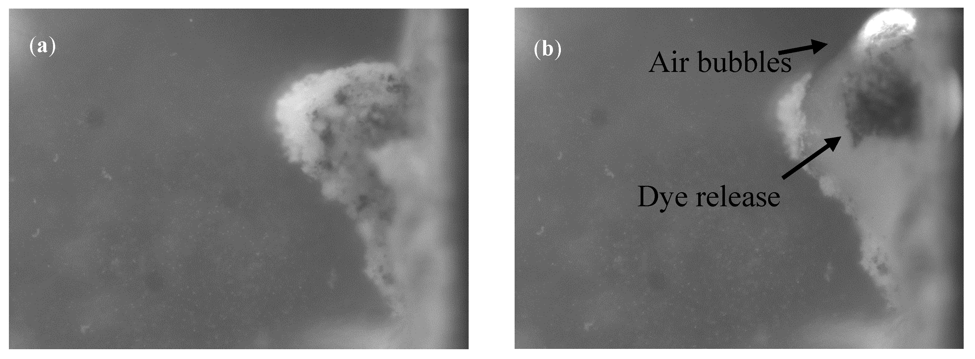
| Dye | Phantom Macroscopic Aspect | Before Activation | After Laser Activation |
|---|---|---|---|
| Hydrophilic(Blue methylene) |  |  |  |
| Lipophilic(Sudan III) |  |  |  |
© 2020 by the authors. Licensee MDPI, Basel, Switzerland. This article is an open access article distributed under the terms and conditions of the Creative Commons Attribution (CC BY) license (http://creativecommons.org/licenses/by/4.0/).
Share and Cite
Lopes, J.; Coelho, J.M.P.; Vieira, P.M.C.; Viana, A.S.; Gaspar, M.M.; Reis, C. Preliminary Assays towards Melanoma Cells Using Phototherapy with Gold-Based Nanomaterials. Nanomaterials 2020, 10, 1536. https://doi.org/10.3390/nano10081536
Lopes J, Coelho JMP, Vieira PMC, Viana AS, Gaspar MM, Reis C. Preliminary Assays towards Melanoma Cells Using Phototherapy with Gold-Based Nanomaterials. Nanomaterials. 2020; 10(8):1536. https://doi.org/10.3390/nano10081536
Chicago/Turabian StyleLopes, Joana, João Miguel Pinto Coelho, Pedro Manuel Cardoso Vieira, Ana Silveira Viana, Maria Manuela Gaspar, and Catarina Reis. 2020. "Preliminary Assays towards Melanoma Cells Using Phototherapy with Gold-Based Nanomaterials" Nanomaterials 10, no. 8: 1536. https://doi.org/10.3390/nano10081536