

Screening of Potential α-Glucosidase Inhibitors from the Roots and Rhizomes of Panax Ginseng by Affinity Ultrafiltration Screening Coupled with UPLC-ESI-Orbitrap-MS Method

Abstract
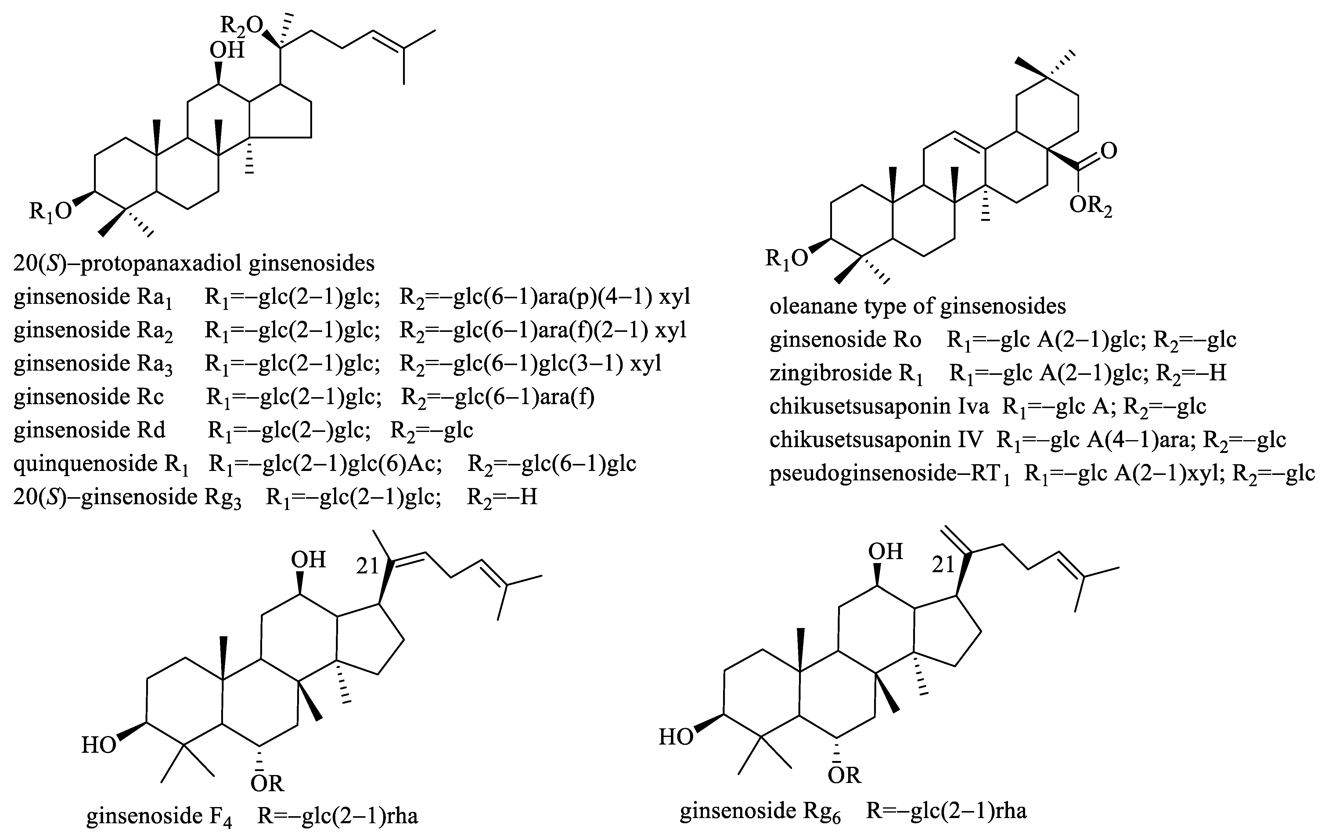
:
1. Introduction
2. Results and Discussion
2.1. Selection of α-Glucosidase Inhibitors by Affinity Ultrafiltration Screening–LC-UPLC-ESI-Orbitrap-MS
2.2. Molecular Docking of α-Glucosidase and Ligands
2.3. α-Glucosidase Inhibitory Activity of Ligands
3. Materials and Methods
3.1. Samples, Reference Standards, and Reagents
3.2. Sample Preparations
3.3. Screening of α-Glucosidase Inhibitors from Panax Gingseng by Ultrafiltration-ESI-Orbitrap-MS
3.3.1. Affinity Ultrafiltration Screening
3.3.2. UPLC-ESI-Orbitrap-MS Analysis
3.4. Data Process
- ①
- Extracting all compounds in each specimen
- ②
- Calculating the peak area ration of each compound and t-test
- ③
- Characterizing the structures of ligands
3.5. Molecular Docking of α-Glucosidase and Ligands
3.6. α-Glucosidase Inhibitory Activity Assay
4. Conclusions
Supplementary Materials
Author Contributions
Funding
Institutional Review Board Statement
Informed Consent Statement
Data Availability Statement
Acknowledgments
Conflicts of Interest
References
- Li, M.; Wang, X.; Wang, Y.; Bao, S.C.; Chang, Q.; Liu, L.L.; Zhang, S.; Sun, L.W. Strategies for remodeling the tumor microenvironment using active ingredients of ginseng-A promising approach for cancer therapy. Front. Pharmacol. 2021, 12, 797634. [Google Scholar] [CrossRef] [PubMed]
- Hao, M.Q.; Ding, C.B.; Peng, X.J.; Chen, H.Y.; Dong, L.; Zhang, Y.; Chen, X.Y.; Liu, W.C.; Luo, Y.Q. Ginseng under forest exerts stronger anti-aging effects compared to garden ginseng probably via regulating PI3K/AKT/mTOR pathway, SIRT1/NF-κB pathway and intestinal flora. Phytomedicine 2022, 105, 154365. [Google Scholar] [CrossRef] [PubMed]
- Hyun, S.H.; Kim, S.W.; Seo, H.W.; Youn, S.H.; Kyung, J.S.; Lee, Y.Y.; In, G.; Park, C.K.; Han, C.K. Physiological and pharmacological features of the non-saponin components in Korean Red Ginseng. J. Ginseng Res. 2020, 44, 527–537. [Google Scholar] [CrossRef] [PubMed]
- Sung, W.S.; Kang, H.R.; Jung, C.Y.; Park, S.S.; Lee, S.H.; Kim, E.J. Efficacy of Korean red ginseng (Panax ginseng) for middle-aged and moderate level of chronic fatigue patients: A randomized, double-blind, placebo-controlled trial. Complement. Ther. Med. 2020, 48, 102246. [Google Scholar] [CrossRef] [PubMed]
- Chen, L.X.; Qi, Y.L.; Qi, Z.; Gao, K.; Gong, R.Z.; Shao, Z.J.; Liu, S.X.; Li, S.S.; Sun, Y.S. A comparative study on the effects of different parts of panax ginseng on the immune activity of cyclophosphamide-induced immunosuppressed mice. Molecules 2019, 24, 1096. [Google Scholar] [CrossRef] [PubMed] [Green Version]
- Fang, X.X.; Zhang, X.Y.; Zhang, Y.G.; Zhang, X.; Shan, M.Y.; Guan, S.G.; Qiu, Z.D.; Zhu, D.F.; Luo, H.M. Exploring the potential of ginseng glycoprotein to improve learning and memory in mice via Notch signaling pathway and structural analysis using multi-information fusion based on liquid chromatography-mass spectrometry. J. Ethnopharmacol. 2023, 303, 115978. [Google Scholar] [CrossRef] [PubMed]
- Yao, Y.Q.; Hu, S.; Zhang, C.X.; Zhou, Q.; Wang, H.; Yang, Y.; Liu, C.; Ding, H.Y. Ginsenoside Rd attenuates cerebral ischemia/reperfusion injury by exerting an anti-pyroptotic effect via the miR-139-5p/FoxO1/Keap1/Nrf2 axis. Int. Immunopharmacol. 2022, 105, 108582. [Google Scholar] [CrossRef]
- Mostafa, R.E.; Shaffie, N.M.; Allam, R.M. Panax Ginseng alleviates thioacetamide-induced liver injury in ovariectomized rats: Crosstalk between inflammation and oxidative stress. PLoS ONE 2021, 16, e0260507. [Google Scholar] [CrossRef]
- Jin, D.; Zhang, Y.Q.; Zhang, Y.H.; Duan, L.Y.; Zhou, R.R.; Duan, Y.Y.; Sun, Y.T.; Lian, F.M.; Tong, X.L. Panax Ginseng C.A. Mey. as Medicine: The Potential Use of Panax Ginseng C.A. Mey. as a Remedy for Kidney Protection from a Pharmacological Perspective. Front. Pharmacol. 2021, 12, 734151. [Google Scholar] [CrossRef]
- Lu, J.; Wang, X.; Wu, A.X.; Cao, Y.; Dai, X.L.; Liang, Y.D.; Li, X.F. Ginsenosides in central nervous system diseases: Pharmacological actions, mechanisms, and therapeutics. Phytother. Res. 2022, 36, 1523–1544. [Google Scholar] [CrossRef]
- Zhou, P.; Xie, W.J.; He, S.B.; Sun, Y.F.; Meng, X.B.; Sun, G.B.; Sun, X.B. Ginsenoside Rb1 as an anti-diabetic agent and its underlying mechanism analysis. Cells 2019, 8, 204. [Google Scholar] [CrossRef] [PubMed] [Green Version]
- Park, S.J.; Nam, J.; Ahn, C.W.; Kim, Y.S. Anti-diabetic properties of different fractions of Korean red ginseng. J. Ethnopharmacol. 2019, 236, 220–230. [Google Scholar] [CrossRef] [PubMed]
- Yuan, H.D.; Quan, H.Y.; Jung, M.S.; Kim, S.J.; Huang, B.; Kim, D.Y.; Chung, S.H. Anti-Diabetic Effect of Pectinase-Processed Ginseng Radix (GINST) in High Fat Diet-Fed ICR Mice. J. Ginseng Res. 2011, 35, 308–314. [Google Scholar] [CrossRef] [PubMed] [Green Version]
- Hong, Y.J.; Kim, N.; Lee, K.; Sonn, C.H.; Lee, J.E.; Kim, S.T.; Baeg, I.H.; Lee, K.M. Korean red ginseng (Panax ginseng) ameliorates type 1 diabetes and restores immune cell compartments. J. Ethnopharmacol. 2012, 144, 225–233. [Google Scholar] [CrossRef] [PubMed]
- Peng, M.; Wang, L.M.; Su, H.; Zhang, L.; Yang, Y.; Sun, L.; Wu, Y.; Ran, L.; Liu, S.D.; Yin, M.; et al. Ginsenoside Rg1 improved diabetes through regulating the intestinal microbiota in high-fat diet and streptozotocin-induced type 2 diabetes rats. J. Food Biochem. 2022, 46, e14321. [Google Scholar] [CrossRef] [PubMed]
- Lai, D.M.; Tu, Y.K.; Liu, I.M.; Chen, P.F.; Cheng, J.T. Mediation of β-endorphin by ginsenoside Rh2 to lower plasma glucose in streptozotocin-induced diabetic rats. Planta Med. 2006, 72, 9–13. [Google Scholar] [CrossRef]
- Wang, C.W.; Su, S.C.; Huang, S.F.; Huang, Y.C.; Chan, F.N.; Kuo, Y.H.; Hung, M.W.; Lin, H.C.; Chang, W.L.; Chang, T.C. An essential role of cAMP response element binding protein in ginsenoside Rg1-mediated inhibition of Na+/glucose cotransporter 1 gene expression. Mol. Pharmacol. 2015, 88, 1072–1083. [Google Scholar] [CrossRef] [Green Version]
- Gao, Z.; Li, Q.; Wu, X.; Zhao, X.; Zhao, L.; Tong, X. New Insights into the mechanisms of Chinese herbal products on diabetes: A focus on the bacteriamucosal immunity-inflammation-diabetes axis. J. Immunol. Res. 2017, 2017, 1813086. [Google Scholar] [CrossRef] [Green Version]
- Shang, W.B.; Guo, C.; Zhao, J.; Yu, X.Z.; Zhang, H. Ginsenoside Rb1 upregulates expressions of GLUTs to promote glucose consumption in adiopcytes. China J. Chin. Mater. Med. 2014, 39, 4448–4452. [Google Scholar]
- Liu, Q.; Zhang, F.G.; Zhang, W.S.; Pan, A.; Yang, Y.L.; Liu, J.F.; Li, P.; Liu, B.L.; Qi, L.W. Ginsenoside Rg1 inhibits glucagon-induced hepatic gluconeogenesis through Akt-FoxO1 interaction. Theranostics 2017, 7, 4001–4012. [Google Scholar] [CrossRef] [PubMed] [Green Version]
- Meng, F.; Su, X.; Li, W.; Zheng, Y. Ginsenoside Rb3 Strengthens the hypoglycemic effect through AMPK for inhibition of hepatic gluconeogenesis. Exp. Ther. Med. 2017, 13, 2551–2557. [Google Scholar] [CrossRef] [PubMed] [Green Version]
- Wang, H.P.; Yang, X.B.; Yang, X.W.; Liu, J.X.; Xu, W.; Zhang, Y.B.; Zhang, L.X.; Wang, Y.P. Ginsenjilinol, a new protopanaxatriol-type saponin with inhibitory activity on LPS-activated NO production in macrophage RAW 264.7 cells from the roots and rhizomes of Panax ginseng. J. Asian Nat. Prod. Res. 2013, 15, 579–587. [Google Scholar] [CrossRef] [PubMed]
- Liu, R.J.; Kool, J.; Jian, J.Y.; Wang, J.C.; Zhao, X.L.; Jiang, Z.J.; Zhang, T.T. Rapid screening α-Glucosidase inhibitors from natural products by at-line nanofractionation with parallel mass spectrometry and bioactivity assessment. J. Chromatogr. A 2021, 1635, 461740. [Google Scholar] [CrossRef] [PubMed]
- Zhao, C.F.; Liu, Q.; Cong, D.L.; Zhang, H.; Yu, J.J.; Jiang, Y.; Cui, X.Y.; Sun, J.M. Screening and determination for potential α-glucosidase inhibitory constituents from Dalbergia odorifera T. Chen using ultrafiltration-LC/ESI-MSn. Biomed. Chromatogr. 2013, 27, 1621–1629. [Google Scholar] [CrossRef]
- Zhou, H.; Xing, J.P.; Liu, S.; Song, F.R.; Cai, Z.W.; Pi, Z.F.; Liu, Z.Q.; Liu, S.Y. Screening and determination for potential α-Glucosidase inhibitors from leaves of acanthopanax senticosus Harms by using UF-LC/MS and ESI-MSn. Phytochem. Anal. 2012, 23, 315–323. [Google Scholar] [CrossRef]
- Zhou, X.L.; Liang, J.S.; Zhang, Y.; Zhao, H.D.; Guo, Y.; Shi, S.Y. Separation and purification of α-Glucosidase inhibitors from Polygonatum odoratum by stepwise high-speed counter-current chromatography combined with Sephadex LH-20 chromatography target-guided by ultrafiltration–HPLC screening. J. Chromatogr. B Analyt. Technol. Biomed. Life Sci. 2015, 15, 149–154. [Google Scholar] [CrossRef]
- Wu, B.; Song, H.P.; Zhou, X.; Liu, X.G.; Gao, W.; Dong, X.; Li, H.J.; Li, P.; Yang, H. Screening of minor bioactive compounds from herbal medicines by in silico docking and the trace peak exposure methods. J. Chromatogr. A 2016, 4, 91–99. [Google Scholar] [CrossRef]
- Li, H.L.; Song, F.R.; Xing, J.P.; Tsao, R.; Liu, Z.Q.; Liu, S.Y. Screening and structural characterization of α-Glucosidase inhibitors from Hawthorn leaf flavonoids extract by ultrafiltration LC-DAD-MSn and SORI-CID FTICR MS. J. Am. Soc. Mass Spectrom. 2009, 20, 1496–1503. [Google Scholar] [CrossRef] [Green Version]
- Auiewiriyanukul, W.; Saburi, W.; Kato, K.; Yao, M.; Mori, H. Function and structure of GH13_31 α-glucosidase with high α-(1→4)-glucosidic linkage specificity and transglucosylation activity. FEBS Lett. 2018, 592, 2268–2281. [Google Scholar] [CrossRef] [Green Version]
- Jiang, W.Y.; Kan, H.; Li, P.D.; Liu, S.; Liu, Z.Y. Screening and structural characterization of potential α-glucosidase inhibitors from Radix Astragali flavonoids extract by ultrafiltration LC-DAD-ESI-MSn. Anal. Methods 2015, 7, 123–128. [Google Scholar] [CrossRef]


| No. | tR (min) | Molecular Formula | Calc. MW | PAR Value | Compound Name | Product Ions |
|---|---|---|---|---|---|---|
| Compounds of reference standards commercially available | ||||||
| R1 | 24.01 | C42H66O14 | 794.4453 a | 2.69 ± 1.12 | zingibroside R1 | 631.3844 [M-H-Glc]−, 613.3746 [M-H-Glc-H2O]−, 569.3849 [M-H-Glc-H2O-CO2]−, 455.3534 [M-H-Glc-Glu A]− |
| R2 | 24.64 | C42H72O13 | 830.5026 b | 2.02 ± 0.72 | 20(S)-ginsenoside Rg3 | 621.4362 [M-H-Glc]−, 459.3860 [M-H-2Glc]− |
| R3 | 23.34 | C42H70O12 | 812.4924 b | 1.80 ± 0.18 | ginsenoside Rg6 | 619.4229 [M-H-Rha]−, 457.3707 [M-H-Rha-Glc]− |
| R4 | 19.44 | C47H74O18 | 926.4856 a | 1.68 ± 0.27 | pseudoginsenoside-RT1 | 793.4344 [M-H-Xyl]−, 763.4279 [M-H-Glc]−, 613.3762 [M-H-Xyl-Glc-H2O]−, 455.3551 [M-H-Xyl-Glc-Glu A]− |
| R5 | 19.54 | C47H74O18 | 926.4874 a | 1.59 ± 0.06 | chikusetsusaponin IV | 793.4389 [M-H-Ara]−, 613.3696 [M-H-Ara-Glc-H2O]−, 455.3551 [M-H-Ara-Glc-Glu A]− |
| R6 | 20.24 | C42H66O14 | 794.4451 a | 1.57 ± 0.29 | chikusetsusaponin Iva | 631.3849 [M-H-Glc]−, 455.3526 [M-H-Glc-Glu A]− |
| R7 | 20.44 | C48H82O18 | 992.5542 b | 1.57 ± 0.17 | ginsenoside Rd | 783.4885 [M-H-Glc]−, 621.4365 [M-H-2Glc]−, 459.3841 [M-H-3Glc]− |
| R8 | 18.68 | C48H76O19 | 956.4981 a | 1.52 ± 0.06 | ginsenoside Ro | 793.4385 [M-H-Glc]−, 731.4386 [M-H-Glc-CO2-H2O]−, 613.3731 [M-H-2Glc-H2O]−, 569.3847 [M-H-2Glc-H2O-CO2]−, 455.3528 [M-H-2Glc-Glu A]− |
| R9 | 23.63 | C42H70O12 | 812.4924 b | 1.35 ± 0.13 | ginsenoside F4 | 619.4215 [M-H-Rha]−, 457.3696 [M-H-Rha-Glc]− |
| R10 | 18.52 | C58H98O26 | 1256.6394 b | 1.33 ± 0.15 | ginsenoside Ra1 | 1077.5852 [M-H-Xyl]−, 945.5426 [M-H-Xyl-Ara(p)]−, 783.4900 [M-H-Xyl-Ara(p)-Glc]−, 621.4370 [M-H-Xyl-Ara(p)-2Glc]−, 459.3854 [M-H-Xyl-Ara(p)-3Glc]− |
| R11 | 17.67 | C58H98O26 | 1256.6392 b | 1.25 ± 0.11 | ginsenoside Ra2 | 1077.5844 [M-H-Xyl]−, 945.5393 [M-H-Xyl-Ara(f)]−, 783.4897 [M-H-Xyl-Ara(f)-Glc]−, 621.4370 [M-H-Xyl-Ara(f)-2Glc]−, 459.3846 [M-H-Xyl-Ara(f)-3Glc]− |
| R12 | 19.77 | C56H94O24 | 1196.6185 b | 1.23 ± 0.11 | quinquenoside R1 | 1107.5973 [M-H-Ac]−, 945.5438 [M-H-Ac-Glc]−, 783.4898 [M-H-Ac-2Glc]−, 621.4370 [M-H-Ac-3Glc]−, 459.3844 [M-H-Ac-4Glc]− |
| R13 | 17.85 | C59H100O27 | 1286.6503 b | 1.15 ± 0.11 | ginsenoside Ra3 | 1107.5931 [M-H-Glc]−, 945.5460 [M-H-Glc-Xyl]−, 783.4923 [M-H-2Glc-Xyl]−, 621.4380 [M-H-3Glc-Xyl]−, 459.3846 [M-H-4Glc-Xyl]− |
| R14 | 18.45 | C53H90O22 | 1124.5962 b | 1.14 ± 0.10 | ginsenoside Rc | 945.5475 [M-H-Ara(f)]−, 783.4896 [M-H-Ara(f)-Glc]−, 621.4403 [M-H-Ara(f)-2Glc]−, 459.3858 [M-H-Ara(f)-3Glc]− |
| Compounds of reference standards commercially unavailable | ||||||
| R15 | 17.50 | C48H76O19 | 956.4981 a | 3.81 ± 0.38 | ginsenoside Ro isomer | 793.4376 [M-H-Glc]−, 731.4376 [M-H-Glc-CO2-H2O]−, 613.3737 [M-H-2Glc-H2O]−, 569.3847 [M-H-2Glc-H2O-CO2]−, 455.3534 [M-H-2Glc-Glu A]− |
| R16 | 17.92 | C58H98O26 | 1210.6340 a | 2.38 ± 0.16 | ginsenoside Ra1 isomer/ginsenoside Ra2 isomer | 1077.5844 [M-H-Xyl]−, 945.5478 [M-H-Xyl-Ara]−, 783.4885 [M-H-Xyl-Ara-Glc]−, 621.4399 [M-H-Xyl-Ara-2Glc]−, 459.3831 [M-H-Xyl-Ara-3Glc]− |
| R17 | 23.11 | C50H84O19 | 1034.5664 b | 1.64 ± 0.13 | acetyl-ginsenoside Rd | 945.5446 [M-H-Ac]−, 783.4888 [M-H-Ac-Glc]−, 621.4320 [M-H-Ac-2Glc]−, 459.3841 [M-H-Ac-3Glc]− |
| R18 | 17.97 | C48H76O19 | 956.4982 a | 1.55 ± 0.07 | ginsenoside Ro isomer | 793.4373 [M-H-Glc]−, 731.4371 [M-H-Glc-CO2-H2O]−, 613.3723 [M-H-2Glc-H2O]−, 569.3849 [M-H-2Glc-H2O-CO2]−, 455.3546 [M-H-2Glc-Glu A]− |
| R19 | 20.44 | C52H86O19 | 1060.5427 b | 1.51 ± 0.23 | (E)-but-2-enoyl ginsenoside Rd | 945.5330 [M-H-(E)-but-2-enoyl]−, 783.4881 [M-H-(E)-but-2-enoyl-Glc]−, 621.4363 [M-H-(E)-but-2-enoyl-2Glc]−, 459.3841 [M-H-(E)-but-2-enoyl-3Glc]−, |
| R20 | 19.69 | C48H80O18 | 990.5405 b | 1.39 ± 0.24 | dehydrated-protopanaxatriol + 3Glc | 781.4766 [M-H-Glc]−, 619.4214 [M-H-2Glc]−, 457.3699 [M-H-3Glc]− |
| R21 | 20.74 | C50H84O19 | 988.5601 a | 1.39 ± 0.16 | acetyl-ginsenoside Rd | 945.5449 [M-H-Ac]−, 783.4902 [M-H-Ac-Glc]−, 621.4320 [M-H-Ac-2Glc]−, 459.3843 [M-H-Ac-3Glc]− |
| R22 | 19.50 | C58H98O26 | 1210.6350 a | 1.33 ± 0.20 | ginsenoside Ra1 isomer/ginsenoside Ra2 isomer | 1077.5856 [M-H-Xyl]−, 945.5452 [M-H-Xyl-Ara]−, 783.4890 [M-H-Xyl-Ara-Glc]−, 621.4329 [M-H-Xyl-Ara-2Glc]−, 459.3865 [M-H-Xyl-Ara-3Glc]− |
| R23 | 17.39 | C54H90O23 | 1152.5928 b | 1.31 ± 0.19 | dehydrated-protopanaxatriol + 4Glc | 943.5316 [M-H-Glc]−, 781.4628 [M-H-2Glc]−, 763.4622 [M-H-2Glc-H2O]−, 619.4229 [M-H-3Glc]−, 601.4104 [M-H-3Glc-H2O]−, 457.3698 [M-H-4Glc]− |
| R24 | 18.98 | C56H94O24 | 1150.6135 a | 1.31 ± 0.09 | quinquenoside R1 isomer | 1107.5918 [M-H-Ac]−, 945.5487 [M-H-Ac-Glc]−, 783.4916 [M-H-Ac-2Glc]−, 621.4385 [M-H-Ac-3Glc]−, 459.3836 [M-H-Ac-4Glc]− |
| No. | Compound Name | Affinity (kcal/mol) | IC50 (mM) | Inhibitory (%) |
|---|---|---|---|---|
| R1 | zingibroside R1 | −8.1 | 3.61 | - |
| R2 | 20(S)-ginsenoside Rg3 | −8.2 | - | UT c |
| R3 | ginsenoside Rg6 | −7.8 | - | 27.35% b |
| R4 | pseudoginsenoside-RT1 | −7.8 | 39.30 | - |
| R5 | chikusetsusaponin IV | −8.1 | - | 16.20% b |
| R6 | chikusetsusaponin Iva | −7.8 | 17.33 | - |
| R7 | ginsenoside Rd | −7.7 | - | UT c |
| R8 | ginsenoside Ro | −7.8 | - | 20.23% b |
| R9 | ginsenoside F4 | −8.4 | 22.13 | - |
| R10 | ginsenoside Ra1 | −8.7 | - | 16.36% b |
| R11 | ginsenoside Ra2 | −8.6 | - | 29.54% b |
| R12 | quinquenoside R1 | −7.2 | - | 25.50% a |
| R13 | ginsenoside Ra3 | −9.0 | - | 17.09% a |
| R14 | ginsenoside Rc | −7.9 | 36.83 | - |
| acarbose | −7.1 | 5.25 | - |
Disclaimer/Publisher’s Note: The statements, opinions and data contained in all publications are solely those of the individual author(s) and contributor(s) and not of MDPI and/or the editor(s). MDPI and/or the editor(s) disclaim responsibility for any injury to people or property resulting from any ideas, methods, instructions or products referred to in the content. |
© 2023 by the authors. Licensee MDPI, Basel, Switzerland. This article is an open access article distributed under the terms and conditions of the Creative Commons Attribution (CC BY) license (https://creativecommons.org/licenses/by/4.0/).
Share and Cite
Wang, H.-P.; Fan, C.-L.; Lin, Z.-Z.; Yin, Q.; Zhao, C.; Peng, P.; Zhang, R.; Wang, Z.-J.; Du, J.; Wang, Z.-B. Screening of Potential α-Glucosidase Inhibitors from the Roots and Rhizomes of Panax Ginseng by Affinity Ultrafiltration Screening Coupled with UPLC-ESI-Orbitrap-MS Method. Molecules 2023, 28, 2069. https://doi.org/10.3390/molecules28052069
Wang H-P, Fan C-L, Lin Z-Z, Yin Q, Zhao C, Peng P, Zhang R, Wang Z-J, Du J, Wang Z-B. Screening of Potential α-Glucosidase Inhibitors from the Roots and Rhizomes of Panax Ginseng by Affinity Ultrafiltration Screening Coupled with UPLC-ESI-Orbitrap-MS Method. Molecules. 2023; 28(5):2069. https://doi.org/10.3390/molecules28052069
Chicago/Turabian StyleWang, Hong-Ping, Chun-Lan Fan, Zhao-Zhou Lin, Qiong Yin, Chen Zhao, Ping Peng, Run Zhang, Zi-Jian Wang, Jing Du, and Zhi-Bin Wang. 2023. "Screening of Potential α-Glucosidase Inhibitors from the Roots and Rhizomes of Panax Ginseng by Affinity Ultrafiltration Screening Coupled with UPLC-ESI-Orbitrap-MS Method" Molecules 28, no. 5: 2069. https://doi.org/10.3390/molecules28052069