
Synthesis, Structural Elucidation, and Anti-Inflammatory Activity of a Water-Soluble Derivative of Arctiin
by
Xia Xu
1,
Xiaofeng Huang
1,
Yuedan Zheng
1,
Xiaoling Wang
1,
Jing Xie
2,
Sha Liu
2,3,* and
Kun Guo
1,* 1
College of Pharmacy, Southwest Minzu University, Chengdu 610200, China
2
School of Pharmacy, Chengdu Medical College, Chengdu 610500, China
3
The Second Affiliated Hospital of Chengdu Medical College (China National Nuclear Corporation 416 Hospital), Chengdu 610066, China
*
Authors to whom correspondence should be addressed.
Molecules 2023, 28(4), 1789; https://doi.org/10.3390/molecules28041789
Submission received: 30 December 2022
/
Revised: 19 January 2023
/
Accepted: 30 January 2023
/
Published: 14 February 2023
(This article belongs to the Special Issue Novel Natural and Derivative Products: Synthesis and Pharmacological Potential)
Abstract
:The poor oral bioavailability of arctiin caused by its low water solubility is the biggest obstacle in developing it as a drug. In this work, a new water-soluble glucuronide derivative of arctiin (arctigenin-4′-O-glucuronide) was synthesized through 2,2,6,6-tetramethylpiperidine 1-oxyl mediated oxidation reaction. Subsequently, its anti-inflammatory effect was evaluated by mice acute lung injury model in vivo. The results showed that the glucuronide derivative of arctiin not only had better water solubility but also displayed improved anti-inflammatory activity in vivo, thus serving as an innovative compound in the drug development of arctiin.
1. Introduction
Fructus Arctii, the dried and mature fruit of the compositae plant Arctium lappa L., has a long history of use in traditional Chinese medicine. The earliest record can be traced back to 420 AD. As a crude drug included in the Chinese Pharmacopoeia, F. Arctii was a traditional Chinese medicine used to treat colds, throat irritation, mumps, measles, sores, and eczema. The main active ingredient in F. Arctii was lactone lignans. Among them, arctiin (arctigenin-4′-O-glucoside, Arc) (Figure 1) was considered to be the representative anti-inflammatory active ingredient of F. Arctii [1]. Its anti-inflammatory effect was not only obvious in in vitro models [2,3,4], but also showed certain efficacy in acute lung injury (ALI) [5], glomerulonephritis (GN) [6], and other inflammatory diseases [7,8,9,10]. Although Arc had shown great potential in drug development, its medicinal applicability was limited, mainly due to the poor oral bioavailability of Arc caused by the low water solubility [11], which limited the development of Arc as a drug. Therefore, improving the water solubility of Arc was key to the development of Arc as an innovative drug. The poor druggability resulting from low water solubility was a common shortcoming of natural products. Therefore, many new preparation technologies have been developed by pharmacists, such as solid dispersion [12], liposome [13], cyclodextrin inclusion [14], and nanoparticles [15]. It had been reported that the water solubility of Arc had been improved by preparing Arc self micro-emulsifying drug release system, but unfortunately, the activity had not been studied in this study [16]. In addition, the use of a large number of excipients increased the uncertainty of animal/human safety tests. Pharmaceutical chemists starting from the structure of compounds, improved the water solubility of compounds through a variety of chemical structure modification methods, such as salifying [17], introducing polar groups [18], reducing fat solubility [19], and prodrug modification, including phosphoric acid modified prodrugs [20], amino acid modified prodrugs [21], glycosylated prodrugs [22], carboxylate modified prodrugs [23], amide modified prodrugs [24], etc. Among them, glucuronization was one of the schemes to improve water solubility. As a phase II metabolite of the compound, it could not only improve the water solubility of the compound, but also completely retained the active structure of the compound. Many successful cases had emerged in the development of drugs, such as the glucuronized derivative of morphine [25], the glucuronized derivative of S-8921 [26], and the glucuronized derivative of astragaloside IV [27,28,29,30,31].
2,2,6,6-Tetramethylpiperidine 1-oxyl (TEMPO) was a classic nitroxide radical, which could electively oxidize the primary alcohol to carboxylic acid [27]. In this work, a new water-soluble glucuronide derivative of Arc, arctigenin-4′-O-glucuronide (AG) (Figure 1), was synthesized through TEMPO mediated oxidation reaction. This synthesis method was not only simple in operation, mild in conditions, and small in side-reactions, but also high in yield of products obtained by this method, which was suitable for large-scale production. Subsequently, the anti-inflammatory effect of AG was evaluated by the mice ALI model in vivo. The results showed that AG not only had better water solubility but also had better effective activity in vivo, which was expected to be an innovative compound for the drug development of Arc.
2. Results
2.1. Structure Elucidation of AG
Unambiguous structural assignments were made using a combination of nuclear magnetic resonance techniques in DMSO solution, including 1H-NMR, 13C-NMR, DEPT (θ = 90°, 135°), HSQC, and HMBC. The 1H NMR spectrum of AG showed six aromatic proton signals, three methoxyl signals, and one sugar moiety signal separately. Six proton signals upfield of δ 6.61–6.98 were observed in the 1H NMR spectrum, which was characteristic of two 1,3,4-trisubstituted benzene ring structures. In addition, there were three methoxyl signals at δ 3.69, 3.70, and 3.71 in the 1H NMR spectrum. The 13C NMR spectrum of AG showed two carbonyl signals, twelve aromatic carbon signals, three methoxyl carbons signals, one anomeric carbon signal, and nine saturated carbon signals, separately. Thus, the aglycone of AG could be preliminarily determined as arctigenin based on the 1H NMR and 13C data and the comparison of literature. The 1H and 13C NMR spectra displayed one anomeric signal at δH 4.88 (d, J = 7.56 Hz) and δC 99.87, suggesting that there was one sugar moiety in AG. Further, the carbohydrate could easily be identified as β-d-glucuronic acid by negative mode ESI-MS fragments at m/z 371 [M-H-176]-, and TLC analysis of acidic hydrolysis of AG. The attachment sites of the carbohydrates in AG were determined as C-4′ by glycosylation, which was confirmed by HMBC analysis. Under further verification by DEPT 135, DEPT 90, HSQC, and HMBC spectrums, the structure of the derivative was elucidated as arctigenin-4′-O-glucuronide. The 1H and 13C NMR data of AG are listed in Table 1. All spectrums used above for structure elucidation were available in Supplementary Materials.
2.2. Anti-Inflammatory Activity
2.2.1. Effects of Arc and AG on Concentrations of IL-1β, IL-6 and TNF-α in Bronchoalveolar Lavage Fluid (BALF)
In this study, inflammatory cytokines in BALF were determined to evaluate the anti-inflammatory effects of Arc and AG. Cytokines analyzed including TNF-α, IL-1β, and IL-6 in BALF 6 h after inhalation of lipopolysaccharide (LPS) were apparently raised in LPS-challenged animals when compared to the control group. The treatment of Arc and AG groups efficiently inhibited the LPS-induced increase of the inflammatory factors, as compared with the LPS group (Figure 1). Compared with dexamethasone (Dex), AG-H performed a similar inhibitory effect on inflammatory cytokines, while Arc-H showed that the inhibitory effect on IL-6 and IL-1β was significantly lower than that of the Dex group. Furthermore, for the IL-1β and TNF-α, the low and middle doses of AG performed a more significant inhibitory effect than Arc (Figure 1A,B); For IL-6, compared with the Arc group, the low dose group of AG also showed a more significant effect on decreasing the IL-6 (Figure 1C).
2.2.2. Effects of Arc and AG on Inflammatory Cell Accumulation in Lungs
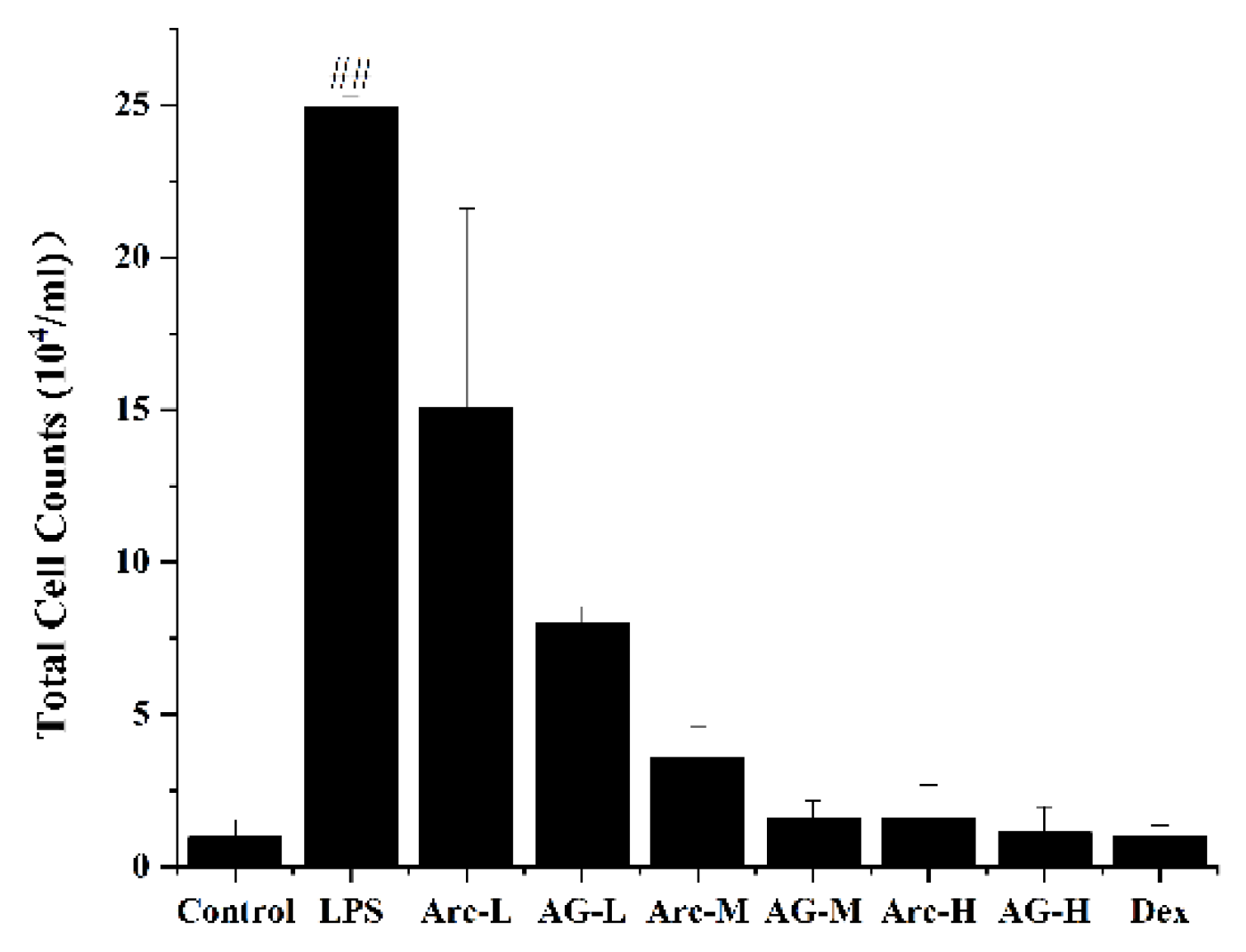
Inhalation of LPS will cause inflammatory cells to flow into the lungs over time. As shown in Figure 2, LPS significantly increased the total number of inflammatory cells in BALF. However, Arc and AG effectively reduced the increase in the total number of inflammatory cells induced by LPS. The high dose of Arc and AG showed similar efficacy when compared with the Dex group; Compared with the Arc group, at the same dose, the AG group had a more effective role in reducing the total number of inflammatory cells (Figure 2).
2.2.3. Effects of Arc and AG on LPS-Induced Lung W/D Ratio
Pulmonary edema was one of the typical characteristics of acute lung injury. As shown in Table 2, after LPS challenged, the lung W/D Ratios of mice were increased significantly. However, the ratios could be reduced effectively by pretreating with Arc and AG. The improvement effect of a high dose of Arc and AG was similar to the Dex group. And the inhibitory effect of the AG group was better than the Arc group at a medium dose.
2.2.4. Effects of Arc and AG on LPS-Mediated Lung Histopathologic Changes
Compared to the control group, lung specimens from the LPS group demonstrated obvious histopathologic abnormalities, which included the severe hemorrhage in the alveolus, infiltration of inflammatory cells into the interstitial and alveolar spaces, interstitial edema, and widespread alveolar wall thickness (Figure 3). With regard to the treatment groups (Arc, AG, and Dex), the severity of histopathologic changes in lung tissues was slimmer than that in the LPS group, especially on inflammatory cell infiltration and hemorrhage. In addition, compared with the Arc group, the AG group showed a better improvement effect at the same dose.
Table 3 showed the average score of each category, and the total score was calculated by adding the individual scores of each category. The average score of each group showed that there was no lesion in the control group, and the lesion in the LPS group was the most serious. In addition, the drug pretreatment of Arc, AG, and Dex could effectively inhibit the pathological changes in lung tissue. According to the Damage degree: 2 > 3 > 4 > 5 > 6 = 7 > 8, the high dose of AG performed the most significant inhibitory action. Both Arc and AG could effectively prevent LPS-induced lung injury, but the AG group showed better Inhibitory action, especially in the treatment of Interstitial edema and hemorrhage, and a high dose of AG even performed better effect than dexamethasone.
2.2.5. Effects of Arc and AG on LPS-Induced Myeloperoxidase (MPO) Activity
As an important index to evaluate the aggregation of inflammatory cells in lung tissue, this study measured the activity of MPO. Compared with the control group, the concentration of MPO in lung tissue was significantly increased after LPS administration. However, the administration of Arc and AG one hour before the LPS challenge significantly inhibited this change. In addition, the inhibitory effect of AG was more significant at low doses (Figure 4).
3. Materials and Methods
3.1. Apparatus and Materials
Arctiin was purchased from Lemeitian (Chengdu, China). Potassium bromide for IR was spectral grade, purchased from Kermel (Tianjin, China). All chemicals used for synthesis were of analytical grade. D101 macroporous adsorption resin purchased from Tianjin Guangfu Fine Chemical Research Institute (Tianjin, China). AG was synthesized from Arctiin with structure characterization by multiple spectroscopic analysis (IR, HR-ESI-MS, 1H-NMR, 13C-NMR, DEPT (θ = 90°, 135°), HSQC, and HMBC). Lipopolysaccharide (LPS) was purchased from Sigma-Aldrich. Mouse TNF-α, IL-6, IL-1β, and MPO ELISA kits were purchased from MULTI SCIENCES (Hangzhou, China).
3.2. Preparation of AG
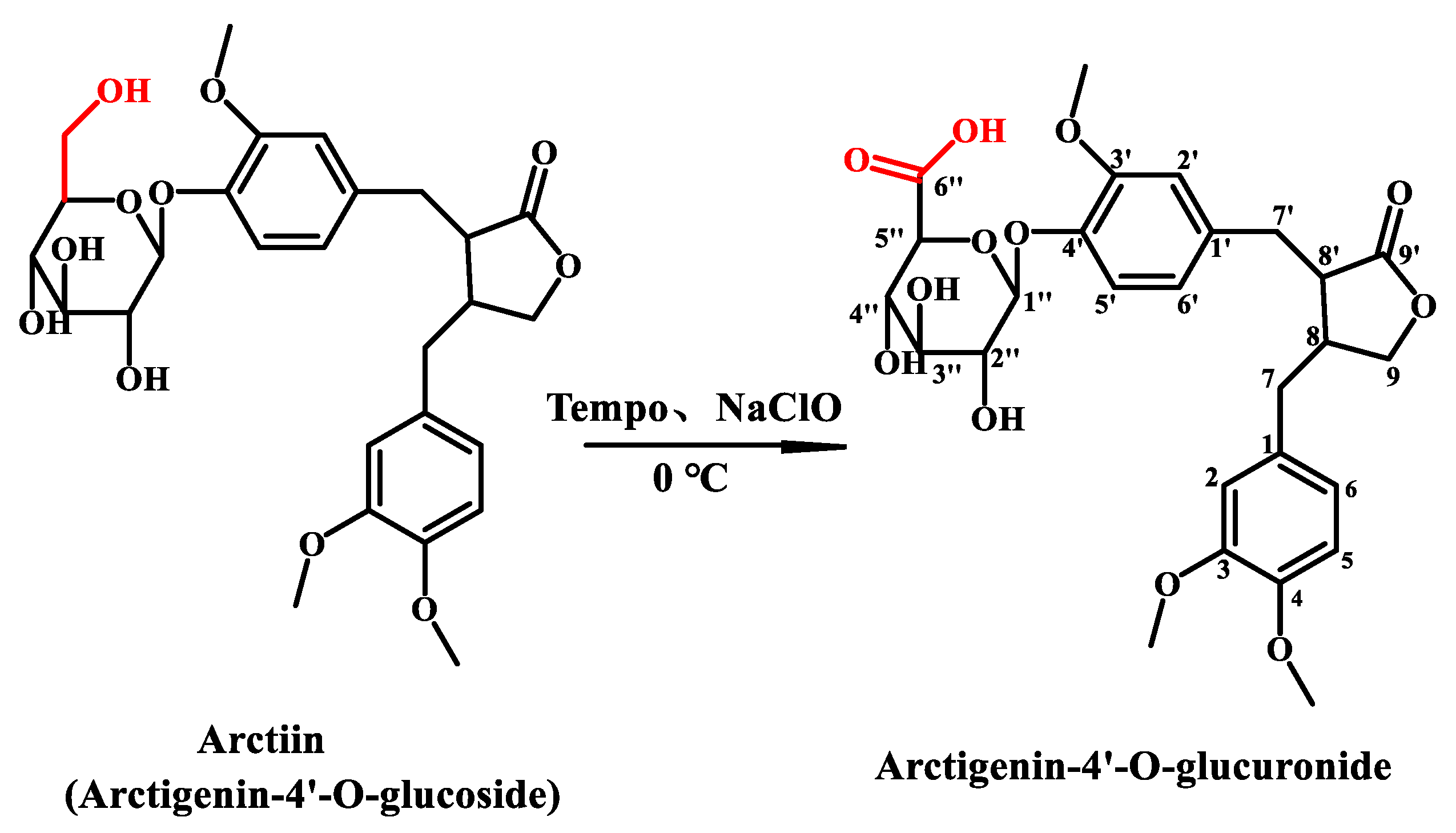
400 mg Arc was added to 50 mL 60% THF (by volume) solution containing 160 mg Na2CO3 (Sinopharm Chemical Reagent Co., Ltd., Shanghai, China), 60 mg NaHCO3 (Sinopharm Chemical Reagent Co., Ltd., Shanghai, China), and 40 mg TEMPO (Sinopharm Chemical Reagent Co., Ltd., Shanghai, China). The reaction mixture was cooled to about 0 °C in ice water, then NaClO was added dropwise until the thin-layer chromatography (TLC) monitored that the starting material was completely reacted, and finally 1.5 mL methanol was added to terminate the reaction. The reaction solution was concentrated under reduced pressure to remove the THF. The residue was purified with D101 macroporous resin using water and 40% ethanol as eluent, respectively. Collect 40% ethanol eluent and removed the solvent to dryness under vacuum to obtain 353 mg of product with a yield of 86%. The reaction was shown in Scheme 1.
AG was obtained as a white powder with [α − 48° (c, 1.0, CHCl3), and IR (KBr) γmax 3419, 2928, 1771, 1609, 1521, 1429, 1271, 1378, 1158, 1075 and 1026 cm−1. Its molecular formula was established as C27H32O12 by HR-ESI-MS (m/z 547.1883 [M-H]–, calcd. for C27H31O12: 547.5359), requiring 12° of unsaturation. The ESI-MS2 fragments were m/z 371 [M-H-glcA]– and 357 [M-glcA-CH3]–. The TLC Rf value was 0.5 (CHCl3-MeOH-H2O-CH3COOH, 2:1:0.025:0.075). The solubility of AG was 227 mg/mL in pure water at 25 °C.
3.3. Evaluation of Anti-Inflammatory Activity
3.3.1. Animals and Experimental Design
Five-week-old male Kunming mice (18–22 g) were purchased from Chengdu Dashuo Experimental Animal Co., Ltd. (Chengdu, China) and raised at a controlled temperature of 22.0 ± 2 °C and relative humidity of 50–60% on a 12 h light/12 h dark cycle with free access to food and drink. The animal experiments were conducted in accordance with the protocol of the National Act on the Use of Experimental Animals (China) and were approved by the Sichuan Committee on Laboratory Animals (approval number SYXK2014−187).
After one week of adaptation to general meals, the mice were divided into the following nine groups randomly (n = 12 mice per group). The groups included the control group, LPS groups, Arc high-dose (Arc-H, 40 mg/kg) group, Arc middle-dose (Arc-M, 20 mg/kg) group, Arc low-dose (Arc-L, 10 mg/kg) group, AG high-dose (AG-H, 40 mg/kg) group, AG middle-dose (AG-M, 20 mg/kg) group, AG low-dose (AG-L, 10 mg/kg) group, and Dexamethasone (Dex, 5 mg/kg) group.
The groups of Arc, AG, and Dex were given corresponding drug pretreatment by gavage 1 h prior to intranasal administration of LPS, while the control group and LPS group were treated intranasally with 50 μL of PBS. After 1 h, all mice except the blank control group were given LPS by intranasal administration. Six hours after LPS stimulation, the mice were euthanized. After that, BALF and lung tissue samples were harvested.
3.3.2. Collection of BALF and Determination of TNF-α, IL-1β, and IL-6 Levels
After the mice were euthanized, the skin of the neck was cut open with scissors, and the neck muscles and subcutaneous tissues were separated longitudinally with curved tweezers so that the trachea could be fully exposed. Then, the trachea was cannulated and 0.5 mL PBS was injected into the lungs of mice. After careful flushing, draw out the liquid and put it into a centrifuge tube. Repeat the flushing process three times to obtain the BALF 1.2–1.5 mL (recovery rate is more than 80%).
The obtained BALF was centrifuged (4 °C, 3000 r/min, 10 min), and the supernatant was used for the subsequent determination of inflammatory factors, while the bottom cells were collected for the counting of inflammatory cells. The level of TNF-α, IL-1β, and IL-6 in BALF were determined by an enzyme-linked immunosorbent assay (ELISA) kit. All procedures are in accordance with the manufacturer’s instructions.
3.3.3. Cell Counting
The bottom cells obtained by BALF centrifugation were rinsed twice with 0.5 mL hank’s solution (without Ca2+, Mg2+) under the same conditions (4 °C, 3000 r/min) for 5 min each time. The washed cell sediment was mixed with 0.5 ml of normal saline for cell counting.
3.3.4. Lung Wet-to-Dry Weight (W/D) Ratio
The mice without BALF obtained were dissected and the right lungs of mice were excised, dried with filter paper, weighed to obtain “wet” weight (W), and then placed in an oven at 80 °C for 72 h to obtain “dry” weight (D). After that, dried it for 5 h at 80 °C, weighed it three times, and checked whether it is weighed. The ratio of the wet lung to the dry lung (W/D) was calculated to assess tissue edema.
3.3.5. Histopathologic Evaluation of the Lung Tissue
Histopathological examination was performed on mice without BALF collection. The left lung tissue samples of mice were fixed in 10% neutral buffered formalin for 48 h, then dehydrated in ascending series ethanol, including 70%, 80%, 90%, 95%, and 100% ethanol, transparent in xylene, soaked in melted paraffin, and finally embedded in paraffin and sliced. embedded in paraffin and sliced. After hematoxylin-eosin (H&E) staining (Follow the instructions), the pathological changes of lung tissue were observed under the microscope.
According to the pathological injury characteristics of each sample, the histological score was carried out. The severity of lung injury ranges from 0 to 4 (the “0” meant normal while the “4” represented severely) and falls into the following categories: neutrophil infiltration, interstitial edema, hemorrhage, and congestion. The total score was calculated by adding the individual scores of each category.
3.3.6. Pulmonary MPO Activity Assay
The right lung of the mice was rinsed with precooled PBS to remove residual blood and inhaled dry on the filter paper. After weighing, the tissue was cut into pieces with scissors. Then the cut tissue was added to PBS in a 1:9 weight volume ratio. The cut tissue was put into a low-temperature homogenizer to make homogenate. Finally, the homogenate was centrifuged at 4 °C at 5000 r/min for 10 min. The supernatant was taken and tested according to MPO Elisa kit.
3.3.7. Statistical Analysis
IBM SPSS Statistics (R26.0.0.0, IBM, Chicago, IL, USA) was used for the statistical analysis of data through one-way ANOVA. The experimental data were expressed as mean ± standard deviation (x ± s); Comparing the difference between groups, p < 0.05 indicates a statistical difference.
4. Discussion
The mouse model of acute lung injury was established to verify the biological activity of AG. ALI may be induced by many factors, such as foreign body inhalation, pathogenic bacteria infection (including novel coronavirus), primary injury, etc. LPS was the main component of the outer membrane of gram-negative bacteria cells, which could stimulate the recruitment and activation of inflammatory cells in the lungs, lead to systemic inflammatory reaction and immune activation, and produce tissue damage [32]. In this experiment, the ALI model was established by intranasal administration of LPS. The pathological changes in the lung tissue of the model group mice showed alveolar hemorrhage, destruction of alveolar tissue, thickening of alveolar septa, and infiltration of inflammatory cells. It could be seen that the model was successfully established, and it was further confirmed by the increase of W/D in lung tissue, the number of inflammatory cells accumulated in the lung, the activity of MPO, and the significant increase in inflammatory factor production.
Hormone therapy was the main method for drug treatment of acute lung injury. Glucocorticoids could effectively reduce the inflammatory reaction, but had no tissue repair effect. Long-term use of hormone drugs would affect the body’s organs and even lead to systemic diseases [33,34]. Therefore, a growing number of researchers turned their attention to traditional Chinese medicine, hoping to find potential drugs for treating ALI.
It had been reported that Arc had a certain inhibitory effect on LPS-induced acute lung injury [5], but the oral bioavailability of Arc was low because of its poor solubility. In this study, the water-soluble derivative of Arc, AG, obtained by tempo-mediated oxidation method, effectively improved the inflammatory cell infiltration, pulmonary edema, pulmonary hemorrhage, interstitial edema, and other symptoms of the model group, effectively inhibited the increase of inflammatory factors in BALF of mice, and compared with the same dose of Arc, the improvement effect and inhibition effect were enhanced. Therefore, the AG obtained by tempo-mediated oxidation did indeed improve their bioavailability by improving the solubility of Arc as expected.
Our research showed that AG was beneficial to ALI. And in mice with ALI induced, AG was more effective than Arc in inhibiting the release of inflammatory factors in the lungs of ALI mice, reducing the inflammatory response, reducing pulmonary edema, and improving lung injury.
Supplementary Materials
The following supporting information can be downloaded at: https://www.mdpi.com/article/10.3390/molecules28041789/s1, Figure S1: HR-ESI-MS spectra; Figure S2: ESI-MS2 spectra; Figure S3: 1H-NMR spectra; Figure S4: 13C-NMR spectra; Figure S5: DEPT (θ = 90°), Figure S6: DEPT (θ = 135°) spectra; Figure S7: HMBC spectra and Figure S8: HSQC spectra.
Author Contributions
Conceptualization, K.G.; Data curation, Y.Z., J.X. and S.L.; Formal analysis, X.X. and X.W.; Funding acquisition, K.G.; Investigation, X.X.; Methodology, X.H. and S.L.; Supervision, X.W. and K.G.; Validation, S.L.; Writing—original draft, X.X.; Writing—review & editing, K.G. All authors have read and agreed to the published version of the manuscript.
Funding
This work was supported by Central Government Funds of Guiding Local Scientific and Technological Development for Sichuan Province (No. 2021ZYD0057).
Institutional Review Board Statement
The animal experiments were conducted in accordance with the protocol of the National Act on the Use of Experimental Animals (China) and were approved by the Sichuan Committee on Laboratory Animals (approval number SYXK2014−187).
Informed Consent Statement
Not applicable.
Data Availability Statement
Not applicable.
Conflicts of Interest
The authors declare no conflict of interest.
Sample Availability
Samples of arctigenin-4′-O-glucuronide is available from the authors.
References
- Gao, Q.; Yang, M.B.; Zuo, Z. Overview of the anti-inflammatory effects, pharmacokinetic properties and clinical efficacies of arctigenin and arctiin from Arctium lappa L. Acta Pharmacol. Sin. 2018, 39, 787–801. [Google Scholar] [CrossRef]
- Park, S.Y.; Hong, S.S.; Han, X.H.; Hwang, J.S.; Lee, D.; Ro, J.S.; Hwang, B.Y. Lignans from Arctium lappa and Their Inhibition of LPS-Induced Nitric Oxide Production. Chem. Pharm. Bull. 2007, 55, 150–152. [Google Scholar] [CrossRef]
- Lee, S.; Shin, S.; Kim, H.; Han, S.; Kim, K.; Kwon, J.; Kwak, J.; Lee, C.; Ha, N.; Yim, D.; et al. Anti-inflammatory function of arctiin by inhibiting COX-2 expression via NF- B pathways. J. Inflamm. 2011, 8, 16–25. [Google Scholar] [CrossRef] [PubMed]
- Lee, J.Y.; Cho, B.J.; Park, T.W.; Psrk, B.E.; Kim, S.J.; Sim, S.S.; Kim, C.J. Dibenzylbutyrolactone Lignans from Forsythia koreana Fruits Attenuate Lipopolysaccharide-Induced Inducible Nitric Oxide Synthetase and Cyclooxygenase-2 Expressions through Activation of Nuclear Factor-kB and Mitogen-Activated Protein Kinase in RAW264.7 Cells. Biol. Pharm. Bull. 2010, 33, 1847–1853. [Google Scholar] [PubMed]
- Zhou, B.; Weng, G.; Huang, Z.; Liu, T.; Dai, F. Arctiin Prevents LPS-Induced Acute Lung Injury via Inhibition of PI3K/AKT Signaling Pathway in Mice. Inflammation 2018, 41, 2129–2135. [Google Scholar] [CrossRef] [PubMed]
- Wu, J.G.; Wu, J.Z.; Sun, L.N.; Han, T.; Du, J.; Ye, Q.; Zhang, H.; Zhang, Y.G. Ameliorative effects of arctiin from Arctium lappa on experimental glomerulonephritis in rats. Phytomedicine 2009, 16, 1033–1041. [Google Scholar] [CrossRef] [PubMed]
- Zhou, B.; Wang, L.; Liang, Y.; Li, J.; Pan, X. Arctiin suppresses H9N2 avian influenza virus-mediated inflammation via activation of Nrf2/HO-1 signaling. BMC Complement. Med. 2021, 21, 289. [Google Scholar] [CrossRef]
- Xu, X.; Zeng, X.Y.; Cui, Y.X.; Li, Y.B.; Cheng, J.H.; Zhao, X.D.; Xu, G.H.; Ma, J.; Piao, H.N.; Jin, X.; et al. Antidepressive Effect of Arctiin by Attenuating Neuroinflammation via HMGB1/TLR4- and TNF-alpha/TNFR1-Mediated NF-kappaB Activation. ACS Chem. Neurosci. 2020, 11, 2214–2230. [Google Scholar] [CrossRef] [PubMed]
- Liu, X.; Wang, J.; Dou, P.; Zhang, X.; Ran, X.; Liu, L.; Dou, D. The Ameliorative Effects of Arctiin and Arctigenin on the Oxidative Injury of Lung Induced by Silica via TLR-4/NLRP3/TGF-beta Signaling Pathway. Oxidative Med. Cell. Longev. 2021, 2021, 5598980. [Google Scholar] [CrossRef]
- Lee, J.H.; Lee, J.Y.; Kim, T.D.; Kim, C.J. Antiasthmatic action of dibenzylbutyrolactone lignans from fruits of Forsythia viridissima on asthmatic responses to ovalbumin challenge in conscious guinea-pigs. Phytother. Res. 2011, 25, 387–395. [Google Scholar] [CrossRef] [PubMed]
- Lu, L.C. Isolation and Purification of Arctiin and Its Protective Mechanism on Vascular Endothelial Cells in Experimental Diabetes Rats; Third Military Medical University: Chongqin, China, 2007. [Google Scholar]
- Liu, J.; Zou, M.; Piao, H.; Liu, Y.; Tang, B.; Gao, Y.; Ma, N.; Cheng, G. Characterization and Pharmacokinetic Study of Aprepitant Solid Dispersions with Soluplus(R). Molecules 2015, 20, 11345–11356. [Google Scholar] [CrossRef] [PubMed]
- Ong, S.G.; Ming, L.C.; Lee, K.S.; Yuen, K.H. Influence of the Encapsulation Efficiency and Size of Liposome on the Oral Bioavailability of Griseofulvin-Loaded Liposomes. Pharmaceutics 2016, 8, 25. [Google Scholar] [CrossRef] [PubMed]
- Ghosh, A.; Biswas, S.; Ghosh, T. Preparation and Evaluation of Silymarin beta-cyclodextrin Molecular Inclusion Complexes. J. Young Pharm. 2011, 3, 205–210. [Google Scholar] [CrossRef] [PubMed]
- Shome, S.; Talukdar, A.D.; Choudhury, M.D.; Bhattacharya, M.K.; Upadhyaya, H. Curcumin as potential therapeutic natural product: A nanobiotechnological perspective. J. Pharm. Pharmacol. 2016, 68, 1481–1500. [Google Scholar] [CrossRef]
- Guan, H.Y.; Zhao, D.H.; Long, J.; Lu, L.C. Preparation and in vitro evaluation of arctiin self microemulsifying drug delivery system. Chin. J. Pharm. 2015, 46, 248–253. [Google Scholar]
- Steffen, P.G.; Dressman, J.B.; Saal, C. Trends in Active Pharmaceutical Ingredient Salt Selection based on Analysis of the Orange Book Database. J. Med. Chem. 2007, 50, 6665–6672. [Google Scholar]
- Nieman, J.A.; Nair, S.K.; Heasley, S.E.; Schultz, B.L.; Zerth, H.M.; Nugent, R.A.; Chen, K.; Stephanski, K.J.; Hopkins, T.A.; Knechtel, M.L.; et al. Modifications of C-2 on the pyrroloquinoline template aimed at the development of potent herpesvirus antivirals with improved aqueous solubility. Bioorg. Med. Chem. Lett. 2010, 20, 3039–3042. [Google Scholar] [CrossRef]
- Press, N.J.; Taylor, R.J.; Fullerton, J.D.; Tranter, P.; McCarthy, C.; Keller, T.H.; Arnold, N.; Beer, D.; Brown, L.; Cheung, R.; et al. Solubility-driven optimization of phosphodiesterase-4 inhibitors leading to a clinical candidate. J. Med. Chem. 2012, 55, 7472–7479. [Google Scholar] [CrossRef]
- Furfine, E.S.; Baker, C.T.; Hale, M.R.; Reynolds, D.J.; Salisbury, J.A.; Searle, A.D.; Studenberg, S.D.; Todd, D.; Tung, R.D.; Spaltenstein, A. Preclinical pharmacology and pharmacokinetics of GW433908, a water-soluble prodrug of the human immunodeficiency virus protease inhibitor amprenavir. Antimicrob. Agents Chemother. 2004, 48, 791–798. [Google Scholar] [CrossRef]
- Vollmann, K.; Qurishi, R.; Hockemeyer, J.; Müller, C.E. Synthesis and Properties of a New Water-Soluble Prodrug of the Adenosine A2A Receptor Antagonist MSX-2. Molecules 2008, 13, 348–359. [Google Scholar] [CrossRef]
- Rodríguez-Pérez, T.; Fernández, S.; Sanghvi, Y.S.; Detorio, M.; Schinazi, R.F.; Gotor, V.; Ferrero, M. Chemoenzymatic Syntheses and Anti-HIV-1 Activity of Glucose-Nucleoside Conjugates as Prodrugs. Bioconj. Chem. 2010, 21, 2239–2249. [Google Scholar] [CrossRef] [PubMed]
- O’Reilly, S.; Rowinsky, E.K. The clinical status of irinotecan (CPT-11), a novel water soluble camptothecin analogue: 1996. Crit. Rev. Oncol. Hemat. 1996, 24, 47–70. [Google Scholar] [CrossRef] [PubMed]
- Vignaroli, G.; Zamperini, C.; Dreassi, E.; Radi, M.; Angelucci, A.; Sanita, P.; Crespan, E.; Kissova, M.; Maga, G.; Schenone, S.; et al. Pyrazolo[3,4-d]pyrimidine Prodrugs: Strategic Optimization of the Aqueous Solubility of Dual Src/Abl Inhibitors. ACS Med. Chem. Lett. 2013, 4, 57–61. [Google Scholar] [CrossRef] [PubMed]
- Klimas, R.; Mikus, G. Morphine-6-glucuronide is responsible for the analgesic effect after morphine administration: A quantitative review of morphine, morphine-6-glucuronide, and morphine-3-glucuronide. Br. J. Anaesth. 2014, 113, 935–944. [Google Scholar] [CrossRef]
- Sakamoto, S.; Kusuhara, H.; Horie, K.; Takahashi, K.; Baba, T.; Ishizaki, J.; Sugiyama, Y. Identification of the transporters involved in the hepatobiliary transport and intestinal efflux of methyl 1-(3,4-dimethoxyphenyl)-3-(3-ethylvaleryl)-4-hydroxy-6,7,8-trimethoxy-2-naphthoat e (S-8921) glucuronide, a pharmacologically active metabolite of S-8921. Drug Metab. Dispos. 2008, 36, 1553–1561. [Google Scholar]
- Qing, L.S.; Peng, S.L.; Liang, J.; Ding, L.S. Astragalosidic Acid: A New Water-Soluble Derivative of Astragaloside IV Prepared Using Remarkably Simple TEMPO-Mediated Oxidation. Molecules 2017, 22, 1275. [Google Scholar] [CrossRef]
- Qing, L.S.; Chen, T.B.; Sun, W.X.; Chen, L.; Luo, P.; Zhang, Z.F.; Ding, L.S. Pharmacokinetics Comparison, Intestinal Absorption and Acute Toxicity Assessment of a Novel Water-Soluble Astragaloside IV Derivative (Astragalosidic Acid, LS-102). Eur. J. Drug Metab. Pharmacokinet. 2019, 44, 251–259. [Google Scholar] [CrossRef] [PubMed]
- Sun, W.X.; Zhang, Z.F.; Xie, J.; He, Y.; Cheng, Y.; Ding, L.S.; Luo, P.; Qing, L.S. Determination of a astragaloside IV derivative LS-102 in plasma by ultra-performance liquid chromatography-tandem mass spectrometry in dog plasma and its application in a pharmacokinetic study. Phytomedicine 2019, 53, 243–251. [Google Scholar] [CrossRef]
- Wu, Y.J.; Li, Z.H.; Li, J.Y.; Zhou, Y.; Wang, R.Y.; Chen, X.Y.; Qing, L.S.; Luo, P. Elucidation of the binding mechanism of astragaloside IV derivative with human serum albumin and its cardiotoxicity in zebrafish embryos. Front. Pharmacol. 2022, 13, 987882. [Google Scholar] [CrossRef]
- Chen, L.; Chen, X.Y.; Wang, Q.L.; Yang, S.J.; Zhou, H.; Ding, L.S.; Qing, L.S.; Luo, P. Astragaloside IV Derivative (LS-102) Alleviated Myocardial Ischemia Reperfusion Injury by Inhibiting Drp1(Ser616) Phosphorylation-Mediated Mitochondrial Fission. Front. Pharmacol. 2020, 11, 1083. [Google Scholar] [CrossRef]
- Dong, Z.; Yuan, Y. Accelerated inflammation and oxidative stress induced by LPS in acute lung injury: Iotanhibition by ST1926. Int. J. Mol. Med. 2018, 41, 3405–3421. [Google Scholar] [CrossRef] [PubMed]
- Wang, H.; Zhou, J.; Guo, X.; Li, Y.; Duan, L.; Si, X.; Zhang, L. Use of glucocorticoids in the management of immunotherapy-related adverse effects. Thorac. Cancer 2020, 11, 3047–3052. [Google Scholar] [CrossRef] [PubMed]
- Oray, M.; Samra, K.A.; Ebrahimiadib, N.; Meese, H.; Foster, C.S. Long-term side effects of glucocorticoids. Expert Opin. Drug Saf. 2016, 15, 457–465. [Google Scholar] [CrossRef] [PubMed]
Figure 1.
Effects of Arc and AG on cytokines of TNF-α (A), IL-1β (B), and IL-6 (C) in the BALF of LPS-induced ALI mice. The groups of Arc, AG, and Dex were pretreated with corresponding drugs 1 h prior to intranasal administration of LPS. The values presented are mean ± SEM of four independent experiments (n = 4 in each group). ## p < 0.01 vs. Model; * p < 0.05, ** p < 0.01 vs. the same dose of Arc group; ▲ p < 0.05 vs. Dex group. Control: Control group; LPS: LPS group; Dex: LPS + Dex (5 mg/kg) group; Arc-L: LPS+Arc-low (10 mg/kg) group; Arc-M: LPS+Arc-middle (20 mg/kg) group; Arc-H: LPS+Arc-high (40 mg/kg) group; AG-L: LPS+AG-low (10 mg/kg) group; AG-M: LPS+AG-middle (20 mg/kg) group; AG-H: LPS+AG-high (40 mg/kg) group.

Figure 2.
Effects of Arc and AG on inflammatory cell accumulation in lungs. The groups of Arc, AG, and Dex were pretreated with corresponding drugs 1 h prior to intranasal administration of LPS. The values presented are mean ± SEM of four independent experiments. (n = 3~4 in each group). ## p < 0.01 vs. Model; Control: Control group; LPS: LPS group; Dex: LPS + Dex (5 mg/kg) group; Arc-L: LPS+Arc-low (10 mg/kg) group; Arc-M: LPS+Arc-middle (20 mg/kg) group; Arc-H: LPS+Arc-high (40 mg/kg) group; AG-L: LPS+AG-low (10 mg/kg) group; AG-M: LPS+AG-middle (20 mg/kg) group; AG-H: LPS+AG-high (40 mg/kg) group.

Figure 3.
Effects of Arc and AG on histopathological changes in lung tissues in LPS-induced mice. These representative histological changes of the lung were obtained from mice of different groups (hematoxylin and eosin staining, original magnification 200×). Control: Control group; LPS: LPS group; Dex: LPS + Dex (5 mg/kg) group; Arc-L: LPS+Arc-low (10 mg/kg) group; Arc-M: LPS+Arc-middle (20 mg/kg) group; Arc-H: LPS+Arc-high (40 mg/kg) group; AG-L: LPS+AG-low (10 mg/kg) group; AG-M: LPS+AG-middle (20 mg/kg) group; AG-H: LPS+AG-high (40 mg/kg) group.

Figure 4.
Effects of Arc and AG on MPO activity in lung tissues of LPS-challenged mice. The groups of Arc, AG, and Dex were pretreated with corresponding drugs 1 h prior to intranasal administration of LPS. The values presented are mean ± SEM of four independent experiments. (n = 3~4 in each group). ## p < 0.01 vs. Model; ** p < 0.01 vs. the same dose of Arc group; Control: Control group; LPS: LPS group; Dex: LPS + Dex (5 mg/kg) group; Arc-L: LPS+Arc-low (10 mg/kg) group; Arc-M: LPS+Arc-middle (20 mg/kg) group; Arc-H: LPS+Arc-high (40 mg/kg) group; AG-L: LPS+AG-low (10 mg/kg) group; AG-M: LPS+AG-middle (20 mg/kg) group; AG-H: LPS+AG-high (40 mg/kg) group.

Scheme 1.
Structures of Arc and AG.


Table 1.
1H-NMR (600 MHz) and 13C-NMR data (150 MHz) of AG in DMSO (δ in ppm).
| Position | δH | δC | DEPT | Position | δH | δC | DEPT |
|---|---|---|---|---|---|---|---|
| 1 | 131.68 | C | 6′ | 6.84 (7.6, d) | 120.4 | CH | |
| 2 | 6.76 (s) | 112.46 | CH | 7′ | 2.45 (m) | 36.83 | CH2 |
| 3 | 148.64 | C | 8′ | 2.59 (m) | 40.62 | CH | |
| 4 | 145.31 | C | 9′ | 178.4 | C | ||
| 5 | 6.65 (8.2, 2.0, dd) | 115.17 | CH | Glu-1″ | 4.88 (7.6, d) | 99.87 | CH |
| 6 | 6.97 (8.3, d) | 121.28 | CH | Glu-2″ | 3.20–3.28 (m) | 76.68 | CH |
| 7 | 2.80 (m) | 33.39 | CH2 | Glu-3″ | 3.20–3.28 (m) | 73.08 | CH |
| 8 | 2.73 (m) | 45.61 | CH | Glu-4″ | 3.20–3.28 (m) | 74.29 | CH |
| 9 | 3.86 (8.8, t), 4.09 (8.2, t) | 70.68 | CH2 | Glu-5″ | 3.52 (m) | 71.99 | CH |
| 1′ | 131.19 | C | Glu-6″ | 173.1 | C | ||
| 2′ | 6.68 (s) | 111.91 | CH | OCH3 | 3.71 (s) | 55.68 | CH3 |
| 3′ | 148.69 | C | OCH3 | 3.70 (s) | 55.51 | ||
| 4′ | 147.34 | C | OCH3 | 3.69 (s) | 55.45 | ||
| 5′ | 6.62 (8.4, 2.0, dd) | 113.94 | CH |
Table 2.
Effects of Arc and AG on pulmonary edema.
| Groups | Dose/(mg·kg−1) | Lung W/D/(%) |
|---|---|---|
| Control | - | 3.92 ± 0.06 |
| LPS | - | 5.02 ± 0.08 ## |
| Arc-L | 10 | 4.84 ± 0.38 |
| AG-L | 10 | 4.73 ± 0.28 |
| Arc-M | 20 | 4.51 ± 0.17 |
| AG-M | 20 | 4.47 ± 0.5 * |
| Arc-H | 40 | 4.32 ± 0.07 |
| AG-H | 50 | 4.29 ± 0.07 |
| Dex | 5 | 4.2 ± 0.12 |
The values presented are mean ± SEM of four independent experiments. (n = 3~4 in each group). The groups of Arc, AG, and Dex were pretreated with corresponding drugs 1 h prior to intranasal administration of LPS. ## p < 0.01 vs. Model; * p < 0.05 vs. the same dose of Arc group. Control: Control group; LPS: LPS group; Dex: LPS + Dex (5 mg/kg) group; Arc-L: LPS+Arc-low (10 mg/kg) group; Arc-M: LPS+Arc-middle (20 mg/kg) group; Arc-H: LPS+Arc-high (40 mg/kg) group; AG-L: LPS+AG-low (10 mg/kg) group; AG-M: LPS+AG-middle (20 mg/kg) group; AG-H: LPS+AG-high (40 mg/kg) group.
Table 3.
Effects of Arc and AG on LPS-induced lung morphology. Lung Histology Score.
| Groups | Neutrophil Infiltration | Interstitial Edema | Hemorrhage | Congestion | Scores |
|---|---|---|---|---|---|
| Control | 0 | 0 | 0 | 0 | 0 |
| LPS | 3 | 3 | 4 | 3 | 13 |
| Arc-L | 2 | 2 | 2.5 | 3 | 9.5 |
| AG-L | 2.5 | 2 | 2.5 | 2 | 9 |
| Arc-M | 2 | 1 | 2 | 2 | 7 |
| AG-M | 2.5 | 0 | 2 | 2 | 6.5 |
| Arc-H | 1 | 1.5 | 2 | 2 | 6.5 |
| AG-H | 0.5 | 0 | 0 | 1 | 1.5 |
| Dex | 1 | 1 | 2 | 1 | 5 |
Lung injury was graded from 0 (normal) to 4 (severe) in four categories: Neutrophil infiltration, Interstitial edema, Hemorrhage, and Congestion. The total lung injury score was calculated by adding up the individual scores of each category. The values presented are mean ± SEM of four independent experiments. (n = 4 in each group). Control: Control group; LPS: LPS group; Dex: LPS + Dex (5 mg/kg) group; Arc-L: LPS+Arc-low (10 mg/kg) group; Arc-M: LPS+Arc-middle (20 mg/kg) group; Arc-H: LPS+Arc-high (40 mg/kg) group; AG-L: LPS+AG-low (10 mg/kg) group; AG-M: LPS+AG-middle (20 mg/kg) group; AG-H: LPS+AG-high (40 mg/kg) group.
Disclaimer/Publisher’s Note: The statements, opinions and data contained in all publications are solely those of the individual author(s) and contributor(s) and not of MDPI and/or the editor(s). MDPI and/or the editor(s) disclaim responsibility for any injury to people or property resulting from any ideas, methods, instructions or products referred to in the content. |
© 2023 by the authors. Licensee MDPI, Basel, Switzerland. This article is an open access article distributed under the terms and conditions of the Creative Commons Attribution (CC BY) license (https://creativecommons.org/licenses/by/4.0/).
Share and Cite
MDPI and ACS Style
Xu, X.; Huang, X.; Zheng, Y.; Wang, X.; Xie, J.; Liu, S.; Guo, K. Synthesis, Structural Elucidation, and Anti-Inflammatory Activity of a Water-Soluble Derivative of Arctiin. Molecules 2023, 28, 1789. https://doi.org/10.3390/molecules28041789
AMA Style
Xu X, Huang X, Zheng Y, Wang X, Xie J, Liu S, Guo K. Synthesis, Structural Elucidation, and Anti-Inflammatory Activity of a Water-Soluble Derivative of Arctiin. Molecules. 2023; 28(4):1789. https://doi.org/10.3390/molecules28041789
Chicago/Turabian StyleXu, Xia, Xiaofeng Huang, Yuedan Zheng, Xiaoling Wang, Jing Xie, Sha Liu, and Kun Guo. 2023. "Synthesis, Structural Elucidation, and Anti-Inflammatory Activity of a Water-Soluble Derivative of Arctiin" Molecules 28, no. 4: 1789. https://doi.org/10.3390/molecules28041789