
Structure-Morphology-Antimicrobial and Antiviral Activity Relationship in Silver-Containing Nanocomposites Based on Polylactide

 , , and
Abstract
:1. Introduction
2. Experimental
2.1. Materials
2.2. Preparation of Polymer Systems
2.3. Experimental Methods
2.3.1. X-ray Diffraction Pattern
2.3.2. TEM Analysis
2.3.3. Thermal Stability by Thermogravimetric Analysis
2.3.4. DSC Analysis
2.3.5. Antimicrobial Activity
2.3.6. Viruses and Cell Culture
2.3.7. The Cytotoxicity Assay
2.3.8. The Antiviral Assay
3. Results and Discussion
3.1. Structural Characterization by X-ray Diffraction
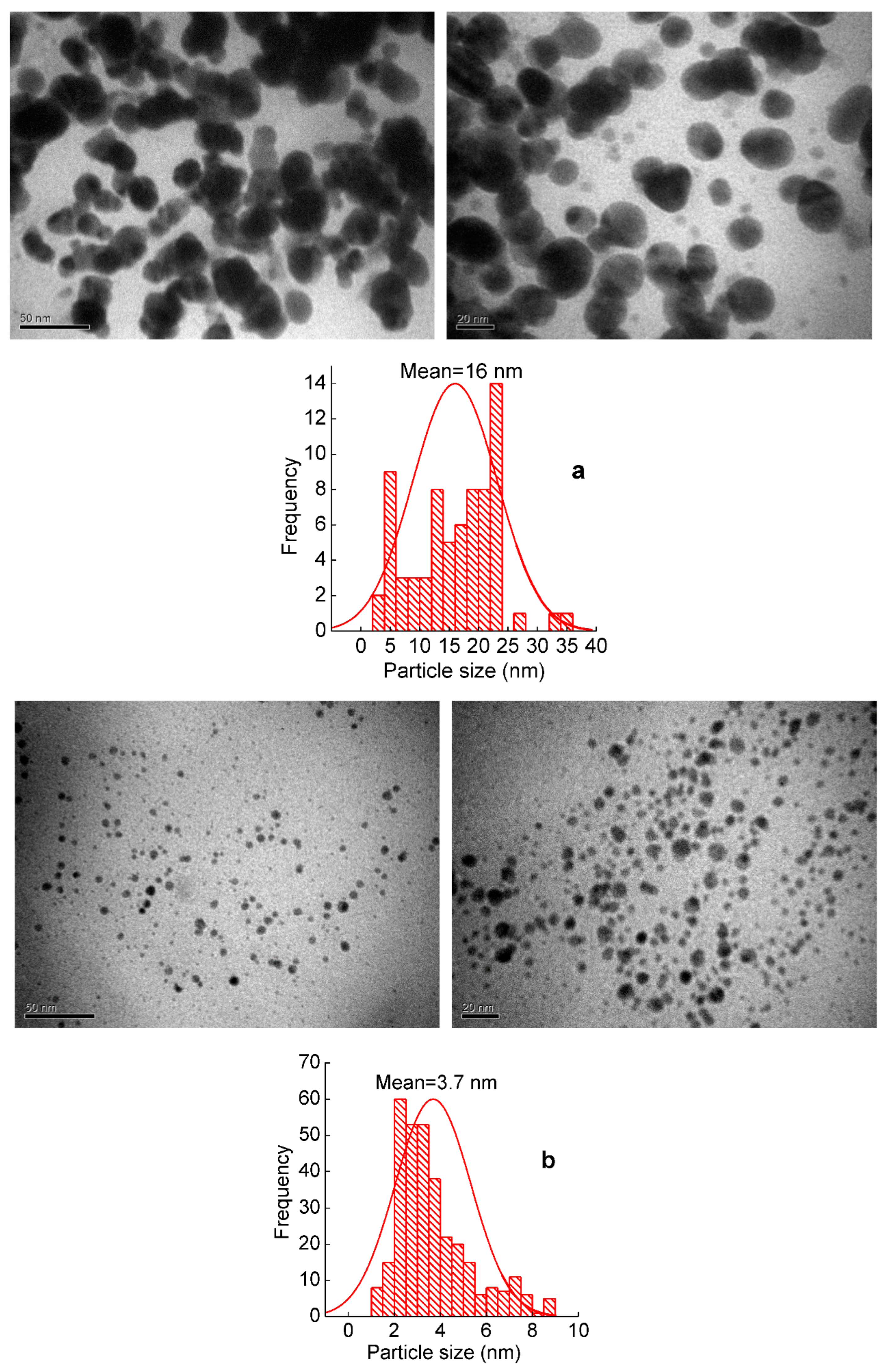
3.2. TEM Analysis
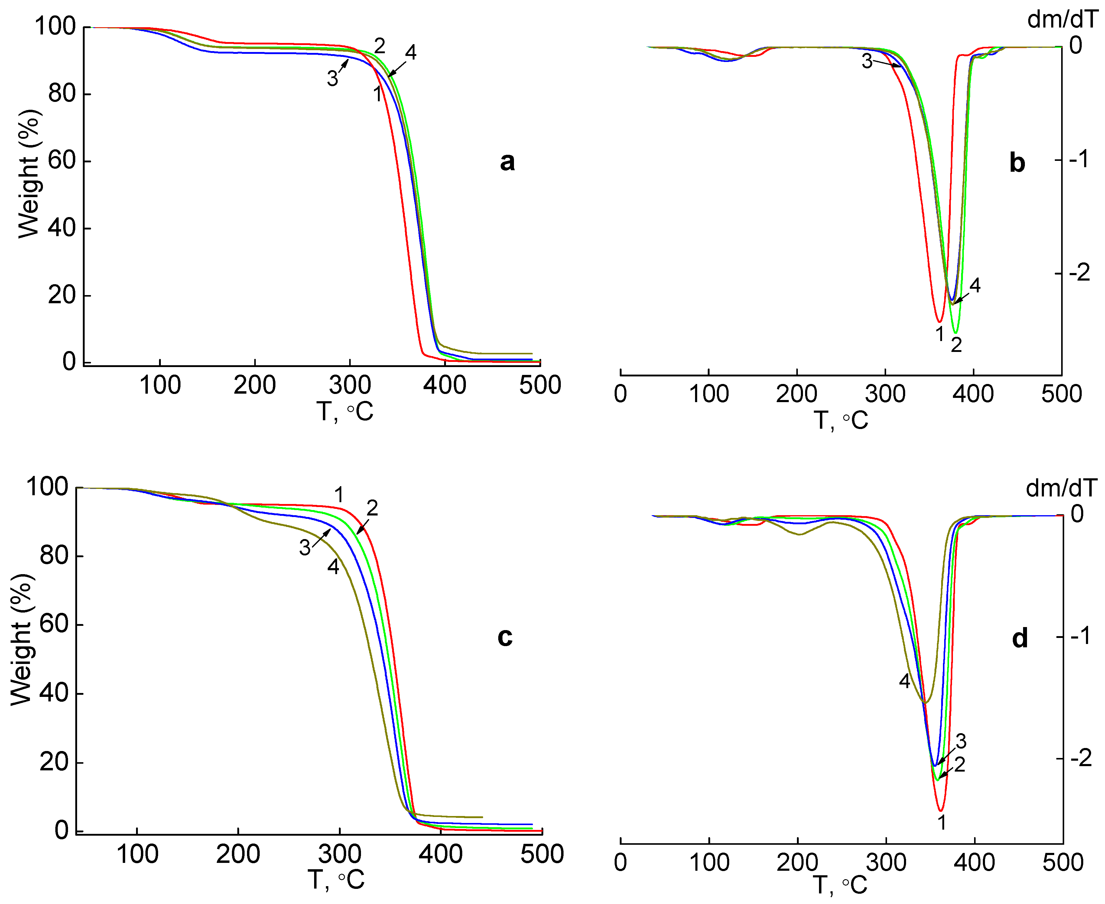
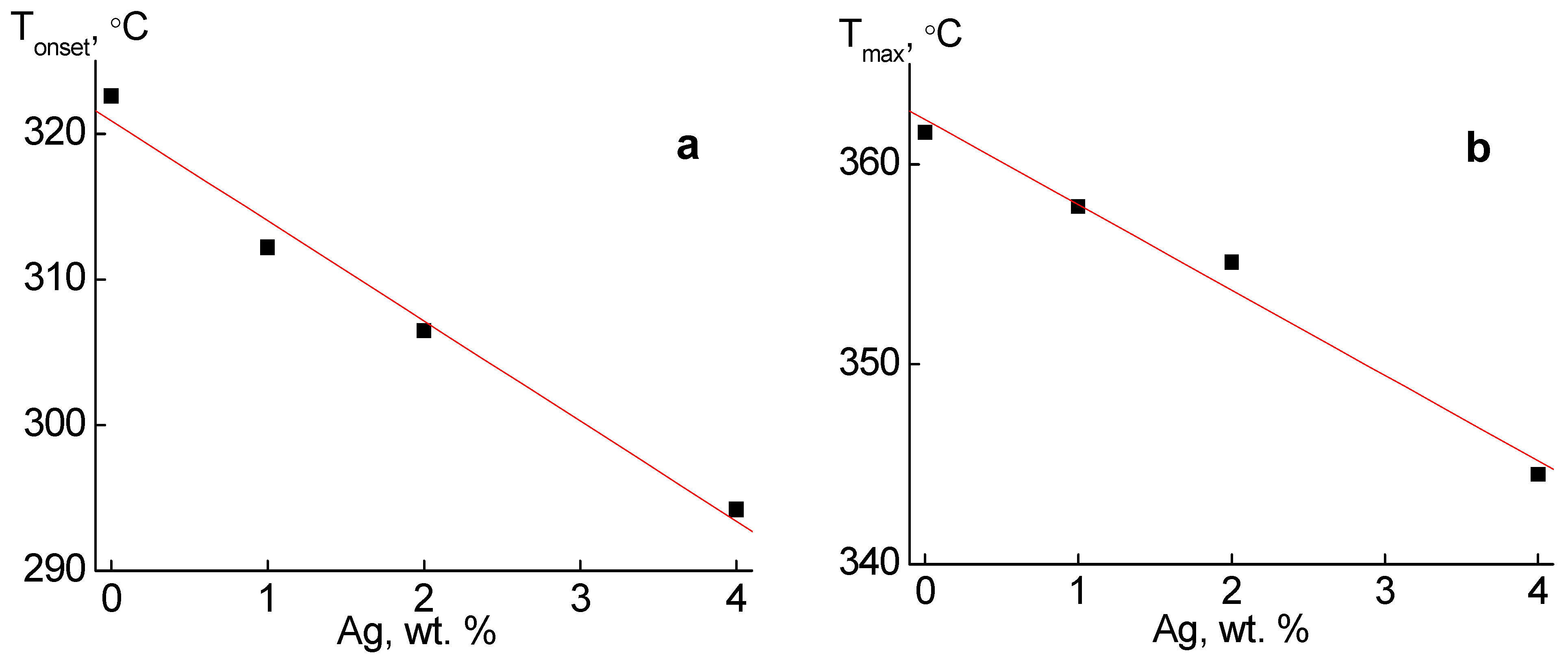
3.3. Thermal Stability by Thermogravimetric Analysis
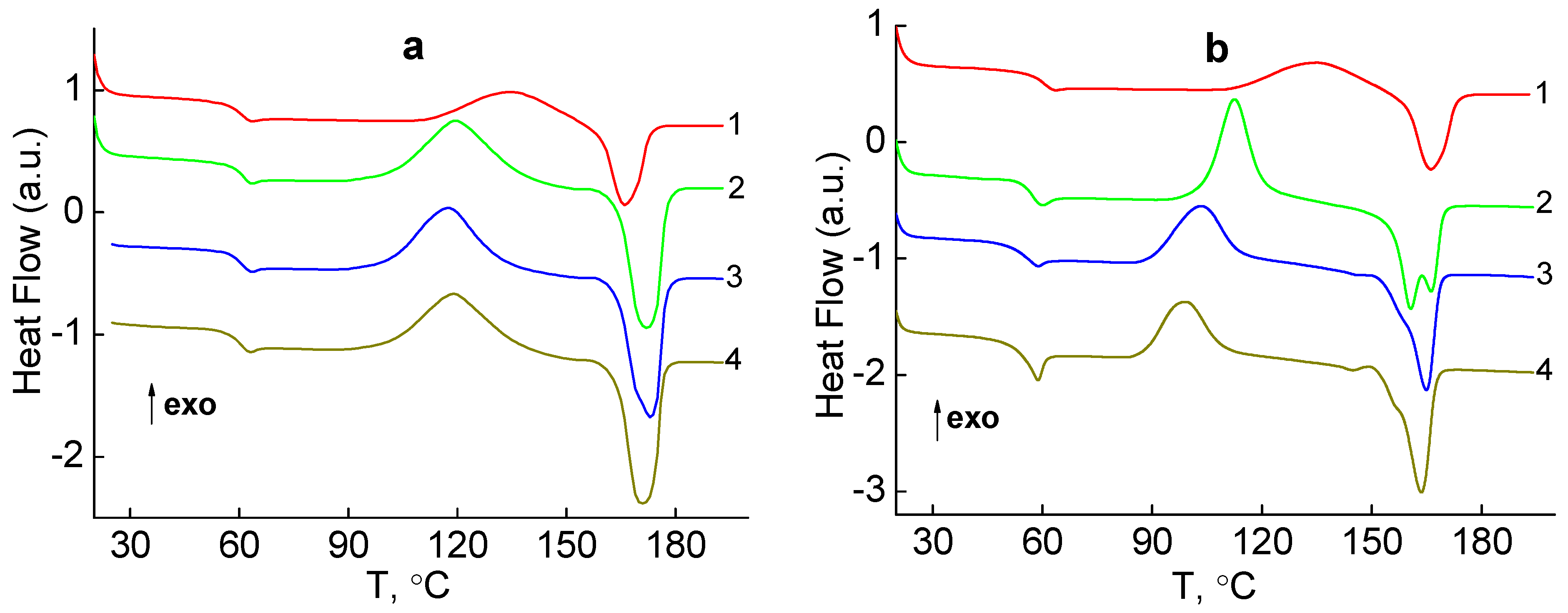
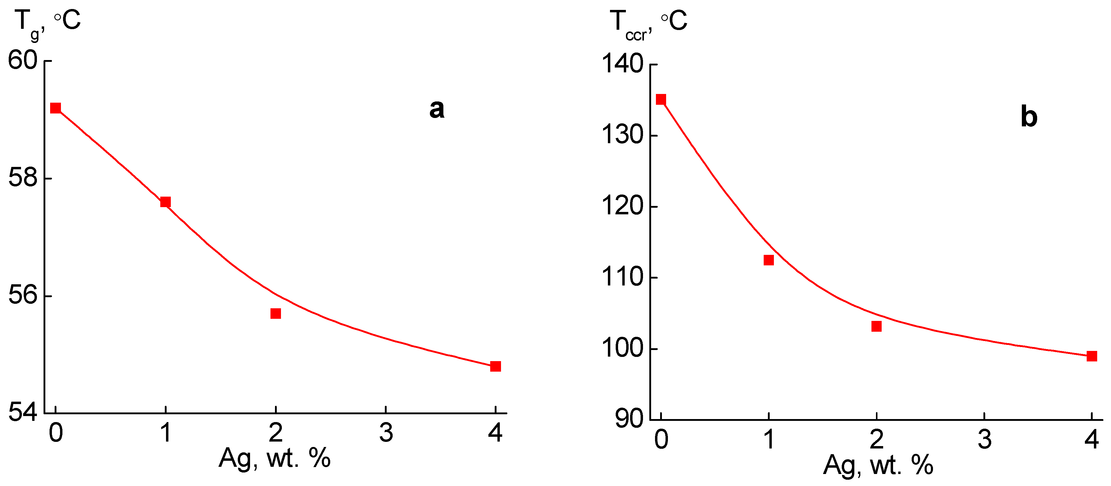
3.4. DSC Analysis
- 1—glass transition in the region of Tg = 57–62 °C;
- 2—cold crystallization in the region of Tccr = 90–150 °C;
- 3—melting in the region of Tm = 160–185 °C.
- 1—glass transition in the region of Tg = 54–61 °C;
- 2—cold crystallization in the region of Tccr = 99–136 °C;
- 3—melting in the region of Tm = 158–167 °C.
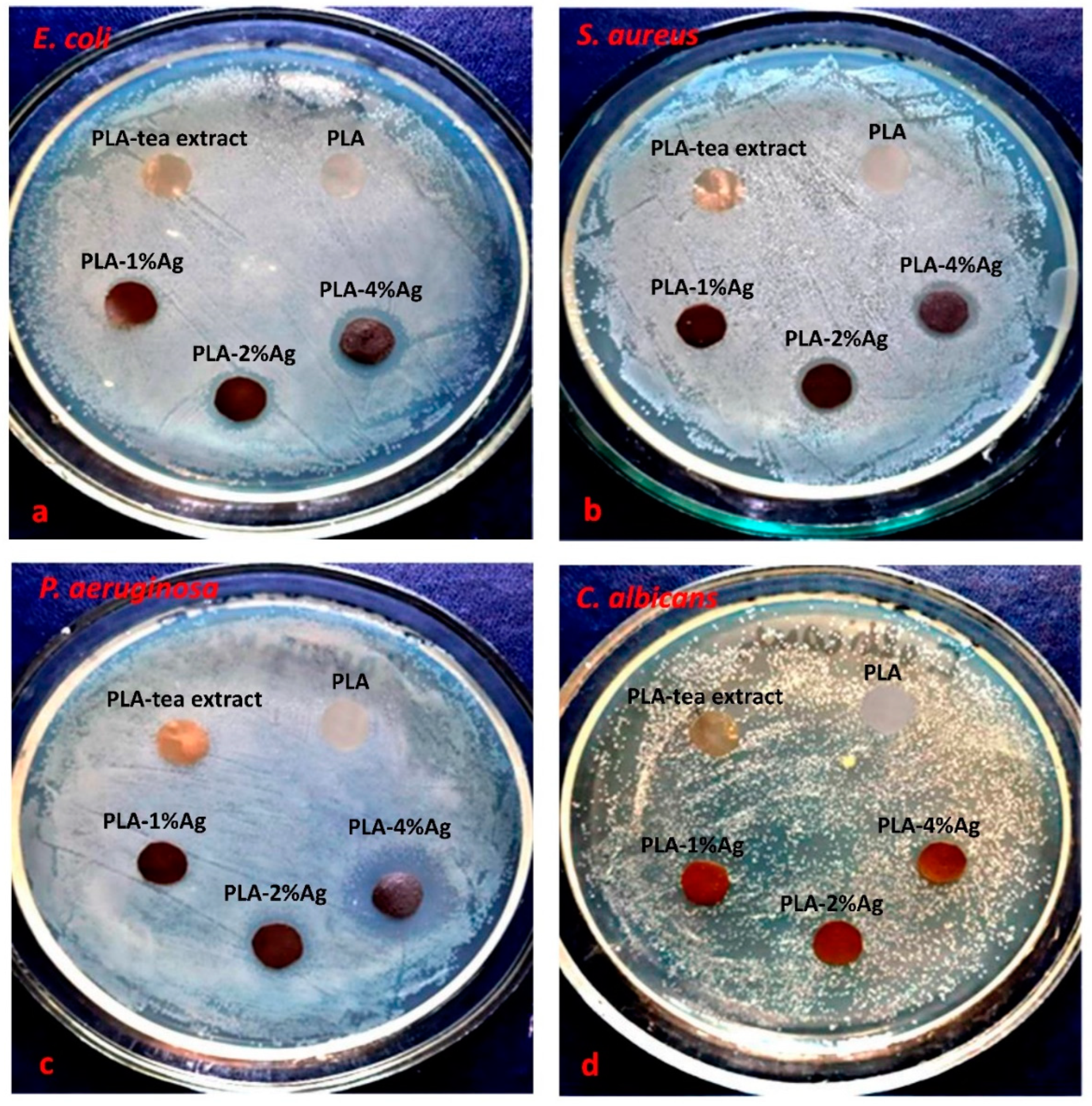
3.5. Antimicrobial Activity of PLA-Ag Nanocomposites
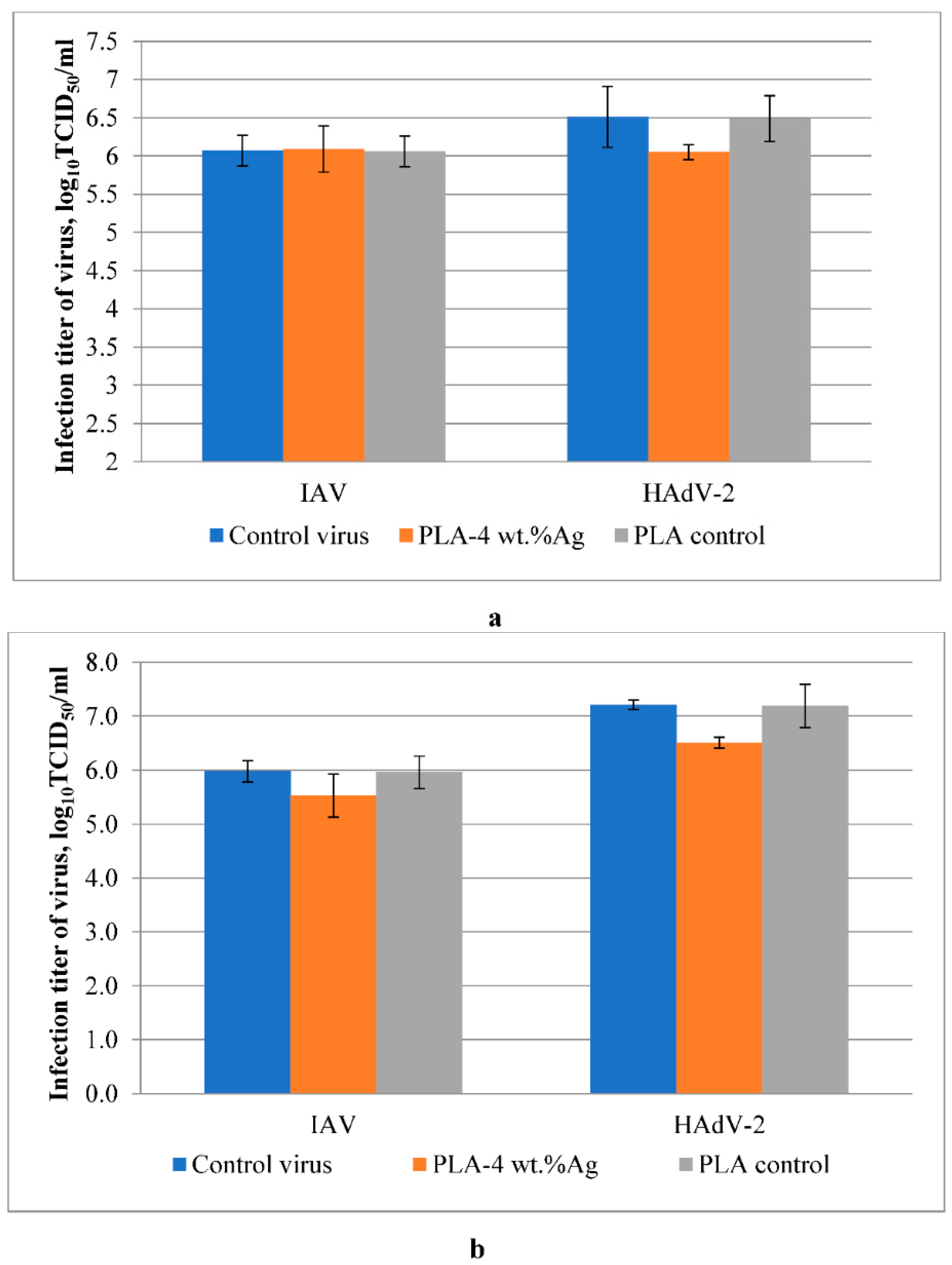
3.6. Antiviral Activity of Ag-Containing Nanocomposites
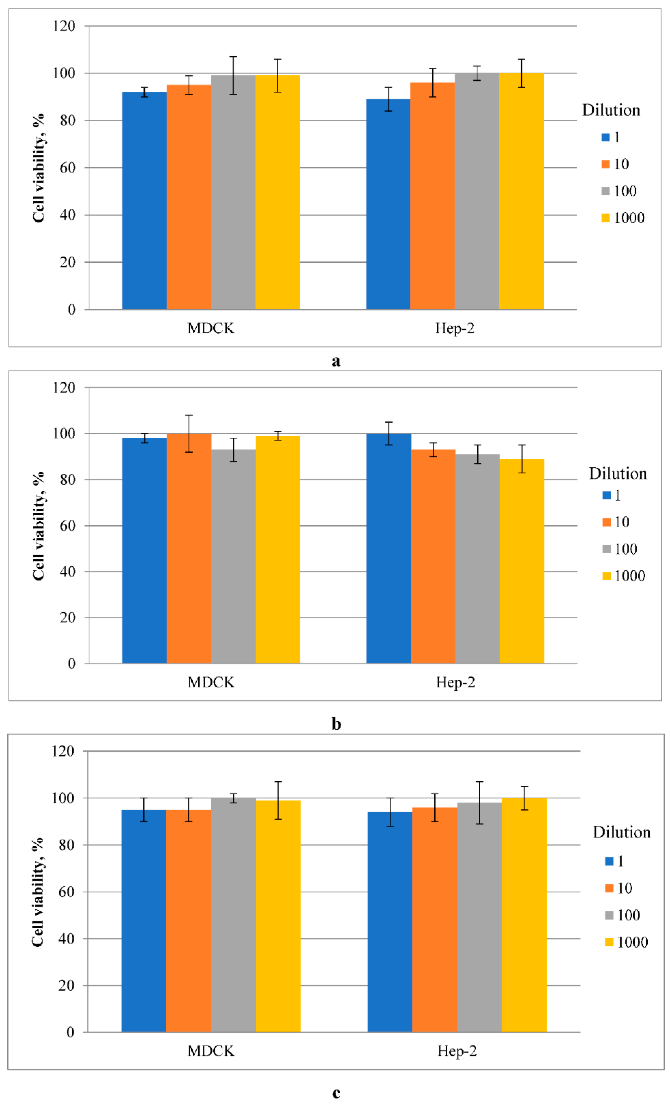
3.7. Cytotoxicity of Ag-Containing Nanocomposites
4. Conclusions
Author Contributions
Funding
Institutional Review Board Statement
Informed Consent Statement
Data Availability Statement
Acknowledgments
Conflicts of Interest
References
- Kuthyar, S.; Anthony, C.L.; Fashina, T.; Yeh, S.; Shantha, J.G. World health organization high priority pathogens: Ophthalmic disease findings and vision health perspectives. Pathogens 2021, 10, 442. [Google Scholar] [CrossRef] [PubMed]
- Rodríguez-Lázaro, D.; Cook, N.; Ruggeri, F.; Sellwood, J.; Nasser, A.; Nascimento, M.; D’Agostino, M.; Santos, R.; Saiz, J.; Rzeżutka, A.; et al. Virus hazards from food, water and other contaminated environments. FEMS Microbiol. Rev. 2012, 36, 786–814. [Google Scholar] [CrossRef] [PubMed]
- Yong, H.; Liu, J. Active packaging films and edible coatings based on polyphenol-rich propolis extract: A review. Compr. Rev. Food Sci. Food Saf. 2021, 20, 2106–2145. [Google Scholar] [CrossRef]
- Lishchynskyi, O.; Shymborska, Y.; Stetsyshyn, Y.; Raczkowska, J.; Skirtach, A.G.; Peretiatko, T.; Budkowski, A. Passive antifouling and active self-disinfecting antiviral surfaces. Chem. Eng. J. 2022, 446, 137048. [Google Scholar] [CrossRef]
- Demchenko, V.; Riabov, S.; Rybalchenko, N.; Goncharenko, L.; Kobylinskyi, S.; Shtompel’, V. X-ray study of structural formation, thermomechanical and antimicrobial properties of copper-containing polymer nanocomposites obtained by the thermal reduction method. Eur. Polym. J. 2017, 96, 326–336. [Google Scholar] [CrossRef]
- Demchenko, V.; Riabov, S.; Sinelnikov, S.; Radchenko, O.; Kobylinskyi, S.; Rybalchenko, N. Novel approach to synthesis of silver nanoparticles in interpolyelectrolyte complexes based on pectin, chitosan, starch and their derivatives. Carbohydr. Polym. 2020, 242, 116431. [Google Scholar] [CrossRef]
- Abbas, M.; Buntinx, M.; Deferme, W.; Peeters, R. (Bio)polymer/ZnO Nanocomposites for Packaging Applications: A Review of Gas Barrier and Mechanical Properties. Nanomaterials 2019, 9, 1494. [Google Scholar] [CrossRef]
- Shevtsova, T.; Cavallaro, G.; Lazzara, G.; Milioto, S.; Donchak, V.; Harhay, K.; Korolko, S.; Budkowski, A.; Stetsyshyn, Y. Temperature-responsive hybrid nanomaterials based on modified halloysite nanotubes uploaded with silver nanoparticles. Colloids Surf. A Physicochem. Eng. Asp. 2022, 641, 128525. [Google Scholar] [CrossRef]
- Merkl, P.; Long, S.; McInerney, G.M.; Sotiriou, G.A. Antiviral activity of silver, copper oxide and zinc oxide nanoparticle coatings against SARS-CoV-2. Nanomaterials 2021, 11, 1312. [Google Scholar] [CrossRef]
- Sikorska, W.; Zięba, M.; Musioł, M.; Kowalczuk, M.; Janeczek, H.; Chaber, P.; Masiuchok, O.; Demchenko, V.; Talanyuk, V.; Iurzhenko, M.; et al. Forensic Engineering of Advanced Polymeric Materials–Part VII: Degradation of Biopolymer Welded Joints. Polymers 2020, 12, 1167. [Google Scholar] [CrossRef]
- Demchenko, V.L.; Iurzhenko, M.V.; Kobylinskyi, S.M.; Goncharenko, L.A. Synthesis and characterization of nanocomposites based on polylactide/silver nanoparticles, obtained by thermochemical reduction of Ag+ ions by natural or synthetic polymers. Chem. Phys. Technol. Surf. 2021, 12, 365–373. [Google Scholar]
- Ahmed, J.; Varshney, S. Polylactides—Chemistry, Properties and Green Packaging Technology: A Review. Int. J. Food Prop. 2011, 14, 37–58. [Google Scholar] [CrossRef]
- Nootsuwan, N.; Sukthavorn, K.; Wattanathana, W.; Jongrungruangchok, S.; Veranitisagul, C.; Koonsaeng, N.; Laobuthee, A. Development of Antimicrobial Hybrid Materials from Polylactic Acid and Nano-silver Coated Chitosan. Orient. J. Chem. 2018, 34, 683–692. [Google Scholar] [CrossRef]
- Demchenko, V.; Kobylinskyi, S.; Iurzhenko, M.; Riabov, S.; Vashchuk, A.; Rybalchenko, N.; Zahorodnia, S.; Naumenko, K.; Demchenko, O.; Adamus, G.; et al. Nanocomposites based on polylactide and silver nanoparticles and their antimicrobial and antiviral applications. React. Funct. Polym. 2022, 170, 105096. [Google Scholar] [CrossRef]
- Zezin, A.A. Synthesis of metal-polymer complexes and functional nanostructures in films and coatings of interpolyelectrolyte complexes. Polym. Sci. 2019, A61, 754–764. [Google Scholar] [CrossRef]
- Zezin, A.A. Synthesis of hybrid materials in polyelectrolyte matrixes: Control over sizes and spatial organization of metallic nanostructures. Polym. Sci. 2016, C58, 118–130. [Google Scholar] [CrossRef]
- Demchenko, V.; Riabov, S.; Kobylinskyi, S.; Goncharenko, L.; Rybalchenko, N.; Kruk, A.; Moskalenko, O.; Shut, M. Effect of the type of reducing agents of silver ions in interpolyelectrolyte-metal complexes on the structure, morphology and properties of silver-containing nanocomposites. Sci. Rep. 2020, 10, 7126. [Google Scholar] [CrossRef]
- Demchenko, V.L.; Kobylinskyi, S.M.; Riabov, S.V.; Shtompel, V.I.; Iurzhenko, M.V.; Rybalchenko, N.P. Novel approach to formation of silver-containing nanocomposites by thermochemical reduction of Ag+ ions in interpolyelectrolyte-metal complexes. Appl. Nanosci. 2020, 10, 5409–5419. [Google Scholar] [CrossRef]
- Zezin, A.A.; Klimov, D.I.; Zezina, E.A.; Mkrtchyan, K.V.; Feldman, V.I. Controlled radiation-chemical synthesis of metal polymer nanocomposites in the films of interpolyelectrolyte complexes: Principles, prospects and implications. Radiat. Phys. Chem. 2020, 169, 108076. [Google Scholar] [CrossRef]
- Alnairat, N.; Dalo, M.A.; Abu-Zurayk, R.; Mallouh, S.A.; Odeh, F.; Al Bawab, A. Green Synthesis of Silver Nanoparticles as an Effective Antibiofouling Material for Polyvinylidene Fluoride (PVDF) Ultrafiltration Membrane. Polymers 2021, 13, 3683. [Google Scholar] [CrossRef]
- Lokanathan, A.R.; Uddin, K.M.A.; Rojas, O.J.; Laine, J. Cellulose Nanocrystal-Mediated Synthesis of Silver Nanoparticles: Role of Sulfate Groups in Nucleation Phenomena. Biomacromolecules 2014, 15, 373–379. [Google Scholar] [CrossRef] [PubMed]
- Demchenko, V.; Shtompel’, V.; Riabov, S.; Lysenkov, E. Constant electric and magnetic fields affect on the structuring and thermomechanical and thermophysical properties of nanocomposites formed from pectin–Cu2+–polyethyleneimine interpolyelectrolyte–metal complexes. Nanoscale Res. Lett. 2015, 10, 479–485. [Google Scholar] [CrossRef] [PubMed]
- Kaewvilai, A.; Wattanathana, W.; Jongrungruangchok, S.; Veranitisagul, S.; Koonsaeng, N.; Laobuthee, A. 3,4-Dihydro-1,3-2H-benzoxazines: Novel reducing agents through one electron donation mechanism and their application as the formation of nano-metallic silver coating. Mater. Chem. Phys. 2015, 167, 9–13. [Google Scholar] [CrossRef]
- Behravan, M.; Panahi, A.H.; Naghizadeh, A.; Ziaee, M.; Mahdavi, R.; Mirzapour, A. Facile green synthesis of silver nanoparticles using Berberis vulgaris leaf and root aqueous extract and its antibacterial activity. Int. J. Biol. Macromol. 2019, 124, 148–154. [Google Scholar] [CrossRef] [PubMed]
- Yousaf, H.; Mehmood, A.; Ahmad, K.S.; Raffi, M. Green synthesis of silver nanoparticles and their applications as an alternative antibacterial and antioxidant agents. Mater. Sci. Eng. C 2020, 112, 110901. [Google Scholar] [CrossRef]
- Flieger, J.; Franus, W.; Panek, R.; Szymańska-Chargot, M.; Flieger, W.; Flieger, M.; Kołodziej, P. Green Synthesis of Silver Nanoparticles Using Natural Extractswith Proven Antioxidant Activity. Molecules 2021, 26, 4986. [Google Scholar] [CrossRef]
- Abd-Elhady, H.M.; Ashor, M.A.; Hazem, A.; Saleh, F.M.; Selim, S.; El Nahhas, N.; Abdel-Hafez, S.H.; Sayed, S.; Hassan, E.A. Biosynthesis and Characterization of Extracellular Silver Nanoparticles from Streptomyces aizuneusis: Antimicrobial, Anti Larval, and Anticancer Activities. Molecules 2022, 27, 212. [Google Scholar] [CrossRef]
- Rizwana, H.; Alwhibi, M.S.; Al-Judaie, R.A.; Aldehaish, H.A.; Alsaggabi, N.S. Sunlight-Mediated Green Synthesis of Silver Nanoparticles Using the Berries of Ribes rubrum (Red Currants): Characterisation and Evaluation of Their Antifungal and Antibacterial Activities. Molecules 2022, 27, 2186. [Google Scholar] [CrossRef]
- Pilaquinga, F.; Amaguaña, D.; Morey, J.; Moncada-Basualto, M.; Pozo-Martínez, J.; Olea-Azar, C.; Fernández, L.; Espinoza-Montero, P.; Jara-Negrete, E.; Meneses, L.; et al. Synthesis of Silver Nanoparticles Using Aqueous Leaf Extract of Mimosa albida (Mimosoideae): Characterization and Antioxidant Activity. Materials 2020, 13, 503. [Google Scholar] [CrossRef]
- Li, Y.; Wang, S.; Yang, X.; Zhang, X. Preparation and characterization of silver palmitate. Int. J. Nanosci. 2009, 8, 97–102. [Google Scholar] [CrossRef]
- Tarani, E.; Črešnar, K.P.; Zemljič, L.F.; Chrissafis, K.; Papageorgiou, G.Z.; Lambropoulou, D.; Zamboulis, A.; Bikiaris, D.N.; Terzopoulou, Z. Cold Crystallization Kinetics and Thermal Degradation of PLA Composites with Metal Oxide Nanofillers. Appl. Sci. 2021, 11, 3004. [Google Scholar] [CrossRef]
- Case, C.L.; Johnson, T.R. Laboratory Experiments in Microbiology; Benjamin Cummings Pub Inc.: San Francisco, CA, USA, 1984. [Google Scholar]
- Toned, A.; Joubert, A.; Cromarty, D. Limitation of the 3-(4,5-dimethylthizol-2-yl)-2,5-diphenyl-2H-tetrazolium bromide (MTT) assay when compared to three commonly used cell enumeration assays. BMC Res. Notes 2015, 8, 47. [Google Scholar]
- Kohn, L.; Foglio, M.; Rodrigues, R.; De O Sousa, I.; Martini, M.; Padilla, M.; Neto, D.D.L.; Arns, C. In-Vitro Antiviral Activities of Extracts of Plants of The Brazilian Cerrado against the Avian Metapneumovirus (aMPV). Braz. J. Poult. Sci. 2015, 17, 275–280. [Google Scholar] [CrossRef]
- Pan, P.; Kai, W.; Zhu, B.; Dong, T.; Inoue, Y. Polymorphous Crystallization and Multiple Melting Behavior of Poly(L-lactide): Molecular Weight Dependence. Macromolecules 2007, 40, 6898–6905. [Google Scholar] [CrossRef]
- Pan, P.; Zhu, B.; Kai, W.; Dong, T.; Inoue, Y. Polymorphic Transition in Disordered Poly(L-lactide) Crystals Induced by Annealing at Elevated Temperatures. Macromolecules 2008, 41, 4296–4304. [Google Scholar] [CrossRef]
- Du, Y.; Wu, T.; Yan, N.; Kortschot, M.T.; Farnood, R. Fabrication and characterization of fully biodegradable natural fiber-reinforced poly(lactic acid) composites. Compos. Part B 2014, 56, 717–723. [Google Scholar] [CrossRef]
- Hodge, I.M. Effects of annealing and prior history on enthalpy relaxation in glassy polymers. 4. Comparison of five polymers. Macromolecules 1983, 16, 898–902. [Google Scholar] [CrossRef]
- Boucher, V.M.; Cangialosi, D.; Alegría, A.; Colmenero, J. Enthalpy recovery of PMMA/Silica nanocomposites. Macromolecules 2010, 43, 7594–7603. [Google Scholar] [CrossRef]
- Shalygina, T.A.; Rudenko, M.S.; Nemtsev, I.V.; Parfenov, V.A.; Voronina, S.Y.; Simonov-Emelyanov, I.D.; Borisova, P.E. Influence of the filler particles’ surface morphology on the polyurethane matrix’s structure formation in the composite. Polymers 2021, 13, 3864. [Google Scholar] [CrossRef]
- Ghasemi, Y.; Rajczakowska, M.; Emborg, M.; Cwirzen, A. Shape-dependent calculation of specific surface area of aggregates versus X-ray microtomography. Mag. Concr. Res. 2018, 72, 1–31. [Google Scholar] [CrossRef]
- Matkovska, L.; Iurzhenko, M.; Mamunya, Y.; Tkachenko, I.; Demchenko, V.; Synyuk, V.; Shadrin, A.; Boiteux, G. Structural Peculiarities of Ion-Conductive Organic-Inorganic Polymer Composites Based on Aliphatic Epoxy Resin and Salt of Lithium Perchlorate. Nanoscale Res. Lett. 2017, 12, 423–431. [Google Scholar] [CrossRef]
- Matkovska, L.; Iurzhenko, M.; Mamunya, Y.; Matkovska, O.; Demchenko, V.; Lebedev, E.; Boiteux, G.; Serghei, A. Electrophysical behavior of ion-conductive organic-inorganic polymer system based on aliphatic epoxy resin and salt of lithium perchlorate. Nanoscale Res. Lett. 2014, 9, 674–682. [Google Scholar] [CrossRef]
- Mijakovic, I.; Petranovic, D.; Mecek, B.; Cepo, T.; Mann, M.; Davies, J. Bacterialsingle-stranded DNA-binding proteins are phosphorylated on tyrosine. Nucleic Acids Res. 2006, 34, 1588–1596. [Google Scholar] [CrossRef]
- Mijakovic, I.; Petranovic, D.; Bottini, N.; Deutscher, J. Protein tyrosine phosphorylation in Bacillus subtilis. J. Mol. Microbiol. Biotechnol. 2005, 9, 189–197. [Google Scholar] [CrossRef] [PubMed]
- Salleh, A.; Naomi, R.; Utami, N.D.; Mohammad, A.W.; Mahmoudi, E.; Mustafa, N.; Fauzi, M.B. The Potential of Silver Nanoparticles for Antiviral and Antibacterial Applications: A Mechanism of Action. Nanomaterials 2020, 10, 1566. [Google Scholar] [CrossRef] [PubMed]
- Baram-Pinto, D.; Shukla, S.; Gedanken, A.; Sarid, R. Inhibition of HSV-1 attachment, entry, and cell-to-cell spread by functionalized multivalent gold nanoparticles. Small 2010, 6, 1044–1050. [Google Scholar] [CrossRef] [PubMed]
- Lara, H.H.; Ayala-Nuñez, N.V.; Ixtepan-Turrent, L.; Rodriguez-Padilla, C. Mode of antiviral action of silver nanoparticles against HIV-1. J. Nanobiotechnol. 2010, 8, 1–10. [Google Scholar] [CrossRef] [PubMed]
- Allawadhi, P.; Singh, V.; Khurana, A.; Khurana, I.; Allwadhi, S.; Kumar, P.; Banothu, A.K.; Thalugula, S.; Barani, P.J.; Naik, R.R.; et al. Silver nanoparticle-based multifunctional approach for combating COVID-19. Sens. Int. 2021, 2, 100101. [Google Scholar] [CrossRef]
- Lu, L.; Sun, R.W.-Y.; Chen, R.; Hui, C.-K.; Ho, C.-M.; Luk, J.M.; Lau, G.K.K.; Che, C.-M. Silver nanoparticles inhibit hepatitis B virus replication. Antivir. Ther. 2008, 13, 253–262. [Google Scholar] [CrossRef] [PubMed]


| Samples | First Stage | Second Stage | ||
|---|---|---|---|---|
| Weight Loss, % | Tmax, °C (DTG) | Tonset, °C | Tmax, °C (DTG) | |
| PLA | 4.8 | 145 | 323 | 362 |
| PLA-1 wt.% Ag | 5.8 | 125 | 341 | 380 |
| PLA-2 wt.% Ag | 7.5 | 121 | 339 | 375 |
| PLA-4 wt.% Ag | 6.0 | 124 | 340 | 376 |
| Samples | First Stage | Second Stage | Third Stage | |||
|---|---|---|---|---|---|---|
| Weight Loss, % | Tmax, °C (DTG) | Weight Loss, % | Tmax, °C (DTG) | Tonset, °C | Tmax, °C (DTG) | |
| PLA | 4.8 | 145 | – | – | 323 | 362 |
| PLA-1 wt.% Ag | 4.5 | 121 | – | – | 312 | 358 |
| PLA-2 wt.% Ag | 3.2 | 115 | 4.7 | 202 | 306 | 355 |
| PLA-4 wt.% Ag | 1.8 | 116 | 8.8 | 202 | 294 | 344 |
| Specimen | Glass Transition | Cold Crystallization | Melting | χC, % | |||
|---|---|---|---|---|---|---|---|
| Tg, °C | ΔCp, J/g·°C | Tccr, °C | ΔHccr, J/g | Tm, °C | ΔHm, J/g | ||
| PLA | 59.2 | 0.412 | 135.1 | 16.74 | 166.1 | 18.12 | 19.5 |
| PLA-1 wt.% Ag (mech. method) | 61.3 | 0.378 | 119.7 | 37.35 | 172.3 | 34.21 | 37.1 |
| PLA-2 wt.% Ag (mech. method) | 61.2 | 0.432 | 117.6 | 34.71 | 173.2 | 32.83 | 36.0 |
| PLA-4 wt.% Ag (mech. method) | 60.5 | 0.443 | 119.0 | 35.0 | 170.9 | 33.58 | 37.6 |
| PLA-1 wt.% Ag (in-situ) | 57.6 | 0.459 | 112.5 | 28.5 | 160/166.4 | 28.12 | 32.0 |
| PLA-2 wt.% Ag (in-situ) | 55.7 | 0.449 | 103.2 | 22.97 | 158.4/165.0 | 23.51 | 27.7 |
| PLA-4 wt.% Ag (in-situ) | 54.8 | 0.394 | 99.0 | 20.35 | 156.5/163.6 | 25.09 | 31.9 |
| Polymer Systems | Diameter of Inhibition Zone, mm | |||
|---|---|---|---|---|
| S. aureus | E. coli | P. aeruginosa | C. albicans | |
| PLA | 0 | 0 | 0 | 0 |
| PLA-tea extract | 0 | 0 | 0 | 0 |
| PLA-1 wt.% Ag | 10.7 ± 0.4 | 11.0 ± 0.5 | 0 | 0 |
| PLA-2 wt.% Ag | 11.0 ± 0.6 | 11.8 ± 1.1 | 10.9 ± 0.6 | 0 |
| PLA-4 wt.% Ag | 11.9 ± 0.8 | 13.1 ± 1.4 | 14.9 ± 1.2 | 10.6 ± 0.7 |
Publisher’s Note: MDPI stays neutral with regard to jurisdictional claims in published maps and institutional affiliations. |
© 2022 by the authors. Licensee MDPI, Basel, Switzerland. This article is an open access article distributed under the terms and conditions of the Creative Commons Attribution (CC BY) license (https://creativecommons.org/licenses/by/4.0/).
Share and Cite
Demchenko, V.; Mamunya, Y.; Kobylinskyi, S.; Riabov, S.; Naumenko, K.; Zahorodnia, S.; Povnitsa, O.; Rybalchenko, N.; Iurzhenko, M.; Adamus, G.; et al. Structure-Morphology-Antimicrobial and Antiviral Activity Relationship in Silver-Containing Nanocomposites Based on Polylactide. Molecules 2022, 27, 3769. https://doi.org/10.3390/molecules27123769
Demchenko V, Mamunya Y, Kobylinskyi S, Riabov S, Naumenko K, Zahorodnia S, Povnitsa O, Rybalchenko N, Iurzhenko M, Adamus G, et al. Structure-Morphology-Antimicrobial and Antiviral Activity Relationship in Silver-Containing Nanocomposites Based on Polylactide. Molecules. 2022; 27(12):3769. https://doi.org/10.3390/molecules27123769
Chicago/Turabian StyleDemchenko, Valeriy, Yevgen Mamunya, Serhii Kobylinskyi, Sergii Riabov, Krystyna Naumenko, Svitlana Zahorodnia, Olga Povnitsa, Nataliya Rybalchenko, Maksym Iurzhenko, Grazyna Adamus, and et al. 2022. "Structure-Morphology-Antimicrobial and Antiviral Activity Relationship in Silver-Containing Nanocomposites Based on Polylactide" Molecules 27, no. 12: 3769. https://doi.org/10.3390/molecules27123769